Next Issue
Volume 12, April-1
Previous Issue
Volume 12, March-1


Cells, Volume 12, Issue 6 (March-2 2023) – 149 articles

Cover Story (view full-size image): 
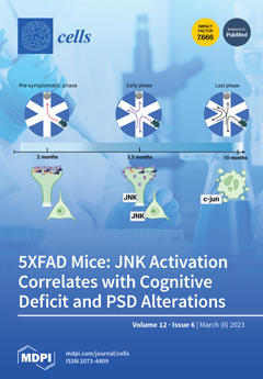
The c-Jun N-terminal kinase (JNK) mediates cellular stress and is deeply involved in Alzheimer’s disease (AD) pathogenesis. In this study, we used the benchmark reference mouse model in the AD field, the 5xFAD mouse model, to further clarify JNK’s role in AD and to understand the link between JNK activation and disease progression. We show a stage-dependent role of JNK in AD pathogenesis: while JNK activation at an early phase of the disease correlates with mild cognitive impairment and with an alteration of the post-synaptic element, the activation of the JNK signalling pathway at a later disease stage activates targets involved in the neuronal death program. This indicates that JNK is an important therapeutic target for the development of new compounds able to tackle synaptic impairment in the early phase of AD pathology. View this paper
  • Issues are regarded as officially published after their release is announced to the table of contents alert mailing list.
  • You may sign up for e-mail alerts to receive table of contents of newly released issues.
  • PDF is the official format for papers published in both, html and pdf forms. To view the papers in pdf format, click on the "PDF Full-text" link, and use the free Adobe Reader to open them.
Order results
Result details
Section
Select all
Export citation of selected articles as:
21 pages, 1620 KiB  
Review
Induced Pluripotent Stem Cells and Their Applications in Amyotrophic Lateral Sclerosis
by Hongmei Du, Zijun Huo, Yanchun Chen, Zhenhan Zhao, Fandi Meng, Xuemei Wang, Shiyue Liu, Haoyun Zhang, Fenghua Zhou, Jinmeng Liu, Lingyun Zhang, Shuanhu Zhou, Yingjun Guan and Xin Wang
Cells 2023, 12(6), 971; https://doi.org/10.3390/cells12060971 - 22 Mar 2023
Cited by 7 | Viewed by 3274
Abstract
Amyotrophic lateral sclerosis (ALS) is a progressive neurodegenerative disease that results in the loss of motor function in the central nervous system (CNS) and ultimately death. The mechanisms underlying ALS pathogenesis have not yet been fully elucidated, and ALS cannot be treated effectively. [...] Read more.
Amyotrophic lateral sclerosis (ALS) is a progressive neurodegenerative disease that results in the loss of motor function in the central nervous system (CNS) and ultimately death. The mechanisms underlying ALS pathogenesis have not yet been fully elucidated, and ALS cannot be treated effectively. Most studies have applied animal or single-gene intervention cell lines as ALS disease models, but they cannot accurately reflect the pathological characteristics of ALS. Induced pluripotent stem cells (iPSCs) can be reprogrammed from somatic cells, possessing the ability to self-renew and differentiate into a variety of cells. iPSCs can be obtained from ALS patients with different genotypes and phenotypes, and the genetic background of the donor cells remains unchanged during reprogramming. iPSCs can differentiate into neurons and glial cells related to ALS. Therefore, iPSCs provide an excellent method to evaluate the impact of diseases on ALS patients. Moreover, patient-derived iPSCs are obtained from their own somatic cells, avoiding ethical concerns and posing only a low risk of immune rejection. The iPSC technology creates new hope for ALS treatment. Here, we review recent studies on iPSCs and their applications in disease modeling, drug screening and cell therapy in ALS, with a particular focus on the potential for ALS treatment. Full article
(This article belongs to the Special Issue iPS Cells (iPSCs) for Modelling and Treatment of Human Diseases 2022)
Show Figures

Figure 1

16 pages, 1366 KiB  
Article
HTR1A, TPH2, and 5-HTTLPR Polymorphisms and Their Impact on the Severity of Depressive Symptoms and on the Concentration of Tryptophan Catabolites during Hepatitis C Treatment with Pegylated Interferon-α2a and Oral Ribavirin (PEG-IFN-α2a/RBV)
by Tomasz Pawlowski, Krzysztof Malyszczak, Dariusz Pawlak, Małgorzata Inglot, Małgorzata Zalewska, Anna Grzywacz, Marek Radkowski, Tomasz Laskus, Justyna Janocha-Litwin and Dorota Frydecka
Cells 2023, 12(6), 970; https://doi.org/10.3390/cells12060970 - 22 Mar 2023
Cited by 1 | Viewed by 1523
Abstract
Background: Seeing that there are no data about associations between serotonin gene polymorphism and tryptophan catabolite concentration during PEG-IFN-α2a treatment, the aim of the current study is to examine (a) the associations between polymorphisms within the HTR1A, TPH2, and 5-HTT genes and the [...] Read more.
Background: Seeing that there are no data about associations between serotonin gene polymorphism and tryptophan catabolite concentration during PEG-IFN-α2a treatment, the aim of the current study is to examine (a) the associations between polymorphisms within the HTR1A, TPH2, and 5-HTT genes and the severity of depression symptoms and (b) the relationships among rs6295, rs4570625, and 5-HTTLPR rs25531polymorphisms and indoleamine 2,3-dioxygenase (IDO) activity, as well as kynurenine (KYN), tryptophan (TRP), kynurenic acid (KA), and anthranilic acid (AA) concentrations. Materials and methods: The study followed a prospective, longitudinal, single-center cohort design. The severity of the depressive symptoms of 101 adult patients with chronic HCV infections was measured during PEG-IFN-α2a/RBV treatment. We used the Montgomery–Åsberg Depression Rating Scale (MADRS) to assess the severity of depressive symptoms. The subjects were evaluated six times—at baseline and at weeks 2, 4, 8, 12, and 24. At all the time points, MADRS score, as well as KYN, TRP, KA, and AA concentrations, and IDO activity were measured. At baseline, rs6295, rs4570625, and 5-HTTLPR rs25531polymorphisms were assessed. Results: Subjects with C/C genotypes of 5-HT1A and lower-expressing alleles (S/S, LG/LG, and S/LG) of 5-HTTLPR scored the highest total MADRS scores and recorded the highest increase in MADRS scores during treatment. We found associations between TRP concentrations and the TPH-2 and 5-HTTLPR rs25531 genotypes. Conclusions: Our findings provide new data that we believe can help better understand infection-induced depression as a distinct type of depression. Full article
Show Figures

Figure 1

15 pages, 1919 KiB  
Article
Computational and Functional Analysis of Structural Features in the ZAKα Kinase
by Valdemar Brimnes Ingemann Johansen, Goda Snieckute, Anna Constance Vind, Melanie Blasius and Simon Bekker-Jensen
Cells 2023, 12(6), 969; https://doi.org/10.3390/cells12060969 - 22 Mar 2023
Cited by 2 | Viewed by 1840
Abstract
The kinase ZAKα acts as the proximal sensor of translational impairment and ribotoxic stress, which results in the activation of the MAP kinases p38 and JNK. Despite recent insights into the functions and binding partners of individual protein domains in ZAKα, the mechanisms [...] Read more.
The kinase ZAKα acts as the proximal sensor of translational impairment and ribotoxic stress, which results in the activation of the MAP kinases p38 and JNK. Despite recent insights into the functions and binding partners of individual protein domains in ZAKα, the mechanisms by which ZAKα binds ribosomes and becomes activated have remained elusive. Here, we highlight a short, thrice-repeated, and positively charged peptide motif as critical for the ribotoxic stress-sensing function of the Sensor (S) domain of ZAKα. We use this insight to demonstrate that the mutation of the SAM domain uncouples ZAKα activity from ribosome binding. Finally, we use 3D structural comparison to identify and functionally characterize an additional folded domain in ZAKα with structural homology to YEATS domains. These insights allow us to formulate a model for ribosome-templated ZAKα activation based on the re-organization of interactions between modular protein domains. In sum, our work both advances our understanding of the protein domains and 3D architecture of the ZAKα kinase and furthers our understanding of how the ribotoxic stress response is activated. Full article
Show Figures

Figure 1

21 pages, 4336 KiB  
Article
Chromatin Accessibility and Transcriptional Landscape during Inhibition of Salmonella enterica by Lactobacillus reuteri in IPEC-J2 Cells
by Weiyun Qin, Zhanshi Ren, Chao Xu, Ya-nan Cao, Ming-an Sun, Ruihua Huang and Wenbin Bao
Cells 2023, 12(6), 968; https://doi.org/10.3390/cells12060968 - 22 Mar 2023
Cited by 4 | Viewed by 1346
Abstract
Lactobacillus reuteri is a probiotic with bacteriostatic effects, which can effectively inhibit the activity of pathogens. However, the molecular mechanism underlying the inhibition of pathogens by L. reuteri in intestinal cells remains unclear. Using the porcine intestinal cell line IPEC-J2 as a model, [...] Read more.
Lactobacillus reuteri is a probiotic with bacteriostatic effects, which can effectively inhibit the activity of pathogens. However, the molecular mechanism underlying the inhibition of pathogens by L. reuteri in intestinal cells remains unclear. Using the porcine intestinal cell line IPEC-J2 as a model, we combined RNA-seq and ATAC-seq methods to delineate the porcine genome-wide changes in biological processes and chromatin accessibility in IPEC-J2 cells stimulated by Salmonella enterica BNCC186354, as well as L. reuteri ATCC 53608. Overall, we found that many porcine transcripts were altered after S. enterica BNCC186354 treatment, while L. reuteri ATCC 53608 treatment partially restored this alteration, such as salmonella infection and PI3K/AKT and MAPK pathways. Combined analysis of these two datasets revealed that 26 genes with similar trends overlapped between gene expression and chromatin accessibility. In addition, we identified potential host functional transcription factors (TFs), such as GATA1, TAL1, TBP, RUNX1, Gmeb1, Gfi1b, RARA, and RXRG, in IPEC-J2 cells that might play a critical role and are targeted by L. reuteri ATCC 53608. Moreover, we verified that PI3K/AKT, MAPK, and apoptosis pathways are potentially regulated by S. enterica BNCC186354 but restored by L. reuteri ATCC 53608. The PI3K/AKT pathway was activated by L. reuteri ATCC 53608, thereby potentially inhibiting S. enterica BNCC186354 infection. In conclusion, our data provide new insights into the expression pattern of functional genes and the epigenetic alterations in IPEC-J2 cells underlying the bacteriostatic action of L. reuteri ATCC 53608. Full article
(This article belongs to the Section Cell Signaling)
Show Figures

Figure 1

18 pages, 4693 KiB  
Article
VEGF Stimulates Activation of ERK5 in the Absence of C-Terminal Phosphorylation Preventing Nuclear Localization and Facilitating AKT Activation in Endothelial Cells
by Anil Kumar Mondru, Mohammad A. Aljasir, Ahmed Alrumayh, Gopika N. Nithianandarajah, Katie Ahmed, Jurgen Muller, Christopher E. P. Goldring, Bettina Wilm and Michael J. Cross
Cells 2023, 12(6), 967; https://doi.org/10.3390/cells12060967 - 22 Mar 2023
Viewed by 2040
Abstract
Extracellular-signal-regulated kinase 5 (ERK5) is critical for normal cardiovascular development. Previous studies have defined a canonical pathway for ERK5 activation, showing that ligand stimulation leads to MEK5 activation resulting in dual phosphorylation of ERK5 on Thr218/Tyr220 residues within the activation loop. ERK5 then [...] Read more.
Extracellular-signal-regulated kinase 5 (ERK5) is critical for normal cardiovascular development. Previous studies have defined a canonical pathway for ERK5 activation, showing that ligand stimulation leads to MEK5 activation resulting in dual phosphorylation of ERK5 on Thr218/Tyr220 residues within the activation loop. ERK5 then undergoes a conformational change, facilitating phosphorylation on residues in the C-terminal domain and translocation to the nucleus where it regulates MEF2 transcriptional activity. Our previous research into the importance of ERK5 in endothelial cells highlighted its role in VEGF-mediated tubular morphogenesis and cell survival, suggesting that ERK5 played a unique role in endothelial cells. Our current data show that in contrast to EGF-stimulated HeLa cells, VEGF-mediated ERK5 activation in human dermal microvascular endothelial cells (HDMECs) does not result in C-terminal phosphorylation of ERK5 and translocation to the nucleus, but instead to a more plasma membrane/cytoplasmic localisation. Furthermore, the use of small-molecule inhibitors to MEK5 and ERK5 shows that instead of regulating MEF2 activity, VEGF-mediated ERK5 is important for regulating AKT activity. Our data define a novel pathway for ERK5 activation in endothelial cells leading to cell survival. Full article
(This article belongs to the Special Issue The ERK5 Signaling Pathway in Health and Disease)
Show Figures

Figure 1

14 pages, 6259 KiB  
Article
Identification of YWHAH as a Novel Brain-Derived Extracellular Vesicle Marker Post Long-Term Midazolam Exposure during Early Development
by Nghi M. Nguyen, Daniel Meyer, Luke Meyer, Subhash Chand, Sankarasubramanian Jagadesan, Maireen Miravite, Chittibabu Guda, Sowmya V. Yelamanchili and Gurudutt Pendyala
Cells 2023, 12(6), 966; https://doi.org/10.3390/cells12060966 - 22 Mar 2023
Cited by 1 | Viewed by 1691
Abstract
Recently, the long-term use of sedative agents in the neonatal intensive care unit (NICU) has raised concerns about neurodevelopmental outcomes in exposed neonates. Midazolam (MDZ), a common neonatal sedative in the NICU, has been suggested to increase learning disturbances and cognitive impairment in [...] Read more.
Recently, the long-term use of sedative agents in the neonatal intensive care unit (NICU) has raised concerns about neurodevelopmental outcomes in exposed neonates. Midazolam (MDZ), a common neonatal sedative in the NICU, has been suggested to increase learning disturbances and cognitive impairment in children. However, molecular mechanisms contributing to such outcomes with long-term MDZ use during the early stages of life remain unclear. In this study, we for the first time elucidate the role of brain-derived extracellular vesicles (BDEVs), including mining the BDEV proteome post long-term MDZ exposure during early development. Employing our previously established rodent model system that mimics the exposure of MDZ in the NICU using an increasing dosage regimen, we isolated BDEVs from postnatal 21-days-old control and MDZ groups using a differential sucrose density gradient. BDEVs from the control and MDZ groups were then characterized using a ZetaView nanoparticle tracking analyzer and transmission electron microscopy analysis. Next, using RT-qPCR, we examined the expression of key ESCRT-related genes involved in EV biogenesis. Lastly, using quantitative mass spectrometry-based proteomics, we mined the BDEV protein cargo that revealed key differentially expressed proteins and associated molecular pathways to be altered post long-term MDZ exposure. Our study characterized the proteome in BDEV cargo from long-term MDZ exposure at early development. Importantly, we identified and validated the expression of YWHAH as a potential target for further characterization of its downstream mechanism and a potential biomarker for the early onset of neurodevelopment and neurodegenerative diseases. Overall, the present study demonstrated long-term exposure to MDZ at early development stages could influence BDEV protein cargo, which potentially impact neural functions and behavior at later stages of development. Full article
(This article belongs to the Special Issue Extracellular Vesicles (EVs) in Diseases)
Show Figures

Figure 1

21 pages, 2505 KiB  
Review
The Similar and Distinct Roles of Satellite Glial Cells and Spinal Astrocytes in Neuropathic Pain
by Aidan McGinnis and Ru-Rong Ji
Cells 2023, 12(6), 965; https://doi.org/10.3390/cells12060965 - 22 Mar 2023
Cited by 7 | Viewed by 4949
Abstract
Preclinical studies have identified glial cells as pivotal players in the genesis and maintenance of neuropathic pain after nerve injury associated with diabetes, chemotherapy, major surgeries, and virus infections. Satellite glial cells (SGCs) in the dorsal root and trigeminal ganglia of the peripheral [...] Read more.
Preclinical studies have identified glial cells as pivotal players in the genesis and maintenance of neuropathic pain after nerve injury associated with diabetes, chemotherapy, major surgeries, and virus infections. Satellite glial cells (SGCs) in the dorsal root and trigeminal ganglia of the peripheral nervous system (PNS) and astrocytes in the central nervous system (CNS) express similar molecular markers and are protective under physiological conditions. They also serve similar functions in the genesis and maintenance of neuropathic pain, downregulating some of their homeostatic functions and driving pro-inflammatory neuro-glial interactions in the PNS and CNS, i.e., “gliopathy”. However, the role of SGCs in neuropathic pain is not simply as “peripheral astrocytes”. We delineate how these peripheral and central glia participate in neuropathic pain by producing different mediators, engaging different parts of neurons, and becoming active at different stages following nerve injury. Finally, we highlight the recent findings that SGCs are enriched with proteins related to fatty acid metabolism and signaling such as Apo-E, FABP7, and LPAR1. Targeting SGCs and astrocytes may lead to novel therapeutics for the treatment of neuropathic pain. Full article
(This article belongs to the Special Issue Role of Glial Cells in Neuropathic Pain)
Show Figures

Figure 1

25 pages, 4065 KiB  
Review
Adult and Pediatric Nail Unit Melanoma: Epidemiology, Diagnosis, and Treatment
by Jade Conway, Jane S. Bellet, Adam I. Rubin and Shari R. Lipner
Cells 2023, 12(6), 964; https://doi.org/10.3390/cells12060964 - 22 Mar 2023
Cited by 3 | Viewed by 15301
Abstract
Nail unit melanoma (NUM) is an uncommon form of melanoma and is often diagnosed at later stages. Approximately two-thirds of NUMs are present clinically as longitudinal melanonychia, but longitudinal melanonychia has a broad differential diagnosis. Clinical examination and dermoscopy are valuable for identifying [...] Read more.
Nail unit melanoma (NUM) is an uncommon form of melanoma and is often diagnosed at later stages. Approximately two-thirds of NUMs are present clinically as longitudinal melanonychia, but longitudinal melanonychia has a broad differential diagnosis. Clinical examination and dermoscopy are valuable for identifying nail findings concerning malignancy, but a biopsy with histopathology is necessary to confirm a diagnosis of NUM. Surgical treatment options for NUM include en bloc excision, digit amputation, and Mohs micrographic surgery. Newer treatments for advanced NUM include targeted and immune systemic therapies. NUM in pediatric patients is extremely rare and diagnosis is challenging since both qualitative and quantitative parameters have only been studied in adults. There is currently no consensus on management in children; for less concerning melanonychia, some physicians recommend close follow-up. However, some dermatologists argue that the “wait and see” approach can cause delayed diagnosis. This article serves to enhance the familiarity of NUM by highlighting its etiology, clinical presentations, diagnosis, and treatment options in both adults and children. Full article
(This article belongs to the Special Issue Melanoma: From Molecular Mechanisms to Therapeutic Opportunities)
Show Figures

Figure 1

20 pages, 1718 KiB  
Review
Psychedelic Targeting of Metabotropic Glutamate Receptor 2 and Its Implications for the Treatment of Alcoholism
by Kevin Domanegg, Wolfgang H. Sommer and Marcus W. Meinhardt
Cells 2023, 12(6), 963; https://doi.org/10.3390/cells12060963 - 22 Mar 2023
Cited by 6 | Viewed by 3812
Abstract
Alcohol abuse is a leading risk factor for the public health burden worldwide. Approved pharmacotherapies have demonstrated limited effectiveness over the last few decades in treating alcohol use disorders (AUD). New therapeutic approaches are therefore urgently needed. Historical and recent clinical trials using [...] Read more.
Alcohol abuse is a leading risk factor for the public health burden worldwide. Approved pharmacotherapies have demonstrated limited effectiveness over the last few decades in treating alcohol use disorders (AUD). New therapeutic approaches are therefore urgently needed. Historical and recent clinical trials using psychedelics in conjunction with psychotherapy demonstrated encouraging results in reducing heavy drinking in AUD patients, with psilocybin being the most promising candidate. While psychedelics are known to induce changes in gene expression and neuroplasticity, we still lack crucial information about how this specifically counteracts the alterations that occur in neuronal circuits throughout the course of addiction. This review synthesizes well-established knowledge from addiction research about pathophysiological mechanisms related to the metabotropic glutamate receptor 2 (mGlu2), with findings and theories on how mGlu2 connects to the major signaling pathways induced by psychedelics via serotonin 2A receptors (2AR). We provide literature evidence that mGlu2 and 2AR are able to regulate each other’s downstream signaling pathways, either through monovalent crosstalk or through the formation of a 2AR-mGlu2 heteromer, and highlight epigenetic mechanisms by which 2ARs can modulate mGlu2 expression. Lastly, we discuss how these pathways might be targeted therapeutically to restore mGlu2 function in AUD patients, thereby reducing the propensity to relapse. Full article
(This article belongs to the Special Issue The Role of Metabotropic Glutamate Receptors in Health and Disease)
Show Figures

Graphical abstract

20 pages, 1730 KiB  
Review
Biomarkers Assessing Endothelial Dysfunction in Alzheimer’s Disease
by Antía Custodia, Marta Aramburu-Núñez, Mariña Rodríguez-Arrizabalaga, Juan Manuel Pías-Peleteiro, Laura Vázquez-Vázquez, Javier Camino-Castiñeiras, José Manuel Aldrey, José Castillo, Alberto Ouro, Tomás Sobrino and Daniel Romaus-Sanjurjo
Cells 2023, 12(6), 962; https://doi.org/10.3390/cells12060962 - 22 Mar 2023
Cited by 6 | Viewed by 2436
Abstract
Alzheimer’s disease (AD) is the most common degenerative disorder in the elderly in developed countries. Currently, growing evidence is pointing at endothelial dysfunction as a key player in the cognitive decline course of AD. As a main component of the blood–brain barrier (BBB), [...] Read more.
Alzheimer’s disease (AD) is the most common degenerative disorder in the elderly in developed countries. Currently, growing evidence is pointing at endothelial dysfunction as a key player in the cognitive decline course of AD. As a main component of the blood–brain barrier (BBB), the dysfunction of endothelial cells driven by vascular risk factors associated with AD allows the passage of toxic substances to the cerebral parenchyma, producing chronic hypoperfusion that eventually causes an inflammatory and neurotoxic response. In this process, the levels of several biomarkers are disrupted, such as an increase in adhesion molecules that allow the passage of leukocytes to the cerebral parenchyma, increasing the permeability of the BBB; moreover, other vascular players, including endothelin-1, also mediate artery inflammation. As a consequence of the disruption of the BBB, a progressive neuroinflammatory response is produced that, added to the astrogliosis, eventually triggers neuronal degeneration (possibly responsible for cognitive deterioration). Recently, new molecules have been proposed as early biomarkers for endothelial dysfunction that can constitute new therapeutic targets as well as early diagnostic and prognostic markers for AD. Full article
Show Figures

Figure 1

24 pages, 6637 KiB  
Article
Social Isolation Activates Dormant Mammary Tumors, and Modifies Inflammatory and Mitochondrial Metabolic Pathways in the Rat Mammary Gland
by Fabia de Oliveira Andrade, Lu Jin, Robert Clarke, Imani Wood, MaryAnn Dutton, Chezaray Anjorin, Grace Rubin, Audrey Gao, Surojeet Sengupta, Kevin FitzGerald and Leena Hilakivi-Clarke
Cells 2023, 12(6), 961; https://doi.org/10.3390/cells12060961 - 21 Mar 2023
Cited by 1 | Viewed by 1734
Abstract
Although multifactorial in origin, one of the most impactful consequences of social isolation is an increase in breast cancer mortality. How this happens is unknown, but many studies have shown that social isolation increases circulating inflammatory cytokines and impairs mitochondrial metabolism. Using a [...] Read more.
Although multifactorial in origin, one of the most impactful consequences of social isolation is an increase in breast cancer mortality. How this happens is unknown, but many studies have shown that social isolation increases circulating inflammatory cytokines and impairs mitochondrial metabolism. Using a preclinical Sprague Dawley rat model of estrogen receptor-positive breast cancer, we investigated whether social isolation impairs the response to tamoxifen therapy and increases the risk of tumors emerging from dormancy, and thus their recurrence. We also studied which signaling pathways in the mammary glands may be affected by social isolation in tamoxifen treated rats, and whether an anti-inflammatory herbal mixture blocks the effects of social isolation. Social isolation increased the risk of dormant mammary tumor recurrence after tamoxifen therapy. The elevated recurrence risk was associated with changes in multiple signaling pathways including an upregulation of IL6/JAK/STAT3 signaling in the mammary glands and tumors and suppression of the mitochondrial oxidative phosphorylation (OXPHOS) pathway. In addition, social isolation increased the expression of receptor for advanced glycation end-products (RAGE), consistent with impaired insulin sensitivity and weight gain linked to social isolation. In socially isolated animals, the herbal product inhibited IL6/JAK/STAT3 signaling, upregulated OXPHOS signaling, suppressed the expression of RAGE ligands S100a8 and S100a9, and prevented the increase in recurrence of dormant mammary tumors. Increased breast cancer mortality among socially isolated survivors may be most effectively prevented by focusing on the period following the completion of hormone therapy using interventions that simultaneously target several different pathways including inflammatory and mitochondrial metabolism pathways. Full article
Show Figures

Graphical abstract

23 pages, 4521 KiB  
Article
Reduced Levels of Misfolded and Aggregated Mutant p53 by Proteostatic Activation
by Evelyne Naus, Marleen Derweduwe, Youlia Lampi, Annelies Claeys, Jarne Pauwels, Tobias Langenberg, Filip Claes, Jie Xu, Veerle Haemels, Zeynep Kalender Atak, Rob van der Kant, Joost Van Durme, Greet De Baets, Keith L. Ligon, Mark Fiers, Kris Gevaert, Stein Aerts, Frederic Rousseau, Joost Schymkowitz and Frederik De Smet
Cells 2023, 12(6), 960; https://doi.org/10.3390/cells12060960 - 21 Mar 2023
Viewed by 1797
Abstract
In malignant cancer, excessive amounts of mutant p53 often lead to its aggregation, a feature that was recently identified as druggable. Here, we describe that induction of a heat shock-related stress response mediated by Foldlin, a small-molecule tool compound, reduces the protein levels [...] Read more.
In malignant cancer, excessive amounts of mutant p53 often lead to its aggregation, a feature that was recently identified as druggable. Here, we describe that induction of a heat shock-related stress response mediated by Foldlin, a small-molecule tool compound, reduces the protein levels of misfolded/aggregated mutant p53, while contact mutants or wild-type p53 remain largely unaffected. Foldlin also prevented the formation of stress-induced p53 nuclear inclusion bodies. Despite our inability to identify a specific molecular target, Foldlin also reduced protein levels of aggregating SOD1 variants. Finally, by screening a library of 778 FDA-approved compounds for their ability to reduce misfolded mutant p53, we identified the proteasome inhibitor Bortezomib with similar cellular effects as Foldlin. Overall, the induction of a cellular heat shock response seems to be an effective strategy to deal with pathological protein aggregation. It remains to be seen however, how this strategy can be translated to a clinical setting. Full article
Show Figures

Figure 1

22 pages, 2709 KiB  
Article
MicroRNA-223 Dampens Pulmonary Inflammation during Pneumococcal Pneumonia
by Cengiz Goekeri, Peter Pennitz, Wibke Groenewald, Ulrike Behrendt, Holger Kirsten, Christian M. Zobel, Sarah Berger, Gitta A. Heinz, Mir-Farzin Mashreghi, Sandra-Maria Wienhold, Kristina Dietert, Anca Dorhoi, Achim D. Gruber, Markus Scholz, Gernot Rohde, Norbert Suttorp, CAPNETZ Study Group, Martin Witzenrath and Geraldine Nouailles
Cells 2023, 12(6), 959; https://doi.org/10.3390/cells12060959 - 21 Mar 2023
Cited by 2 | Viewed by 2318
Abstract
Community-acquired pneumonia remains a major contributor to global communicable disease-mediated mortality. Neutrophils play a leading role in trying to contain bacterial lung infection, but they also drive detrimental pulmonary inflammation, when dysregulated. Here we aimed at understanding the role of microRNA-223 in orchestrating [...] Read more.
Community-acquired pneumonia remains a major contributor to global communicable disease-mediated mortality. Neutrophils play a leading role in trying to contain bacterial lung infection, but they also drive detrimental pulmonary inflammation, when dysregulated. Here we aimed at understanding the role of microRNA-223 in orchestrating pulmonary inflammation during pneumococcal pneumonia. Serum microRNA-223 was measured in patients with pneumococcal pneumonia and in healthy subjects. Pulmonary inflammation in wild-type and microRNA-223-knockout mice was assessed in terms of disease course, histopathology, cellular recruitment and evaluation of inflammatory protein and gene signatures following pneumococcal infection. Low levels of serum microRNA-223 correlated with increased disease severity in pneumococcal pneumonia patients. Prolonged neutrophilic influx into the lungs and alveolar spaces was detected in pneumococci-infected microRNA-223-knockout mice, possibly accounting for aggravated histopathology and acute lung injury. Expression of microRNA-223 in wild-type mice was induced by pneumococcal infection in a time-dependent manner in whole lungs and lung neutrophils. Single-cell transcriptome analyses of murine lungs revealed a unique profile of antimicrobial and cellular maturation genes that are dysregulated in neutrophils lacking microRNA-223. Taken together, low levels of microRNA-223 in human pneumonia patient serum were associated with increased disease severity, whilst its absence provoked dysregulation of the neutrophil transcriptome in murine pneumococcal pneumonia. Full article
(This article belongs to the Special Issue Immunopathogenesis of Bacterial Infection)
Show Figures

Figure 1

19 pages, 2902 KiB  
Article
High-Throughput Screening Assay for Detecting Drug-Induced Changes in Synchronized Neuronal Oscillations and Potential Seizure Risk Based on Ca2+ Fluorescence Measurements in Human Induced Pluripotent Stem Cell (hiPSC)-Derived Neuronal 2D and 3D Cultures
by Hua-Rong Lu, Manabu Seo, Mohamed Kreir, Tetsuya Tanaka, Rie Yamoto, Cristina Altrocchi, Karel van Ammel, Fetene Tekle, Ly Pham, Xiang Yao, Ard Teisman and David J. Gallacher
Cells 2023, 12(6), 958; https://doi.org/10.3390/cells12060958 - 21 Mar 2023
Cited by 1 | Viewed by 5394
Abstract
Drug-induced seizure liability is a significant safety issue and the basis for attrition in drug development. Occurrence in late development results in increased costs, human risk, and delayed market availability of novel therapeutics. Therefore, there is an urgent need for biologically relevant, in [...] Read more.
Drug-induced seizure liability is a significant safety issue and the basis for attrition in drug development. Occurrence in late development results in increased costs, human risk, and delayed market availability of novel therapeutics. Therefore, there is an urgent need for biologically relevant, in vitro high-throughput screening assays (HTS) to predict potential risks for drug-induced seizure early in drug discovery. We investigated drug-induced changes in neural Ca2+ oscillations, using fluorescent dyes as a potential indicator of seizure risk, in hiPSC-derived neurons co-cultured with human primary astrocytes in both 2D and 3D forms. The dynamics of synchronized neuronal calcium oscillations were measured with an FDSS kinetics reader. Drug responses in synchronized Ca2+ oscillations were recorded in both 2D and 3D hiPSC-derived neuron/primary astrocyte co-cultures using positive controls (4-aminopyridine and kainic acid) and negative control (acetaminophen). Subsequently, blinded tests were carried out for 25 drugs with known clinical seizure incidence. Positive predictive value (accuracy) based on significant changes in the peak number of Ca2+ oscillations among 25 reference drugs was 91% in 2D vs. 45% in 3D hiPSC-neuron/primary astrocyte co-cultures. These data suggest that drugs that alter neuronal activity and may have potential risk for seizures can be identified with high accuracy using an HTS approach using the measurements of Ca2+ oscillations in hiPSC-derived neurons co-cultured with primary astrocytes in 2D. Full article
(This article belongs to the Special Issue Feature Papers in "Stem Cells" 2023)
Show Figures

Figure 1

23 pages, 1598 KiB  
Review
Brain Vascular Health in ALS Is Mediated through Motor Cortex Microvascular Integrity
by Stefanie Schreiber, Jose Bernal, Philipp Arndt, Frank Schreiber, Patrick Müller, Lorena Morton, Rüdiger Christian Braun-Dullaeus, Maria Del Carmen Valdés-Hernández, Roberto Duarte, Joanna Marguerite Wardlaw, Sven Günther Meuth, Grazia Mietzner, Stefan Vielhaber, Ildiko Rita Dunay, Alexander Dityatev, Solveig Jandke and Hendrik Mattern
Cells 2023, 12(6), 957; https://doi.org/10.3390/cells12060957 - 21 Mar 2023
Cited by 3 | Viewed by 2639
Abstract
Brain vascular health appears to be critical for preventing the development of amyotrophic lateral sclerosis (ALS) and slowing its progression. ALS patients often demonstrate cardiovascular risk factors and commonly suffer from cerebrovascular disease, with evidence of pathological alterations in their small cerebral blood [...] Read more.
Brain vascular health appears to be critical for preventing the development of amyotrophic lateral sclerosis (ALS) and slowing its progression. ALS patients often demonstrate cardiovascular risk factors and commonly suffer from cerebrovascular disease, with evidence of pathological alterations in their small cerebral blood vessels. Impaired vascular brain health has detrimental effects on motor neurons: vascular endothelial growth factor levels are lowered in ALS, which can compromise endothelial cell formation and the integrity of the blood–brain barrier. Increased turnover of neurovascular unit cells precedes their senescence, which, together with pericyte alterations, further fosters the failure of toxic metabolite removal. We here provide a comprehensive overview of the pathogenesis of impaired brain vascular health in ALS and how novel magnetic resonance imaging techniques can aid its detection. In particular, we discuss vascular patterns of blood supply to the motor cortex with the number of branches from the anterior and middle cerebral arteries acting as a novel marker of resistance and resilience against downstream effects of vascular risk and events in ALS. We outline how certain interventions adapted to patient needs and capabilities have the potential to mechanistically target the brain microvasculature towards favorable motor cortex blood supply patterns. Through this strategy, we aim to guide novel approaches to ALS management and a better understanding of ALS pathophysiology. Full article
Show Figures

Figure 1

37 pages, 3303 KiB  
Review
Crosstalk between Autophagy and RLR Signaling
by Po-Yuan Ke
Cells 2023, 12(6), 956; https://doi.org/10.3390/cells12060956 - 21 Mar 2023
Cited by 4 | Viewed by 2434
Abstract
Autophagy plays a homeostatic role in regulating cellular metabolism by degrading unwanted intracellular materials and acts as a host defense mechanism by eliminating infecting pathogens, such as viruses. Upon viral infection, host cells often activate retinoic acid-inducible gene I (RIG-I)-like receptor (RLR) signaling [...] Read more.
Autophagy plays a homeostatic role in regulating cellular metabolism by degrading unwanted intracellular materials and acts as a host defense mechanism by eliminating infecting pathogens, such as viruses. Upon viral infection, host cells often activate retinoic acid-inducible gene I (RIG-I)-like receptor (RLR) signaling to induce the transcription of type I interferons, thus establishing the first line of the innate antiviral response. In recent years, numerous studies have shown that virus-mediated autophagy activation may benefit viral replication through different actions on host cellular processes, including the modulation of RLR-mediated innate immunity. Here, an overview of the functional molecules and regulatory mechanism of the RLR antiviral immune response as well as autophagy is presented. Moreover, a summary of the current knowledge on the biological role of autophagy in regulating RLR antiviral signaling is provided. The molecular mechanisms underlying the crosstalk between autophagy and RLR innate immunity are also discussed. Full article
(This article belongs to the Special Issue Autophagy in Cell Survival and Growth)
Show Figures

Graphical abstract

19 pages, 2432 KiB  
Article
In Vivo Efficacy and Safety Evaluations of Therapeutic Splicing Correction Using U1 snRNA in the Mouse Retina
by Sebastian Swirski, Oliver May, Malte Ahlers, Bernd Wissinger, Martin Greschner, Christoph Jüschke and John Neidhardt
Cells 2023, 12(6), 955; https://doi.org/10.3390/cells12060955 - 21 Mar 2023
Cited by 1 | Viewed by 1693
Abstract
Efficacy and safety considerations constitute essential steps during development of in vivo gene therapies. Herein, we evaluated efficacy and safety of splice factor-based treatments to correct mutation-induced splice defects in an Opa1 mutant mouse line. We applied adeno-associated viruses to the retina. The [...] Read more.
Efficacy and safety considerations constitute essential steps during development of in vivo gene therapies. Herein, we evaluated efficacy and safety of splice factor-based treatments to correct mutation-induced splice defects in an Opa1 mutant mouse line. We applied adeno-associated viruses to the retina. The viruses transduced retinal cells with an engineered U1 snRNA splice factor designed to correct the Opa1 splice defect. We found the treatment to be efficient in increasing wild-type Opa1 transcripts. Correspondingly, Opa1 protein levels increased significantly in treated eyes. Measurements of retinal morphology and function did not reveal therapy-related side-effects supporting the short-term safety of the treatment. Alterations of potential off-target genes were not detected. Our data suggest that treatments of splice defects applying engineered U1 snRNAs represent a promising in vivo therapeutic approach. The therapy increased wild-type Opa1 transcripts and protein levels without detectable morphological, functional or genetic side-effects in the mouse eye. The U1 snRNA-based therapy can be tailored to specific disease gene mutations, hence, raising the possibility of a wider applicability of this promising technology towards treatment of different inherited retinal diseases. Full article
(This article belongs to the Special Issue Retinal Cell Biology in Health and Disease)
Show Figures

Figure 1

16 pages, 4282 KiB  
Article
Impact of Translocator Protein 18 kDa (TSPO) Deficiency on Mitochondrial Function and the Inflammatory State of Human C20 Microglia Cells
by Stefanie Bader, Thea Würfel, Tatjana Jahner, Caroline Nothdurfter, Rainer Rupprecht, Vladimir M. Milenkovic and Christian H. Wetzel
Cells 2023, 12(6), 954; https://doi.org/10.3390/cells12060954 - 21 Mar 2023
Cited by 2 | Viewed by 1575
Abstract
Microglia are the resident immune cells of the central nervous system. Upon stimulus presentation, microglia polarize from a resting to an activated state. Microglial translocator protein 18 kDa (TSPO) is considered a marker of inflammation. Here, we characterized the role of TSPO by [...] Read more.
Microglia are the resident immune cells of the central nervous system. Upon stimulus presentation, microglia polarize from a resting to an activated state. Microglial translocator protein 18 kDa (TSPO) is considered a marker of inflammation. Here, we characterized the role of TSPO by investigating the impact of TSPO deficiency on human microglia. We used TSPO knockout (TSPO−/−) variants of the human C20 microglia cell line. We found a significant reduction in the TSPO-associated protein VDAC1 in TSPO−/− cells compared to control cells. Moreover, we assessed the impact of TSPO deficiency on calcium levels and the mitochondrial membrane potential. Cytosolic and mitochondrial calcium concentrations were increased in TSPO−/− cell lines, whereas the mitochondrial membrane potential tended to be lower. Assessment of the mitochondrial DNA copy number via RT-PCR revealed a decreased amount of mtDNA in the TSPO−/− cells when compared to controls. Moreover, the metabolic profiles of C20 cells were strongly dependent on the glycolytic pathway. However, TSPO depletion did not affect the cellular metabolic profile. Measurement of the mRNA expression levels of the pro-inflammatory mediators revealed an attenuated response to pro-inflammatory stimuli in TSPO-depleted cells, implying a role for the TSPO protein in the process of microglial polarization. Full article
(This article belongs to the Section Mitochondria)
Show Figures

Figure 1

13 pages, 2476 KiB  
Article
Genomic Redistribution of Metal-Response Transcription Factor-1 (MTF-1) in Cadmium Resistant Cells
by Gregory M. Wright and Joshua C. Black
Cells 2023, 12(6), 953; https://doi.org/10.3390/cells12060953 - 21 Mar 2023
Viewed by 1327
Abstract
(1) Background: Metal homeostasis is an important part of cellular programs and is disrupted when cells are exposed to carcinogenic heavy metals. Metal response is mediated by the metal response element transcription factor MTF-1. However, where MTF-1 binds and how that binding changes [...] Read more.
(1) Background: Metal homeostasis is an important part of cellular programs and is disrupted when cells are exposed to carcinogenic heavy metals. Metal response is mediated by the metal response element transcription factor MTF-1. However, where MTF-1 binds and how that binding changes in response to heavy metals, such as cadmium, remains unknown. (2) Methods: To investigate the effects of prolonged cadmium exposure on the genomic distribution of MTF-1, we performed MTF-1 CUT&RUN, RNA-seq and ATAC-seq on control and cadmium-resistant cells. (3) Results: Changes in MTF-1 binding primarily occur distal to the transcription start sight. Newly occupied MTF-1 sites are enriched for FOS/JUN DNA binding motifs, while regions that lose MTF-1 binding in cadmium are enriched for the FOX transcription factor family member DNA binding sites. (4) Conclusions: Relocalization of MTF-1 to new genomic loci does not alter the accessibility of these locations. Our results support a model whereby MTF-1 is relocalized to accessible FOS/JUN-bound genomic locations in response to cadmium. Full article
(This article belongs to the Section Cell Nuclei: Function, Transport and Receptors)
Show Figures

Figure 1

15 pages, 1432 KiB  
Review
Targeting ARID1A-Deficient Cancers: An Immune-Metabolic Perspective
by Timofey Lebedev, Rubina Kousar, Bbumba Patrick, Muhammad Usama, Meng-Kuei Lee, Ming Tan and Xing-Guo Li
Cells 2023, 12(6), 952; https://doi.org/10.3390/cells12060952 - 21 Mar 2023
Cited by 2 | Viewed by 2700
Abstract
Epigenetic remodeling and metabolic reprogramming, two well-known cancer hallmarks, are highly intertwined. In addition to their abilities to confer cancer cell growth advantage, these alterations play a critical role in dynamically shaping the tumor microenvironment and antitumor immunity. Recent studies point toward the [...] Read more.
Epigenetic remodeling and metabolic reprogramming, two well-known cancer hallmarks, are highly intertwined. In addition to their abilities to confer cancer cell growth advantage, these alterations play a critical role in dynamically shaping the tumor microenvironment and antitumor immunity. Recent studies point toward the interplay between epigenetic regulation and metabolic rewiring as a potentially targetable Achilles’ heel in cancer. In this review, we explore the key metabolic mechanisms that underpin the immunomodulatory role of AT-rich interaction domain 1A (ARID1A), the most frequently mutated epigenetic regulator across human cancers. We will summarize the recent advances in targeting ARID1A-deficient cancers by harnessing immune-metabolic vulnerability elicited by ARID1A deficiency to stimulate antitumor immune response, and ultimately, to improve patient outcome. Full article
(This article belongs to the Special Issue Epigenetic and Metabolic Regulation of Cancer)
Show Figures

Graphical abstract

12 pages, 565 KiB  
Review
Human Stem Cell Use in Androgenetic Alopecia: A Systematic Review
by Katarzyna Krefft-Trzciniecka, Zuzanna Piętowska, Danuta Nowicka and Jacek C. Szepietowski
Cells 2023, 12(6), 951; https://doi.org/10.3390/cells12060951 - 21 Mar 2023
Cited by 6 | Viewed by 3630
Abstract
Androgenetic alopecia is a condition that results in hair loss in both men and women. This can have a significant impact on a person’s psychological well-being, which can lead to a decreased quality of life. We conducted a systematic review to evaluate the [...] Read more.
Androgenetic alopecia is a condition that results in hair loss in both men and women. This can have a significant impact on a person’s psychological well-being, which can lead to a decreased quality of life. We conducted a systematic review to evaluate the efficacy of using stem cells in androgenic alopecia. The search was conducted in MEDLINE via PubMed, Web of Science, and Scopus databases. The review was performed on data pertaining to the efficacy of using different types of stem cells in androgenic alopecia: quantitative results of stem cell usage were compared to the control treatment or, different types of treatment for female and male androgenetic alopecia. Of the outcomes, the density of hair was analyzed. Fourteen articles were selected for this review. During and after treatment with stem cells, no major side effects were reported by patients with alopecia. The use of stem cells in androgenic alopecia seems to be a promising alternative to the standard treatment or it could play the role of complementary therapy to improve the effect of primary treatment. However, these results should be interpreted with caution until they can be reproduced in larger and more representative samples. Full article
(This article belongs to the Special Issue Regeneration of Tissue with Mesenchymal Stem Cells)
Show Figures

Figure 1

3 pages, 1353 KiB  
Correction
Correction: Furlan, G. and Galupa R. Mechanisms of Choice in X-Chromosome Inactivation. Cells 2022, 11, 535
by Giulia Furlan and Rafael Galupa
Cells 2023, 12(6), 950; https://doi.org/10.3390/cells12060950 - 21 Mar 2023
Viewed by 952
Abstract
The authors wish to make the following changes to their paper [...] Full article
Show Figures

Figure 1

20 pages, 3001 KiB  
Article
Enrichment of Human Dermal Stem Cells from Primary Cell Cultures through the Elimination of Fibroblasts
by Christin Starzonek, Mouna Mhamdi-Ghodbani, Stefan Henning, Marc Bender, Sarah Degenhardt, I-Peng Chen, Mohamed Said, Rüdiger Greinert and Beate Volkmer
Cells 2023, 12(6), 949; https://doi.org/10.3390/cells12060949 - 21 Mar 2023
Cited by 3 | Viewed by 1812
Abstract
Dermal stem cells (DSCs), which are progenitor cells of melanocytes, are isolated from human foreskin and cultivated as mixed cultures containing both DSCs and fibroblasts in varying proportions. These contaminating fibroblasts may have an impact on the results of experimental studies and are [...] Read more.
Dermal stem cells (DSCs), which are progenitor cells of melanocytes, are isolated from human foreskin and cultivated as mixed cultures containing both DSCs and fibroblasts in varying proportions. These contaminating fibroblasts may have an impact on the results of experimental studies and are a serious limitation for certain applications. The aim of the present study was to purify or enrich DSCs—an indispensable step towards future investigations. Applying different methods, we demonstrated that highly enriched DSCs with a good recovery rate can be obtained through positive selection with MACS® immunomagnetic cell sorting. These DSCs remain vital and proliferate constantly in culture, maintaining a high level of purity after enrichment. Other approaches such as treatment with Geneticin or selective detachment were not suitable to purify DSC-fibroblast co-cultures. Overall, enriched DSCs represent a novel and unique model to study the effects of UV radiation on the differentiation of DSCs into melanocytes and their potential relevance in the genesis of malignant melanoma. Full article
Show Figures

Figure 1

28 pages, 4788 KiB  
Article
Transcriptional and Chromatin Accessibility Profiling of Neural Stem Cells Differentiating into Astrocytes Reveal Dynamic Signatures Affected under Inflammatory Conditions
by Maria Angeliki S. Pavlou, Kartikeya Singh, Srikanth Ravichandran, Rashi Halder, Nathalie Nicot, Cindy Birck, Luc Grandbarbe, Antonio del Sol and Alessandro Michelucci
Cells 2023, 12(6), 948; https://doi.org/10.3390/cells12060948 - 21 Mar 2023
Viewed by 2141
Abstract
Astrocytes arise from multipotent neural stem cells (NSCs) and represent the most abundant cell type of the central nervous system (CNS), playing key roles in the developing and adult brain. Since the differentiation of NSCs towards a gliogenic fate is a precisely timed [...] Read more.
Astrocytes arise from multipotent neural stem cells (NSCs) and represent the most abundant cell type of the central nervous system (CNS), playing key roles in the developing and adult brain. Since the differentiation of NSCs towards a gliogenic fate is a precisely timed and regulated process, its perturbation gives rise to dysfunctional astrocytic phenotypes. Inflammation, which often underlies neurological disorders, including neurodevelopmental disorders and brain tumors, disrupts the accurate developmental process of NSCs. However, the specific consequences of an inflammatory environment on the epigenetic and transcriptional programs underlying NSCs’ differentiation into astrocytes is unexplored. Here, we address this gap by profiling in mice glial precursors from neural tissue derived from early embryonic stages along their astrocytic differentiation trajectory in the presence or absence of tumor necrosis factor (TNF), a master pro-inflammatory cytokine. By using a combination of RNA- and ATAC-sequencing approaches, together with footprint and integrated gene regulatory network analyses, we here identify key differences during the differentiation of NSCs into astrocytes under physiological and inflammatory settings. In agreement with its role to turn cells resistant to inflammatory challenges, we detect Nrf2 as a master transcription factor supporting the astrocytic differentiation under TNF exposure. Further, under these conditions, we unravel additional transcriptional regulatory hubs, including Stat3, Smad3, Cebpb, and Nfkb2, highlighting the interplay among pathways underlying physiological astrocytic developmental processes and those involved in inflammatory responses, resulting in discrete astrocytic phenotypes. Overall, our study reports key transcriptional and epigenetic changes leading to the identification of molecular regulators of astrocytic differentiation. Furthermore, our analyses provide a valuable resource for understanding inflammation-induced astrocytic phenotypes that might contribute to the development and progression of CNS disorders with an inflammatory component. Full article
(This article belongs to the Special Issue Astrocytes in CNS Disorders)
Show Figures

Figure 1

21 pages, 1817 KiB  
Review
Endothelial Autophagy Dysregulation in Diabetes
by Yann Salemkour and Olivia Lenoir
Cells 2023, 12(6), 947; https://doi.org/10.3390/cells12060947 - 21 Mar 2023
Cited by 4 | Viewed by 2138
Abstract
Diabetes mellitus is a major public health issue that affected 537 million people worldwide in 2021, a number that is only expected to increase in the upcoming decade. Diabetes is a systemic metabolic disease with devastating macro- and microvascular complications. Endothelial dysfunction is [...] Read more.
Diabetes mellitus is a major public health issue that affected 537 million people worldwide in 2021, a number that is only expected to increase in the upcoming decade. Diabetes is a systemic metabolic disease with devastating macro- and microvascular complications. Endothelial dysfunction is a key determinant in the pathogenesis of diabetes. Dysfunctional endothelium leads to vasoconstriction by decreased nitric oxide bioavailability and increased expression of vasoconstrictor factors, vascular inflammation through the production of pro-inflammatory cytokines, a loss of microvascular density leading to low organ perfusion, procoagulopathy, and/or arterial stiffening. Autophagy, a lysosomal recycling process, appears to play an important role in endothelial cells, ensuring endothelial homeostasis and functions. Previous reports have provided evidence of autophagic flux impairment in patients with type I or type II diabetes. In this review, we report evidence of endothelial autophagy dysfunction during diabetes. We discuss the mechanisms driving endothelial autophagic flux impairment and summarize therapeutic strategies targeting autophagy in diabetes. Full article
(This article belongs to the Special Issue The Autophagic Process in Human Physiology and Pathogenesis)
Show Figures

Figure 1

23 pages, 4867 KiB  
Review
Cytological Diagnosis of Classic Myeloproliferative Neoplasms at the Age of Molecular Biology
by Sophie Combaluzier, Julie Quessada, Norman Abbou, Robin Arcani, Antoine Tichadou, Jean Gabert, Régis Costello, Marie Loosveld, Geoffroy Venton and Yaël Berda-Haddad
Cells 2023, 12(6), 946; https://doi.org/10.3390/cells12060946 - 20 Mar 2023
Cited by 2 | Viewed by 2480
Abstract
Myeloproliferative neoplasms (MPN) are clonal hematopoietic stem cell-derived disorders characterized by uncontrolled proliferation of differentiated myeloid cells. Two main groups of MPN, BCR::ABL1-positive (Chronic Myeloid Leukemia) and BCR::ABL1-negative (Polycythemia Vera, Essential Thrombocytosis, Primary Myelofibrosis) are distinguished. For many years, cytomorphologic and [...] Read more.
Myeloproliferative neoplasms (MPN) are clonal hematopoietic stem cell-derived disorders characterized by uncontrolled proliferation of differentiated myeloid cells. Two main groups of MPN, BCR::ABL1-positive (Chronic Myeloid Leukemia) and BCR::ABL1-negative (Polycythemia Vera, Essential Thrombocytosis, Primary Myelofibrosis) are distinguished. For many years, cytomorphologic and histologic features were the only proof of MPN and attempted to distinguish the different entities of the subgroup BCR::ABL1-negative MPN. World Health Organization (WHO) classification of myeloid neoplasms evolves over the years and increasingly considers molecular abnormalities to prove the clonal hematopoiesis. In addition to morphological clues, the detection of JAK2, MPL and CALR mutations are considered driver events belonging to the major diagnostic criteria of BCR::ABL1-negative MPN. This highlights the preponderant place of molecular features in the MPN diagnosis. Moreover, the advent of next-generation sequencing (NGS) allowed the identification of additional somatic mutations involved in clonal hematopoiesis and playing a role in the prognosis of MPN. Nowadays, careful cytomorphology and molecular biology are inseparable and complementary to provide a specific diagnosis and to permit the best follow-up of these diseases. Full article
(This article belongs to the Special Issue The Myeloid Diseases from the Biology to the Clinic)
Show Figures

Figure 1

2 pages, 182 KiB  
Editorial
Cellular Mechanisms of Skin Diseases
by Mohamad Goldust
Cells 2023, 12(6), 945; https://doi.org/10.3390/cells12060945 - 20 Mar 2023
Viewed by 883
Abstract
Skin plays an important role in protecting and enhancing health [...] Full article
(This article belongs to the Special Issue Cellular Mechanisms of Skin Diseases)
21 pages, 3437 KiB  
Article
Identification of Myelin Basic Protein Proximity Interactome Using TurboID Labeling Proteomics
by Evgeniya V. Smirnova, Tatiana V. Rakitina, Rustam H. Ziganshin, George A. Saratov, Georgij P. Arapidi, Alexey A. Belogurov, Jr. and Anna A. Kudriaeva
Cells 2023, 12(6), 944; https://doi.org/10.3390/cells12060944 - 20 Mar 2023
Cited by 1 | Viewed by 3078
Abstract
Myelin basic protein (MBP) is one of the key structural elements of the myelin sheath and has autoantigenic properties in multiple sclerosis (MS). Its intracellular interaction network is still partially deconvoluted due to the unfolded structure, abnormally basic charge, and specific cellular localization. [...] Read more.
Myelin basic protein (MBP) is one of the key structural elements of the myelin sheath and has autoantigenic properties in multiple sclerosis (MS). Its intracellular interaction network is still partially deconvoluted due to the unfolded structure, abnormally basic charge, and specific cellular localization. Here we used the fusion protein of MBP with TurboID, an engineered biotin ligase that uses ATP to convert biotin to reactive biotin-AMP that covalently attaches to nearby proteins, to determine MBP interactome. Despite evident benefits, the proximity labeling proteomics technique generates high background noise, especially in the case of proteins tending to semi-specific interactions. In order to recognize unique MBP partners, we additionally mapped protein interaction networks for deaminated MBP variant and cyclin-dependent kinase inhibitor 1 (p21), mimicking MBP in terms of natively unfolded state, size and basic amino acid clusters. We found that in the plasma membrane region, MBP is colocalized with adhesion proteins occludin and myelin protein zero-like protein 1, solute carrier family transporters ZIP6 and SNAT1, Eph receptors ligand Ephrin-B1, and structural components of the vesicle transport machinery—synaptosomal-associated protein 23 (SNAP23), vesicle-associated membrane protein 3 (VAMP3), protein transport protein hSec23B and cytoplasmic dynein 1 heavy chain 1. We also detected that MBP potentially interacts with proteins involved in Fe2+ and lipid metabolism, namely, ganglioside GM2 activator protein, long-chain-fatty-acid-CoA ligase 4 (ACSL4), NADH-cytochrome b5 reductase 1 (CYB5R1) and metalloreductase STEAP3. Assuming the emerging role of ferroptosis and vesicle cargo docking in the development of autoimmune neurodegeneration, MBP may recruit and regulate the activity of these processes, thus, having a more inclusive role in the integrity of the myelin sheath. Full article
(This article belongs to the Special Issue Proteomic Applications in Ageing and Neurodegenerative Conditions)
Show Figures

Figure 1

21 pages, 7724 KiB  
Article
The Molecular Mechanism of the TEAD1 Gene and miR-410-5p Affect Embryonic Skeletal Muscle Development: A miRNA-Mediated ceRNA Network Analysis
by Wenping Hu, Xinyue Wang, Yazhen Bi, Jingjing Bao, Mingyu Shang and Li Zhang
Cells 2023, 12(6), 943; https://doi.org/10.3390/cells12060943 - 20 Mar 2023
Cited by 2 | Viewed by 1501
Abstract
Muscle development is a complex biological process involving an intricate network of multiple factor interactions. Through the analysis of transcriptome data and molecular biology confirmation, this study aims to reveal the molecular mechanism underlying sheep embryonic skeletal muscle development. The RNA sequencing of [...] Read more.
Muscle development is a complex biological process involving an intricate network of multiple factor interactions. Through the analysis of transcriptome data and molecular biology confirmation, this study aims to reveal the molecular mechanism underlying sheep embryonic skeletal muscle development. The RNA sequencing of embryos was conducted, and microRNA (miRNA)-mediated competitive endogenous RNA (ceRNA) networks were constructed. qRT-PCR, siRNA knockdown, CCK-8 assay, scratch assay, and dual luciferase assay were used to carry out gene function identification. Through the analysis of the ceRNA networks, three miRNAs (miR-493-3p, miR-3959-3p, and miR-410-5p) and three genes (TEAD1, ZBTB34, and POGLUT1) were identified. The qRT-PCR of the DE-miRNAs and genes in the muscle tissues of sheep showed that the expression levels of the TEAD1 gene and miR-410-5p were correlated with the growth rate. The knockdown of the TEAD1 gene by siRNA could significantly inhibit the proliferation of sheep primary embryonic myoblasts, and the expression levels of SLC1A5, FoxO3, MyoD, and Pax7 were significantly downregulated. The targeting relationship between miR-410-5p and the TEAD1 gene was validated by a dual luciferase assay, and miR-410-5p can significantly downregulate the expression of TEAD1 in sheep primary embryonic myoblasts. We proved the regulatory relationship between miR-410-5p and the TEAD1 gene, which was related to the proliferation of sheep embryonic myoblasts. The results provide a reference and molecular basis for understanding the molecular mechanism of embryonic muscle development. Full article
Show Figures

Figure 1

23 pages, 1360 KiB  
Review
Caveolae Mechanotransduction at the Interface between Cytoskeleton and Extracellular Matrix
by Laura Sotodosos-Alonso, Marta Pulgarín-Alfaro and Miguel A. del Pozo
Cells 2023, 12(6), 942; https://doi.org/10.3390/cells12060942 - 20 Mar 2023
Cited by 9 | Viewed by 3859
Abstract
The plasma membrane (PM) is subjected to multiple mechanical forces, and it must adapt and respond to them. PM invaginations named caveolae, with a specific protein and lipid composition, play a crucial role in this mechanosensing and mechanotransduction process. They respond to PM [...] Read more.
The plasma membrane (PM) is subjected to multiple mechanical forces, and it must adapt and respond to them. PM invaginations named caveolae, with a specific protein and lipid composition, play a crucial role in this mechanosensing and mechanotransduction process. They respond to PM tension changes by flattening, contributing to the buffering of high-range increases in mechanical tension, while novel structures termed dolines, sharing Caveolin1 as the main component, gradually respond to low and medium forces. Caveolae are associated with different types of cytoskeletal filaments, which regulate membrane tension and also initiate multiple mechanotransduction pathways. Caveolar components sense the mechanical properties of the substrate and orchestrate responses that modify the extracellular matrix (ECM) according to these stimuli. They perform this function through both physical remodeling of ECM, where the actin cytoskeleton is a central player, and via the chemical alteration of the ECM composition by exosome deposition. Here, we review mechanotransduction regulation mediated by caveolae and caveolar components, focusing on how mechanical cues are transmitted through the cellular cytoskeleton and how caveolae respond and remodel the ECM. Full article
(This article belongs to the Special Issue Cellular Integrity under Mechanical Stress)
Show Figures

Figure 1

Previous Issue
Next Issue
Back to TopTop