


Cells 2024, 13(9), 756; https://doi.org/10.3390/cells13090756 (registering DOI) - 27 Apr 2024
Abstract
Integrin α4β7+ T cells perpetuate tissue injury in chronic inflammatory diseases, yet their role in hepatic fibrosis progression remains poorly understood. Here, we report increased accumulation of α4β7+ T cells in the liver of people
[...] Read more.
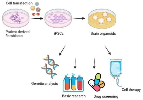
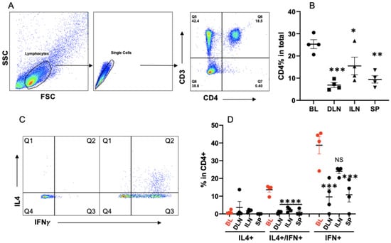
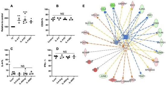
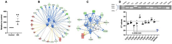
Integrin α4β7+ T cells perpetuate tissue injury in chronic inflammatory diseases, yet their role in hepatic fibrosis progression remains poorly understood. Here, we report increased accumulation of α4β7+ T cells in the liver of people with cirrhosis relative to disease controls. Similarly, hepatic fibrosis in the established mouse model of CCl4-induced liver fibrosis was associated with enrichment of intrahepatic α4β7+ CD4 and CD8 T cells. Monoclonal antibody (mAb)-mediated blockade of α4β7 or its ligand mucosal addressin cell adhesion molecule (MAdCAM)-1 attenuated hepatic inflammation and prevented fibrosis progression in CCl4-treated mice. Improvement in liver fibrosis was associated with a significant decrease in the infiltration of α4β7+ CD4 and CD8 T cells, suggesting that α4β7/MAdCAM-1 axis regulates both CD4 and CD8 T cell recruitment to the fibrotic liver, and α4β7+ T cells promote hepatic fibrosis progression. Analysis of hepatic α4β7+ and α4β7- CD4 T cells revealed that α4β7+ CD4 T cells were enriched for markers of activation and proliferation, demonstrating an effector phenotype. The findings suggest that α4β7+ T cells play a critical role in promoting hepatic fibrosis progression, and mAb-mediated blockade of α4β7 or MAdCAM-1 represents a promising therapeutic strategy for slowing hepatic fibrosis progression in chronic liver diseases.
Full article
(This article belongs to the Topic Molecular and Cellular Mechanisms of Diseases: Liver Diseases)


