


Cells 2024, 13(9), 761; https://doi.org/10.3390/cells13090761 - 29 Apr 2024
Viewed by 190
Abstract
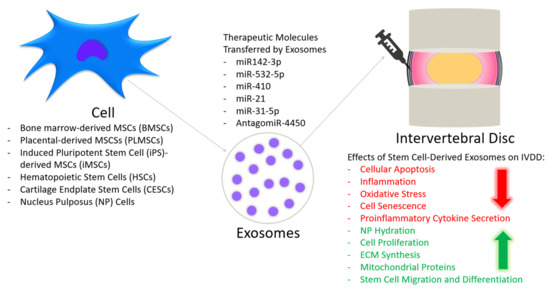
Spinal fusion, a common surgery performed for degenerative lumbar conditions, often uses recombinant human bone morphogenetic protein 2 (rhBMP-2) that is associated with adverse effects. Mesenchymal stromal/stem cells (MSCs) and their extracellular vesicles (EVs), particularly exosomes, have demonstrated efficacy in bone and cartilage
[...] Read more.
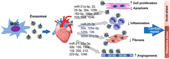
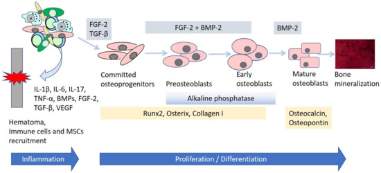
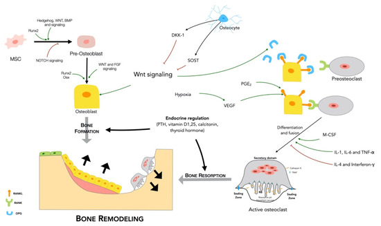
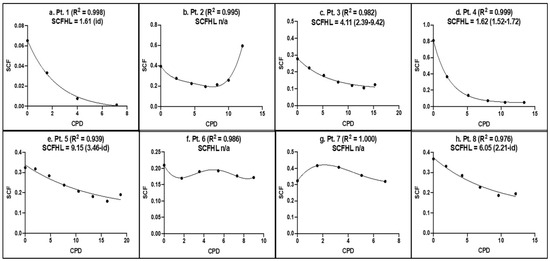
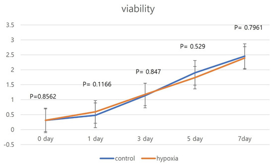
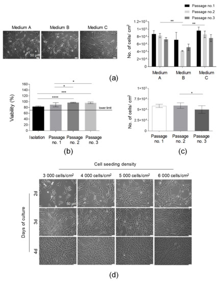
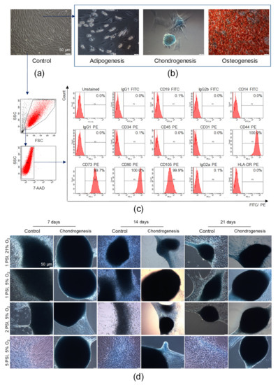
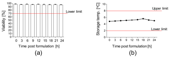
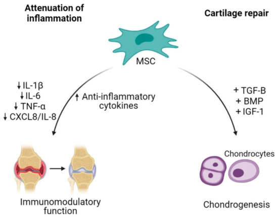
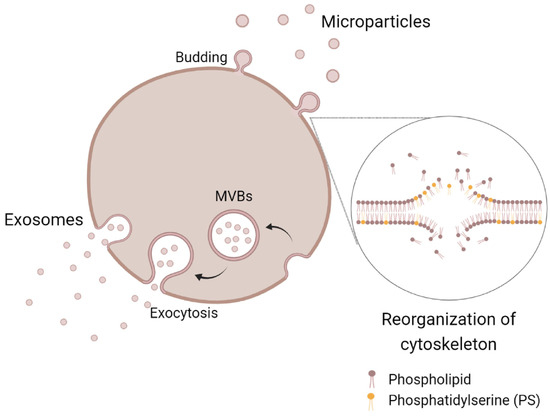
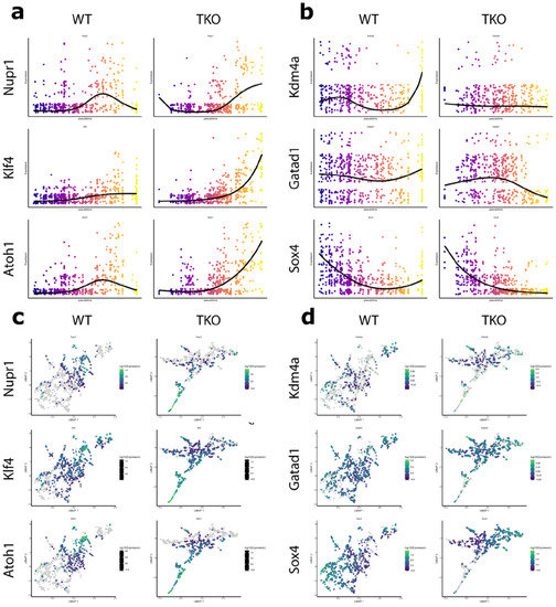
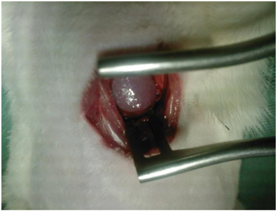
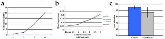
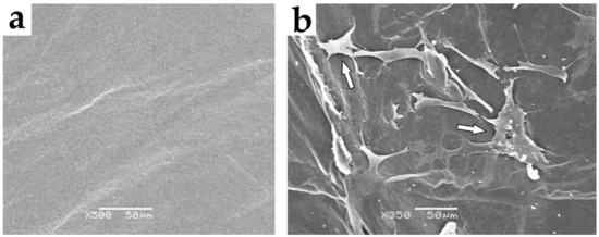
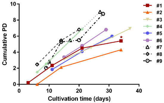
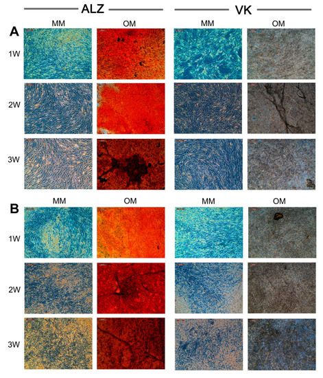
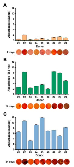
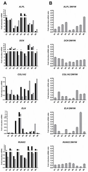
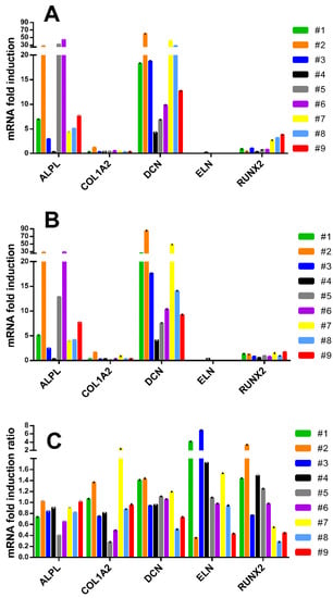
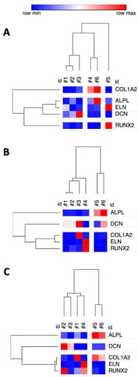
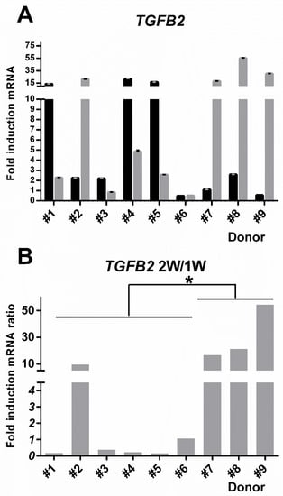
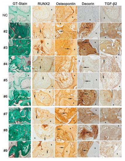
Spinal fusion, a common surgery performed for degenerative lumbar conditions, often uses recombinant human bone morphogenetic protein 2 (rhBMP-2) that is associated with adverse effects. Mesenchymal stromal/stem cells (MSCs) and their extracellular vesicles (EVs), particularly exosomes, have demonstrated efficacy in bone and cartilage repair. However, the efficacy of MSC exosomes in spinal fusion remains to be ascertained. This study investigates the fusion efficacy of MSC exosomes delivered via an absorbable collagen sponge packed in a poly Ɛ-caprolactone tricalcium phosphate (PCL–TCP) scaffold in a rat posterolateral spinal fusion model. Herein, it is shown that a single implantation of exosome-supplemented collagen sponge packed in PCL–TCP scaffold enhanced spinal fusion and improved mechanical stability by inducing bone formation and bridging between the transverse processes, as evidenced by significant improvements in fusion score and rate, bone structural parameters, histology, stiffness, and range of motion. This study demonstrates for the first time that MSC exosomes promote bone formation to enhance spinal fusion and mechanical stability in a rat model, supporting its translational potential for application in spinal fusion.
Full article

