Next Issue
Volume 12, April-2
Previous Issue
Volume 12, March-2


Cells, Volume 12, Issue 7 (April-1 2023) – 129 articles

Cover Story (view full-size image): 
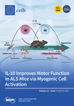
ALS is the most common adult motor neuron disease with a poor prognosis, unmet therapeutic needs, and high healthcare costs. The current strategies aimed at protecting motor neurons have failed to counteract irreversible muscular atrophy. Recent research has shown the pivotal role of macrophages in driving skeletal muscle regeneration. We investigated whether modulating macrophage muscle response and enhancing satellite cell differentiation could counteract muscle dysfunction in ALS mice. The results showed that IL-10 intramuscular administration improved motor performance by delaying muscle atrophy and motor neuron loss. These findings suggest a promising therapeutic approach to targeting muscle pathology in ALS. View this paper
  • Issues are regarded as officially published after their release is announced to the table of contents alert mailing list.
  • You may sign up for e-mail alerts to receive table of contents of newly released issues.
  • PDF is the official format for papers published in both, html and pdf forms. To view the papers in pdf format, click on the "PDF Full-text" link, and use the free Adobe Reader to open them.
Order results
Result details
Section
Select all
Export citation of selected articles as:
15 pages, 3266 KiB  
Article
DPP8 Selective Inhibitor Tominostat as a Novel and Broad-Spectrum Anticancer Agent against Hematological Malignancies
by Shohei Kikuchi, Akinori Wada, Yusuke Kamihara, Kosuke Okazaki, Paras Jawaid, Mati Ur Rehman, Eiji Kobayashi, Takeshi Susukida, Tomoki Minemura, Yoshimi Nabe, Noriaki Iwao, Tatsuhiko Ozawa, Ryo Hatano, Mitsugu Yamada, Hiroyuki Kishi, Yuji Matsuya, Mineyuki Mizuguchi, Yoshihiro Hayakawa, Nam H. Dang, Yasumitsu Sakamoto, Chikao Morimoto and Tsutomu Satoadd Show full author list remove Hide full author list
Cells 2023, 12(7), 1100; https://doi.org/10.3390/cells12071100 - 06 Apr 2023
Viewed by 2310
Abstract
DPP8/9 inhibition induces either pyroptotic or apoptotic cell death in hematological malignancies. We previously reported that treatment with the DPP8/9 inhibitor 1G244 resulted in apoptotic cell death in myeloma, and our current study further evaluates the mechanism of action of 1G244 in different [...] Read more.
DPP8/9 inhibition induces either pyroptotic or apoptotic cell death in hematological malignancies. We previously reported that treatment with the DPP8/9 inhibitor 1G244 resulted in apoptotic cell death in myeloma, and our current study further evaluates the mechanism of action of 1G244 in different blood cancer cell lines. Specifically, 1G244 inhibited DPP9 to induce GSDMD-mediated-pyroptosis at low concentrations and inhibited DPP8 to cause caspase-3-mediated-apoptosis at high concentrations. HCK expression is necessary to induce susceptibility to pyroptosis but does not participate in the induction of apoptosis. To further characterize this DPP8-dependent broad-spectrum apoptosis induction effect, we evaluated the potential antineoplastic role for an analog of 1G244 with higher DPP8 selectivity, tominostat (also known as 12 m). In vitro studies demonstrated that the cytotoxic effect of 1G244 at high concentrations was enhanced in tominostat. Meanwhile, in vivo work showed tominostat exhibited antitumor activity that was more effective on a cell line sensitive to 1G244, and at higher doses, it was also effective on a cell line resistant to 1G244. Importantly, the weight loss morbidity associated with increasing doses of 1G244 was not observed with tominostat. These results suggest the possible development of novel drugs with antineoplastic activity against selected hematological malignancies by refining and increasing the DPP8 selectivity of tominostat. Full article
Show Figures

Figure 1

15 pages, 4255 KiB  
Article
The αMSH-Dependent PI3K Pathway Supports Energy Metabolism, via Glucose Uptake, in Melanoma Cells
by Giorgia Cardinali, Daniela Kovacs, Sarah Mosca, Barbara Bellei, Enrica Flori, Aldo Morrone, Anna Maria Mileo and Vittoria Maresca
Cells 2023, 12(7), 1099; https://doi.org/10.3390/cells12071099 - 06 Apr 2023
Cited by 1 | Viewed by 1520
Abstract
Stimulation of melanocytes and murine melanoma cells with αMSH plus the PI3K inhibitor LY294002 resulted in ROS increase, oxidative DNA damage, and pigment retention. We performed cellular and molecular biology assays (Western blot, FACS, immunofluorescence analysis, scratch assay) on murine and human melanoma [...] Read more.
Stimulation of melanocytes and murine melanoma cells with αMSH plus the PI3K inhibitor LY294002 resulted in ROS increase, oxidative DNA damage, and pigment retention. We performed cellular and molecular biology assays (Western blot, FACS, immunofluorescence analysis, scratch assay) on murine and human melanoma cells. Treatment with αMSH plus LY294002 altered cortical actin architecture. Given that cytoskeleton integrity requires energy, we next evaluated ATP levels and we observed a drop in ATP after exposure to αMSH plus LY294002. To evaluate if the αMSH-activated PI3K pathway could modulate energy metabolism, we focused on glucose uptake by analyzing the expression of the Glut-1 glucose translocator. Compared with cells treated with αMSH alone, those exposed to combined treatment showed a reduction of Glut-1 on the plasma membrane. This metabolic alteration was associated with changes in mitochondrial mass. A significant decrease of the cell migratory potential was also observed. We demonstrated that the αMSH-dependent PI3K pathway acts as a regulator of energy metabolism via glucose uptake, influencing the actin cytoskeleton, which is involved in melanosome release and cell motility. Hence, these results could constitute the basis for innovative therapeutical strategies. Full article
(This article belongs to the Special Issue Melanoma: From Molecular Mechanisms to Therapeutic Opportunities)
Show Figures

Figure 1

19 pages, 13668 KiB  
Article
Glucagon Promotes Gluconeogenesis through the GCGR/PKA/CREB/PGC-1α Pathway in Hepatocytes of the Japanese Flounder Paralichthys olivaceus
by Mengxi Yang, Mingzhu Pan, Dong Huang, Jiahuan Liu, Yanlin Guo, Yue Liu and Wenbing Zhang
Cells 2023, 12(7), 1098; https://doi.org/10.3390/cells12071098 - 06 Apr 2023
Cited by 3 | Viewed by 1991
Abstract
In order to investigate the mechanism of glucagon regulation of gluconeogenesis, primary hepatocytes of the Japanese flounder (Paralichthys olivaceus) were incubated with synthesized glucagon, and methods based on inhibitors and gene overexpression were employed. The results indicated that glucagon promoted glucose [...] Read more.
In order to investigate the mechanism of glucagon regulation of gluconeogenesis, primary hepatocytes of the Japanese flounder (Paralichthys olivaceus) were incubated with synthesized glucagon, and methods based on inhibitors and gene overexpression were employed. The results indicated that glucagon promoted glucose production and increased the mRNA levels of glucagon receptor (gcgr), guanine nucleotide-binding protein Gs α subunit (gnas), adenylate cyclase 2 (adcy2), protein kinase A (pka), cAMP response element-binding protein 1 (creb1), peroxisome proliferator-activated receptor-γ coactivator 1α (pgc-1α), phosphoenolpyruvate carboxykinase 1 (pck1), and glucose-6-phosphatase (g6pc) in the hepatocytes. An inhibitor of GCGR decreased the mRNA expression of gcgr, gnas, adcy2, pka, creb1, pgc-1α, pck1, g6pc, the protein expression of phosphorylated CREB and PGC-1α, and glucose production. The overexpression of gcgr caused the opposite results. An inhibitor of PKA decreased the mRNA expression of pgc-1α, pck1, g6pc, the protein expression of phosphorylated-CREB, and glucose production in hepatocytes. A CREB-targeted inhibitor significantly decreased the stimulation by glucagon of the mRNA expression of creb1, pgc-1α, and gluconeogenic genes, and glucose production decreased accordingly. After incubating the hepatocytes with an inhibitor of PGC-1α, the glucagon-activated mRNA expression of pck1 and g6pc was significantly down-regulated. Together, these results demonstrate that glucagon promotes gluconeogenesis through the GCGR/PKA/CREB/PGC-1α pathway in the Japanese flounder. Full article
(This article belongs to the Section Cell Signaling)
Show Figures

Figure 1

16 pages, 6006 KiB  
Brief Report
Crystal Structure of a Classical MHC Class I Molecule in Dogs; Comparison of DLA-88*0 and DLA-88*5 Category Molecules
by Yujiao Sun, Lizhen Ma, Shen Li, Yawen Wang, Ruiqi Xiao, Junqi Yang, Johannes M. Dijkstra and Chun Xia
Cells 2023, 12(7), 1097; https://doi.org/10.3390/cells12071097 - 06 Apr 2023
Cited by 1 | Viewed by 1384
Abstract
DLA-88 is a classical major histocompatibility complex (MHC) class I gene in dogs, and allelic DLA-88 molecules have been divided into two categories named “DLA-88*0” and “DLA-88*5.” The defining difference between the two categories concerns an LQW motif in the α2 domain helical [...] Read more.
DLA-88 is a classical major histocompatibility complex (MHC) class I gene in dogs, and allelic DLA-88 molecules have been divided into two categories named “DLA-88*0” and “DLA-88*5.” The defining difference between the two categories concerns an LQW motif in the α2 domain helical region of the DLA-88*5 molecules that includes the insertion of an extra amino acid compared to MHC class I consensus length. We here show that this motif has been exchanged by recombination between different DLA-88 evolutionary lineages. Previously, with pDLA-88*508:01, the structure of a molecule of the DLA-88*5 category was elucidated. The present study is the first to elucidate a structure, using X-ray crystallography, of the DLA-88*0 category, namely DLA-88*001:04 complexed with β2m and a nonamer peptide derived from canine distemper virus (CDV). The LQW motif that distinguishes DLA-88*5 from DLA-88*0 causes a shallower peptide binding groove (PBG) and a leucine exposed at the top of the α2 domain helix expected to affect T cell selection. Peptide ligand amino acid substitution and pMHC-I complex formation and stability analyses revealed that P2 and P3 are the major anchor residue positions for binding to DLA-88*001:04. We speculate that the distribution pattern of the LQW motif among canine classical MHC class I alleles represents a strategy to enhance allogeneic rejection by T cells of transmissible cancers such as canine transmissible venereal tumor (CTVT). Full article
(This article belongs to the Special Issue Major Histocompatibility Complex (MHC) in Health and Disease 2022)
Show Figures

Figure 1

21 pages, 10188 KiB  
Article
Comprehensive Analysis of BrHMPs Reveals Potential Roles in Abiotic Stress Tolerance and Pollen–Stigma Interaction in Brassica rapa
by Lin Yang, Xiaoyu Wu, Shangjia Liu, Lina Zhang, Ting Li, Yunyun Cao and Qiaohong Duan
Cells 2023, 12(7), 1096; https://doi.org/10.3390/cells12071096 - 06 Apr 2023
Cited by 1 | Viewed by 1333
Abstract
Heavy metal-associated proteins (HMPs) participate in heavy metal detoxification. Although HMPs have been identified in several plants, no studies to date have identified the HMPs in Brassica rapa (B. rapa). Here, we identified 85 potential HMPs in B. rapa by bioinformatic [...] Read more.
Heavy metal-associated proteins (HMPs) participate in heavy metal detoxification. Although HMPs have been identified in several plants, no studies to date have identified the HMPs in Brassica rapa (B. rapa). Here, we identified 85 potential HMPs in B. rapa by bioinformatic methods. The promoters of the identified genes contain many elements associated with stress responses, including response to abscisic acid, low-temperature, and methyl jasmonate. The expression levels of BrHMP14, BrHMP16, BrHMP32, BrHMP41, and BrHMP42 were upregulated under Cu2+, Cd2+, Zn2+, and Pb2+ stresses. BrHMP06, BrHMP30, and BrHMP41 were also significantly upregulated after drought treatment. The transcripts of BrHMP06 and BrHMP11 increased mostly under cold stress. After applying salt stress, the expression of BrHMP02, BrHMP16, and BrHMP78 was induced. We observed increased BrHMP36 expression during the self-incompatibility (SI) response and decreased expression in the compatible pollination (CP) response during pollen–stigma interactions. These changes in expression suggest functions for these genes in HMPs include participating in heavy metal transport, detoxification, and response to abiotic stresses, with the potential for functions in sexual reproduction. We found potential co-functional partners of these key players by protein–protein interaction (PPI) analysis and found that some of the predicted protein partners are known to be involved in corresponding stress responses. Finally, phosphorylation investigation revealed many phosphorylation sites in BrHMPs, suggesting post-translational modification may occur during the BrHMP-mediated stress response. This comprehensive analysis provides important clues for the study of the molecular mechanisms of BrHMP genes in B. rapa, especially for abiotic stress and pollen–stigma interactions. Full article
(This article belongs to the Section Plant, Algae and Fungi Cell Biology)
Show Figures

Figure 1

3 pages, 1266 KiB  
Correction
Correction: Peng et al. Distinct Dominant Lineage from In Vitro Expanded Adipose-Derived Stem Cells (ASCs) Exhibits Enhanced Wound Healing Properties. Cells 2022, 11, 1236
by Qiuyue Peng, Guoqiang Ren, Zongzhe Xuan, Martyna Duda, Cristian Pablo Pennisi, Simone Riis Porsborg, Trine Fink and Vladimir Zachar
Cells 2023, 12(7), 1095; https://doi.org/10.3390/cells12071095 - 06 Apr 2023
Viewed by 780
Abstract
In the original publication [...] Full article
Show Figures

Figure 4

20 pages, 6679 KiB  
Article
Early Alterations of RNA Binding Protein (RBP) Homeostasis and ER Stress-Mediated Autophagy Contributes to Progressive Retinal Degeneration in the rd10 Mouse Model of Retinitis Pigmentosa (RP)
by Alfred Yamoah, Priyanka Tripathi, Haihong Guo, Leonie Scheve, Peter Walter, Sandra Johnen, Frank Müller, Joachim Weis and Anand Goswami
Cells 2023, 12(7), 1094; https://doi.org/10.3390/cells12071094 - 06 Apr 2023
Cited by 5 | Viewed by 1973
Abstract
The retinal degeneration 10 (rd10) mouse model is widely used to study retinitis pigmentosa (RP) pathomechanisms. It offers a rather unique opportunity to study trans-neuronal degeneration because the cell populations in question are separated anatomically and the mutated Pde6b gene is [...] Read more.
The retinal degeneration 10 (rd10) mouse model is widely used to study retinitis pigmentosa (RP) pathomechanisms. It offers a rather unique opportunity to study trans-neuronal degeneration because the cell populations in question are separated anatomically and the mutated Pde6b gene is selectively expressed in rod photoreceptors. We hypothesized that RNA binding protein (RBP) aggregation and abnormal autophagy might serve as early pathogenic events, damaging non-photoreceptor retinal cell types that are not primarily targeted by the Pde6b gene defect. We used a combination of immunohistochemistry (DAB, immunofluorescence), electron microscopy (EM), subcellular fractionation, and Western blot analysis on the retinal preparations obtained from both rd10 and wild-type mice. We found early, robust increases in levels of the protective endoplasmic reticulum (ER) calcium (Ca2+) buffering chaperone Sigma receptor 1 (SigR1) together with other ER-Ca2+ buffering proteins in both photoreceptors and non-photoreceptor neuronal cells before any noticeable photoreceptor degeneration. In line with this, we found markedly altered expression of the autophagy proteins p62 and LC3, together with abnormal ER widening and large autophagic vacuoles as detected by EM. Interestingly, these changes were accompanied by early, prominent cytoplasmic and nuclear aggregation of the key RBPs including pTDP-43 and FET family RBPs and stress granule formation. We conclude that progressive neurodegeneration in the rd10 mouse retina is associated with early disturbances of proteostasis and autophagy, along with abnormal cytoplasmic RBP aggregation. Full article
(This article belongs to the Special Issue Model Organisms to Study Autophagy)
Show Figures

Figure 1

17 pages, 2796 KiB  
Article
Integrated Placental Modelling of Histology with Gene Expression to Identify Functional Impact on Fetal Growth
by Hannah Ee Juen Yong, Katarzyna Maksym, Muhammad Ashraf Bin Yusoff, Esteban Salazar-Petres, Tatiana Nazarenko, Alexey Zaikin, Anna L. David, Sara L. Hillman and Amanda N. Sferruzzi-Perri
Cells 2023, 12(7), 1093; https://doi.org/10.3390/cells12071093 - 06 Apr 2023
Cited by 2 | Viewed by 1823
Abstract
Fetal growth restriction (FGR) is a leading cause of perinatal morbidity and mortality. Altered placental formation and functional capacity are major contributors to FGR pathogenesis. Relating placental structure to function across the placenta in healthy and FGR pregnancies remains largely unexplored but could [...] Read more.
Fetal growth restriction (FGR) is a leading cause of perinatal morbidity and mortality. Altered placental formation and functional capacity are major contributors to FGR pathogenesis. Relating placental structure to function across the placenta in healthy and FGR pregnancies remains largely unexplored but could improve understanding of placental diseases. We investigated integration of these parameters spatially in the term human placenta using predictive modelling. Systematic sampling was able to overcome heterogeneity in placental morphological and molecular features. Defects in villous development, elevated fibrosis, and reduced expression of growth and functional marker genes (IGF2, VEGA, SLC38A1, and SLC2A3) were seen in age-matched term FGR versus healthy control placentas. Characteristic histopathological changes with specific accompanying molecular signatures could be integrated through computational modelling to predict if the placenta came from a healthy or FGR pregnancy. Our findings yield new insights into the spatial relationship between placental structure and function and the etiology of FGR. Full article
(This article belongs to the Special Issue Molecular and Cellular Mechanisms of Fetal Growth and Preterm Birth)
Show Figures

Figure 1

24 pages, 4083 KiB  
Article
Lung Inflammation Induced by Inactivated SARS-CoV-2 in C57BL/6 Female Mice Is Controlled by Intranasal Instillation of Vitamin D
by William Danilo Fernandes de Souza, Sofia Fernanda Gonçalves Zorzella-Pezavento, Marina Caçador Ayupe, Caio Loureiro Salgado, Bernardo de Castro Oliveira, Francielly Moreira, Guilherme William da Silva, Stefanie Primon Muraro, Gabriela Fabiano de Souza, José Luiz Proença-Módena, Joao Pessoa Araujo Junior, Denise Morais da Fonseca and Alexandrina Sartori
Cells 2023, 12(7), 1092; https://doi.org/10.3390/cells12071092 - 06 Apr 2023
Viewed by 2135
Abstract
The COVID-19 pandemic was triggered by the coronavirus SARS-CoV-2, whose peak occurred in the years 2020 and 2021. The main target of this virus is the lung, and the infection is associated with an accentuated inflammatory process involving mainly the innate arm of [...] Read more.
The COVID-19 pandemic was triggered by the coronavirus SARS-CoV-2, whose peak occurred in the years 2020 and 2021. The main target of this virus is the lung, and the infection is associated with an accentuated inflammatory process involving mainly the innate arm of the immune system. Here, we described the induction of a pulmonary inflammatory process triggered by the intranasal (IN) instillation of UV-inactivated SARS-CoV-2 in C57BL/6 female mice, and then the evaluation of the ability of vitamin D (VitD) to control this process. The assays used to estimate the severity of lung involvement included the total and differential number of cells in the bronchoalveolar lavage fluid (BALF), histopathological analysis, quantification of T cell subsets, and inflammatory mediators by RT-PCR, cytokine quantification in lung homogenates, and flow cytometric analysis of cells recovered from lung parenchyma. The IN instillation of inactivated SARS-CoV-2 triggered a pulmonary inflammatory process, consisting of various cell types and mediators, resembling the typical inflammation found in transgenic mice infected with SARS-CoV-2. This inflammatory process was significantly decreased by the IN delivery of VitD, but not by its IP administration, suggesting that this hormone could have a therapeutic potential in COVID-19 if locally applied. To our knowledge, the local delivery of VitD to downmodulate lung inflammation in COVID-19 is an original proposition. Full article
(This article belongs to the Topic Inflammation: The Cause of All Diseases)
Show Figures

Figure 1

0 pages, 454 KiB  
Review
Asthma: The Use of Animal Models and Their Translational Utility
by Jane Seymour Woodrow, M. Katie Sheats, Bethanie Cooper and Rosemary Bayless
Cells 2023, 12(7), 1091; https://doi.org/10.3390/cells12071091 - 05 Apr 2023
Cited by 15 | Viewed by 4351
Abstract
Asthma is characterized by chronic lower airway inflammation that results in airway remodeling, which can lead to a permanent decrease in lung function. The pathophysiology driving the development of asthma is complex and heterogenous. Animal models have been and continue to be essential [...] Read more.
Asthma is characterized by chronic lower airway inflammation that results in airway remodeling, which can lead to a permanent decrease in lung function. The pathophysiology driving the development of asthma is complex and heterogenous. Animal models have been and continue to be essential for the discovery of molecular pathways driving the pathophysiology of asthma and novel therapeutic approaches. Animal models of asthma may be induced or naturally occurring. Species used to study asthma include mouse, rat, guinea pig, cat, dog, sheep, horse, and nonhuman primate. Some of the aspects to consider when evaluating any of these asthma models are cost, labor, reagent availability, regulatory burden, relevance to natural disease in humans, type of lower airway inflammation, biological samples available for testing, and ultimately whether the model can answer the research question(s). This review aims to discuss the animal models most available for asthma investigation, with an emphasis on describing the inciting antigen/allergen, inflammatory response induced, and its translation to human asthma. Full article
(This article belongs to the Special Issue Asthma: Pathogenesis, Diagnosis, and Treatment)
14 pages, 1050 KiB  
Review
Preclinical Large Animal Porcine Models for Cardiac Regeneration and Its Clinical Translation: Role of hiPSC-Derived Cardiomyocytes
by Divya Sridharan, Nooruddin Pracha, Schaza Javed Rana, Salmman Ahmed, Anam J. Dewani, Syed Baseeruddin Alvi, Muhamad Mergaye, Uzair Ahmed and Mahmood Khan
Cells 2023, 12(7), 1090; https://doi.org/10.3390/cells12071090 - 05 Apr 2023
Cited by 3 | Viewed by 2088
Abstract
Myocardial Infarction (MI) occurs due to a blockage in the coronary artery resulting in ischemia and necrosis of cardiomyocytes in the left ventricular heart muscle. The dying cardiac tissue is replaced with fibrous scar tissue, causing a decrease in myocardial contractility and thus [...] Read more.
Myocardial Infarction (MI) occurs due to a blockage in the coronary artery resulting in ischemia and necrosis of cardiomyocytes in the left ventricular heart muscle. The dying cardiac tissue is replaced with fibrous scar tissue, causing a decrease in myocardial contractility and thus affecting the functional capacity of the myocardium. Treatments, such as stent placements, cardiac bypasses, or transplants are beneficial but with many limitations, and may decrease the overall life expectancy due to related complications. In recent years, with the advent of human induced pluripotent stem cells (hiPSCs), newer avenues using cell-based approaches for the treatment of MI have emerged as a potential for cardiac regeneration. While hiPSCs and their derived differentiated cells are promising candidates, their translatability for clinical applications has been hindered due to poor preclinical reproducibility. Various preclinical animal models for MI, ranging from mice to non-human primates, have been adopted in cardiovascular research to mimic MI in humans. Therefore, a comprehensive literature review was essential to elucidate the factors affecting the reproducibility and translatability of large animal models. In this review article, we have discussed different animal models available for studying stem-cell transplantation in cardiovascular applications, mainly focusing on the highly translatable porcine MI model. Full article
Show Figures

Figure 1

13 pages, 2303 KiB  
Article
High-Throughput Microscopy Analysis of Mitochondrial Membrane Potential in 2D and 3D Models
by Caterina Vianello, Federica Dal Bello, Sang Hun Shin, Sara Schiavon, Camilla Bean, Ana Paula Magalhães Rebelo, Tomáš Knedlík, Emad Norouzi Esfahani, Veronica Costiniti, Rodrigo S. Lacruz, Giuseppina Covello, Fabio Munari, Tommaso Scolaro, Antonella Viola, Elena Rampazzo, Luca Persano, Sara Zumerle, Luca Scorrano, Alessio Gianelle and Marta Giacomello
Cells 2023, 12(7), 1089; https://doi.org/10.3390/cells12071089 - 05 Apr 2023
Cited by 3 | Viewed by 3787
Abstract
Recent proteomic, metabolomic, and transcriptomic studies have highlighted a connection between changes in mitochondria physiology and cellular pathophysiological mechanisms. Secondary assays to assess the function of these organelles appear fundamental to validate these -omics findings. Although mitochondrial membrane potential is widely recognized as [...] Read more.
Recent proteomic, metabolomic, and transcriptomic studies have highlighted a connection between changes in mitochondria physiology and cellular pathophysiological mechanisms. Secondary assays to assess the function of these organelles appear fundamental to validate these -omics findings. Although mitochondrial membrane potential is widely recognized as an indicator of mitochondrial activity, high-content imaging-based approaches coupled to multiparametric to measure it have not been established yet. In this paper, we describe a methodology for the unbiased high-throughput quantification of mitochondrial membrane potential in vitro, which is suitable for 2D to 3D models. We successfully used our method to analyze mitochondrial membrane potential in monolayers of human fibroblasts, neural stem cells, spheroids, and isolated muscle fibers. Moreover, by combining automated image analysis and machine learning, we were able to discriminate melanoma cells from macrophages in co-culture and to analyze the subpopulations separately. Our data demonstrated that our method is a widely applicable strategy for large-scale profiling of mitochondrial activity. Full article
(This article belongs to the Special Issue Mitochondria: New Findings from Single Cells to Organs)
Show Figures

Figure 1

24 pages, 1503 KiB  
Review
High Mobility Group Box 1 (HMGB1): Potential Target in Sepsis-Associated Encephalopathy
by Bram DeWulf, Laurens Minsart, Franck Verdonk, Véronique Kruys, Michael Piagnerelli, Mervyn Maze and Sarah Saxena
Cells 2023, 12(7), 1088; https://doi.org/10.3390/cells12071088 - 04 Apr 2023
Cited by 3 | Viewed by 2043
Abstract
Sepsis-associated encephalopathy (SAE) remains a challenge for intensivists that is exacerbated by lack of an effective diagnostic tool and an unambiguous definition to properly identify SAE patients. Risk factors for SAE development include age, genetic factors as well as pre-existing neuropsychiatric conditions. Sepsis [...] Read more.
Sepsis-associated encephalopathy (SAE) remains a challenge for intensivists that is exacerbated by lack of an effective diagnostic tool and an unambiguous definition to properly identify SAE patients. Risk factors for SAE development include age, genetic factors as well as pre-existing neuropsychiatric conditions. Sepsis due to certain infection sites/origins might be more prone to encephalopathy development than other cases. Currently, ICU management of SAE is mainly based on non-pharmacological support. Pre-clinical studies have described the role of the alarmin high mobility group box 1 (HMGB1) in the complex pathogenesis of SAE. Although there are limited data available about the role of HMGB1 in neuroinflammation following sepsis, it has been implicated in other neurologic disorders, where its translocation from the nucleus to the extracellular space has been found to trigger neuroinflammatory reactions and disrupt the blood–brain barrier. Negating the inflammatory cascade, by targeting HMGB1, may be a strategy to complement non-pharmacologic interventions directed against encephalopathy. This review describes inflammatory cascades implicating HMGB1 and strategies for its use to mitigate sepsis-induced encephalopathy. Full article
(This article belongs to the Special Issue High Mobility Group Box-1 (HMGB1) in a Neuroimmune Crosstalk)
Show Figures

Figure 1

29 pages, 1063 KiB  
Review
Maternal Inflammation with Elevated Kynurenine Metabolites Is Related to the Risk of Abnormal Brain Development and Behavioral Changes in Autism Spectrum Disorder
by Yuki Murakami, Yukio Imamura, Yoshiyuki Kasahara, Chihiro Yoshida, Yuta Momono, Ke Fang, Daisuke Sakai, Yukuo Konishi and Toshimasa Nishiyama
Cells 2023, 12(7), 1087; https://doi.org/10.3390/cells12071087 - 04 Apr 2023
Cited by 4 | Viewed by 2091
Abstract
Several studies show that genetic and environmental factors contribute to the onset and progression of neurodevelopmental disorders. Maternal immune activation (MIA) during gestation is considered one of the major environmental factors driving this process. The kynurenine pathway (KP) is a major route of [...] Read more.
Several studies show that genetic and environmental factors contribute to the onset and progression of neurodevelopmental disorders. Maternal immune activation (MIA) during gestation is considered one of the major environmental factors driving this process. The kynurenine pathway (KP) is a major route of the essential amino acid L-tryptophan (Trp) catabolism in mammalian cells. Activation of the KP following neuro-inflammation can generate various endogenous neuroactive metabolites that may impact brain functions and behaviors. Additionally, neurotoxic metabolites and excitotoxicity cause long-term changes in the trophic support, glutamatergic system, and synaptic function following KP activation. Therefore, investigating the role of KP metabolites during neurodevelopment will likely promote further understanding of additional pathophysiology of neurodevelopmental disorders, including autism spectrum disorder (ASD). In this review, we describe the changes in KP metabolism in the brain during pregnancy and represent how maternal inflammation and genetic factors influence the KP during development. We overview the patients with ASD clinical data and animal models designed to verify the role of perinatal KP elevation in long-lasting biochemical, neuropathological, and behavioral deficits later in life. Our review will help shed light on new therapeutic strategies and interventions targeting the KP for neurodevelopmental disorders. Full article
(This article belongs to the Special Issue Kynurenine Pathway in Health and Disease)
Show Figures

Figure 1

12 pages, 3312 KiB  
Communication
Copy Number Variation in Inflammatory Breast Cancer
by Aditi Hazra, Andrea O’Hara, Kornelia Polyak, Faina Nakhlis, Beth T. Harrison, Antonio Giordano, Beth Overmoyer and Filipa Lynce
Cells 2023, 12(7), 1086; https://doi.org/10.3390/cells12071086 - 04 Apr 2023
Cited by 1 | Viewed by 1980
Abstract
Identification of a unique genomic biomarker in de novo inflammatory breast cancer (IBC) may provide an insight into the biology of this aggressive disease. The goal of our study was to elucidate biomarkers associated with IBC. We examined breast biopsies collected from Dana–Farber [...] Read more.
Identification of a unique genomic biomarker in de novo inflammatory breast cancer (IBC) may provide an insight into the biology of this aggressive disease. The goal of our study was to elucidate biomarkers associated with IBC. We examined breast biopsies collected from Dana–Farber Cancer Institute patients with IBC prior to initiating preoperative systemic treatment (30 samples were examined, of which 14 were eligible). Patients without available biopsies (n = 1), with insufficient tumor epithelial cells (n = 10), or insufficient DNA yield (n = 5) were excluded from the analysis. Molecular subtype and tumor grade were abstracted from a medical records’ review. Ten IBC tumors were estrogen-receptor-positive (ER+) and human epidermal growth factor receptor 2 (HER2)-negative (n = 10 out of 14). Sufficient RNA and DNA were simultaneously extracted from 14 biopsy specimens using the Qiagen AllPrep Kit. RNA was amplified using the Sensation kit and profiled using the Affymetrix Human Transcriptome Array 2.0. DNA was profiled for genome-wide copy number variation (CNV) using the Affymetrix OncoScan Array and analyzed using the Nexus Chromosome Analysis Suite. Among the 14 eligible samples, we first confirmed biological concordance and quality control metrics using replicates and gene expression data. Second, we examined CNVs and gene expression change by IBC subtype. We identified significant CNVs in IBC patients after adjusting for multiple comparisons. Next, to assess whether the CNVs were unique to IBC, we compared the IBC CNV data to fresh-frozen non-IBC CNV data from The Cancer Genome Atlas (n = 388). On chromosome 7p11.2, we identified significant CN gain located at position 58,019,983-58,025,423 in 8 ER+ IBC samples compared to 338 non-IBC ER+ samples (region length: 5440 bp gain and 69,039 bp, False Discovery Rate (FDR) p-value = 3.12 × 10⁻¹⁰) and at position 57,950,944–58,025,423 in 3 TN-IBC samples compared to 50 non-IBC TN samples (74,479 base pair, gain, FDR p-value = 4.27 × 10⁻⁵; near the EGFR gene). We also observed significant CN loss on chromosome 21, located at position 9,648,315–9,764,385 (p-value = 4.27 × 10⁻⁵). Secondarily, differential gene expression in IBC patients with 7p11.2 CN gain compared to SUM149 were explored after FDR correction for multiple testing (p-value = 0.0016), but the results should be interpreted with caution due to the small sample size. Finally, the data presented are hypothesis-generating. Validation of CNVs that contribute to the unique presentation and biological features associated with IBC in larger datasets may lead to the optimization of treatment strategies. Full article
Show Figures

Figure 1

17 pages, 15192 KiB  
Article
Quadra-Stable Dynamics of p53 and PTEN in the DNA Damage Response
by Shantanu Gupta, Pritam Kumar Panda, Daner A. Silveira, Rajeev Ahuja and Ronaldo F. Hashimoto
Cells 2023, 12(7), 1085; https://doi.org/10.3390/cells12071085 - 04 Apr 2023
Cited by 5 | Viewed by 2457
Abstract
Cell fate determination is a complex process that is frequently described as cells traveling on rugged pathways, beginning with DNA damage response (DDR). Tumor protein p53 (p53) and phosphatase and tensin homolog (PTEN) are two critical players in this process. Although both of [...] Read more.
Cell fate determination is a complex process that is frequently described as cells traveling on rugged pathways, beginning with DNA damage response (DDR). Tumor protein p53 (p53) and phosphatase and tensin homolog (PTEN) are two critical players in this process. Although both of these proteins are known to be key cell fate regulators, the exact mechanism by which they collaborate in the DDR remains unknown. Thus, we propose a dynamic Boolean network. Our model incorporates experimental data obtained from NSCLC cells and is the first of its kind. Our network’s wild-type system shows that DDR activates the G2/M checkpoint, and this triggers a cascade of events, involving p53 and PTEN, that ultimately lead to the four potential phenotypes: cell cycle arrest, senescence, autophagy, and apoptosis (quadra-stable dynamics). The network predictions correspond with the gain-and-loss of function investigations in the additional two cell lines (HeLa and MCF-7). Our findings imply that p53 and PTEN act as molecular switches that activate or deactivate specific pathways to govern cell fate decisions. Thus, our network facilitates the direct investigation of quadruplicate cell fate decisions in DDR. Therefore, we concluded that concurrently controlling PTEN and p53 dynamics may be a viable strategy for enhancing clinical outcomes. Full article
(This article belongs to the Special Issue Systems Biology of Apoptotic and Non-apoptotic Signaling)
Show Figures

Figure 1

12 pages, 8146 KiB  
Article
Identification of Neurocan and Phosphacan as Early Biomarkers for Open Neural Tube Defects
by Karolina Janik, George M. Smith and Barbara Krynska
Cells 2023, 12(7), 1084; https://doi.org/10.3390/cells12071084 - 04 Apr 2023
Cited by 1 | Viewed by 1385
Abstract
Open neural tube defects (NTDs) such as myelomeningocele (MMC) are debilitating and the most common congenital defects of the central nervous system. Despite their apparent clinical importance, the existing early prenatal diagnostic options for these defects remain limited. Using a well-accepted retinoic-acid-induced model [...] Read more.
Open neural tube defects (NTDs) such as myelomeningocele (MMC) are debilitating and the most common congenital defects of the central nervous system. Despite their apparent clinical importance, the existing early prenatal diagnostic options for these defects remain limited. Using a well-accepted retinoic-acid-induced model of MMC established in fetal rats, we discovered that neurocan and phosphacan, the secreted chondroitin sulfate proteoglycans of the developing nervous system, are released into the amniotic fluid (AF) of fetal rats displaying spinal cord defects. In contrast to normal controls, elevated AF levels of neurocan and phosphacan were detected in MMC fetuses early in gestation and continued to increase during MMC progression, reaching the highest level in near-term fetuses. The molecular forms of neurocan and phosphacan identified in the AF of MMC fetuses and those found in MMC spinal cords were qualitatively similar. In summary, this is the first report demonstrating the presence of neurocan and phosphacan in the AF of MMC fetuses. The identification of elevated levels of neurocan and phosphacan in the AF of MMC fetuses provides two prospective biomarkers with the potential for early prenatal diagnosis of open NTDs. Full article
(This article belongs to the Collection Cell Biology of Spinal Cord Injury and Repair)
Show Figures

Figure 1

22 pages, 14529 KiB  
Article
α-Synuclein Preformed Fibrils Bind to β-Neurexins and Impair β-Neurexin-Mediated Presynaptic Organization
by Benjamin Feller, Aurélie Fallon, Wen Luo, Phuong Trang Nguyen, Irina Shlaifer, Alfred Kihoon Lee, Nicolas Chofflet, Nayoung Yi, Husam Khaled, Samer Karkout, Steve Bourgault, Thomas M. Durcan and Hideto Takahashi
Cells 2023, 12(7), 1083; https://doi.org/10.3390/cells12071083 - 04 Apr 2023
Cited by 4 | Viewed by 1873
Abstract
Synucleinopathies form a group of neurodegenerative diseases defined by the misfolding and aggregation of α-synuclein (α-syn). Abnormal accumulation and spreading of α-syn aggregates lead to synapse dysfunction and neuronal cell death. Yet, little is known about the synaptic mechanisms underlying the α-syn pathology. [...] Read more.
Synucleinopathies form a group of neurodegenerative diseases defined by the misfolding and aggregation of α-synuclein (α-syn). Abnormal accumulation and spreading of α-syn aggregates lead to synapse dysfunction and neuronal cell death. Yet, little is known about the synaptic mechanisms underlying the α-syn pathology. Here we identified β-isoforms of neurexins (β-NRXs) as presynaptic organizing proteins that interact with α-syn preformed fibrils (α-syn PFFs), toxic α-syn aggregates, but not α-syn monomers. Our cell surface protein binding assays and surface plasmon resonance assays reveal that α-syn PFFs bind directly to β-NRXs through their N-terminal histidine-rich domain (HRD) at the nanomolar range (KD: ~500 nM monomer equivalent). Furthermore, our artificial synapse formation assays show that α-syn PFFs diminish excitatory and inhibitory presynaptic organization induced by a specific isoform of neuroligin 1 that binds only β-NRXs, but not α-isoforms of neurexins. Thus, our data suggest that α-syn PFFs interact with β-NRXs to inhibit β-NRX-mediated presynaptic organization, providing novel molecular insight into how α-syn PFFs induce synaptic pathology in synucleinopathies such as Parkinson’s disease and dementia with Lewy bodies. Full article
Show Figures

Figure 1

10 pages, 3660 KiB  
Communication
Lectin Receptor-like Kinase Signaling during Engineered Ectomycorrhiza Colonization
by Him Shrestha, Tao Yao, Zhenzhen Qiao, Wellington Muchero, Robert L. Hettich, Jin-Gui Chen and Paul E. Abraham
Cells 2023, 12(7), 1082; https://doi.org/10.3390/cells12071082 - 04 Apr 2023
Cited by 3 | Viewed by 1899
Abstract
Mutualistic association can improve a plant’s health and productivity. G-type lectin receptor-like kinase (PtLecRLK1) is a susceptibility factor in Populus trichocarpa that permits root colonization by a beneficial fungus, Laccaria bicolor. Engineering PtLecRLK1 also permits L. bicolor root colonization in non-host plants [...] Read more.
Mutualistic association can improve a plant’s health and productivity. G-type lectin receptor-like kinase (PtLecRLK1) is a susceptibility factor in Populus trichocarpa that permits root colonization by a beneficial fungus, Laccaria bicolor. Engineering PtLecRLK1 also permits L. bicolor root colonization in non-host plants similar to Populus trichocarpa. The intracellular signaling reprogramed by PtLecRLK1 upon recognition of L. bicolor to allow for the development and maintenance of symbiosis is yet to be determined. In this study, phosphoproteomics was utilized to identify phosphorylation-based relevant signaling pathways associated with PtLecRLK1 recognition of L. bicolor in transgenic switchgrass roots. Our finding shows that PtLecRLK1 in transgenic plants modifies the chitin-triggered plant defense and MAPK signaling along with a significant adjustment in phytohormone signaling, ROS balance, endocytosis, cytoskeleton movement, and proteasomal degradation in order to facilitate the establishment and maintenance of L. bicolor colonization. Moreover, protein–protein interaction data implicate a cGMP-dependent protein kinase as a potential substrate of PtLecRLK1. Full article
(This article belongs to the Section Plant, Algae and Fungi Cell Biology)
Show Figures

Figure 1

24 pages, 2720 KiB  
Review
Insights on the Biomarker Potential of Exosomal Non-Coding RNAs in Colorectal Cancer: An In Silico Characterization of Related Exosomal lncRNA/circRNA–miRNA–Target Axis
by Maria Mezher, Samira Abdallah, Ohanes Ashekyan, Ayman Al Shoukari, Hayat Choubassy, Abdallah Kurdi, Sally Temraz and Rihab Nasr
Cells 2023, 12(7), 1081; https://doi.org/10.3390/cells12071081 - 04 Apr 2023
Cited by 4 | Viewed by 2727
Abstract
Colorectal cancer (CRC) is one of the most common cancer types, ranking third after lung and breast cancers. As such, it demands special attention for better characterization, which may eventually result in the development of early detection strategies and preventive measures. Currently, components [...] Read more.
Colorectal cancer (CRC) is one of the most common cancer types, ranking third after lung and breast cancers. As such, it demands special attention for better characterization, which may eventually result in the development of early detection strategies and preventive measures. Currently, components of bodily fluids, which may reflect various disease states, are being increasingly researched for their biomarker potential. One of these components is the circulating extracellular vesicles, namely, exosomes, which are demonstrated to carry various cargo. Of importance, the non-coding RNA cargo of circulating exosomes, especially long non-coding RNAs (lncRNAs), circular RNAs (circRNAs), and micro RNAs (miRNAs), may potentially serve as significant diagnostic and prognostic/predictive biomarkers. In this review, we present existing evidence on the diagnostic and prognostic/predictive biomarker value of exosomal non-coding RNAs in CRC. In addition, taking advantage of the miRNA sponging functionality of lncRNAs and circRNAs, we demonstrate an experimentally validated CRC exosomal non-coding RNA-regulated target gene axis benefiting from published miRNA sponging studies in CRC. Hence, we present a set of target genes and pathways downstream of the lncRNA/circRNA–miRNA–target axis along with associated significant Gene Ontology (GO) terms and Kyoto Encyclopedia of Genes and Genomes (KEGG) pathways, which may collectively serve to better characterize CRC and shed light on the significance of exosomal non-coding RNAs in CRC diagnosis and prognosis/prediction. Full article
Show Figures

Figure 1

18 pages, 5326 KiB  
Article
Photobiomodulation at 660 nm Stimulates In Vitro Diabetic Wound Healing via the Ras/MAPK Pathway
by Patricia Kasowanjete, Heidi Abrahamse and Nicolette N. Houreld
Cells 2023, 12(7), 1080; https://doi.org/10.3390/cells12071080 - 04 Apr 2023
Cited by 4 | Viewed by 1431
Abstract
Diabetic foot ulcers (DFUs) are open chronic wounds that affect diabetic patients due to hyperglycaemia. DFUs are known for their poor response to treatment and frequently require amputation, which may result in premature death. The present study evaluated the effect of photobiomodulation (PBM) [...] Read more.
Diabetic foot ulcers (DFUs) are open chronic wounds that affect diabetic patients due to hyperglycaemia. DFUs are known for their poor response to treatment and frequently require amputation, which may result in premature death. The present study evaluated the effect of photobiomodulation (PBM) at 660 nm on wound healing via activation of Ras/MAPK signalling in diabetic wounded cells in vitro. This study used four human skin fibroblast cell (WS1) models, namely normal (N), wounded (W), diabetic (D), and diabetic wounded (DW). Cells were irradiated at 660 nm with 5 J/cm2. Non-irradiated cells (0 J/cm2) served as controls. Cells were incubated for 24 and 48 h post-irradiation, and the effect of PBM on cellular morphology and migration rate, viability, and proliferation was assessed. Basic fibroblast growth factor (bFGF), its phosphorylated (activated) receptor FGFR, and phosphorylated target proteins (Ras, MEK1/2 and MAPK) were determined by enzyme-linked immunosorbent assay (ELISA) and Western blotting; nuclear translocation of p-MAPK was determined by immunofluorescence. PBM resulted in an increase in bFGF and a subsequent increase in FGFR activation. There was also an increase in downstream proteins, p-Ras, p-MEK1/2 and p-MAPK. PBM at 660 nm led to increased viability, proliferation, and migration as a result of increased bFGF and subsequent activation of the Ras/MAPK signalling pathway. Therefore, this study can conclude that PBM at 660 nm stimulates in vitro diabetic wound healing via the bFGF-activated Ras/MAPK pathway. Full article
(This article belongs to the Topic Cell Signaling Pathways)
Show Figures

Figure 1

18 pages, 40486 KiB  
Article
KDEL Receptor Trafficking to the Plasma Membrane Is Regulated by ACBD3 and Rab4A-GTP
by Chuanting Tan, Yulei Du, Lianhui Zhu, Shuaiyang Jing, Jingkai Gao, Yi Qian, Xihua Yue and Intaek Lee
Cells 2023, 12(7), 1079; https://doi.org/10.3390/cells12071079 - 04 Apr 2023
Viewed by 1624
Abstract
KDEL receptor-1 maintains homeostasis in the early secretory pathway by capturing and retrieving ER chaperones to the ER during heavy secretory activity. Unexpectedly, a fraction of the receptor is also known to reside in the plasma membrane (PM), although it is largely unknown [...] Read more.
KDEL receptor-1 maintains homeostasis in the early secretory pathway by capturing and retrieving ER chaperones to the ER during heavy secretory activity. Unexpectedly, a fraction of the receptor is also known to reside in the plasma membrane (PM), although it is largely unknown exactly how the KDEL receptor gets exported from the Golgi and travels to the PM. We have previously shown that a Golgi scaffolding protein (ACBD3) facilitates KDEL receptor localization at the Golgi via the regulating cargo wave-induced cAMP/PKA-dependent signaling pathway. Upon endocytosis, surface-expressed KDEL receptor undergoes highly complex itineraries through the Golgi and the endo-lysosomal compartments, where the endocytosed receptor utilizes Rab14A- and Rab11A-positive recycling endosomes and clathrin-decorated tubulovesicular carriers. In this study, we sought to investigate the mechanism through which the KDEL receptor gets exported from the Golgi en route to the PM. We report here that ACBD3 depletion results in greatly increased trafficking of KDEL receptor to the PM via Rab4A-positive tubular carriers emanating from the Golgi. Expression of constitutively activated Rab4A mutant (Q72L) increases the surface expression of KDEL receptor up to 2~3-fold, whereas Rab4A knockdown or the expression of GDP-locked Rab4A mutant (S27N) inhibits KDEL receptor targeting of the PM. Importantly, KDELR trafficking from the Golgi to the PM is independent of PKA- and Src kinase-mediated mechanisms. Taken together, these results reveal that ACBD3 and Rab4A play a key role in regulating KDEL receptor trafficking to the cell surface. Full article
(This article belongs to the Section Intracellular and Plasma Membranes)
Show Figures

Figure 1

17 pages, 4817 KiB  
Article
Development of Cyclic Peptides Targeting the Epidermal Growth Factor Receptor in Mesenchymal Triple-Negative Breast Cancer Subtype
by Nancy Nisticò, Annamaria Aloisio, Antonio Lupia, Anna Maria Zimbo, Selena Mimmi, Domenico Maisano, Rossella Russo, Fabiola Marino, Mariangela Scalise, Emanuela Chiarella, Teresa Mancuso, Giuseppe Fiume, Daniela Omodei, Antonella Zannetti, Giuliana Salvatore, Ileana Quinto and Enrico Iaccino
Cells 2023, 12(7), 1078; https://doi.org/10.3390/cells12071078 - 03 Apr 2023
Cited by 2 | Viewed by 2286
Abstract
Triple-negative breast cancer (TNBC) is an aggressive malignancy characterized by the lack of expression of estrogen and progesterone receptors and amplification of human epidermal growth factor receptor 2 (HER2). Being the Epidermal Growth Factor Receptor (EGFR) highly expressed in mesenchymal TNBC and correlated [...] Read more.
Triple-negative breast cancer (TNBC) is an aggressive malignancy characterized by the lack of expression of estrogen and progesterone receptors and amplification of human epidermal growth factor receptor 2 (HER2). Being the Epidermal Growth Factor Receptor (EGFR) highly expressed in mesenchymal TNBC and correlated with aggressive growth behavior, it represents an ideal target for anticancer drugs. Here, we have applied the phage display for selecting two highly specific peptide ligands for targeting the EGFR overexpressed in MDA-MB-231 cells, a human TNBC cell line. Molecular docking predicted the peptide-binding affinities and sites in the extracellular domain of EGFR. The binding of the FITC-conjugated peptides to human and murine TNBC cells was validated by flow cytometry. Confocal microscopy confirmed the peptide binding specificity to EGFR-positive MDA-MB-231 tumor xenograft tissues and their co-localization with the membrane EGFR. Further, the peptide stimulation did not affect the cell cycle of TNBC cells, which is of interest for their utility for tumor targeting. Our data indicate that these novel peptides are highly specific ligands for the EGFR overexpressed in TNBC cells, and thus they could be used in conjugation with nanoparticles for tumor-targeted delivery of anticancer drugs. Full article
(This article belongs to the Special Issue Advances in Cellular and Molecular Research in Breast Cancer)
Show Figures

Figure 1

18 pages, 5212 KiB  
Article
New Findings: Hindlimb Unloading Causes Nucleocytoplasmic Ca2+ Overload and DNA Damage in Skeletal Muscle
by Huajian Yang, Huiping Wang, Fangyang Pan, Yuxi Guo, Liqi Cao, Wenjing Yan and Yunfang Gao
Cells 2023, 12(7), 1077; https://doi.org/10.3390/cells12071077 - 03 Apr 2023
Cited by 1 | Viewed by 1879
Abstract
Disuse atrophy of skeletal muscle is associated with a severe imbalance in cellular Ca2+ homeostasis and marked increase in nuclear apoptosis. Nuclear Ca2+ is involved in the regulation of cellular Ca2+ homeostasis. However, it remains unclear whether nuclear Ca2+ [...] Read more.
Disuse atrophy of skeletal muscle is associated with a severe imbalance in cellular Ca2+ homeostasis and marked increase in nuclear apoptosis. Nuclear Ca2+ is involved in the regulation of cellular Ca2+ homeostasis. However, it remains unclear whether nuclear Ca2+ levels change under skeletal muscle disuse conditions, and whether changes in nuclear Ca2+ levels are associated with nuclear apoptosis. In this study, changes in Ca2+ levels, Ca2+ transporters, and regulatory factors in the nucleus of hindlimb unloaded rat soleus muscle were examined to investigate the effects of disuse on nuclear Ca2+ homeostasis and apoptosis. Results showed that, after hindlimb unloading, the nuclear envelope Ca2+ levels ([Ca2+]NE) and nucleocytoplasmic Ca2+ levels ([Ca2+]NC) increased by 78% (p < 0.01) and 106% (p < 0.01), respectively. The levels of Ca2+-ATPase type 2 (Ca2+-ATPase2), Ryanodine receptor 1 (RyR1), Inositol 1,4,5-tetrakisphosphate receptor 1 (IP3R1), Cyclic ADP ribose hydrolase (CD38) and Inositol 1,4,5-tetrakisphosphate (IP3) increased by 470% (p < 0.001), 94% (p < 0.05), 170% (p < 0.001), 640% (p < 0.001) and 12% (p < 0.05), respectively, and the levels of Na+/Ca2+ exchanger 3 (NCX3), Ca2+/calmodulin dependent protein kinase II (CaMK II) and Protein kinase A (PKA) decreased by 54% (p < 0.001), 33% (p < 0.05) and 5% (p > 0.05), respectively. In addition, DNase X is mainly localized in the myonucleus and its activity is elevated after hindlimb unloading. Overall, our results suggest that enhanced Ca2+ uptake from cytoplasm is involved in the increase in [Ca2+]NE after hindlimb unloading. Moreover, the increase in [Ca2+]NC is attributed to increased Ca2+ release into nucleocytoplasm and weakened Ca2+ uptake from nucleocytoplasm. DNase X is activated due to elevated [Ca2+]NC, leading to DNA fragmentation in myonucleus, ultimately initiating myonuclear apoptosis. Nucleocytoplasmic Ca2+ overload may contribute to the increased incidence of myonuclear apoptosis in disused skeletal muscle. Full article
Show Figures

Figure 1

19 pages, 28034 KiB  
Article
Effect of Commonly Used Cosmetic Preservatives on Healthy Human Skin Cells
by Patrycja Głaz, Agata Rosińska, Sylwia Woźniak, Anna Boguszewska-Czubara, Anna Biernasiuk and Dariusz Matosiuk
Cells 2023, 12(7), 1076; https://doi.org/10.3390/cells12071076 - 03 Apr 2023
Cited by 5 | Viewed by 3765
Abstract
Cosmetic products contain preservatives to prevent microbial growth. The various types of preservatives present in skincare products applied on the skin induce many side effects. We tested several types of preservatives such as phenoxyethanol, methyl paraben, propyl paraben, imidazolidinyl urea (IU), the composition [...] Read more.
Cosmetic products contain preservatives to prevent microbial growth. The various types of preservatives present in skincare products applied on the skin induce many side effects. We tested several types of preservatives such as phenoxyethanol, methyl paraben, propyl paraben, imidazolidinyl urea (IU), the composition of gluconolactone and sodium benzoate (GSB), diazolidinyl urea (DU), and two grapefruit essential oils, one of which was industrially produced and a second which was freshly distilled from fresh grapefruit peels. This study aimed to find the relationship between preservative concentration, cell growth, collagen secretion, and cell viability. We hypothesized that these products induced a decrease in collagen secretion from human dermal fibroblasts. Our research, for the first time, addressed the overall effect of other preservatives on skin extracellular matrix (ECM) by studying their effect on metalloproteinase-2 (MMP-2) activity. Except for cytotoxicity and contact sensitivity tests, there are no studies of their effect on skin ECM in the available literature. These studies show potential antimicrobial activity, especially from the compounds IU and DU towards reference bacteria and the compounds methyl paraben and propyl paraben against reference fungi. The MTS test showed that fibroblasts are more sensitive to the tested group of preservatives than keratinocytes, which could be caused by the differences between the cells’ structures. The grapefruit oils exhibited the most cytotoxicity to both tested cell lines compared to all considered preservatives. The most destructive influence of preservatives on collagen synthesis was observed in the case of IU and DU. In this case, the homemade grapefruit oil turned out to be the mildest one. The results from a diverse group of preservatives show that whether they are natural or synthesized compounds, they require controlled use. Appropriate dosages and evaluation of preservative efficacy should not be the only aspects considered. The complex effect of preservatives on skin processes and cytotoxicity is an important topic for modern people. Full article
(This article belongs to the Special Issue Cellular Mechanisms of Skin Diseases)
Show Figures

Figure 1

14 pages, 1705 KiB  
Review
HAT1: Landscape of Biological Function and Role in Cancer
by Vincenza Capone, Laura Della Torre, Daniela Carannante, Mehrad Babaei, Lucia Altucci, Rosaria Benedetti and Vincenzo Carafa
Cells 2023, 12(7), 1075; https://doi.org/10.3390/cells12071075 - 02 Apr 2023
Cited by 1 | Viewed by 2213
Abstract
Histone modifications, as key chromatin regulators, play a pivotal role in the pathogenesis of several diseases, such as cancer. Acetylation, and more specifically lysine acetylation, is a reversible epigenetic process with a fundamental role in cell life, able to target histone and non-histone [...] Read more.
Histone modifications, as key chromatin regulators, play a pivotal role in the pathogenesis of several diseases, such as cancer. Acetylation, and more specifically lysine acetylation, is a reversible epigenetic process with a fundamental role in cell life, able to target histone and non-histone proteins. This epigenetic modification regulates transcriptional processes and protein activity, stability, and localization. Several studies highlight a specific role for HAT1 in regulating molecular pathways, which are altered in several pathologies, among which is cancer. HAT1 is the first histone acetyltransferase discovered; however, to date, its biological characterization is still unclear. In this review, we summarize and update the current knowledge about the biological function of this acetyltransferase, highlighting recent advances of HAT1 in the pathogenesis of cancer. Full article
(This article belongs to the Special Issue Cell Biology: State-of-the-Art and Perspectives in Italy)
Show Figures

Figure 1

17 pages, 1302 KiB  
Article
A Deep-Learning-Computed Cancer Score for the Identification of Human Hepatocellular Carcinoma Area Based on a Six-Colour Multiplex Immunofluorescence Panel
by Axel Dievernich, Johannes Stegmaier, Pascal Achenbach, Svetlana Warkentin, Till Braunschweig, Ulf Peter Neumann and Uwe Klinge
Cells 2023, 12(7), 1074; https://doi.org/10.3390/cells12071074 - 02 Apr 2023
Cited by 1 | Viewed by 3066
Abstract
Liver cancer is one of the most frequently diagnosed and fatal cancers worldwide, with hepatocellular carcinoma (HCC) being the most common primary liver cancer. Hundreds of studies involving thousands of patients have now been analysed across different cancer types, including HCC, regarding the [...] Read more.
Liver cancer is one of the most frequently diagnosed and fatal cancers worldwide, with hepatocellular carcinoma (HCC) being the most common primary liver cancer. Hundreds of studies involving thousands of patients have now been analysed across different cancer types, including HCC, regarding the effects of immune infiltrates on the prognosis of cancer patients. However, for these analyses, an unambiguous delineation of the cancer area is paramount, which is difficult due to the strong heterogeneity and considerable inter-operator variability induced by qualitative visual assessment and manual assignment. Nowadays, however, multiplex analyses allow the simultaneous evaluation of multiple protein markers, which, in conjunction with recent machine learning approaches, may offer great potential for the objective, enhanced identification of cancer areas with further in situ analysis of prognostic immune parameters. In this study, we, therefore, used an exemplary five-marker multiplex immunofluorescence panel of commonly studied markers for prognosis (CD3 T, CD4 T helper, CD8 cytotoxic T, FoxP3 regulatory T, and PD-L1) and DAPI to assess which analytical approach is best suited to combine morphological and immunohistochemical data into a cancer score to identify the cancer area that best matches an independent pathologist’s assignment. For each cell, a total of 68 individual cell features were determined, which were used as input for 4 different approaches for computing a cancer score: a correlation-based selection of individual cell features, a MANOVA-based selection of features, a multilayer perceptron, and a convolutional neural network (a U-net). Accuracy was used to evaluate performance. With a mean accuracy of 75%, the U-net was best capable of identifying the cancer area. Although individual cell features showed a strong heterogeneity between patients, the spatial representations obtained with the computed cancer scores delineate HCC well from non-cancer liver tissues. Future analyses with larger sample sizes will help to improve the model and enable direct, in-depth investigations of prognostic parameters, ultimately enabling precision medicine. Full article
Show Figures

Graphical abstract

17 pages, 2277 KiB  
Article
The Ability of Airborne Microalgae and Cyanobacteria to Survive and Transfer the Carcinogenic Benzo(a)pyrene in Coastal Regions
by Kinga A. Wiśniewska, Anita U. Lewandowska, Sylwia Śliwińska-Wilczewska, Marta Staniszewska and Gracjana Budzałek
Cells 2023, 12(7), 1073; https://doi.org/10.3390/cells12071073 - 02 Apr 2023
Cited by 2 | Viewed by 1575
Abstract
Air pollution has been a significant problem threatening human health for years. One commonly reported air pollutant is benzo(a)pyrene, a dangerous compound with carcinogenic properties. Values which exceed normative values for benzo(a)pyrene concentration in the air are often noted in many regions of [...] Read more.
Air pollution has been a significant problem threatening human health for years. One commonly reported air pollutant is benzo(a)pyrene, a dangerous compound with carcinogenic properties. Values which exceed normative values for benzo(a)pyrene concentration in the air are often noted in many regions of the world. Studies on the worldwide spread of COVID-19 since 2020, as well as avian flu, measles, and SARS, have proven that viruses and bacteria are more dangerous to human health when they occur in polluted air. Regarding cyanobacteria and microalgae, little is known about their relationship with benzo(a)pyrene. The question is whether these microorganisms can pose a threat when present in poor quality air. We initially assessed whether cyanobacteria and microalgae isolated from the atmosphere are sensitive to changes in PAH concentrations and whether they can accumulate or degrade PAHs. The presence of B(a)P has significantly affected both the quantity of cyanobacteria and microalgae cells as well as their chlorophyll a (chl a) content and their ability to fluorescence. For many cyanobacteria and microalgae, an increase in cell numbers was observed after the addition of B(a)P. Therefore, even slight air pollution with benzo(a)pyrene is likely to facilitate the growth of airborne cyanobacteria and microalgae. The results provided an assessment of the organisms that are most susceptible to cellular stress following exposure to benzo(a)pyrene, as well as the potential consequences for the environment. Additionally, the results indicated that green algae have the greatest potential for degrading PAHs, making their use a promising bioremediation approach. Kirchneriella sp. demonstrated the highest average degradation of B(a)P, with the above-mentioned research indicating it can even degrade up to 80% of B(a)P. The other studied green algae exhibited a lower, yet still significant, B(a)P degradation rate exceeding 50% when compared to cyanobacteria and diatoms. Full article
(This article belongs to the Section Plant, Algae and Fungi Cell Biology)
Show Figures

Figure 1

12 pages, 2103 KiB  
Review
Impaired Mitochondrial Function in T-Lymphocytes as a Result of Exposure to HIV and ART
by Alexander V. Blagov, Vasily N. Sukhorukov, Shuzhen Guo, Dongwei Zhang, Mikhail A. Popov and Alexander N. Orekhov
Cells 2023, 12(7), 1072; https://doi.org/10.3390/cells12071072 - 02 Apr 2023
Cited by 2 | Viewed by 1504
Abstract
Mitochondrial dysfunction is a described phenomenon for a number of chronic and infectious diseases. At the same time, the question remains open: is this condition a consequence or a cause of the progression of the disease? In this review, we consider the role [...] Read more.
Mitochondrial dysfunction is a described phenomenon for a number of chronic and infectious diseases. At the same time, the question remains open: is this condition a consequence or a cause of the progression of the disease? In this review, we consider the role of the development of mitochondrial dysfunction in the progression of HIV (human immunodeficiency viruses) infection and the onset of AIDS (acquired immunodeficiency syndrome), as well as the direct impact of HIV on mitochondria. In addition, we will touch upon such an important issue as the effect of ART (Antiretroviral Therapy) drugs on mitochondria, since ART is currently the only effective way to curb the progression of HIV in infected patients, and because the identification of potential side effects can help to more consciously approach the development of new drugs in the treatment of HIV infection. Full article
(This article belongs to the Special Issue Mitochondria at the Crossroad of Health and Disease)
Show Figures

Figure 1

18 pages, 989 KiB  
Review
Ocular Delivery of Therapeutic Agents by Cell-Penetrating Peptides
by Nguyễn Thị Thanh Nhàn, Daniel E. Maidana and Kaori H. Yamada
Cells 2023, 12(7), 1071; https://doi.org/10.3390/cells12071071 - 01 Apr 2023
Cited by 8 | Viewed by 3932
Abstract
Cell-penetrating peptides (CPPs) are short peptides with the ability to translocate through the cell membrane to facilitate their cellular uptake. CPPs can be used as drug-delivery systems for molecules that are difficult to uptake. Ocular drug delivery is challenging due to the structural [...] Read more.
Cell-penetrating peptides (CPPs) are short peptides with the ability to translocate through the cell membrane to facilitate their cellular uptake. CPPs can be used as drug-delivery systems for molecules that are difficult to uptake. Ocular drug delivery is challenging due to the structural and physiological complexity of the eye. CPPs may be tailored to overcome this challenge, facilitating cellular uptake and delivery to the targeted area. Retinal diseases occur at the posterior pole of the eye; thus, intravitreal injections are needed to deliver drugs at an effective concentration in situ. However, frequent injections have risks of causing vision-threatening complications. Recent investigations have focused on developing long-acting drugs and drug delivery systems to reduce the frequency of injections. In fact, conjugation with CPP could deliver FDA-approved drugs to the back of the eye, as seen by topical application in animal models. This review summarizes recent advances in CPPs, protein/peptide-based drugs for eye diseases, and the use of CPPs for drug delivery based on systematic searches in PubMed and clinical trials. We highlight targeted therapies and explore the potential of CPPs and peptide-based drugs for eye diseases. Full article
Show Figures

Figure 1

Previous Issue
Next Issue
Back to TopTop