Next Issue
Volume 12, March-2
Previous Issue
Volume 12, February-2


Cells, Volume 12, Issue 5 (March-1 2023) – 147 articles

Cover Story (view full-size image): 
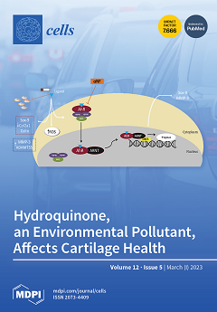
We know that pollution can negatively affect human and animal health; however, its impact on musculoskeletal health remains vastly unknown. Our study aimed to investigate whether hydroquinone (HQ), an environmental pollutant, could affect the homeostasis of articular cartilage. Our data showed that HQ could exacerbate the pro-degenerative effect of inflammatory molecules in the tissue, promoting the degradation and reducing the content of proteoglycans, and increasing oxidative stress. We showed that HQ mediates catabolic activity through the activation of the aryl hydrocarbon receptor. In summary, our findings demonstrate the harmful effects of HQ on articular cartilage health, showing how exposure to pollutants can favor the onset and/or sustain the progression of articular diseases. View this paper
  • Issues are regarded as officially published after their release is announced to the table of contents alert mailing list.
  • You may sign up for e-mail alerts to receive table of contents of newly released issues.
  • PDF is the official format for papers published in both, html and pdf forms. To view the papers in pdf format, click on the "PDF Full-text" link, and use the free Adobe Reader to open them.
Order results
Result details
Section
Select all
Export citation of selected articles as:
19 pages, 9482 KiB  
Article
Fermented Soybean Paste Attenuates Biogenic Amine-Induced Liver Damage in Obese Mice
by Ju-Hwan Yang, Eun-Hye Byeon, Dawon Kang, Seong-Geun Hong, Jinsung Yang, Deok-Ryong Kim, Seung-Pil Yun, Sang-Won Park, Hyun-Joon Kim, Jae-Won Huh, So-Yong Kim, Young-Wan Kim and Dong-Kun Lee
Cells 2023, 12(5), 822; https://doi.org/10.3390/cells12050822 - 06 Mar 2023
Cited by 4 | Viewed by 2038
Abstract
Biogenic amines are cellular components produced by the decarboxylation of amino acids; however, excessive biogenic amine production causes adverse health problems. The relationship between hepatic damage and biogenic amine levels in nonalcoholic fatty liver disease (NAFLD) remains unclear. In this study, mice were [...] Read more.
Biogenic amines are cellular components produced by the decarboxylation of amino acids; however, excessive biogenic amine production causes adverse health problems. The relationship between hepatic damage and biogenic amine levels in nonalcoholic fatty liver disease (NAFLD) remains unclear. In this study, mice were fed a high-fat diet (HFD) for 10 weeks to induce obesity, presenting early-stage of NAFLD. We administered histamine (20 mg/kg) + tyramine (100 mg/kg) via oral gavage for 6 days to mice with HFD-induced early-stage NAFLD. The results showed that combined histamine and tyramine administration increased cleaved PARP-1 and IL-1β in the liver, as well as MAO-A, total MAO, CRP, and AST/ALT levels. In contrast, the survival rate decreased in HFD-induced NAFLD mice. Treatment with manufactured or traditional fermented soybean paste decreased biogenically elevated hepatic cleaved PARP-1 and IL-1β expression and blood plasma MAO-A, CRP, and AST/ALT levels in HFD-induced NAFLD mice. Additionally, the biogenic amine-induced reduction in survival rate was alleviated by fermented soybean paste in HFD-induced NAFLD mice. These results show that biogenic amine-induced liver damage can be exacerbated by obesity and may adversely affect life conservation. However, fermented soybean paste can reduce biogenic amine-induced liver damage in NAFLD mice. These results suggest a beneficial effect of fermented soybean paste on biogenic amine-induced liver damage and provide a new research perspective on the relationship between biogenic amines and obesity. Full article
(This article belongs to the Special Issue Liver Diseases: From Molecular Mechanism to Therapeutic Aspect)
Show Figures

Figure 1

17 pages, 2019 KiB  
Article
Electrophysiological Activity of Primary Cortical Neuron-Glia Mixed Cultures
by Noah Goshi, Hyehyun Kim, Gregory Girardi, Alexander Gardner and Erkin Seker
Cells 2023, 12(5), 821; https://doi.org/10.3390/cells12050821 - 06 Mar 2023
Cited by 3 | Viewed by 2748
Abstract
Neuroinflammation plays a central role in many neurological disorders, ranging from traumatic brain injuries to neurodegeneration. Electrophysiological activity is an essential measure of neuronal function, which is influenced by neuroinflammation. In order to study neuroinflammation and its electrophysiological fingerprints, there is a need [...] Read more.
Neuroinflammation plays a central role in many neurological disorders, ranging from traumatic brain injuries to neurodegeneration. Electrophysiological activity is an essential measure of neuronal function, which is influenced by neuroinflammation. In order to study neuroinflammation and its electrophysiological fingerprints, there is a need for in vitro models that accurately capture the in vivo phenomena. In this study, we employed a new tri-culture of primary rat neurons, astrocytes, and microglia in combination with extracellular electrophysiological recording techniques using multiple electrode arrays (MEAs) to determine the effect of microglia on neural function and the response to neuroinflammatory stimuli. Specifically, we established the tri-culture and its corresponding neuron-astrocyte co-culture (lacking microglia) counterpart on custom MEAs and monitored their electrophysiological activity for 21 days to assess culture maturation and network formation. As a complementary assessment, we quantified synaptic puncta and averaged spike waveforms to determine the difference in excitatory to inhibitory neuron ratio (E/I ratio) of the neurons. The results demonstrate that the microglia in the tri-culture do not disrupt neural network formation and stability and may be a better representation of the in vivo rat cortex due to its more similar E/I ratio as compared to more traditional isolated neuron and neuron-astrocyte co-cultures. In addition, only the tri-culture displayed a significant decrease in both the number of active channels and spike frequency following pro-inflammatory lipopolysaccharide exposure, highlighting the critical role of microglia in capturing electrophysiological manifestations of a representative neuroinflammatory insult. We expect the demonstrated technology to assist in studying various brain disease mechanisms. Full article
Show Figures

Figure 1

14 pages, 2561 KiB  
Article
Increased Radiation Sensitivity in Patients with Phelan-McDermid Syndrome
by Sarah Jesse, Lukas Kuhlmann, Laura S. Hildebrand, Henriette Magelssen, Martina Schmaus, Beate Timmermann, Stephanie Andres, Rainer Fietkau and Luitpold V. Distel
Cells 2023, 12(5), 820; https://doi.org/10.3390/cells12050820 - 06 Mar 2023
Viewed by 1335
Abstract
Phelan-McDermid syndrome is an inherited global developmental disorder commonly associated with autism spectrum disorder. Due to a significantly increased radiosensitivity, measured before the start of radiotherapy of a rhabdoid tumor in a child with Phelan-McDermid syndrome, the question arose whether other patients with [...] Read more.
Phelan-McDermid syndrome is an inherited global developmental disorder commonly associated with autism spectrum disorder. Due to a significantly increased radiosensitivity, measured before the start of radiotherapy of a rhabdoid tumor in a child with Phelan-McDermid syndrome, the question arose whether other patients with this syndrome also have increased radiosensitivity. For this purpose, the radiation sensitivity of blood lymphocytes after irradiation with 2Gray was examined using the G0 three-color fluorescence in situ hybridization assay in a cohort of 20 patients with Phelan-McDermid syndrome from blood samples. The results were compared to healthy volunteers, breast cancer patients and rectal cancer patients. Independent of age and gender, all but two patients with Phelan-McDermid syndrome showed significantly increased radiosensitivity, with an average of 0.653 breaks per metaphase. These results correlated neither with the individual genetic findings nor with the individual clinical course, nor with the respective clinical severity of the disease. In our pilot study, we saw a significantly increased radiosensitivity in lymphocytes from patients with Phelan-McDermid syndrome, so pronounced that a dose reduction would be recommended if radiotherapy had to be performed. Ultimately, the question arises as to the interpretation of these data. There does not appear to be an increased risk of tumors in these patients, since tumors are rare overall. The question, therefore, arose as to whether our results could possibly be the basis for processes, such as aging/preaging, or, in this context, neurodegeneration. There are no data on this so far, but this issue should be pursued in further fundamentally based studies in order to better understand the pathophysiology of the syndrome. Full article
(This article belongs to the Special Issue Advances in Cancer Genomics)
Show Figures

Graphical abstract

7 pages, 1695 KiB  
Commentary
Molecular Regulation of Autophagy and Asymmetric Cell Division by Cancer Stem Cell Marker CD133
by Hideki Izumi, Yasuhiko Kaneko and Akira Nakagawara
Cells 2023, 12(5), 819; https://doi.org/10.3390/cells12050819 - 06 Mar 2023
Cited by 2 | Viewed by 1739
Abstract
CD133, also called prominin-1, is widely known as a cancer stem cell marker, and its high expression correlates with a poor prognosis in many cancers. CD133 was originally discovered as a plasma membranous protein in stem/progenitor cells. It is now known that Src [...] Read more.
CD133, also called prominin-1, is widely known as a cancer stem cell marker, and its high expression correlates with a poor prognosis in many cancers. CD133 was originally discovered as a plasma membranous protein in stem/progenitor cells. It is now known that Src family kinases phosphorylate the C-terminal of CD133. However, when Src kinase activity is low, CD133 is not phosphorylated by Src and is preferentially downregulated into cells through endocytosis. Endosomal CD133 then associates with HDAC6, thereby recruiting it to the centrosome via dynein motors. Thus, CD133 protein is now known to localize to the centrosome as endosomes as well as to the plasma membrane. More recently, a mechanism to explain the involvement of CD133 endosomes in asymmetric cell division was reported. Here, we would like to introduce the relationship between autophagy regulation and asymmetric cell division mediated by CD133 endosomes. Full article
(This article belongs to the Special Issue Cell Biology: State of the Art and Perspectives in Japan)
Show Figures

Figure 1

18 pages, 2635 KiB  
Article
Intermittent Lead Exposure Induces Behavioral and Cardiovascular Alterations Associated with Neuroinflammation
by Liana Shvachiy, Ângela Amaro-Leal, Tiago F. Outeiro, Isabel Rocha and Vera Geraldes
Cells 2023, 12(5), 818; https://doi.org/10.3390/cells12050818 - 06 Mar 2023
Cited by 5 | Viewed by 1711
Abstract
The nervous system is the primary target for lead exposure and the developing brain appears to be especially susceptible, namely the hippocampus. The mechanisms of lead neurotoxicity remain unclear, but microgliosis and astrogliosis are potential candidates, leading to an inflammatory cascade and interrupting [...] Read more.
The nervous system is the primary target for lead exposure and the developing brain appears to be especially susceptible, namely the hippocampus. The mechanisms of lead neurotoxicity remain unclear, but microgliosis and astrogliosis are potential candidates, leading to an inflammatory cascade and interrupting the pathways involved in hippocampal functions. Moreover, these molecular changes can be impactful as they may contribute to the pathophysiology of behavioral deficits and cardiovascular complications observed in chronic lead exposure. Nevertheless, the health effects and the underlying influence mechanism of intermittent lead exposure in the nervous and cardiovascular systems are still vague. Thus, we used a rat model of intermittent lead exposure to determine the systemic effects of lead and on microglial and astroglial activation in the hippocampal dentate gyrus throughout time. In this study, the intermittent group was exposed to lead from the fetal period until 12 weeks of age, no exposure (tap water) until 20 weeks, and a second exposure from 20 to 28 weeks of age. A control group (without lead exposure) matched in age and sex was used. At 12, 20 and 28 weeks of age, both groups were submitted to a physiological and behavioral evaluation. Behavioral tests were performed for the assessment of anxiety-like behavior and locomotor activity (open-field test), and memory (novel object recognition test). In the physiological evaluation, in an acute experiment, blood pressure, electrocardiogram, and heart and respiratory rates were recorded, and autonomic reflexes were evaluated. The expression of GFAP, Iba-1, NeuN and Synaptophysin in the hippocampal dentate gyrus was assessed. Intermittent lead exposure induced microgliosis and astrogliosis in the hippocampus of rats and changes in behavioral and cardiovascular function. We identified increases in GFAP and Iba1 markers together with presynaptic dysfunction in the hippocampus, concomitant with behavioral changes. This type of exposure produced significant long-term memory dysfunction. Regarding physiological changes, hypertension, tachypnea, baroreceptor reflex impairment and increased chemoreceptor reflex sensitivity were observed. In conclusion, the present study demonstrated the potential of lead intermittent exposure inducing reactive astrogliosis and microgliosis, along with a presynaptic loss that was accompanied by alterations of homeostatic mechanisms. This suggests that chronic neuroinflammation promoted by intermittent lead exposure since fetal period may increase the susceptibility to adverse events in individuals with pre-existing cardiovascular disease and/or in the elderly. Full article
Show Figures

Graphical abstract

19 pages, 2469 KiB  
Article
Nucleolin Regulates Pulmonary Artery Smooth Muscle Cell Proliferation under Hypoxia by Modulating miRNA Expression
by Jihui Lee and Hara Kang
Cells 2023, 12(5), 817; https://doi.org/10.3390/cells12050817 - 06 Mar 2023
Cited by 1 | Viewed by 1478
Abstract
Hypoxia induces the abnormal proliferation of vascular smooth muscle cells (VSMCs), resulting in the pathogenesis of various vascular diseases. RNA-binding proteins (RBPs) are involved in a wide range of biological processes, including cell proliferation and responses to hypoxia. In this study, we observed [...] Read more.
Hypoxia induces the abnormal proliferation of vascular smooth muscle cells (VSMCs), resulting in the pathogenesis of various vascular diseases. RNA-binding proteins (RBPs) are involved in a wide range of biological processes, including cell proliferation and responses to hypoxia. In this study, we observed that the RBP nucleolin (NCL) was downregulated by histone deacetylation in response to hypoxia. We evaluated its regulatory effects on miRNA expression under hypoxic conditions in pulmonary artery smooth muscle cells (PASMCs). miRNAs associated with NCL were assessed using RNA immunoprecipitation in PASMCs and small RNA sequencing. The expression of a set of miRNAs was increased by NCL but reduced by hypoxia-induced downregulation of NCL. The downregulation of miR-24-3p and miR-409-3p promoted PASMC proliferation under hypoxic conditions. These results clearly demonstrate the significance of NCL–miRNA interactions in the regulation of hypoxia-induced PASMC proliferation and provide insight into the therapeutic value of RBPs for vascular diseases. Full article
(This article belongs to the Section Cell Proliferation and Division)
Show Figures

Figure 1

24 pages, 2454 KiB  
Review
Pathogenesis Underlying Neurological Manifestations of Long COVID Syndrome and Potential Therapeutics
by Albert Leng, Manuj Shah, Syed Ameen Ahmad, Lavienraj Premraj, Karin Wildi, Gianluigi Li Bassi, Carlos A. Pardo, Alex Choi and Sung-Min Cho
Cells 2023, 12(5), 816; https://doi.org/10.3390/cells12050816 - 06 Mar 2023
Cited by 46 | Viewed by 10183
Abstract
The development of long-term symptoms of coronavirus disease 2019 (COVID-19) more than four weeks after primary infection, termed “long COVID” or post-acute sequela of COVID-19 (PASC), can implicate persistent neurological complications in up to one third of patients and present as fatigue, “brain [...] Read more.
The development of long-term symptoms of coronavirus disease 2019 (COVID-19) more than four weeks after primary infection, termed “long COVID” or post-acute sequela of COVID-19 (PASC), can implicate persistent neurological complications in up to one third of patients and present as fatigue, “brain fog”, headaches, cognitive impairment, dysautonomia, neuropsychiatric symptoms, anosmia, hypogeusia, and peripheral neuropathy. Pathogenic mechanisms of these symptoms of long COVID remain largely unclear; however, several hypotheses implicate both nervous system and systemic pathogenic mechanisms such as SARS-CoV2 viral persistence and neuroinvasion, abnormal immunological response, autoimmunity, coagulopathies, and endotheliopathy. Outside of the CNS, SARS-CoV-2 can invade the support and stem cells of the olfactory epithelium leading to persistent alterations to olfactory function. SARS-CoV-2 infection may induce abnormalities in innate and adaptive immunity including monocyte expansion, T-cell exhaustion, and prolonged cytokine release, which may cause neuroinflammatory responses and microglia activation, white matter abnormalities, and microvascular changes. Additionally, microvascular clot formation can occlude capillaries and endotheliopathy, due to SARS-CoV-2 protease activity and complement activation, can contribute to hypoxic neuronal injury and blood–brain barrier dysfunction, respectively. Current therapeutics target pathological mechanisms by employing antivirals, decreasing inflammation, and promoting olfactory epithelium regeneration. Thus, from laboratory evidence and clinical trials in the literature, we sought to synthesize the pathophysiological pathways underlying neurological symptoms of long COVID and potential therapeutics. Full article
(This article belongs to the Special Issue Insights into Molecular and Cellular Mechanisms of NeuroCOVID)
Show Figures

Figure 1

20 pages, 2358 KiB  
Systematic Review
The Role of Preservation Solutions upon Saphenous Vein Endothelial Integrity and Function: Systematic Review and UK Practice Survey
by Georgia R. Layton, Shameem S. Ladak, Riccardo Abbasciano, Liam W. McQueen, Sarah J. George, Gavin J. Murphy and Mustafa Zakkar
Cells 2023, 12(5), 815; https://doi.org/10.3390/cells12050815 - 06 Mar 2023
Cited by 1 | Viewed by 1519
Abstract
The long saphenous vein is the most used conduit in cardiac surgery, but its long-term patency is limited by vein graft disease (VGD). Endothelial dysfunction is a key driver of VGD; its aetiology is multi-factorial. However emerging evidence identifies vein conduit harvest technique [...] Read more.
The long saphenous vein is the most used conduit in cardiac surgery, but its long-term patency is limited by vein graft disease (VGD). Endothelial dysfunction is a key driver of VGD; its aetiology is multi-factorial. However emerging evidence identifies vein conduit harvest technique and preservation fluids as causal in their onset and propagation. This study aims to comprehensively review published data on the relationship between preservation solutions, endothelial cell integrity and function, and VGD in human saphenous veins harvested for CABG. The review was registered with PROSPERO (CRD42022358828). Electronic searches of Cochrane Central Register of Controlled Trials, MEDLINE, and EMBASE databases were undertaken from inception until August 2022. Papers were evaluated in line with registered inclusion and exclusion criteria. Searches identified 13 prospective, controlled studies for inclusion in the analysis. All studies used saline as a control solution. Intervention solutions included heparinised whole blood and saline, DuraGraft, TiProtec, EuroCollins, University of Wisconsin (UoW), buffered, cardioplegic and Pyruvate solutions. Most studies demonstrated that normal saline appears to have negative effects on venous endothelium and the most effective preservation solutions identified in this review were TiProtec and DuraGraft. The most used preservation solutions in the UK are heparinised saline or autologous whole blood. There is substantial heterogeneity both in practice and reporting of trials evaluating vein graft preservation solutions, and the quality of existing evidence is low. There is an unmet need for high quality trials evaluating the potential for these interventions to improve long-term patency in venous bypass grafts. Full article
Show Figures

Figure 1

3 pages, 200 KiB  
Editorial
Large-Scale Multi-Omic Approaches and Atlas-Based Profiling for Cellular, Molecular, and Functional Specificity, Heterogeneity, and Diversity in the Endocannabinoid System
by Jun Aoki and Masako Isokawa
Cells 2023, 12(5), 814; https://doi.org/10.3390/cells12050814 - 06 Mar 2023
Viewed by 801
Abstract
The endocannabinoid system (ECS) is a widely-recognized lipid messenger system involved in many aspects of our our lives in health and diseases [...] Full article
18 pages, 3260 KiB  
Article
17⍺-Estradiol Protects against HIV-1 Tat-Induced Endolysosome Dysfunction and Dendritic Impairments in Neurons
by Gaurav Datta, Nicole M. Miller and Xuesong Chen
Cells 2023, 12(5), 813; https://doi.org/10.3390/cells12050813 - 06 Mar 2023
Cited by 1 | Viewed by 1278
Abstract
HIV-1 Tat continues to play an important role in the development of HIV-associated neurocognitive disorders (HAND), which persist in 15–55% of people living with HIV even with virological control. In the brain, Tat is present on neurons, where Tat exerts direct neuronal damaging [...] Read more.
HIV-1 Tat continues to play an important role in the development of HIV-associated neurocognitive disorders (HAND), which persist in 15–55% of people living with HIV even with virological control. In the brain, Tat is present on neurons, where Tat exerts direct neuronal damaging effects by, at least in part, disrupting endolysosome functions, a pathological feature present in HAND. In this study, we determined the protective effects of 17α-estradiol (17αE2), the predominant form of estrogen in the brain, against Tat-induced endolysosome dysfunction and dendritic impairment in primary cultured hippocampal neurons. We demonstrated that pre-treatment with 17αE2 protected against Tat-induced endolysosome dysfunction and reduction in dendritic spine density. Estrogen receptor alpha (ERα) knockdown impairs the ability of 17αE2 to protect against Tat-induced endolysosome dysfunction and reduction in dendritic spine density. Furthermore, over-expressing an ERα mutant that fails to localize on endolysosomes impairs 17αE2′s protective effects against Tat-induced endolysosome dysfunction and reduction in dendritic spine density. Our findings demonstrate that 17αE2 protects against Tat-induced neuronal injury via a novel ERα-mediated and endolysosome-dependent pathway, and such a finding might lead to the development of novel adjunct therapeutics against HAND. Full article
(This article belongs to the Topic Molecular and Cellular Mechanisms of Diseases: HIV)
Show Figures

Figure 1

13 pages, 2739 KiB  
Communication
Phosphorylation of LKB1 by PDK1 Inhibits Cell Proliferation and Organ Growth by Decreased Activation of AMPK
by Sarah Borkowsky, Maximilian Gass, Azadeh Alavizargar, Johannes Hanewinkel, Ina Hallstein, Pavel Nedvetsky, Andreas Heuer and Michael P. Krahn
Cells 2023, 12(5), 812; https://doi.org/10.3390/cells12050812 - 06 Mar 2023
Cited by 1 | Viewed by 2810
Abstract
The master kinase LKB1 is a key regulator of se veral cellular processes, including cell proliferation, cell polarity and cellular metabolism. It phosphorylates and activates several downstream kinases, including AMP-dependent kinase, AMPK. Activation of AMPK by low energy supply and phosphorylation of LKB1 [...] Read more.
The master kinase LKB1 is a key regulator of se veral cellular processes, including cell proliferation, cell polarity and cellular metabolism. It phosphorylates and activates several downstream kinases, including AMP-dependent kinase, AMPK. Activation of AMPK by low energy supply and phosphorylation of LKB1 results in an inhibition of mTOR, thus decreasing energy-consuming processes, in particular translation and, thus, cell growth. LKB1 itself is a constitutively active kinase, which is regulated by posttranslational modifications and direct binding to phospholipids of the plasma membrane. Here, we report that LKB1 binds to Phosphoinositide-dependent kinase (PDK1) by a conserved binding motif. Furthermore, a PDK1-consensus motif is located within the kinase domain of LKB1 and LKB1 gets phosphorylated by PDK1 in vitro. In Drosophila, knockin of phosphorylation-deficient LKB1 results in normal survival of the flies, but an increased activation of LKB1, whereas a phospho-mimetic LKB1 variant displays decreased AMPK activation. As a functional consequence, cell growth as well as organism size is decreased in phosphorylation-deficient LKB1. Molecular dynamics simulations of PDK1-mediated LKB1 phosphorylation revealed changes in the ATP binding pocket, suggesting a conformational change upon phosphorylation, which in turn can alter LKB1’s kinase activity. Thus, phosphorylation of LKB1 by PDK1 results in an inhibition of LKB1, decreased activation of AMPK and enhanced cell growth. Full article
(This article belongs to the Topic Cell Signaling Pathways)
Show Figures

Graphical abstract

13 pages, 1976 KiB  
Article
Functional Deficiency of Interneurons and Negative BOLD fMRI Response
by Daniil P. Aksenov, Limin Li, Natalya A. Serdyukova, David A. Gascoigne, Evan D. Doubovikov and Alexander Drobyshevsky
Cells 2023, 12(5), 811; https://doi.org/10.3390/cells12050811 - 06 Mar 2023
Cited by 1 | Viewed by 1425
Abstract
The functional deficiency of the inhibitory system typically appears during development and can progress to psychiatric disorders or epilepsy, depending on its severity, in later years. It is known that interneurons, the major source of GABAergic inhibition in the cerebral cortex, can make [...] Read more.
The functional deficiency of the inhibitory system typically appears during development and can progress to psychiatric disorders or epilepsy, depending on its severity, in later years. It is known that interneurons, the major source of GABAergic inhibition in the cerebral cortex, can make direct connections with arterioles and participate in the regulation of vasomotion. The goal of this study was to mimic the functional deficiency of interneurons through the use of localized microinjections of the GABA antagonist, picrotoxin, in such a concentration that it did not elicit epileptiform neuronal activity. First, we recorded the dynamics of resting-state neuronal activity in response to picrotoxin injections in the somatosensory cortex of an awake rabbit; second, we assessed the altered neuronal and hemodynamic responses to whisker stimulation using BOLD fMRI and electrophysiology recordings; third, we evaluated brain tissue oxygen levels before and after picrotoxin injection. Our results showed that neuronal activity typically increased after picrotoxin administration, the BOLD responses to stimulation became negative, and the oxygen response was nearly abolished. Vasoconstriction during the resting baseline was not observed. These results indicate that picrotoxin provoked imbalanced hemodynamics either due to increased neuronal activity, decreased vascular response, or a combination of both. Full article
(This article belongs to the Special Issue Remodeling and Recovery in the Neurovascular Unit)
Show Figures

Figure 1

37 pages, 1061 KiB  
Review
Targeting Autophagy Using Long Non-Coding RNAs (LncRNAs): New Landscapes in the Arena of Cancer Therapeutics
by Aviral Kumar, Sosmitha Girisa, Mohammed S. Alqahtani, Mohamed Abbas, Mangala Hegde, Gautam Sethi and Ajaikumar B. Kunnumakkara
Cells 2023, 12(5), 810; https://doi.org/10.3390/cells12050810 - 06 Mar 2023
Cited by 8 | Viewed by 2702
Abstract
Cancer has become a global health hazard accounting for 10 million deaths in the year 2020. Although different treatment approaches have increased patient overall survival, treatment for advanced stages still suffers from poor clinical outcomes. The ever-increasing prevalence of cancer has led to [...] Read more.
Cancer has become a global health hazard accounting for 10 million deaths in the year 2020. Although different treatment approaches have increased patient overall survival, treatment for advanced stages still suffers from poor clinical outcomes. The ever-increasing prevalence of cancer has led to a reanalysis of cellular and molecular events in the hope to identify and develop a cure for this multigenic disease. Autophagy, an evolutionary conserved catabolic process, eliminates protein aggregates and damaged organelles to maintain cellular homeostasis. Accumulating evidence has implicated the deregulation of autophagic pathways to be associated with various hallmarks of cancer. Autophagy exhibits both tumor-promoting and suppressive effects based on the tumor stage and grades. Majorly, it maintains the cancer microenvironment homeostasis by promoting viability and nutrient recycling under hypoxic and nutrient-deprived conditions. Recent investigations have discovered long non-coding RNAs (lncRNAs) as master regulators of autophagic gene expression. lncRNAs, by sequestering autophagy-related microRNAs, have been known to modulate various hallmarks of cancer, such as survival, proliferation, EMT, migration, invasion, angiogenesis, and metastasis. This review delineates the mechanistic role of various lncRNAs involved in modulating autophagy and their related proteins in different cancers. Full article
(This article belongs to the Special Issue Autophagy and Inflammation in Chronic Disease)
Show Figures

Figure 1

23 pages, 2301 KiB  
Article
Large-Scale Polymorphism Analysis of Dog Leukocyte Antigen Class I and Class II Genes (DLA-88, DLA-12/88L and DLA-DRB1) and Comparison of the Haplotype Diversity between Breeds in Japan
by Jiro Miyamae, Masaharu Okano, Fumihiko Katakura, Jerzy K. Kulski, Tadaaki Moritomo and Takashi Shiina
Cells 2023, 12(5), 809; https://doi.org/10.3390/cells12050809 - 06 Mar 2023
Cited by 1 | Viewed by 1901
Abstract
Polymorphisms of canine leukocyte antigen (DLA) class I (DLA-88 and DLA-12/88L) and class II (DLA-DRB1) genes are important for disease susceptibility studies, but information on the genetic diversity among dog breeds is still lacking. To better elucidate the polymorphism [...] Read more.
Polymorphisms of canine leukocyte antigen (DLA) class I (DLA-88 and DLA-12/88L) and class II (DLA-DRB1) genes are important for disease susceptibility studies, but information on the genetic diversity among dog breeds is still lacking. To better elucidate the polymorphism and genetic diversity between breeds, we genotyped DLA-88, DLA-12/88L, and DLA-DRB1 loci using 829 dogs of 59 breeds in Japan. Genotyping by Sanger sequencing identified 89, 43, and 61 alleles in DLA-88, DLA-12/88L, and DLA-DRB1 loci, respectively, and a total of 131 DLA-88DLA-12/88LDLA-DRB1 haplotypes (88-12/88L-DRB1) were detected more than once. Of the 829 dogs, 198 were homozygotes for one of the 52 different 88-12/88L-DRB1 haplotypes (homozygosity rate: 23.8%). Statistical modeling suggests that 90% of the DLA homozygotes or heterozygotes with one or other of the 52 different 88-12/88L-DRB1 haplotypes within somatic stem cell lines would benefit graft outcome after 88-12/88L-DRB1-matched transplantation. As previously reported for DLA class II haplotypes, the diversity of 88-12/88L-DRB1 haplotypes varied remarkably between breeds but was relatively conserved within most breeds. Therefore, the genetic characteristics of high DLA homozygosity rate and poor DLA diversity within a breed are useful for transplantation therapy, but they may affect biological fitness as homozygosity progresses. Full article
(This article belongs to the Special Issue Major Histocompatibility Complex (MHC) in Health and Disease 2022)
Show Figures

Figure 1

14 pages, 14481 KiB  
Article
Estrogen Mediates the Sexual Dimorphism of GT1b-Induced Central Pain Sensitization
by Jaesung Lee, Seohyun Chung, Minkyu Hwang, Yeongkag Kwon, Seung Hyun Han and Sung Joong Lee
Cells 2023, 12(5), 808; https://doi.org/10.3390/cells12050808 - 06 Mar 2023
Cited by 2 | Viewed by 1691
Abstract
We have previously reported that the intrathecal (i.t.) administration of GT1b, a ganglioside, induces spinal cord microglia activation and central pain sensitization as an endogenous agonist of Toll-like receptor 2 on microglia. In this study, we investigated the sexual dimorphism of GT1b-induced central [...] Read more.
We have previously reported that the intrathecal (i.t.) administration of GT1b, a ganglioside, induces spinal cord microglia activation and central pain sensitization as an endogenous agonist of Toll-like receptor 2 on microglia. In this study, we investigated the sexual dimorphism of GT1b-induced central pain sensitization and the underlying mechanisms. GT1b administration induced central pain sensitization only in male but not in female mice. Spinal tissue transcriptomic comparison between male and female mice after GT1b injection suggested the putative involvement of estrogen (E2)-mediated signaling in the sexual dimorphism of GT1b-induced pain sensitization. Upon ovariectomy-reducing systemic E2, female mice became susceptible to GT1b-induced central pain sensitization, which was completely reversed by systemic E2 supplementation. Meanwhile, orchiectomy of male mice did not affect pain sensitization. As an underlying mechanism, we present evidence that E2 inhibits GT1b-induced inflammasome activation and subsequent IL-1β production. Our findings demonstrate that E2 is responsible for sexual dimorphism in GT1b-induced central pain sensitization. Full article
(This article belongs to the Special Issue Role of Glial Cells in Neuropathic Pain)
Show Figures

Figure 1

24 pages, 7673 KiB  
Article
Perfusion Air Culture of Precision-Cut Tumor Slices: An Ex Vivo System to Evaluate Individual Drug Response under Controlled Culture Conditions
by Meng Dong, Kathrin Böpple, Julia Thiel, Bernd Winkler, Chunguang Liang, Julia Schueler, Emma J. Davies, Simon T. Barry, Tauno Metsalu, Thomas E. Mürdter, Georg Sauer, German Ott, Matthias Schwab and Walter E. Aulitzky
Cells 2023, 12(5), 807; https://doi.org/10.3390/cells12050807 - 04 Mar 2023
Cited by 2 | Viewed by 2514
Abstract
Precision-cut tumor slices (PCTS) maintain tissue heterogeneity concerning different cell types and preserve the tumor microenvironment (TME). Typically, PCTS are cultured statically on a filter support at an air–liquid interface, which gives rise to intra-slice gradients during culture. To overcome this problem, we [...] Read more.
Precision-cut tumor slices (PCTS) maintain tissue heterogeneity concerning different cell types and preserve the tumor microenvironment (TME). Typically, PCTS are cultured statically on a filter support at an air–liquid interface, which gives rise to intra-slice gradients during culture. To overcome this problem, we developed a perfusion air culture (PAC) system that can provide a continuous and controlled oxygen medium, and drug supply. This makes it an adaptable ex vivo system for evaluating drug responses in a tissue-specific microenvironment. PCTS from mouse xenografts (MCF-7, H1437) and primary human ovarian tumors (primary OV) cultured in the PAC system maintained the morphology, proliferation, and TME for more than 7 days, and no intra-slice gradients were observed. Cultured PCTS were analyzed for DNA damage, apoptosis, and transcriptional biomarkers for the cellular stress response. For the primary OV slices, cisplatin treatment induced a diverse increase in the cleavage of caspase-3 and PD-L1 expression, indicating a heterogeneous response to drug treatment between patients. Immune cells were preserved throughout the culturing period, indicating that immune therapy can be analyzed. The novel PAC system is suitable for assessing individual drug responses and can thus be used as a preclinical model to predict in vivo therapy responses. Full article
(This article belongs to the Collection Advances in 3D Cell Culture)
Show Figures

Graphical abstract

15 pages, 2686 KiB  
Article
Changes in Liver Lipidomic Profile in G2019S-LRRK2 Mouse Model of Parkinson’s Disease
by Yaiza Corral Nieto, Sokhna M. S. Yakhine-Diop, Paula Moreno-Cruz, Laura Manrique García, Amanda Gabrielly Pereira, José A. Morales-García, Mireia Niso-Santano, Rosa A. González-Polo, Elisabet Uribe-Carretero, Sylvère Durand, Maria Chiara Maiuri, Marta Paredes-Barquero, Eva Alegre-Cortés, Saray Canales-Cortés, Adolfo López de Munain, Jordi Pérez-Tur, Ana Pérez-Castillo, Guido Kroemer, José M. Fuentes and José M. Bravo-San Pedro
Cells 2023, 12(5), 806; https://doi.org/10.3390/cells12050806 - 04 Mar 2023
Cited by 2 | Viewed by 2597
Abstract
The identification of Parkinson’s disease (PD) biomarkers has become a main goal for the diagnosis of this neurodegenerative disorder. PD has not only been intrinsically related to neurological problems, but also to a series of alterations in peripheral metabolism. The purpose of this [...] Read more.
The identification of Parkinson’s disease (PD) biomarkers has become a main goal for the diagnosis of this neurodegenerative disorder. PD has not only been intrinsically related to neurological problems, but also to a series of alterations in peripheral metabolism. The purpose of this study was to identify metabolic changes in the liver in mouse models of PD with the scope of finding new peripheral biomarkers for PD diagnosis. To achieve this goal, we used mass spectrometry technology to determine the complete metabolomic profile of liver and striatal tissue samples from WT mice, 6-hydroxydopamine-treated mice (idiopathic model) and mice affected by the G2019S-LRRK2 mutation in LRRK2/PARK8 gene (genetic model). This analysis revealed that the metabolism of carbohydrates, nucleotides and nucleosides was similarly altered in the liver from the two PD mouse models. However, long-chain fatty acids, phosphatidylcholine and other related lipid metabolites were only altered in hepatocytes from G2019S-LRRK2 mice. In summary, these results reveal specific differences, mainly in lipid metabolism, between idiopathic and genetic PD models in peripheral tissues and open up new possibilities to better understand the etiology of this neurological disorder. Full article
(This article belongs to the Special Issue Cell Biology: State-of-the-Art and Perspectives in Spain II)
Show Figures

Graphical abstract

40 pages, 4215 KiB  
Review
LIM Kinases, LIMK1 and LIMK2, Are Crucial Node Actors of the Cell Fate: Molecular to Pathological Features
by Elodie Villalonga, Christine Mosrin, Thierry Normand, Caroline Girardin, Amandine Serrano, Bojan Žunar, Michel Doudeau, Fabienne Godin, Hélène Bénédetti and Béatrice Vallée
Cells 2023, 12(5), 805; https://doi.org/10.3390/cells12050805 - 04 Mar 2023
Cited by 5 | Viewed by 3861
Abstract
LIM kinase 1 (LIMK1) and LIM kinase 2 (LIMK2) are serine/threonine and tyrosine kinases and the only two members of the LIM kinase family. They play a crucial role in the regulation of cytoskeleton dynamics by controlling actin filaments and microtubule turnover, especially [...] Read more.
LIM kinase 1 (LIMK1) and LIM kinase 2 (LIMK2) are serine/threonine and tyrosine kinases and the only two members of the LIM kinase family. They play a crucial role in the regulation of cytoskeleton dynamics by controlling actin filaments and microtubule turnover, especially through the phosphorylation of cofilin, an actin depolymerising factor. Thus, they are involved in many biological processes, such as cell cycle, cell migration, and neuronal differentiation. Consequently, they are also part of numerous pathological mechanisms, especially in cancer, where their involvement has been reported for a few years and has led to the development of a wide range of inhibitors. LIMK1 and LIMK2 are known to be part of the Rho family GTPase signal transduction pathways, but many more partners have been discovered over the decades, and both LIMKs are suspected to be part of an extended and various range of regulation pathways. In this review, we propose to consider the different molecular mechanisms involving LIM kinases and their associated signalling pathways, and to offer a better understanding of their variety of actions within the physiology and physiopathology of the cell. Full article
(This article belongs to the Special Issue LIM Kinases: From Molecular to Pathological Features)
Show Figures

Graphical abstract

12 pages, 1114 KiB  
Review
Polyunsaturated Fatty Acids Drive Lipid Peroxidation during Ferroptosis
by Michael S. Mortensen, Jimena Ruiz and Jennifer L. Watts
Cells 2023, 12(5), 804; https://doi.org/10.3390/cells12050804 - 04 Mar 2023
Cited by 16 | Viewed by 4983
Abstract
Ferroptosis is a form of regulated cell death that is intricately linked to cellular metabolism. In the forefront of research on ferroptosis, the peroxidation of polyunsaturated fatty acids has emerged as a key driver of oxidative damage to cellular membranes leading to cell [...] Read more.
Ferroptosis is a form of regulated cell death that is intricately linked to cellular metabolism. In the forefront of research on ferroptosis, the peroxidation of polyunsaturated fatty acids has emerged as a key driver of oxidative damage to cellular membranes leading to cell death. Here, we review the involvement of polyunsaturated fatty acids (PUFAs), monounsaturated fatty acids (MUFAs), lipid remodeling enzymes and lipid peroxidation in ferroptosis, highlighting studies revealing how using the multicellular model organism Caenorhabditis elegans contributes to the understanding of the roles of specific lipids and lipid mediators in ferroptosis. Full article
(This article belongs to the Special Issue Cellular and Molecular Control of Lipid Metabolism)
Show Figures

Figure 1

16 pages, 331 KiB  
Article
Relationship between Oxidative Stress and Left Ventricle Markers in Patients with Chronic Heart Failure
by Aušra Mongirdienė, Agnė Liuizė, Dovilė Karčiauskaitė, Eglė Mazgelytė, Arūnas Liekis and Ilona Sadauskienė
Cells 2023, 12(5), 803; https://doi.org/10.3390/cells12050803 - 04 Mar 2023
Cited by 4 | Viewed by 1355
Abstract
Oxidative stress is proposed in the literature as an important player in the development of CHF and correlates with left ventricle (LV) dysfunction and hypertrophy in the failing heart. In this study, we aimed to verify if the serum oxidative stress markers differ [...] Read more.
Oxidative stress is proposed in the literature as an important player in the development of CHF and correlates with left ventricle (LV) dysfunction and hypertrophy in the failing heart. In this study, we aimed to verify if the serum oxidative stress markers differ in chronic heart failure (CHF) patients’ groups depending on the LV geometry and function. Patients were stratified into two groups according to left ventricular ejection fraction (LVEF) values: HFrEF (<40% (n = 27)) and HFpEF (≥40% (n = 33)). Additionally, patients were stratified into four groups according to LV geometry: NG–normal left ventricle geometry (n = 7), CR–concentric remodeling (n = 14), cLVH–concentric LV hypertrophy (n = 16), and eLVF–eccentric LV hypertrophy (n = 23). We measured protein (protein carbonyl (PC), nitrotyrosine (NT-Tyr), dityrosine), lipid (malondialdehyde (MDA), oxidizes (HDL) oxidation and antioxidant (catalase activity, total plasma antioxidant capacity (TAC) markers in serum. Transthoracic echocardiogram analysis and lipidogram were also performed. We found that oxidative (NT-Tyr, dityrosine, PC, MDA, oxHDL) and antioxidative (TAC, catalase) stress marker levels did not differ between the groups according to LVEF or LV geometry. NT-Tyr correlated with PC (rs = 0.482, p = 0.000098), and oxHDL (rs = 0.278, p = 0.0314). MDA correlated with total (rs = 0.337, p = 0.008), LDL (rs = 0.295, p = 0.022) and non-HDL (rs = 0.301, p = 0.019) cholesterol. NT-Tyr negatively correlated with HDL cholesterol (rs = -0.285, p = 0.027). LV parameters did not correlate with oxidative/antioxidative stress markers. Significant negative correlations were found between the end-diastolic volume of the LV and the end-systolic volume of the LV and HDL-cholesterol (rs = –0.935, p < 0.0001; rs = –0.906, p < 0.0001, respectively). Significant positive correlations between both the thickness of the interventricular septum and the thickness of the LV wall and the levels of triacylglycerol in serum (rs = 0.346, p = 0.007; rs = 0.329, p = 0.010, respectively) were found. In conclusions, we did not find a difference in serum concentrations of both oxidant (NT-Tyr, PC, MDA) and antioxidant (TAC and catalase) concentrations in CHF patients’ groups according to LV function and geometry was found. The geometry of the LV could be related to lipid metabolism in CHF patients, and no correlation between oxidative/antioxidant and LV markers in CHF patients was found. Full article
(This article belongs to the Special Issue The Role of Oxidative Stress in Cardiovascular Diseases)
15 pages, 1567 KiB  
Review
Cancer-Associated Fibroblast: Role in Prostate Cancer Progression to Metastatic Disease and Therapeutic Resistance
by Martina Bedeschi, Noemi Marino, Elena Cavassi, Filippo Piccinini and Anna Tesei
Cells 2023, 12(5), 802; https://doi.org/10.3390/cells12050802 - 04 Mar 2023
Cited by 9 | Viewed by 2297
Abstract
Prostate cancer (PCa) is one of the most common cancers in European males. Although therapeutic approaches have changed in recent years, and several new drugs have been approved by the Food and Drug Administration (FDA), androgen deprivation therapy (ADT) remains the standard of [...] Read more.
Prostate cancer (PCa) is one of the most common cancers in European males. Although therapeutic approaches have changed in recent years, and several new drugs have been approved by the Food and Drug Administration (FDA), androgen deprivation therapy (ADT) remains the standard of care. Currently, PCa represents a clinical and economic burden due to the development of resistance to ADT, paving the way to cancer progression, metastasis, and to long-term side effects induced by ADT and radio-chemotherapeutic regimens. In light of this, a growing number of studies are focusing on the tumor microenvironment (TME) because of its role in supporting tumor growth. Cancer-associated fibroblasts (CAFs) have a central function in the TME because they communicate with prostate cancer cells, altering their metabolism and sensitivity to drugs; hence, targeted therapy against the TME, and, in particular, CAFs, could represent an alternative therapeutic approach to defeat therapy resistance in PCa. In this review, we focus on different CAF origins, subsets, and functions to highlight their potential in future therapeutic strategies for prostate cancer. Full article
(This article belongs to the Special Issue Molecular and Cellular Mechanisms of Cancers: Prostate Cancer)
Show Figures

Figure 1

12 pages, 2125 KiB  
Communication
Tubule-Derived Follistatin Is Increased in the Urine of Rats with Renal Ischemia and Reflects the Severity of Acute Tubular Damage
by Izumi Nagayama, Kaori Takayanagi, Hajime Hasegawa and Akito Maeshima
Cells 2023, 12(5), 801; https://doi.org/10.3390/cells12050801 - 04 Mar 2023
Cited by 1 | Viewed by 1787
Abstract
Activin A, a member of the TGF-beta superfamily, is a negative regulator of tubular regeneration after renal ischemia. Activin action is controlled by an endogenous antagonist, follistatin. However, the role of follistatin in the kidney is not fully understood. In the present study, [...] Read more.
Activin A, a member of the TGF-beta superfamily, is a negative regulator of tubular regeneration after renal ischemia. Activin action is controlled by an endogenous antagonist, follistatin. However, the role of follistatin in the kidney is not fully understood. In the present study, we examined the expression and localization of follistatin in normal and ischemic rat kidneys and measured urinary follistatin in rats with renal ischemia to assess whether urinary follistatin could serve as a biomarker for acute kidney injury. Using vascular clamps, renal ischemia was induced for 45 min in 8-week-old male Wistar rats. In normal kidneys, follistatin was localized in distal tubules of the cortex. In contrast, in ischemic kidneys, follistatin was localized in distal tubules of both the cortex and outer medulla. Follistatin mRNA was mainly present in the descending limb of Henle of the outer medulla in normal kidneys but was upregulated in the descending limb of Henle of both the outer and inner medulla after renal ischemia. Urinary follistatin, which was undetectable in normal rats, was significantly increased in ischemic rats and peaked 24 h after reperfusion. There was no correlation between urinary follistatin and serum follistatin. Urinary follistatin levels were increased according to ischemic duration and were significantly correlated with the follistatin-positive area as well as the acute tubular damage area. These results suggest that follistatin normally produced by renal tubules increases and becomes detectable in urine after renal ischemia. Urinary follistatin might be useful to assess the severity of acute tubular damage. Full article
(This article belongs to the Special Issue Recent Advances in Development and Progression of Kidney Diseases)
Show Figures

Figure 1

23 pages, 5352 KiB  
Article
Study of the Bcl-2 Interactome by BiFC Reveals Differences in the Activation Mechanism of Bax and Bak
by Óscar Gonzalo, Andrea Benedi, Laura Vela, Alberto Anel, Javier Naval and Isabel Marzo
Cells 2023, 12(5), 800; https://doi.org/10.3390/cells12050800 - 03 Mar 2023
Cited by 1 | Viewed by 1365
Abstract
Evasion of apoptosis is one of the hallmarks of cancer cells. Proteins of the Bcl-2 family are key regulators of the intrinsic pathway of apoptosis, and alterations in some of these proteins are frequently found in cancer cells. Permeabilization of the outer mitochondrial [...] Read more.
Evasion of apoptosis is one of the hallmarks of cancer cells. Proteins of the Bcl-2 family are key regulators of the intrinsic pathway of apoptosis, and alterations in some of these proteins are frequently found in cancer cells. Permeabilization of the outer mitochondrial membrane, regulated by pro- and antiapoptotic members of the Bcl-2 family of proteins, is essential for the release of apoptogenic factors leading to caspase activation, cell dismantlement, and death. Mitochondrial permeabilization depends on the formation of oligomers of the effector proteins Bax and Bak after an activation event mediated by BH3-only proteins and regulated by antiapoptotic members of the Bcl-2 family. In the present work, we have studied interactions between different members of the Bcl-2 family in living cells via the BiFC technique. Despite the limitations of this technique, present data suggest that native proteins of the Bcl-2 family acting inside living cells establish a complex network of interactions, which would fit nicely into “mixed” models recently proposed by others. Furthermore, our results point to differences in the regulation of Bax and Bak activation by proteins of the antiapoptotic and BH3-only subfamilies. We have also applied the BiFC technique to explore the different molecular models proposed for Bax and Bak oligomerization. Bax and Bak’s mutants lacking the BH3 domain were still able to associate and give BiFC signals, suggesting the existence of alternative surfaces of interaction between two Bax or Bak molecules. These results agree with the widely accepted symmetric model for the dimerization of these proteins and also suggest that other regions, different from the α6 helix, could be involved in the oligomerization of BH3-in groove dimers. Full article
(This article belongs to the Special Issue Regulation of Apoptosis in Health and Disease)
Show Figures

Figure 1

16 pages, 3467 KiB  
Article
Melatonin Inhibits VEGF-Induced Endothelial Progenitor Cell Angiogenesis in Neovascular Age-Related Macular Degeneration
by Liang-Wei Lin, Shih-Wei Wang, Wei-Chien Huang, Thanh Kieu Huynh, Chao-Yang Lai, Chih-Yuan Ko, Yi-Chin Fong, Jie-Jen Lee, Shun-Fa Yang and Chih-Hsin Tang
Cells 2023, 12(5), 799; https://doi.org/10.3390/cells12050799 - 03 Mar 2023
Cited by 1 | Viewed by 2081
Abstract
Neovascular age-related macular degeneration (AMD) is described as abnormal angiogenesis in the retina and the leaking of fluid and blood that generates a huge, dark, blind spot in the center of the visual field, causing severe vision loss in over 90% of patients. [...] Read more.
Neovascular age-related macular degeneration (AMD) is described as abnormal angiogenesis in the retina and the leaking of fluid and blood that generates a huge, dark, blind spot in the center of the visual field, causing severe vision loss in over 90% of patients. Bone marrow-derived endothelial progenitor cells (EPCs) contribute to pathologic angiogenesis. Gene expression profiles downloaded from the eyeIntegration v1.0 database for healthy retinas and retinas from patients with neovascular AMD identified significantly higher levels of EPC-specific markers (CD34, CD133) and blood vessel markers (CD31, VEGF) in the neovascular AMD retinas compared with healthy retinas. Melatonin is a hormone that is mainly secreted by the pineal gland, and is also produced in the retina. Whether melatonin affects vascular endothelial growth factor (VEGF)-induced EPC angiogenesis in neovascular AMD is unknown. Our study revealed that melatonin inhibits VEGF-induced stimulation of EPC migration and tube formation. By directly binding with the VEGFR2 extracellular domain, melatonin significantly and dose-dependently inhibited VEGF-induced PDGF-BB expression and angiogenesis in EPCs via c-Src and FAK, NF-κB and AP-1 signaling. The corneal alkali burn model demonstrated that melatonin markedly inhibited EPC angiogenesis and neovascular AMD. Melatonin appears promising for reducing EPC angiogenesis in neovascular AMD. Full article
Show Figures

Figure 1

22 pages, 1062 KiB  
Review
Transcriptional Response to Hypoxia: The Role of HIF-1-Associated Co-Regulators
by Angelos Yfantis, Ilias Mylonis, Georgia Chachami, Marios Nikolaidis, Grigorios D. Amoutzias, Efrosyni Paraskeva and George Simos
Cells 2023, 12(5), 798; https://doi.org/10.3390/cells12050798 - 03 Mar 2023
Cited by 19 | Viewed by 3413
Abstract
The Hypoxia Inducible Factor 1 (HIF-1) plays a major role in the cellular response to hypoxia by regulating the expression of many genes involved in adaptive processes that allow cell survival under low oxygen conditions. Adaptation to the hypoxic tumor micro-environment is also [...] Read more.
The Hypoxia Inducible Factor 1 (HIF-1) plays a major role in the cellular response to hypoxia by regulating the expression of many genes involved in adaptive processes that allow cell survival under low oxygen conditions. Adaptation to the hypoxic tumor micro-environment is also critical for cancer cell proliferation and therefore HIF-1 is also considered a valid therapeutical target. Despite the huge progress in understanding regulation of HIF-1 expression and activity by oxygen levels or oncogenic pathways, the way HIF-1 interacts with chromatin and the transcriptional machinery in order to activate its target genes is still a matter of intense investigation. Recent studies have identified several different HIF-1- and chromatin-associated co-regulators that play important roles in the general transcriptional activity of HIF-1, independent of its expression levels, as well as in the selection of binding sites, promoters and target genes, which, however, often depends on cellular context. We review here these co-regulators and examine their effect on the expression of a compilation of well-characterized HIF-1 direct target genes in order to assess the range of their involvement in the transcriptional response to hypoxia. Delineating the mode and the significance of the interaction between HIF-1 and its associated co-regulators may offer new attractive and specific targets for anticancer therapy. Full article
(This article belongs to the Special Issue Gene Regulation by HIFs during Hypoxia 2022)
Show Figures

Figure 1

16 pages, 2941 KiB  
Article
Maternal and Intrauterine Influences on Feto-Placental Growth Are Accompanied by Sexually Dimorphic Changes in Placental Mitochondrial Respiration, and Metabolic Signalling Pathways
by Esteban Salazar-Petres, Daniela Pereira-Carvalho, Jorge Lopez-Tello and Amanda N. Sferruzzi-Perri
Cells 2023, 12(5), 797; https://doi.org/10.3390/cells12050797 - 03 Mar 2023
Cited by 2 | Viewed by 1897
Abstract
Adverse maternal environments such as small size, malnutrition, and metabolic conditions are known to influence fetal growth outcomes. Similarly, fetal growth and metabolic alterations may alter the intrauterine environment and affect all fetuses in multiple gestation/litter-bearing species. The placenta is the site of [...] Read more.
Adverse maternal environments such as small size, malnutrition, and metabolic conditions are known to influence fetal growth outcomes. Similarly, fetal growth and metabolic alterations may alter the intrauterine environment and affect all fetuses in multiple gestation/litter-bearing species. The placenta is the site of convergence between signals derived from the mother and the developing fetus/es. Its functions are fuelled by energy generated by mitochondrial oxidative phosphorylation (OXPHOS). The aim of this study was to delineate the role of an altered maternal and/or fetal/intrauterine environment in feto-placental growth and placental mitochondrial energetic capacity. To address this, in mice, we used disruptions of the gene encoding phosphoinositol 3-kinase (PI3K) p110α, a growth and metabolic regulator to perturb the maternal and/or fetal/intrauterine environment and study the impact on wildtype conceptuses. We found that feto-placental growth was modified by a perturbed maternal and intrauterine environment, and effects were most evident for wildtype males compared to females. However, placental mitochondrial complex I+II OXPHOS and total electron transport system (ETS) capacity were similarly reduced for both fetal sexes, yet reserve capacity was additionally decreased in males in response to the maternal and intrauterine perturbations. These were also sex-dependent differences in the placental abundance of mitochondrial-related proteins (e.g., citrate synthase and ETS complexes), and activity of growth/metabolic signalling pathways (AKT and MAPK) with maternal and intrauterine alterations. Our findings thus identify that the mother and the intrauterine environment provided by littermates modulate feto-placental growth, placental bioenergetics, and metabolic signalling in a manner dependent on fetal sex. This may have relevance for understanding the pathways leading to reduced fetal growth, particularly in the context of suboptimal maternal environments and multiple gestation/litter-bearing species. Full article
(This article belongs to the Special Issue Signaling Pathways in Pregnancy)
Show Figures

Figure 1

16 pages, 901 KiB  
Review
No Time to Die—How Islets Meet Their Demise in Transplantation
by Atharva Kale and Natasha M. Rogers
Cells 2023, 12(5), 796; https://doi.org/10.3390/cells12050796 - 03 Mar 2023
Cited by 5 | Viewed by 2136
Abstract
Islet transplantation represents an effective treatment for patients with type 1 diabetes mellitus (T1DM) and severe hypoglycaemia unawareness, capable of circumventing impaired counterregulatory pathways that no longer provide protection against low blood glucose levels. The additional beneficial effect of normalizing metabolic glycaemic control [...] Read more.
Islet transplantation represents an effective treatment for patients with type 1 diabetes mellitus (T1DM) and severe hypoglycaemia unawareness, capable of circumventing impaired counterregulatory pathways that no longer provide protection against low blood glucose levels. The additional beneficial effect of normalizing metabolic glycaemic control is the minimisation of further complications related to T1DM and insulin administration. However, patients require allogeneic islets from up to three donors, and the long-term insulin independence is inferior to that achieved with solid organ (whole pancreas) transplantation. This is likely due to the fragility of islets caused by the isolation process, innate immune responses following portal infusion, auto- and allo-immune-mediated destruction and β-cell exhaustion following transplantation. This review covers the specific challenges related to islet vulnerability and dysfunction that affect long-term cell survival following transplantation. Full article
Show Figures

Figure 1

13 pages, 2073 KiB  
Article
Methylglyoxal-Modified Albumin Effects on Endothelial Arginase Enzyme and Vascular Function
by Ebaa M. Alzayadneh, Alia Shatanawi, R. William Caldwell and Ruth B. Caldwell
Cells 2023, 12(5), 795; https://doi.org/10.3390/cells12050795 - 03 Mar 2023
Cited by 2 | Viewed by 1575
Abstract
Advanced glycation end products (AGEs) contribute significantly to vascular dysfunction (VD) in diabetes. Decreased nitric oxide (NO) is a hallmark in VD. In endothelial cells, NO is produced by endothelial NO synthase (eNOS) from L-arginine. Arginase competes with NOS for L-arginine to produce [...] Read more.
Advanced glycation end products (AGEs) contribute significantly to vascular dysfunction (VD) in diabetes. Decreased nitric oxide (NO) is a hallmark in VD. In endothelial cells, NO is produced by endothelial NO synthase (eNOS) from L-arginine. Arginase competes with NOS for L-arginine to produce urea and ornithine, limiting NO production. Arginase upregulation was reported in hyperglycemia; however, AGEs’ role in arginase regulation is unknown. Here, we investigated the effects of methylglyoxal-modified albumin (MGA) on arginase activity and protein expression in mouse aortic endothelial cells (MAEC) and on vascular function in mice aortas. Exposure of MAEC to MGA increased arginase activity, which was abrogated by MEK/ERK1/2 inhibitor, p38 MAPK inhibitor, and ABH (arginase inhibitor). Immunodetection of arginase revealed MGA-induced protein expression for arginase I. In aortic rings, MGA pretreatment impaired acetylcholine (ACh)-induced vasorelaxation, which was reversed by ABH. Intracellular NO detection by DAF-2DA revealed blunted ACh-induced NO production with MGA treatment that was reversed by ABH. In conclusion, AGEs increase arginase activity probably through the ERK1/2/p38 MAPK pathway due to increased arginase I expression. Furthermore, AGEs impair vascular function that can be reversed by arginase inhibition. Therefore, AGEs may be pivotal in arginase deleterious effects in diabetic VD, providing a novel therapeutic target. Full article
Show Figures

Figure 1

14 pages, 20243 KiB  
Article
An Innovative Drug Repurposing Approach to Restrain Endometrial Cancer Metastatization
by Federica Torricelli, Elisabetta Sauta, Veronica Manicardi, Vincenzo Dario Mandato, Andrea Palicelli, Alessia Ciarrocchi and Gloria Manzotti
Cells 2023, 12(5), 794; https://doi.org/10.3390/cells12050794 - 03 Mar 2023
Cited by 2 | Viewed by 1577
Abstract
Background: Endometrial cancer (EC) is the most common gynecologic tumor and the world’s fourth most common cancer in women. Most patients respond to first-line treatments and have a low risk of recurrence, but refractory patients, and those with metastatic cancer at diagnosis, remain [...] Read more.
Background: Endometrial cancer (EC) is the most common gynecologic tumor and the world’s fourth most common cancer in women. Most patients respond to first-line treatments and have a low risk of recurrence, but refractory patients, and those with metastatic cancer at diagnosis, remain with no treatment options. Drug repurposing aims to discover new clinical indications for existing drugs with known safety profiles. It provides ready-to-use new therapeutic options for highly aggressive tumors for which standard protocols are ineffective, such as high-risk EC. Methods: Here, we aimed at defining new therapeutic opportunities for high-risk EC using an innovative and integrated computational drug repurposing approach. Results: We compared gene-expression profiles, from publicly available databases, of metastatic and non-metastatic EC patients being metastatization the most severe feature of EC aggressiveness. A comprehensive analysis of transcriptomic data through a two-arm approach was applied to obtain a robust prediction of drug candidates. Conclusions: Some of the identified therapeutic agents are already successfully used in clinical practice to treat other types of tumors. This highlights the potential to repurpose them for EC and, therefore, the reliability of the proposed approach. Full article
Show Figures

Figure 1

26 pages, 3795 KiB  
Review
Gut-Microbiota-Derived Metabolites Maintain Gut and Systemic Immune Homeostasis
by Juanjuan Wang, Ningning Zhu, Xiaomin Su, Yunhuan Gao and Rongcun Yang
Cells 2023, 12(5), 793; https://doi.org/10.3390/cells12050793 - 02 Mar 2023
Cited by 31 | Viewed by 5061
Abstract
The gut microbiota, including bacteria, archaea, fungi, viruses and phages, inhabits the gastrointestinal tract. This commensal microbiota can contribute to the regulation of host immune response and homeostasis. Alterations of the gut microbiota have been found in many immune-related diseases. The metabolites generated [...] Read more.
The gut microbiota, including bacteria, archaea, fungi, viruses and phages, inhabits the gastrointestinal tract. This commensal microbiota can contribute to the regulation of host immune response and homeostasis. Alterations of the gut microbiota have been found in many immune-related diseases. The metabolites generated by specific microorganisms in the gut microbiota, such as short-chain fatty acids (SCFAs), tryptophan (Trp) and bile acid (BA) metabolites, not only affect genetic and epigenetic regulation but also impact metabolism in the immune cells, including immunosuppressive and inflammatory cells. The immunosuppressive cells (such as tolerogenic macrophages (tMacs), tolerogenic dendritic cells (tDCs), myeloid-derived suppressive cells (MDSCs), regulatory T cells (Tregs), regulatory B cells (Breg) and innate lymphocytes (ILCs)) and inflammatory cells (such as inflammatory Macs (iMacs), DCs, CD4 T helper (Th)1, CD4Th2, Th17, natural killer (NK) T cells, NK cells and neutrophils) can express different receptors for SCFAs, Trp and BA metabolites from different microorganisms. Activation of these receptors not only promotes the differentiation and function of immunosuppressive cells but also inhibits inflammatory cells, causing the reprogramming of the local and systemic immune system to maintain the homeostasis of the individuals. We here will summarize the recent advances in understanding the metabolism of SCFAs, Trp and BA in the gut microbiota and the effects of SCFAs, Trp and BA metabolites on gut and systemic immune homeostasis, especially on the differentiation and functions of the immune cells. Full article
Show Figures

Figure 1

Previous Issue
Next Issue
Back to TopTop