Next Issue
Volume 12, March-1
Previous Issue
Volume 12, February-1


Cells, Volume 12, Issue 4 (February-2 2023) – 161 articles

Cover Story (view full-size image): 
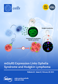
A severe form of autoimmune encephalitis, termed “Ophelia syndrome”, is characterized by the concurrence of acute neuropsychiatric symptoms, Hodgkin's lymphoma, and antibodies to the metabotropic glutamate 5 receptor (mGluR5). However, little is known about the pathogenetic link between these symptoms and the role played by anti-mGluR5-antibodies. This study provides evidence for the association between encephalitis and lymphoma in Ophelia syndrome. It demonstrates mGluR5 expression in Hodgkin lymphoma cells, which likely not only drives tumor progression but also triggers anti-mGluR5 encephalitis even before Hodgkin lymphoma is clinically detected. Therefore, rapid diagnosis of Ophelia syndrome is of paramount importance both for the treatment of anti-mGluR5 encephalitis and for the earliest possible recognition of subsequent Hodgkin lymphoma. View this paper
  • Issues are regarded as officially published after their release is announced to the table of contents alert mailing list.
  • You may sign up for e-mail alerts to receive table of contents of newly released issues.
  • PDF is the official format for papers published in both, html and pdf forms. To view the papers in pdf format, click on the "PDF Full-text" link, and use the free Adobe Reader to open them.
Order results
Result details
Section
Select all
Export citation of selected articles as:
12 pages, 660 KiB  
Review
Enzymology of Ca2+-Mobilizing Second Messengers Derived from NAD: From NAD Glycohydrolases to (Dual) NADPH Oxidases
by Andreas H. Guse
Cells 2023, 12(4), 675; https://doi.org/10.3390/cells12040675 - 20 Feb 2023
Cited by 3 | Viewed by 1656
Abstract
Nicotinamide adenine dinucleotide (NAD) and its 2′-phosphorylated cousin NADP are precursors for the enzymatic formation of the Ca2+-mobilizing second messengers adenosine diphosphoribose (ADPR), 2′-deoxy-ADPR, cyclic ADPR, and nicotinic acid adenine dinucleotide phosphate (NAADP). The enzymes involved are either NAD glycohydrolases CD38 [...] Read more.
Nicotinamide adenine dinucleotide (NAD) and its 2′-phosphorylated cousin NADP are precursors for the enzymatic formation of the Ca2+-mobilizing second messengers adenosine diphosphoribose (ADPR), 2′-deoxy-ADPR, cyclic ADPR, and nicotinic acid adenine dinucleotide phosphate (NAADP). The enzymes involved are either NAD glycohydrolases CD38 or sterile alpha toll/interleukin receptor motif containing-1 (SARM1), or (dual) NADPH oxidases (NOX/DUOX). Enzymatic function(s) are reviewed and physiological role(s) in selected cell systems are discussed. Full article
Show Figures

Figure 1

20 pages, 4380 KiB  
Review
Recent Clinical Advances on Long Non-Coding RNAs in Triple-Negative Breast Cancer
by Desh Deepak Singh, Hae-Jeung Lee and Dharmendra Kumar Yadav
Cells 2023, 12(4), 674; https://doi.org/10.3390/cells12040674 - 20 Feb 2023
Cited by 2 | Viewed by 2393
Abstract
Triple-negative breast cancer (TNBC) is a more aggressive type of breast cancer due to its heterogeneity and complex molecular mechanisms. TNBC has a high risk for metastasis, and it is difficult to manage clinical conditions of the patients. Various investigations are being conducted [...] Read more.
Triple-negative breast cancer (TNBC) is a more aggressive type of breast cancer due to its heterogeneity and complex molecular mechanisms. TNBC has a high risk for metastasis, and it is difficult to manage clinical conditions of the patients. Various investigations are being conducted to overcome these challenges using RNA, DNA, and proteins for early diagnosis and treatment. Recently, long non-coding RNAs (lncRNAs) have emerged as a novel target to treat the multistep process of TNBC. LncRNAs regulate epigenetic expression levels, cell proliferation and apoptosis, and tumour invasiveness and metastasis. Thus, lncRNA-based early diagnosis and treatment options could be helpful, especially for patients with severe TNBC. lncRNAs are expressed in a highly specific manner in cells and tissues and are involved in TNBC progression and development. lncRNAs could be used as sensitive and specific targets for diagnosis, treatment, and monitoring of patients with TNBC. Therefore, the exploration of novel diagnostic and prognostic biomarkers is of extreme importance. Here, we discuss the molecular advances on lncRNA regulation of TNBC and lncRNA-based early diagnosis, treatment, and drug resistance. Full article
(This article belongs to the Special Issue Long Non-coding RNAs in Cancer Metastasis)
Show Figures

Figure 1

17 pages, 6521 KiB  
Article
Effects of IL-11/IL-11 Receptor Alpha on Proliferation and Steroidogenesis in Ovarian Granulosa Cells of Dairy Cows
by Hanxiao Wu, Peihao Sun, Ce Lv, Xinzhe Zhao, Mingxiao Liu, Qunli Zhou, Jiaomei Tang, Liguo Yang and Aixin Liang
Cells 2023, 12(4), 673; https://doi.org/10.3390/cells12040673 - 20 Feb 2023
Viewed by 1893
Abstract
Granulosa cells (GCs) are essential for follicular growth, oocyte maturation, and steroidogenesis in the ovaries. Interleukin (IL)-11 is known to play a crucial role in the decidualization of the uterus, however, the expression of the IL-11 system (IL-11, IL-11Rα, and gp130) in the [...] Read more.
Granulosa cells (GCs) are essential for follicular growth, oocyte maturation, and steroidogenesis in the ovaries. Interleukin (IL)-11 is known to play a crucial role in the decidualization of the uterus, however, the expression of the IL-11 system (IL-11, IL-11Rα, and gp130) in the bovine ovary and its exact role in GCs have not been extensively studied. In this study, we identified the IL-11 signaling receptor complex in the bovine ovary and investigated the regulatory effects and underlying mechanism of IL-11Rα on the proliferation and steroidogenesis of GCs. We observed that the IL-11 complex was highly expressed in the GCs of large follicles. IL-11Rα knockdown significantly inhibited GC proliferation by inducing cell cycle arrest at the G1 phase, along with a significant downregulation of proliferating cell nuclear antigen (PCNA) and Cyclin D1 (CCND1) protein, and induced GC apoptosis by significantly upregulating the ratio of BCL-2-associated X protein (BAX) and B-cell lymphoma-2 (BCL-2). In addition, IL-11Rα knockdown attenuated the Janus kinase (JAK) 1–signal transducer and activator of transcription 3 (STAT3) signaling, which is related to cell proliferation and apoptosis. Furthermore, the enzyme-linked immunosorbent assay (ELISA) indicated that IL-11Rα silencing decreased the basal and forskolin (FSK)-stimulated secretions of estradiol and progesterone in GC culture medium concomitantly with a remarkable decrease in cytochrome P450 family 19 subfamily A member 1 (CYP19A1) and steroidogenic acute regulatory protein (StAR). We subsequently determined that this reduction in steroidogenesis was in parallel with the decrease in phosphorylations of protein kinase A (PKA) substrates, cAMP-response element binding protein (CREB), extracellular regulated protein kinase (ERK) 1/2, and p38 mitogen-activated protein kinase (MAPK). Taken together, these data indicate that the effects of IL-11/IL-11Rα on the proliferation and steroidogenesis in bovine GCs is mediated by the JAK1-STAT3, PKA-CREB, p38MAPK, and ERK1/2 signaling pathways. Our findings provide important insights into the local action of the IL-11 system in regulating ovarian function. Full article
(This article belongs to the Section Reproductive Cells and Development)
Show Figures

Figure 1

15 pages, 18855 KiB  
Article
Two Novel lncRNAs Regulate Primordial Germ Cell Development in Zebrafish
by Wenjing Li, Wei Liu, Chengyu Mo, Meisheng Yi and Jianfang Gui
Cells 2023, 12(4), 672; https://doi.org/10.3390/cells12040672 - 20 Feb 2023
Cited by 2 | Viewed by 1582
Abstract
Long noncoding RNAs (lncRNAs) are regulatory transcripts in various biological processes. However, the role of lncRNAs in germline development remains poorly understood, especially for fish primordial germ cell (PGC) development. In this study, the lncRNA profile of zebrafish PGC was revealed by single [...] Read more.
Long noncoding RNAs (lncRNAs) are regulatory transcripts in various biological processes. However, the role of lncRNAs in germline development remains poorly understood, especially for fish primordial germ cell (PGC) development. In this study, the lncRNA profile of zebrafish PGC was revealed by single cell RNA-sequencing and bioinformatic prediction. We established the regulation network of lncRNA-mRNA associated with PGC development, from which we identified three novel lncRNAs—lnc172, lnc196, and lnc304—highly expressing in PGCs and gonads. Fluorescent in situ hybridization indicated germline-specific localization of lnc196 and lnc304 in the cytoplasm and nucleus of spermatogonia, spermatocyte, and occyte, and they were co-localized with vasa in the cytoplasm of the spermatogonia. By contrast, lnc172 was localized in the cytoplasm of male germline, myoid cells and ovarian somatic cells. Loss- and gain-of-function experiments demonstrated that knockdown and PGC-specific overexpression of lnc304 as well as universal overexpression of lnc172 significantly disrupted PGC development. In summary, the present study revealed the lncRNA profile of zebrafish PGC and identified two novel lncRNAs associated with PGC development, providing new insights for understanding the regulatory mechanism of PGC development. Full article
(This article belongs to the Special Issue Germ Cells, Their Regulation and Their Niches)
Show Figures

Figure 1

25 pages, 918 KiB  
Review
In Vitro Drug Repurposing: Focus on Vasodilators
by Eduarda Ribeiro, Bárbara Costa, Francisco Vasques-Nóvoa and Nuno Vale
Cells 2023, 12(4), 671; https://doi.org/10.3390/cells12040671 - 20 Feb 2023
Cited by 2 | Viewed by 2473
Abstract
Drug repurposing aims to identify new therapeutic uses for drugs that have already been approved for other conditions. This approach can save time and resources compared to traditional drug development, as the safety and efficacy of the repurposed drug have already been established. [...] Read more.
Drug repurposing aims to identify new therapeutic uses for drugs that have already been approved for other conditions. This approach can save time and resources compared to traditional drug development, as the safety and efficacy of the repurposed drug have already been established. In the context of cancer, drug repurposing can lead to the discovery of new treatments that can target specific cancer cell lines and improve patient outcomes. Vasodilators are a class of drugs that have been shown to have the potential to influence various types of cancer. These medications work by relaxing the smooth muscle of blood vessels, increasing blood flow to tumors, and improving the delivery of chemotherapy drugs. Additionally, vasodilators have been found to have antiproliferative and proapoptotic effects on cancer cells, making them a promising target for drug repurposing. Research on vasodilators for cancer treatment has already shown promising results in preclinical and clinical studies. However, additionally research is needed to fully understand the mechanisms of action of vasodilators in cancer and determine the optimal dosing and combination therapy for patients. In this review, we aim to explore the molecular mechanisms of action of vasodilators in cancer cell lines and the current state of research on their repurposing as a treatment option. With the goal of minimizing the effort and resources required for traditional drug development, we hope to shed light on the potential of vasodilators as a viable therapeutic strategy for cancer patients. Full article
Show Figures

Figure 1

15 pages, 1416 KiB  
Article
Peripheral Immunophenotype in IgG4-Related Disease and Its Association with Clinical Phenotypes and Disease Activity
by Eduardo Martín-Nares, Gabriela Hernández-Molina, Ángel A. Priego-Ranero, Isela Chan-Campos, Gladys S. Herrera-Noguera, Fidel López-Verdugo and Janette Furuzawa-Carballeda
Cells 2023, 12(4), 670; https://doi.org/10.3390/cells12040670 - 20 Feb 2023
Cited by 3 | Viewed by 2050
Abstract
Diverse immune cell subsets have been described in IgG4-related disease (IgG4-RD). If there is a different immunophenotype according to clinical phenotype and activity status is not known. Levels of IL-4-, IL-13-, IL-5-, and IL-21-producing CD4+ T cells (Th2 subsets), CD4+ cytotoxic [...] Read more.
Diverse immune cell subsets have been described in IgG4-related disease (IgG4-RD). If there is a different immunophenotype according to clinical phenotype and activity status is not known. Levels of IL-4-, IL-13-, IL-5-, and IL-21-producing CD4+ T cells (Th2 subsets), CD4+ cytotoxic T lymphocytes (CD4+CTLs), T helper 9 cells, T follicular helper cells (Tfh; Tfh1/Tfh2/Tfh17/Tf regulatory [Tfr]), Foxp3+ regulatory T cells, Type 1 regulatory T cells (Tr1), T helper 3 regulatory cells (Th3), IL-10-producing regulatory B cells (Bregs), IL-10-expressing regulatory plasmacytoid dendritic (pDC IL-10+) cells, and M1 and M2 monocytes were determined by flow cytometry in 43 IgG4-RD patients and 12 controls. All immune subsets were higher in patients vs. controls. CD4+/IL-4+, CD4+/IL-5+, CD4+CTLs, Tfh2, Tfh17, Tfr, and M1 monocyte cell number was different among IgG4-RD clinical phenotypes. The pancreato-hepato-biliary phenotype was characterized by a higher CD4+CTLs, Tfh17, Tfh2, and Tfr and lower M1 cell number. An increased CD4+CTLs and Th3 cell number distinguished the head and neck-limited phenotype, while the retroperitoneal/aortic and Mikulicz/systemic phenotypes were characterized by increased Th2 subsets. Tfh17, Tr1, Th3, pDC, M1, and M2 monocytes were augmented in active patients. In summary, the clinical heterogeneity of IgG4-RD might be driven by the participation of different immunophenotypes and, consequently, by a different fibroinflammatory process. Full article
(This article belongs to the Special Issue T Cell Responses in Human Health and Disease - Second Edition)
Show Figures

Figure 1

12 pages, 3597 KiB  
Article
Diallyl Trisulfide, a Biologically Active Component of Garlic Essential Oil, Decreases Male Fertility in Sitotroga cerealella by Impairing Dimorphic Spermatogenesis, Sperm Motility and Lipid Homeostasis
by Sakhawat Shah, Karam Khamis Elgizawy, Chun-Mei Shi, Hucheng Yao, Wen-Han Yan, Yu Li, Xiao-Ping Wang, Gang Wu and Feng-Lian Yang
Cells 2023, 12(4), 669; https://doi.org/10.3390/cells12040669 - 20 Feb 2023
Cited by 4 | Viewed by 1462
Abstract
Diallyl trisulfide (DAT) is a biologically active component of garlic essential oil and exhibits multi-targeted activity against many organisms. The current study tested the capacity of DAT to decrease the male fertility of Sitotroga cerealella. The effects on testis morphology, sperm number, [...] Read more.
Diallyl trisulfide (DAT) is a biologically active component of garlic essential oil and exhibits multi-targeted activity against many organisms. The current study tested the capacity of DAT to decrease the male fertility of Sitotroga cerealella. The effects on testis morphology, sperm number, motility, and lipid homeostasis were observed in adult males fumigated with DAT at a dose of 0.01 μL/L in air. The results indicated that the DAT significantly decreased the dimorphic sperm number. Meanwhile, the ultrastructural analysis of the sperm showed that the DAT caused malformed and aberrant structures of mitochondrial derivatives of dimorphic sperm. Additionally, the lipid homeostasis and ATP contents in the male adults were significantly decreased after treatment. Moreover, the total sperm motility was reduced, while the wave-propagation velocity, amplitude, frequency, and wavelength were significantly decreased compared with the controls. Overall, this study reported, for the first time, that DAT impairs energy metabolism, inhibits dimorphic spermatogenesis, and decreases sperm motility, while these abnormalities in sperm lead to adult-male infertility. Full article
(This article belongs to the Special Issue Cellular Events in Insect Development, Immunity, and Reproduction)
Show Figures

Figure 1

15 pages, 1593 KiB  
Review
Autophagosome Biogenesis
by Yan Zhen and Harald Stenmark
Cells 2023, 12(4), 668; https://doi.org/10.3390/cells12040668 - 20 Feb 2023
Cited by 7 | Viewed by 3059
Abstract
Autophagy–the lysosomal degradation of cytoplasm–plays a central role in cellular homeostasis and protects cells from potentially harmful agents that may accumulate in the cytoplasm, including pathogens, protein aggregates, and dysfunctional organelles. This process is initiated by the formation of a phagophore membrane, which [...] Read more.
Autophagy–the lysosomal degradation of cytoplasm–plays a central role in cellular homeostasis and protects cells from potentially harmful agents that may accumulate in the cytoplasm, including pathogens, protein aggregates, and dysfunctional organelles. This process is initiated by the formation of a phagophore membrane, which wraps around a portion of cytoplasm or cargo and closes to form a double-membrane autophagosome. Upon the fusion of the autophagosome with a lysosome, the sequestered material is degraded by lysosomal hydrolases in the resulting autolysosome. Several alternative membrane sources of autophagosomes have been proposed, including the plasma membrane, endosomes, mitochondria, endoplasmic reticulum, lipid droplets, hybrid organelles, and de novo synthesis. Here, we review recent progress in our understanding of how the autophagosome is formed and highlight the proposed role of vesicles that contain the lipid scramblase ATG9 as potential seeds for phagophore biogenesis. We also discuss how the phagophore is sealed by the action of the endosomal sorting complex required for transport (ESCRT) proteins. Full article
(This article belongs to the Special Issue Exclusive Review Papers in Autophagy)
Show Figures

Figure 1

21 pages, 5701 KiB  
Article
Differential Expression of AP-2 Transcription Factors Family in Lung Adenocarcinoma and Lung Squamous Cell Carcinoma—A Bioinformatics Study
by Dagmara Szmajda-Krygier, Adrian Krygier, Marta Żebrowska-Nawrocka, Jacek Pietrzak, Rafał Świechowski, Agnieszka Wosiak, Agnieszka Jeleń and Ewa Balcerczak
Cells 2023, 12(4), 667; https://doi.org/10.3390/cells12040667 - 20 Feb 2023
Cited by 1 | Viewed by 1820
Abstract
Members of the activator protein 2 (AP-2) transcription factor (TF) family are known to play a role in both physiological processes and cancer development. The family comprises five DNA-binding proteins encoded by the TFAP2A to TFAP2E genes. Numerous scientific reports describe differential expression [...] Read more.
Members of the activator protein 2 (AP-2) transcription factor (TF) family are known to play a role in both physiological processes and cancer development. The family comprises five DNA-binding proteins encoded by the TFAP2A to TFAP2E genes. Numerous scientific reports describe differential expression of these TF and their genes in various types of cancer, identifying among them a potential oncogene or suppressor like TFAP2A or TFAP2C. Other reports suggest their influence on disease development and progression, as well as response to treatment. Not all members of this AP-2 family have been comprehensively studied thus far. The aim of the present article is to gather and discuss knowledge available in bioinformatics databases regarding all five members of this family and to differentiate them in relation to the two most common lung cancer subtypes: adenocarcinoma (LUAD) and squamous cell carcinoma (LUSC). In addition, to assess the difference in levels depending on a number of clinicopathological factors, the impact on patient survival and interactions with tumor-infiltrating immune cells. This article may help to identify the target for further original research that may contribute to the discovery of new diagnostic biomarkers and define the molecular differences between LUAD and LUSC, which may affect the therapy effectiveness improvement and longer survival. Full article
(This article belongs to the Special Issue Determination of AP-2 Transcription Factors Role in Carcinogenesis)
Show Figures

Figure 1

17 pages, 5459 KiB  
Article
Estrogen Signaling Influences Nephron Segmentation of the Zebrafish Embryonic Kidney
by Hannah M. Wesselman, Allison E. Gatz, Mairead R. Pfaff, Liana Arceri and Rebecca A. Wingert
Cells 2023, 12(4), 666; https://doi.org/10.3390/cells12040666 - 20 Feb 2023
Cited by 6 | Viewed by 1962
Abstract
Despite significant advances in understanding nephron segment patterning, many questions remain about the underlying genes and signaling pathways that orchestrate renal progenitor cell fate choices and regulate differentiation. In an effort to identify elusive regulators of nephron segmentation, our lab conducted a high-throughput [...] Read more.
Despite significant advances in understanding nephron segment patterning, many questions remain about the underlying genes and signaling pathways that orchestrate renal progenitor cell fate choices and regulate differentiation. In an effort to identify elusive regulators of nephron segmentation, our lab conducted a high-throughput drug screen using a bioactive chemical library and developing zebrafish, which are a conserved vertebrate model and particularly conducive to large-scale screening approaches. 17β-estradiol (E2), which is the dominant form of estrogen in vertebrates, was a particularly interesting hit from this screen. E2 has been extensively studied in the context of gonad development, but roles for E2 in nephron development were unknown. Here, we report that exogenous estrogen treatments affect distal tubule composition, namely, causing an increase in the distal early segment and a decrease in the neighboring distal late. These changes were noted early in development but were not due to changes in cell dynamics. Interestingly, exposure to the xenoestrogens ethinylestradiol and genistein yielded the same changes in distal segments. Further, upon treatment with an estrogen receptor 2 (Esr2) antagonist, PHTPP, we observed the opposite phenotypes. Similarly, genetic deficiency of the Esr2 analog, esr2b, revealed phenotypes consistent with that of PHTPP treatment. Inhibition of E2 signaling also resulted in decreased expression of essential distal transcription factors, irx3b and its target irx1a. These data suggest that estrogenic compounds are essential for distal segment fate during nephrogenesis in the zebrafish pronephros and expand our fundamental understanding of hormone function during kidney organogenesis. Full article
(This article belongs to the Special Issue Feature Papers in "Stem Cells" 2023)
Show Figures

Figure 1

11 pages, 2440 KiB  
Article
Inhibition of Soluble Epoxide Hydrolase Does Not Promote or Aggravate Pulmonary Hypertension in Rats
by Matthieu Leuillier, Valentin Platel, Ly Tu, Guillaume Feugray, Raphaël Thuillet, Déborah Groussard, Hind Messaoudi, Mina Ottaviani, Mustapha Chelgham, Lionel Nicol, Paul Mulder, Marc Humbert, Vincent Richard, Christophe Morisseau, Valéry Brunel, Thomas Duflot, Christophe Guignabert and Jérémy Bellien
Cells 2023, 12(4), 665; https://doi.org/10.3390/cells12040665 - 20 Feb 2023
Viewed by 1571
Abstract
Inhibitors of soluble epoxide hydrolase (sEH), which catalyzes the hydrolysis of various natural epoxides to their corresponding diols, present an opportunity for developing oral drugs for a range of human cardiovascular and inflammatory diseases, including, among others, diabetes and neuropathic pain. However, some [...] Read more.
Inhibitors of soluble epoxide hydrolase (sEH), which catalyzes the hydrolysis of various natural epoxides to their corresponding diols, present an opportunity for developing oral drugs for a range of human cardiovascular and inflammatory diseases, including, among others, diabetes and neuropathic pain. However, some evidence suggests that their administration may precipitate the development of pulmonary hypertension (PH). We thus evaluated the impact of chronic oral administration of the sEH inhibitor TPPU (N-[1-(1-Oxopropyl)-4-piperidinyl]-N′-[4-(trifluoromethoxy)phenyl]-urea) on hemodynamics, pulmonary vascular reactivity, and remodeling, as well as on right ventricular (RV) dimension and function at baseline and in the Sugen (SU5416) + hypoxia (SuHx) rat model of severe PH. Treatment with TPPU started 5 weeks after SU5416 injection for 3 weeks. No differences regarding the increase in pulmonary vascular resistance, remodeling, and inflammation, nor the abolishment of phenylephrine-induced pulmonary artery constriction, were noted in SuHx rats. In addition, TPPU did not modify the development of RV dysfunction, hypertrophy, and fibrosis in SuHx rats. Similarly, none of these parameters were affected by TPPU in normoxic rats. Complementary in vitro data demonstrated that TPPU reduced the proliferation of cultured human pulmonary artery-smooth muscle cells (PA-SMCs). This study demonstrates that inhibition of sEH does not induce nor aggravate the development of PH and RV dysfunction in SuHx rats. In contrast, a potential beneficial effect against pulmonary artery remodeling in humans is suggested. Full article
Show Figures

Figure 1

16 pages, 2700 KiB  
Article
SB202190 Predicts BRAF-Activating Mutations in Primary Colorectal Cancer Organoids via Erk1-2 Modulation
by Delfina Costa, Roberta Venè, Simona Coco, Luca Longo, Francesca Tosetti, Stefano Scabini, Luca Mastracci, Federica Grillo, Alessandro Poggi and Roberto Benelli
Cells 2023, 12(4), 664; https://doi.org/10.3390/cells12040664 - 20 Feb 2023
Cited by 2 | Viewed by 2151
Abstract
The p38 inhibitor SB202190 is a necessary component of the medium used for normal colorectal mucosa cultures. Sato et al. suggested that the primary activity of SB202190 may be EGFR signaling stabilization, causing an increased phosphorylation of Erk1-2 sustaining organoid proliferation. However, the [...] Read more.
The p38 inhibitor SB202190 is a necessary component of the medium used for normal colorectal mucosa cultures. Sato et al. suggested that the primary activity of SB202190 may be EGFR signaling stabilization, causing an increased phosphorylation of Erk1-2 sustaining organoid proliferation. However, the growth of some colorectal cancer (CRC)-derived organoid cultures is inhibited by this molecule via an unknown mechanism. We biochemically investigated SB202190 activity on a collection of 25 primary human CRC organoids, evaluating EGFR, Akt and Erk1-2 activation using Western blot. We found that Erk1-2 phosphorylation was induced by SB202190 in 20 organoid cultures and inhibited in 5 organoid cultures. A next-generation sequencing (NGS) analysis revealed that the inhibition of p-Erk1-2 signaling corresponded to the cultures with BRAF mutations (with four different hits, one being undescribed), while p-Erk1-2 induction was apparently unrelated to other mutations involving the EGFR pathway (Her2, KRAS and NRAS). We found that SB202190 mirrored the biochemical activity of the BRAF inhibitor Dabrafenib, known to induce the paradoxical activation of p-Erk1-2 signaling in BRAF wild-type cells. SB202190 was a more effective inhibitor of BRAF-mutated organoid growth in the long term than the specific BRAF inhibitors Dabrafenib and PLX8394. Overall, SB202190 can predict BRAF-activating mutations in patient-derived organoids, as well as allowing for the identification of new BRAF variants, preceding and enforcing NGS data. Full article
Show Figures

Graphical abstract

18 pages, 2439 KiB  
Review
Sirtuin 6—A Key Regulator of Hepatic Lipid Metabolism and Liver Health
by X. Charlie Dong
Cells 2023, 12(4), 663; https://doi.org/10.3390/cells12040663 - 19 Feb 2023
Cited by 8 | Viewed by 5522
Abstract
Sirtuin 6 (SIRT6) is an NAD-dependent deacetylase/deacylase/mono-ADP ribosyltransferase, a member of the sirtuin protein family. SIRT6 has been implicated in hepatic lipid homeostasis and liver health. Hepatic lipogenesis is driven by several master regulators including liver X receptor (LXR), carbohydrate response element binding [...] Read more.
Sirtuin 6 (SIRT6) is an NAD-dependent deacetylase/deacylase/mono-ADP ribosyltransferase, a member of the sirtuin protein family. SIRT6 has been implicated in hepatic lipid homeostasis and liver health. Hepatic lipogenesis is driven by several master regulators including liver X receptor (LXR), carbohydrate response element binding protein (ChREBP), and sterol regulatory element binding protein 1 (SREBP1). Interestingly, these three transcription factors can be negatively regulated by SIRT6 through direct deacetylation. Fatty acid oxidation is regulated by peroxisome proliferator activated receptor alpha (PPARα) in the liver. SIRT6 can promote fatty acid oxidation by the activation of PPARα or the suppression of miR-122. SIRT6 can also directly modulate acyl-CoA synthetase long chain family member 5 (ACSL5) activity for fatty acid oxidation. SIRT6 also plays a critical role in the regulation of total cholesterol and low-density lipoprotein (LDL)-cholesterol through the regulation of SREBP2 and proprotein convertase subtilisin/kexin type 9 (PCSK9), respectively. Hepatic deficiency of Sirt6 in mice has been shown to cause hepatic steatosis, inflammation, and fibrosis, hallmarks of alcoholic and nonalcoholic steatohepatitis. SIRT6 can dampen hepatic inflammation through the modulation of macrophage polarization from M1 to M2 type. Hepatic stellate cells are a key cell type in hepatic fibrogenesis. SIRT6 plays a strong anti-fibrosis role by the suppression of multiple fibrogenic pathways including the transforming growth factor beta (TGFβ)-SMAD family proteins and Hippo pathways. The role of SIRT6 in liver cancer is quite complicated, as both tumor-suppressive and tumor-promoting activities have been documented in the literature. Overall, SIRT6 has multiple salutary effects on metabolic homeostasis and liver health, and it may serve as a therapeutic target for hepatic metabolic diseases. To date, numerous activators and inhibitors of SIRT6 have been developed for translational research. Full article
(This article belongs to the Special Issue Lipid Metabolism and Metabolic Disorders)
Show Figures

Graphical abstract

24 pages, 1563 KiB  
Review
A Wrong Fate Decision in Adipose Stem Cells upon Obesity
by Yiu-Ming Cheung, Chui-Yiu-Bamboo Chook, Hoi-Wa Yeung, Fung-Ping Leung and Wing-Tak Wong
Cells 2023, 12(4), 662; https://doi.org/10.3390/cells12040662 - 19 Feb 2023
Cited by 2 | Viewed by 2009
Abstract
Progress has been made in identifying stem cell aging as a pathological manifestation of a variety of diseases, including obesity. Adipose stem cells (ASCs) play a core role in adipocyte turnover, which maintains tissue homeostasis. Given aberrant lineage determination as a feature of [...] Read more.
Progress has been made in identifying stem cell aging as a pathological manifestation of a variety of diseases, including obesity. Adipose stem cells (ASCs) play a core role in adipocyte turnover, which maintains tissue homeostasis. Given aberrant lineage determination as a feature of stem cell aging, failure in adipogenesis is a culprit of adipose hypertrophy, resulting in adiposopathy and related complications. In this review, we elucidate how ASC fails in entering adipogenic lineage, with a specific focus on extracellular signaling pathways, epigenetic drift, metabolic reprogramming, and mechanical stretch. Nonetheless, such detrimental alternations can be reversed by guiding ASCs towards adipogenesis. Considering the pathological role of ASC aging in obesity, targeting adipogenesis as an anti-obesity treatment will be a key area of future research, and a strategy to rejuvenate tissue stem cell will be capable of alleviating metabolic syndrome. Full article
(This article belongs to the Special Issue Metabolic Regulation of Stem Cell Behavior, Function and Aging)
Show Figures

Figure 1

15 pages, 5215 KiB  
Article
Gbb Regulates Blood Cell Proliferation and Differentiation through JNK and EGFR Signaling Pathways in the Drosophila Lymph Gland
by Wenhao Zhang, Dongmei Wang, Jingjing Si, Li Hua Jin and Yangguang Hao
Cells 2023, 12(4), 661; https://doi.org/10.3390/cells12040661 - 19 Feb 2023
Cited by 1 | Viewed by 1717
Abstract
The Drosophila lymph gland is an ideal model for studying hematopoiesis, and unraveling the mechanisms of Drosophila hematopoiesis can improve our understanding of the pathogenesis of human hematopoietic malignancies. Bone morphogenetic protein (BMP) signaling is involved in a variety of biological processes and [...] Read more.
The Drosophila lymph gland is an ideal model for studying hematopoiesis, and unraveling the mechanisms of Drosophila hematopoiesis can improve our understanding of the pathogenesis of human hematopoietic malignancies. Bone morphogenetic protein (BMP) signaling is involved in a variety of biological processes and is highly conserved between Drosophila and mammals. Decapentaplegic (Dpp)/BMP signaling is known to limit posterior signaling center (PSC) cell proliferation by repressing the protooncogene dmyc. However, the role of two other TGF-β family ligands, Glass bottom boat (Gbb) and Screw (Scw), in Drosophila hematopoiesis is currently largely unknown. Here, we showed that the loss of Gbb in the cortical zone (CZ) induced lamellocyte differentiation by overactivation of the EGFR and JNK pathways and caused excessive differentiation of plasmatocytes, mainly by the hyperactivation of EGFR. Furthermore, we found that Gbb was also required for preventing the hyperproliferation of the lymph glands by inhibiting the overactivation of the Epidermal Growth Factor Receptor (EGFR) and c-Jun N-terminal Kinase (JNK) pathways. These results further advance our understanding of the roles of Gbb protein and the BMP signaling in Drosophila hematopoiesis and the regulatory relationship between the BMP, EGFR, and JNK pathways in the proliferation and differentiation of lymph gland hemocytes. Full article
(This article belongs to the Section Cell Signaling)
Show Figures

Figure 1

30 pages, 1255 KiB  
Review
The Vitamin D Receptor as a Potential Target for the Treatment of Age-Related Neurodegenerative Diseases Such as Alzheimer’s and Parkinson’s Diseases: A Narrative Review
by Władysław Lasoń, Danuta Jantas, Monika Leśkiewicz, Magdalena Regulska and Agnieszka Basta-Kaim
Cells 2023, 12(4), 660; https://doi.org/10.3390/cells12040660 - 19 Feb 2023
Cited by 22 | Viewed by 3894
Abstract
The vitamin D receptor (VDR) belongs to the nuclear receptor superfamily of transcription factors. The VDR is expressed in diverse brain regions and has been implicated in the neuroprotective, antiaging, prosurvival, and anti-inflammatory action of vitamin D. Accordingly, a relationship between vitamin D [...] Read more.
The vitamin D receptor (VDR) belongs to the nuclear receptor superfamily of transcription factors. The VDR is expressed in diverse brain regions and has been implicated in the neuroprotective, antiaging, prosurvival, and anti-inflammatory action of vitamin D. Accordingly, a relationship between vitamin D insufficiency and susceptibility to neurodegenerative diseases has been suggested. However, due to the multitargeted mechanisms of vitamin D and its often overlapping genomic and nongenomic effects, the role of the VDR in brain pathologies remains obscure. In this narrative review, we present progress in deciphering the molecular mechanism of nuclear VDR-mediated vitamin D effects on prosurvival and anti-inflammatory signaling pathway activity within the central nervous system. In line with the concept of the neurovascular unit in pathomechanisms of neurodegenerative diseases, a discussion of the role of the VDR in regulating the immune and vascular brain systems is also included. Next, we discuss the results of preclinical and clinical studies evaluating the significance of vitamin D status and the efficacy of vitamin D supplementation in the treatment of Parkinson’s and Alzheimer’s diseases, emphasizing the possible role of the VDR in these phenomena. Finally, the associations of some VDR polymorphisms with higher risks and severity of these neurodegenerative disorders are briefly summarized. Full article
(This article belongs to the Collection Functions of Nuclear Receptors)
Show Figures

Figure 1

30 pages, 1693 KiB  
Review
Doxorubicin—An Agent with Multiple Mechanisms of Anticancer Activity
by Mateusz Kciuk, Adrianna Gielecińska, Somdutt Mujwar, Damian Kołat, Żaneta Kałuzińska-Kołat, Ismail Celik and Renata Kontek
Cells 2023, 12(4), 659; https://doi.org/10.3390/cells12040659 - 19 Feb 2023
Cited by 54 | Viewed by 5112
Abstract
Doxorubicin (DOX) constitutes the major constituent of anti-cancer treatment regimens currently in clinical use. However, the precise mechanisms of DOX’s action are not fully understood. Emerging evidence points to the pleiotropic anticancer activity of DOX, including its contribution to DNA damage, reactive oxygen [...] Read more.
Doxorubicin (DOX) constitutes the major constituent of anti-cancer treatment regimens currently in clinical use. However, the precise mechanisms of DOX’s action are not fully understood. Emerging evidence points to the pleiotropic anticancer activity of DOX, including its contribution to DNA damage, reactive oxygen species (ROS) production, apoptosis, senescence, autophagy, ferroptosis, and pyroptosis induction, as well as its immunomodulatory role. This review aims to collect information on the anticancer mechanisms of DOX as well as its influence on anti-tumor immune response, providing a rationale behind the importance of DOX in modern cancer therapy. Full article
(This article belongs to the Section Cellular Immunology)
Show Figures

Figure 1

16 pages, 1268 KiB  
Review
Extracellular Vesicles and MicroRNA in Myelodysplastic Syndromes
by Mathieu Meunier, David Laurin and Sophie Park
Cells 2023, 12(4), 658; https://doi.org/10.3390/cells12040658 - 19 Feb 2023
Cited by 1 | Viewed by 2000
Abstract
The bone marrow niche plays an increasing role in the pathophysiogenesis of myelodysplastic syndromes. More specifically, mesenchymal stromal cells, which can secrete extracellular vesicles and their miRNA contents, modulate the fate of hematopoietic stem cells leading to leukemogenesis. Extracellular vesicles can mediate their [...] Read more.
The bone marrow niche plays an increasing role in the pathophysiogenesis of myelodysplastic syndromes. More specifically, mesenchymal stromal cells, which can secrete extracellular vesicles and their miRNA contents, modulate the fate of hematopoietic stem cells leading to leukemogenesis. Extracellular vesicles can mediate their miRNA and protein contents between nearby cells but also in the plasma of the patients, being potent tools for diagnosis and prognostic markers in MDS. They can be targeted by antisense miRNA or by modulators of the secretion of extracellular vesicles and could lead to future therapeutic directions in MDS. Full article
(This article belongs to the Special Issue Extracellular Vesicle-Associated Non-Coding RNAs)
Show Figures

Figure 1

29 pages, 7001 KiB  
Review
Mechanisms of Blood–Brain Barrier Protection by Microbiota-Derived Short-Chain Fatty Acids
by Ekaterina Fock and Rimma Parnova
Cells 2023, 12(4), 657; https://doi.org/10.3390/cells12040657 - 18 Feb 2023
Cited by 28 | Viewed by 4739
Abstract
Impairment of the blood–brain barrier (BBB) integrity is implicated in the numerous neurological disorders associated with neuroinflammation, neurodegeneration and aging. It is now evident that short-chain fatty acids (SCFAs), mainly acetate, butyrate and propionate, produced by anaerobic bacterial fermentation of the dietary fiber [...] Read more.
Impairment of the blood–brain barrier (BBB) integrity is implicated in the numerous neurological disorders associated with neuroinflammation, neurodegeneration and aging. It is now evident that short-chain fatty acids (SCFAs), mainly acetate, butyrate and propionate, produced by anaerobic bacterial fermentation of the dietary fiber in the intestine, have a key role in the communication between the gastrointestinal tract and nervous system and are critically important for the preservation of the BBB integrity under different pathological conditions. The effect of SCFAs on the improvement of the compromised BBB is mainly based on the decrease in paracellular permeability via restoration of junctional complex proteins affecting their transcription, intercellular localization or proteolytic degradation. This review is focused on the revealed and putative underlying mechanisms of the direct and indirect effects of SCFAs on the improvement of the barrier function of brain endothelial cells. We consider G-protein-coupled receptor-mediated effects of SCFAs, SCFAs-stimulated acetylation of histone and non-histone proteins via inhibition of histone deacetylases, and crosstalk of these signaling pathways with transcriptional factors NF-κB and Nrf2 as mainstream mechanisms of SCFA’s effect on the preservation of the BBB integrity. Full article
(This article belongs to the Special Issue Exclusive Review Papers in "Cell Signaling")
Show Figures

Figure 1

12 pages, 742 KiB  
Review
Vestibular Disorders and Hormonal Dysregulations: State of the Art and Clinical Perspectives
by Rhizlane El Khiati, Brahim Tighilet, Stéphane Besnard and Christian Chabbert
Cells 2023, 12(4), 656; https://doi.org/10.3390/cells12040656 - 18 Feb 2023
Cited by 2 | Viewed by 1834
Abstract
The interaction between endocrine and vestibular systems remains poorly documented so far, despite numerous observations in humans and animals revealing direct links between the two systems. For example, dizziness or vestibular instabilities often accompany the menstrual cycle and are highly associated with the [...] Read more.
The interaction between endocrine and vestibular systems remains poorly documented so far, despite numerous observations in humans and animals revealing direct links between the two systems. For example, dizziness or vestibular instabilities often accompany the menstrual cycle and are highly associated with the pre-menopause period, while sex hormones, together with their specific receptors, are expressed at key places of the vestibular sensory network. Similarly, other hormones may be associated with vestibular disorders either as causal/inductive factors or as correlates of the pathology. This review was carried out according to the PRISMA method, covering the last two decades and using the MEDLINE and COCHRANE databases in order to identify studies associating the terms vestibular system and/or vestibular pathologies and hormones. Our literature search identified 646 articles, 67 of which referred directly to vestibular dysfunction associated with hormonal variations. While we noted specific hormonal profiles depending on the pathology considered, very few clinical studies attempted to establish a direct link between the expression of the vestibular syndrome and the level of circulating hormones. This review also proposes different approaches to shed new light on the link between hormones and vestibular disorders, and to improve both the diagnosis and the therapeutic management of dizzy patients. Full article
Show Figures

Figure 1

12 pages, 1059 KiB  
Perspective
Promising Perspectives of the Antiproliferative GPER Inverse Agonist ERα17p in Breast Cancer
by Marilena Kampa, Rosamaria Lappano, Fedora Grande, Bruno Rizzuti, Marcello Maggiolini, Elias Castanas and Yves Jacquot
Cells 2023, 12(4), 653; https://doi.org/10.3390/cells12040653 - 18 Feb 2023
Cited by 2 | Viewed by 1388
Abstract
The estrogen receptor α (ERα) corresponds to a large platform in charge of the recruitment of a panel of molecules, including steroids and related heterocyclic derivatives, oligonucleotides, peptides and proteins. Its 295–311 region is particularly targeted by post-translational modifications, suggesting that it could [...] Read more.
The estrogen receptor α (ERα) corresponds to a large platform in charge of the recruitment of a panel of molecules, including steroids and related heterocyclic derivatives, oligonucleotides, peptides and proteins. Its 295–311 region is particularly targeted by post-translational modifications, suggesting that it could be crucial for the control of transcription. In addition to anionic phospholipids, the ERα 295–311 fragment interacts with Ca2+-calmodulin, the heat shock protein 70 (Hsp70), ERα and possibly importins. More recently, we have demonstrated that it is prone to interacting with the G-protein-coupled estrogen receptor (GPER). In light of these observations, the pharmacological profile of the corresponding peptide, namely ERα17p, has been explored in breast cancer cells. Remarkably, it exerts apoptosis through GPER and induces a significant decrease (more than 50%) of the size of triple-negative breast tumor xenografts in mice. Herein, we highlight not only the promising therapeutic perspectives in the use of the first peptidic GPER modulator ERα17p, but also the opportunity to modulate GPER for clinical purposes. Full article
Show Figures

Figure 1

16 pages, 715 KiB  
Article
COVID-19 Affects Serum Brain-Derived Neurotrophic Factor and Neurofilament Light Chain in Aged Men: Implications for Morbidity and Mortality
by Carla Petrella, Maria Antonella Zingaropoli, Flavio Maria Ceci, Patrizia Pasculli, Tiziana Latronico, Grazia Maria Liuzzi, Maria Rosa Ciardi, Antonio Angeloni, Evaristo Ettorre, Michela Menghi, Christian Barbato, Giampiero Ferraguti, Antonio Minni and Marco Fiore
Cells 2023, 12(4), 655; https://doi.org/10.3390/cells12040655 - 17 Feb 2023
Cited by 10 | Viewed by 1667
Abstract
Background and Methods: Severe COVID-19 is known to induce neurological damage (NeuroCOVID), mostly in aged individuals, by affecting brain-derived neurotrophic factor (BDNF), matrix metalloproteinases (MMP) 2 and 9 and the neurofilament light chain (NFL) pathways. Thus, the aim of this pilot study was [...] Read more.
Background and Methods: Severe COVID-19 is known to induce neurological damage (NeuroCOVID), mostly in aged individuals, by affecting brain-derived neurotrophic factor (BDNF), matrix metalloproteinases (MMP) 2 and 9 and the neurofilament light chain (NFL) pathways. Thus, the aim of this pilot study was to investigate BDNF, MMP-2, MMP-9, and NFL in the serum of aged men affected by COVID-19 at the beginning of the hospitalization period and characterized by different outcomes, i.e., attending a hospital ward or an intensive care unit (ICU) or with a fatal outcome. As a control group, we used a novelty of the study, unexposed age-matched men. We also correlated these findings with the routine blood parameters of the recruited individuals. Results: We found in COVID-19 individuals with severe or lethal outcomes disrupted serum BDNF, NFL, and MMP-2 presence and gross changes in ALT, GGT, LDH, IL-6, ferritin, and CRP. We also confirmed and extended previous data, using ROC analyses, showing that the ratio MMPs (2 and 9) versus BDNF and NFL might be a useful tool to predict a fatal COVID-19 outcome. Conclusions: Serum BDNF and NFL and/or their ratios with MMP-2 and MMP-9 could represent early predictors of NeuroCOVID in aged men. Full article
(This article belongs to the Special Issue Insights into Molecular and Cellular Mechanisms of NeuroCOVID)
Show Figures

Figure 1

13 pages, 1573 KiB  
Article
Analysis of the Anticipatory Behavior Formation Mechanism Induced by Methamphetamine Using a Single Hair
by Riku Sato, Megumi Kanai, Yukina Yoshida, Shiori Fukushima, Masahiro Nogami, Takeshi Yamaguchi, Norio Iijima, Kenneth Sutherland, Sanae Haga, Michitaka Ozaki, Kazuko Hamada and Toshiyuki Hamada
Cells 2023, 12(4), 654; https://doi.org/10.3390/cells12040654 - 17 Feb 2023
Viewed by 1357
Abstract
While the suprachiasmatic nucleus (SCN) coordinates many daily rhythms, some circadian patterns of expression are controlled by SCN-independent systems. These include responses to daily methamphetamine (MAP) injections. Scheduled daily injections of MAP resulted in anticipatory activity, with an increase in locomotor activity immediately [...] Read more.
While the suprachiasmatic nucleus (SCN) coordinates many daily rhythms, some circadian patterns of expression are controlled by SCN-independent systems. These include responses to daily methamphetamine (MAP) injections. Scheduled daily injections of MAP resulted in anticipatory activity, with an increase in locomotor activity immediately prior to the time of injection. The MAP-induced anticipatory behavior is associated with the induction and a phase advance in the expression rhythm of the clock gene Period1 (Per1). However, this unique formation mechanism of MAP-induced anticipatory behavior is not well understood. We recently developed a micro-photomultiplier tube (micro-PMT) system to detect a small amount of Per1 expression. In the present study, we used this system to measure the formation kinetics of MAP-induced anticipatory activity in a single whisker hair to reveal the underlying mechanism. Our results suggest that whisker hairs respond to daily MAP administration, and that Per1 expression is affected. We also found that elevated Per1 expression in a single whisker hair is associated with the occurrence of anticipatory behavior rhythm. The present results suggest that elevated Per1 expression in hairs might be a marker of anticipatory behavior formation. Full article
(This article belongs to the Special Issue Molecular Mechanism of Stress, Stress Response, and Adaptation)
Show Figures

Figure 1

24 pages, 3960 KiB  
Article
Post-Stroke Environmental Enrichment Improves Neurogenesis and Cognitive Function and Reduces the Generation of Aberrant Neurons in the Mouse Hippocampus
by Florus Woitke, Antonia Blank, Anna-Lena Fleischer, Shanshan Zhang, Gina-Marie Lehmann, Julius Broesske, Madlen Haase, Christoph Redecker, Christian W. Schmeer and Silke Keiner
Cells 2023, 12(4), 652; https://doi.org/10.3390/cells12040652 - 17 Feb 2023
Cited by 9 | Viewed by 1531
Abstract
Ischemic lesions stimulate adult neurogenesis in the dentate gyrus, however, this is not associated with better cognitive function. Furthermore, increased neurogenesis is associated with the formation of aberrant neurons. In a previous study, we showed that a running task after a stroke not [...] Read more.
Ischemic lesions stimulate adult neurogenesis in the dentate gyrus, however, this is not associated with better cognitive function. Furthermore, increased neurogenesis is associated with the formation of aberrant neurons. In a previous study, we showed that a running task after a stroke not only increases neurogenesis but also the number of aberrant neurons without improving general performance. Here, we determined whether stimulation in an enriched environment after a lesion could increase neurogenesis and cognitive function without enhancing the number of aberrant neurons. After an ischemic stroke induced by MCAO, animals were transferred to an enriched environment containing a running wheel, tunnels and nest materials. A GFP-retroviral vector was delivered on day 3 post-stroke and a modified water maze test was performed 6 weeks after the lesion. We found that the enriched environment significantly increased the number of new neurons compared with the unstimulated stroke group but not the number of aberrant cells after a lesion. Increased neurogenesis after environmental enrichment was associated with improved cognitive function. Our study showed that early placement in an enriched environment after a stroke lesion markedly increased neurogenesis and flexible learning but not the formation of aberrant neurons, indicating that rehabilitative training, as a combination of running wheel training and enriched environment housing, improved functional and structural outcomes after a stroke. Full article
Show Figures

Figure 1

30 pages, 2755 KiB  
Review
Role of Deubiquitinases in Parkinson’s Disease—Therapeutic Perspectives
by Pernille Y. Ø. Nielsen, Justyna Okarmus and Morten Meyer
Cells 2023, 12(4), 651; https://doi.org/10.3390/cells12040651 - 17 Feb 2023
Cited by 3 | Viewed by 2414
Abstract
Parkinson’s disease (PD) is a neurodegenerative disorder that has been associated with mitochondrial dysfunction, oxidative stress, and defects in mitophagy as well as α-synuclein-positive inclusions, termed Lewy bodies (LBs), which are a common pathological hallmark in PD. Mitophagy is a process that maintains [...] Read more.
Parkinson’s disease (PD) is a neurodegenerative disorder that has been associated with mitochondrial dysfunction, oxidative stress, and defects in mitophagy as well as α-synuclein-positive inclusions, termed Lewy bodies (LBs), which are a common pathological hallmark in PD. Mitophagy is a process that maintains cellular health by eliminating dysfunctional mitochondria, and it is triggered by ubiquitination of mitochondrial-associated proteins—e.g., through the PINK1/Parkin pathway—which results in engulfment by the autophagosome and degradation in lysosomes. Deubiquitinating enzymes (DUBs) can regulate this process at several levels by deubiquitinating mitochondrial substrates and other targets in the mitophagic pathway, such as Parkin. Moreover, DUBs can affect α-synuclein aggregation through regulation of degradative pathways, deubiquitination of α-synuclein itself, and/or via co-localization with α-synuclein in inclusions. DUBs with a known association to PD are described in this paper, along with their function. Of interest, DUBs could be useful as novel therapeutic targets against PD through regulation of PD-associated defects. Full article
Show Figures

Figure 1

18 pages, 576 KiB  
Review
Peroxisome Proliferator-Activated Receptor-Targeted Therapies: Challenges upon Infectious Diseases
by In Soo Kim, Prashanta Silwal and Eun-Kyeong Jo
Cells 2023, 12(4), 650; https://doi.org/10.3390/cells12040650 - 17 Feb 2023
Cited by 5 | Viewed by 3279
Abstract
Peroxisome proliferator-activated receptors (PPARs) α, β, and γ are nuclear receptors that orchestrate the transcriptional regulation of genes involved in a variety of biological responses, such as energy metabolism and homeostasis, regulation of inflammation, cellular development, and differentiation. The many roles played by [...] Read more.
Peroxisome proliferator-activated receptors (PPARs) α, β, and γ are nuclear receptors that orchestrate the transcriptional regulation of genes involved in a variety of biological responses, such as energy metabolism and homeostasis, regulation of inflammation, cellular development, and differentiation. The many roles played by the PPAR signaling pathways indicate that PPARs may be useful targets for various human diseases, including metabolic and inflammatory conditions and tumors. Accumulating evidence suggests that each PPAR plays prominent but different roles in viral, bacterial, and parasitic infectious disease development. In this review, we discuss recent PPAR research works that are focused on how PPARs control various infections and immune responses. In addition, we describe the current and potential therapeutic uses of PPAR agonists/antagonists in the context of infectious diseases. A more comprehensive understanding of the roles played by PPARs in terms of host-pathogen interactions will yield potential adjunctive personalized therapies employing PPAR-modulating agents. Full article
(This article belongs to the Special Issue The Role of PPARs in Disease II)
Show Figures

Figure 1

20 pages, 4801 KiB  
Article
Dual Effects of miR-181b-2-3p/SOX21 Interaction on Microglia and Neural Stem Cells after Gamma Irradiation
by Hong Wang, Zhao-Wu Ma, Feng-Ming Ho, Gautam Sethi and Feng Ru Tang
Cells 2023, 12(4), 649; https://doi.org/10.3390/cells12040649 - 17 Feb 2023
Cited by 3 | Viewed by 1354
Abstract
Ionizing radiation induces brain inflammation and the impairment of neurogenesis by activating microglia and inducing apoptosis in neurogenic zones. However, the causal relationship between microglial activation and the impairment of neurogenesis as well as the relevant molecular mechanisms involved in microRNA (miR) remain [...] Read more.
Ionizing radiation induces brain inflammation and the impairment of neurogenesis by activating microglia and inducing apoptosis in neurogenic zones. However, the causal relationship between microglial activation and the impairment of neurogenesis as well as the relevant molecular mechanisms involved in microRNA (miR) remain unknown. In the present study, we employed immunohistochemistry and real-time RT-PCR to study the microglial activation and miRNA expression in mouse brains. Real-time RT-PCR, western blot, ELISA, cell proliferation and cytotoxicity assay were used in BV2 and mouse neural stem cells (NSCs). In the mouse model, we found the acute activation of microglia at 1 day and an increased number of microglial cells at 1, 7 and 120 days after irradiation at postnatal day 3 (P3), day 10 (P10) and day 21 (P21), respectively. In cell models, the activation of BV2, a type of microglial cell line, was observed after gamma irradiation. Real-time RT-PCR analysis revealed a deceased expression of miR-181b-2-3p and an increased expression of its target SRY-related high-mobility group box transcription factor 21 (SOX21) in a dose- and time-dependent fashion. The results of the luciferase reporter assay confirmed that SOX21 was the target of miR-181b-2-3p. Furthermore, SOX21 knockdown by siRNA inhibited the activation of microglia, thereby suggesting that the direct interaction of 181b-2-3p with SOX21 might be involved in radiation-induced microglial activation and proliferation. Interestingly, the gamma irradiation of NSCs increased miR-181b-2-3p expression but decreased SOX21 mRNA, which was the opposite of irradiation-induced expression in BV2 cells. As irradiation reduced the viability and proliferation of NSCs, whereas the overexpression of SOX21 restored the impaired cell viability and promoted the proliferation of NSCs, the findings suggest that the radiation-induced interaction of miR-181b-2-3p with SOX21 may play dual roles in microglia and NSCs, respectively, leading to the impairment of brain neurogenesis. Full article
(This article belongs to the Special Issue microRNA as Biomarker II)
Show Figures

Graphical abstract

18 pages, 2165 KiB  
Article
Impact of Chronic Multi-Generational Exposure to an Environmentally Relevant Atrazine Concentration on Testicular Development and Function in Mice
by Nicola D. Kolaitis, Bethany J. Finger, D. Jo Merriner, Joseph Nguyen, Brendan J. Houston, Moira K. O’Bryan, Jessica M. Stringer, Nadeen Zerafa, Ngoc Nguyen, Karla J. Hutt, Gerard A. Tarulli and Mark P. Green
Cells 2023, 12(4), 648; https://doi.org/10.3390/cells12040648 - 17 Feb 2023
Cited by 1 | Viewed by 1884
Abstract
A common herbicide, atrazine, is associated with poor health. Atrazine acts as an endocrine disruptor at supra-environmental levels. Little research, however, has been conducted regarding chronic exposure to environmental atrazine concentrations across generations. This study utilized comprehensive endpoint measures to investigate the effects [...] Read more.
A common herbicide, atrazine, is associated with poor health. Atrazine acts as an endocrine disruptor at supra-environmental levels. Little research, however, has been conducted regarding chronic exposure to environmental atrazine concentrations across generations. This study utilized comprehensive endpoint measures to investigate the effects of chronic exposure to a conservative atrazine concentration (0.02 ng/mL), measured in Australian waterways, on male mice fertility across two generations. Mice were exposed through the maternal line, from the pre-conception period and through the F1 and F2 generations until three or six months of age. Atrazine did not impact sperm function, testicular morphology nor germ cell parameters but did alter the expression of steroidogenic genes in the F1, down-regulating the expression of Cyp17a1 (Cytochrome P450 family 17, subfamily A member 1; p = 0.0008) and Ddx4 (DEAD-box helicase 4; p = 0.007), and up-regulating the expression of Star (Steroidogenic acute regulatory protein; p = 0.017). In the F2, atrazine induced up-regulation in the expression of Star (p = 0.016). The current study demonstrates that chronic exposure to an environmentally relevant atrazine concentration perturbs testicular steroid-associated gene expression that varies across generations. Future studies through the paternal and combined parental lineages should be undertaken to further elucidate the multigenerational effects of atrazine on male fertility. Full article
Show Figures

Figure 1

14 pages, 1487 KiB  
Review
The Role of Peroxisome Proliferator-Activated Receptors in Preeclampsia
by Iason Psilopatis, Kleio Vrettou, Florian Nima Fleckenstein and Stamatios Theocharis
Cells 2023, 12(4), 647; https://doi.org/10.3390/cells12040647 - 17 Feb 2023
Cited by 10 | Viewed by 1868
Abstract
Preeclampsia is a common pregnancy-related hypertensive disorder. Often presenting as preexisting or new-onset hypertension complicated by proteinuria and/or end-organ dysfunction, preeclampsia significantly correlates with maternal and perinatal morbidity and mortality. Peroxisome proliferator-activated receptors (PPARs) are nuclear receptor proteins that regulate gene expression. In [...] Read more.
Preeclampsia is a common pregnancy-related hypertensive disorder. Often presenting as preexisting or new-onset hypertension complicated by proteinuria and/or end-organ dysfunction, preeclampsia significantly correlates with maternal and perinatal morbidity and mortality. Peroxisome proliferator-activated receptors (PPARs) are nuclear receptor proteins that regulate gene expression. In order to investigate the role of PPARs in the pathophysiology of preeclampsia, we conducted a literature review using the MEDLINE and LIVIVO databases. The search terms “peroxisome proliferator-activated receptor”, “PPAR”, and “preeclampsia” were employed and we were able to identify 35 relevant studies published between 2002 and 2022. Different study groups reached contradictory conclusions in terms of PPAR expression in preeclamptic placentae. Interestingly, PPARγ agonists alone, or in combination with well-established pharmaceutical agents, were determined to represent novel, potent anti-preeclamptic treatment alternatives. In conclusion, PPARs seem to play a significant role in preeclampsia. Full article
Show Figures

Figure 1

20 pages, 5418 KiB  
Article
A Drop-on-Demand Bioprinting Approach to Spatially Arrange Multiple Cell Types and Monitor Their Cell-Cell Interactions towards Vascularization Based on Endothelial Cells and Mesenchymal Stem Cells
by Joshua Weygant, Fritz Koch, Katrin Adam, Kevin Tröndle, Roland Zengerle, Günter Finkenzeller, Sabrina Kartmann, Peter Koltay and Stefan Zimmermann
Cells 2023, 12(4), 646; https://doi.org/10.3390/cells12040646 - 17 Feb 2023
Cited by 2 | Viewed by 1856
Abstract
Spheroids, organoids, or cell-laden droplets are often used as building blocks for bioprinting, but so far little is known about the spatio-temporal cellular interactions subsequent to printing. We used a drop-on-demand bioprinting approach to study the biological interactions of such building blocks in [...] Read more.
Spheroids, organoids, or cell-laden droplets are often used as building blocks for bioprinting, but so far little is known about the spatio-temporal cellular interactions subsequent to printing. We used a drop-on-demand bioprinting approach to study the biological interactions of such building blocks in dimensions of micrometers. Highly-density droplets (approximately 700 cells in 10 nL) of multiple cell types were patterned in a 3D hydrogel matrix with a precision of up to 70 μm. The patterns were used to investigate interactions of endothelial cells (HUVECs) and adipose-derived mesenchymal stem cells (ASCs), which are related to vascularization. We demonstrated that a gap of 200 μm between HUVEC and ASC aggregates led to decreased sprouting of HUVECs towards ASCs and increased growth from ASCs towards HUVECs. For mixed aggregates containing both cell types, cellular interconnections of ASCs with lengths of up to approximately 800 µm and inhibition of HUVEC sprouting were observed. When ASCs were differentiated into smooth muscle cells (dASCs), separate HUVEC aggregates displayed decreased sprouting towards dASCs, whereas no cellular interconnections nor inhibition of HUVEC sprouting were detected for mixed dASCs/HUVEC aggregates. These findings demonstrate that our approach could be applied to investigate cell–cell interactions of different cell types in 3D co-cultures. Full article
(This article belongs to the Special Issue 3D Printing in Bone Tissue Engineering Applications)
Show Figures

Figure 1

Previous Issue
Next Issue
Back to TopTop