Next Issue
Volume 11, July-1
Previous Issue
Volume 11, June-1


Cells, Volume 11, Issue 12 (June-2 2022) – 124 articles

Cover Story (view full-size image): 
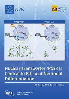
Regulated transport of signaling molecules between the nucleus and cytosol of embryonic stem cells is central to the maintenance of stem cell pluripotency as well as activation of differentiation into specific cell types. This study reports a novel role for the nuclear transporter Importin-13 in regulating differentiation of stem cells into neuronal lineage. Neural progenitor cells generated from Importin-13 knock-out stem cells display reduced neurogenic potential due to impaired Importin-13-dependent nuclear import of the neural development transcription factor Pax6. As a result, the knock-out cells have reduced Pax6 transcriptional activity and decreased expression of key genes required to differentiate progenitor cells into neurons. The study highlights a key mechanism by which Importin-13 potentiates the transcriptional regulatory network underlying neuronal differentiation. View this paper
  • Issues are regarded as officially published after their release is announced to the table of contents alert mailing list.
  • You may sign up for e-mail alerts to receive table of contents of newly released issues.
  • PDF is the official format for papers published in both, html and pdf forms. To view the papers in pdf format, click on the "PDF Full-text" link, and use the free Adobe Reader to open them.
Order results
Result details
Section
Select all
Export citation of selected articles as:
23 pages, 1941 KiB  
Review
The Good and the Bad: Monocytes’ and Macrophages’ Diverse Functions in Inflammation
by Judith Austermann, Johannes Roth and Katarzyna Barczyk-Kahlert
Cells 2022, 11(12), 1979; https://doi.org/10.3390/cells11121979 - 20 Jun 2022
Cited by 40 | Viewed by 7017
Abstract
Monocytes and macrophages are central players of the innate immune response and play a pivotal role in the regulation of inflammation. Thereby, they actively participate in all phases of the immune response, from initiating inflammation and triggering the adaptive immune response, through to [...] Read more.
Monocytes and macrophages are central players of the innate immune response and play a pivotal role in the regulation of inflammation. Thereby, they actively participate in all phases of the immune response, from initiating inflammation and triggering the adaptive immune response, through to the clearance of cell debris and resolution of inflammation. In this review, we described the mechanisms of monocyte and macrophage adaptation to rapidly changing microenvironmental conditions and discussed different forms of macrophage polarization depending on the environmental cues or pathophysiological condition. Therefore, special focus was placed on the tight regulation of the pro- and anti-inflammatory immune response, and the diverse functions of S100A8/S100A9 proteins and the scavenger receptor CD163 were highlighted, respectively. We paid special attention to the function of pro- and anti-inflammatory macrophages under pathological conditions. Full article
Show Figures

Figure 1

15 pages, 1413 KiB  
Article
Programmed Cell Death-1/Programmed Cell Death-1 Ligand as Prognostic Markers of Coronavirus Disease 2019 Severity
by Paulina Niedźwiedzka-Rystwej, Adam Majchrzak, Bogusz Aksak-Wąs, Karol Serwin, Zenon Czajkowski, Ewelina Grywalska, Izabela Korona-Głowniak, Jacek Roliński and Miłosz Parczewski
Cells 2022, 11(12), 1978; https://doi.org/10.3390/cells11121978 - 20 Jun 2022
Cited by 10 | Viewed by 1870
Abstract
Current research proves that immune dysregulation is a common feature of coronavirus disease 2019 (COVID-19), and immune exhaustion is associated with increased disease mortality. Immune checkpoint molecules, including the programmed cell death-1 (PD-1)/PD-1 ligand (PD-L1) axis, may serve as markers of disease severity. [...] Read more.
Current research proves that immune dysregulation is a common feature of coronavirus disease 2019 (COVID-19), and immune exhaustion is associated with increased disease mortality. Immune checkpoint molecules, including the programmed cell death-1 (PD-1)/PD-1 ligand (PD-L1) axis, may serve as markers of disease severity. Accordingly, in this study, we evaluated the expression of PD-1/PD-L1 in patients with COVID-19. Blood immunophenotypes of hospitalized patients with moderate (n = 17, requiring oxygen support) and severe (n = 35, requiring mechanical ventilation in the intensive care setting) COVID-19 were compared and associated with clinical, laboratory, and survival data. The associations between severity and lymphocyte profiles were analysed at baseline and after 7 and 14 days of in-hospital treatment. Forty patients without COVID-19 infection were used as controls. For PD-1-positive T and B lymphocyte subsets, notable increases were observed between controls and patients with moderate or severe COVID-19 for CD4+PD-1+ T cells, CD8+PD-1+ T and CD19+PD-1+ B cells. Similar trends were observed for PD-L1-positive lymphocytes, namely, CD4+PD-L1+ T cells, CD8+PD-L1+ T cells and CD19+PD-L1+ B cells. Importantly, all markers associated with PD-1 and PD-L1 were stable over time for the analysed time points in the moderate and severe COVID-19 groups. Increased abundances of PD-1+ and PD-L1+ lymphocytes were associated with disease severity and mortality and were stable over time in patients with moderate to severe COVID-19. These immune exhaustion parameters may be attractive biomarkers of COVID-19 severity. Full article
(This article belongs to the Special Issue Cutting Edge in Respiratory Disorders Biomarkers Research)
Show Figures

Figure 1

20 pages, 1433 KiB  
Review
Age-Related Lysosomal Dysfunctions
by Lena Guerrero-Navarro, Pidder Jansen-Dürr and Maria Cavinato
Cells 2022, 11(12), 1977; https://doi.org/10.3390/cells11121977 - 20 Jun 2022
Cited by 10 | Viewed by 3744
Abstract
Organismal aging is normally accompanied by an increase in the number of senescent cells, growth-arrested metabolic active cells that affect normal tissue function. These cells present a series of characteristics that have been studied over the last few decades. The damage in cellular [...] Read more.
Organismal aging is normally accompanied by an increase in the number of senescent cells, growth-arrested metabolic active cells that affect normal tissue function. These cells present a series of characteristics that have been studied over the last few decades. The damage in cellular organelles disbalances the cellular homeostatic processes, altering the behavior of these cells. Lysosomal dysfunction is emerging as an important factor that could regulate the production of inflammatory molecules, metabolic cellular state, or mitochondrial function. Full article
(This article belongs to the Special Issue Controversies and Recent Advances in Senescence and Aging)
Show Figures

Figure 1

3 pages, 205 KiB  
Editorial
Fundamental and Applied Advances in Stem Cell Therapeutic Research
by Makram Merimi, Saida Rahmani, Ahmed Afailal Tribak, Fatima Bouhtit, Hassan Fahmi and Mehdi Najar
Cells 2022, 11(12), 1976; https://doi.org/10.3390/cells11121976 - 20 Jun 2022
Viewed by 1488
Abstract
We are pleased to present this Special Issue of Cells, entitled ‘Feature Papers in Stem Cells’ [...] Full article
(This article belongs to the Special Issue Feature Papers in Stem Cells)
17 pages, 28469 KiB  
Article
P-Cadherin Is Expressed by Epithelial Progenitor Cells and Melanocytes in the Human Corneal Limbus
by Naresh Polisetti, Lyne Sharaf, Gottfried Martin, Günther Schlunck and Thomas Reinhard
Cells 2022, 11(12), 1975; https://doi.org/10.3390/cells11121975 - 20 Jun 2022
Cited by 8 | Viewed by 2487
Abstract
Interactions between limbal epithelial progenitor cells (LEPC) and surrounding niche cells, which include limbal mesenchymal stromal cells (LMSC) and melanocytes (LM), are essential for the maintenance of the limbal stem cell niche required for a transparent corneal surface. P-cadherin (P-cad) is a critical [...] Read more.
Interactions between limbal epithelial progenitor cells (LEPC) and surrounding niche cells, which include limbal mesenchymal stromal cells (LMSC) and melanocytes (LM), are essential for the maintenance of the limbal stem cell niche required for a transparent corneal surface. P-cadherin (P-cad) is a critical stem cell niche adhesion molecule at various epithelial stem cell niches; however, conflicting observations were reported on the presence of P-cad in the limbal region. To explore this issue, we assessed the location and phenotype of P-cad+ cells by confocal microscopy of human corneoscleral tissue. In subsequent fluorescence-activated cell sorting (FACS) experiments, we used antibodies against P-cad along with CD90 and CD117 for the enrichment of LEPC, LMSC and LM, respectively. The sorted cells were characterized by immunophenotyping and the repopulation of decellularized limbal scaffolds was evaluated. Our findings demonstrate that P-cad is expressed by epithelial progenitor cells as well as melanocytes in the human limbal epithelial stem cell niche. The modified flow sorting addressing P-cad as well as CD90 and CD117 yielded enriched LEPC (CD90CD117P-cad+) and pure populations of LMSC (CD90+CD117P-cad) and LM (CD90CD117+P-cad+). The enriched LEPC showed the expression of epithelial progenitor markers and better colony-forming ability than their P-cad counterparts. The cultured LEPC and LM exhibited P-cad expression at intercellular junctions and successfully repopulated decellularized limbal scaffolds. These data suggest that P-cad is a critical cell–cell adhesion molecule, connecting LEPC and LM, which may play an important role in the long-term maintenance of LEPC at the limbal stem cell niche; moreover, these findings led to further improvement of cell enrichment protocols to enhance the yield of LEPC. Full article
Show Figures

Figure 1

26 pages, 3421 KiB  
Review
CAR T Cell Locomotion in Solid Tumor Microenvironment
by Duy T. Nguyen, Elizabeth Ogando-Rivas, Ruixuan Liu, Theodore Wang, Jacob Rubin, Linchun Jin, Haipeng Tao, William W. Sawyer, Hector R. Mendez-Gomez, Matthew Cascio, Duane A. Mitchell, Jianping Huang, W. Gregory Sawyer, Elias J. Sayour and Paul Castillo
Cells 2022, 11(12), 1974; https://doi.org/10.3390/cells11121974 - 20 Jun 2022
Cited by 14 | Viewed by 6973
Abstract
The promising outcomes of chimeric antigen receptor (CAR) T cell therapy in hematologic malignancies potentiates its capability in the fight against many cancers. Nevertheless, this immunotherapy modality needs significant improvements for the treatment of solid tumors. Researchers have incrementally identified limitations and constantly [...] Read more.
The promising outcomes of chimeric antigen receptor (CAR) T cell therapy in hematologic malignancies potentiates its capability in the fight against many cancers. Nevertheless, this immunotherapy modality needs significant improvements for the treatment of solid tumors. Researchers have incrementally identified limitations and constantly pursued better CAR designs. However, even if CAR T cells are armed with optimal killer functions, they must overcome and survive suppressive barriers imposed by the tumor microenvironment (TME). In this review, we will discuss in detail the important role of TME in CAR T cell trafficking and how the intrinsic barriers contribute to an immunosuppressive phenotype and cancer progression. It is of critical importance that preclinical models can closely recapitulate the in vivo TME to better predict CAR T activity. Animal models have contributed immensely to our understanding of human diseases, but the intensive care for the animals and unreliable representation of human biology suggest in vivo models cannot be the sole approach to CAR T cell therapy. On the other hand, in vitro models for CAR T cytotoxic assessment offer valuable insights to mechanistic studies at the single cell level, but they often lack in vivo complexities, inter-individual heterogeneity, or physiologically relevant spatial dimension. Understanding the advantages and limitations of preclinical models and their applications would enable more reliable prediction of better clinical outcomes. Full article
Show Figures

Figure 1

14 pages, 130577 KiB  
Article
Transcriptional Profiling of Insulin-like Growth Factor Signaling Components in Embryonic Lung Development and Idiopathic Pulmonary Fibrosis
by Vahid Kheirollahi, Ali Khadim, Georgios Kiliaris, Martina Korfei, Margarida Maria Barroso, Ioannis Alexopoulos, Ana Ivonne Vazquez-Armendariz, Malgorzata Wygrecka, Clemens Ruppert, Andreas Guenther, Werner Seeger, Susanne Herold and Elie El Agha
Cells 2022, 11(12), 1973; https://doi.org/10.3390/cells11121973 - 20 Jun 2022
Cited by 4 | Viewed by 2412
Abstract
Insulin-like growth factor (IGF) signaling controls the development and growth of many organs, including the lung. Loss of function of Igf1 or its receptor Igf1r impairs lung development and leads to neonatal respiratory distress in mice. Although many components of the IGF signaling [...] Read more.
Insulin-like growth factor (IGF) signaling controls the development and growth of many organs, including the lung. Loss of function of Igf1 or its receptor Igf1r impairs lung development and leads to neonatal respiratory distress in mice. Although many components of the IGF signaling pathway have shown to be dysregulated in idiopathic pulmonary fibrosis (IPF), the expression pattern of such components in different cellular compartments of the developing and/or fibrotic lung has been elusive. In this study, we provide a comprehensive transcriptional profile for such signaling components during embryonic lung development in mice, bleomycin-induced pulmonary fibrosis in mice and in human IPF lung explants. During late gestation, we found that Igf1 is upregulated in parallel to Igf1r downregulation in the lung mesenchyme. Lung tissues derived from bleomycin-treated mice and explanted IPF lungs revealed upregulation of IGF1 in parallel to downregulation of IGF1R, in addition to upregulation of several IGF binding proteins (IGFBPs) in lung fibrosis. Finally, treatment of IPF lung fibroblasts with recombinant IGF1 led to myogenic differentiation. Our data serve as a resource for the transcriptional profile of IGF signaling components and warrant further research on the involvement of this pathway in both lung development and pulmonary disease. Full article
(This article belongs to the Special Issue State of the Art in Idiopathic Pulmonary Fibrosis)
Show Figures

Figure 1

17 pages, 1096 KiB  
Review
Vascular Endothelial Glycocalyx Damage and Potential Targeted Therapy in COVID-19
by Duoduo Zha, Mingui Fu and Yisong Qian
Cells 2022, 11(12), 1972; https://doi.org/10.3390/cells11121972 - 19 Jun 2022
Cited by 18 | Viewed by 3956
Abstract
COVID-19 is a highly infectious respiratory disease caused by a new coronavirus known as SARS-CoV-2. COVID-19 is characterized by progressive respiratory failure resulting from diffuse alveolar damage, inflammatory infiltrates, endotheliitis, and pulmonary and systemic coagulopathy forming obstructive microthrombi with multi-organ dysfunction, indicating that [...] Read more.
COVID-19 is a highly infectious respiratory disease caused by a new coronavirus known as SARS-CoV-2. COVID-19 is characterized by progressive respiratory failure resulting from diffuse alveolar damage, inflammatory infiltrates, endotheliitis, and pulmonary and systemic coagulopathy forming obstructive microthrombi with multi-organ dysfunction, indicating that endothelial cells (ECs) play a central role in the pathogenesis of COVID-19. The glycocalyx is defined as a complex gel-like layer of glycosylated lipid–protein mixtures, which surrounds all living cells and acts as a buffer between the cell and the extracellular matrix. The endothelial glycocalyx layer (EGL) plays an important role in vascular homeostasis via regulating vascular permeability, cell adhesion, mechanosensing for hemodynamic shear stresses, and antithrombotic and anti-inflammatory functions. Here, we review the new findings that described EGL damage in ARDS, coagulopathy, and the multisystem inflammatory disease associated with COVID-19. Mechanistically, the inflammatory mediators, reactive oxygen species (ROS), matrix metalloproteases (MMPs), the glycocalyx fragments, and the viral proteins may contribute to endothelial glycocalyx damage in COVID-19. In addition, the potential therapeutic strategies targeting the EGL for the treatment of severe COVID-19 are summarized and discussed. Full article
Show Figures

Figure 1

14 pages, 2025 KiB  
Article
Changes in the Oxidation-Reduction State of Human Dermal Fibroblasts as an Effect of Lomefloxacin Phototoxic Action
by Justyna Kowalska, Klaudia Banach, Zuzanna Rzepka, Jakub Rok, Marta Karkoszka and Dorota Wrześniok
Cells 2022, 11(12), 1971; https://doi.org/10.3390/cells11121971 - 19 Jun 2022
Cited by 5 | Viewed by 1480
Abstract
Phototoxicity induced by antibiotics is a real problem in health care. The discontinuation of antibiotic therapy due to a phototoxic reaction can lead to the development of resistant strains. Fluoroquinolones are widely used antibiotics that exhibit phototoxic activity under UVA radiation. The purpose [...] Read more.
Phototoxicity induced by antibiotics is a real problem in health care. The discontinuation of antibiotic therapy due to a phototoxic reaction can lead to the development of resistant strains. Fluoroquinolones are widely used antibiotics that exhibit phototoxic activity under UVA radiation. The purpose of the study was to examine the redox status of human dermal fibroblasts exposed to UVA radiation and treated with lomefloxacin, the most phototoxic fluoroquinolone. Lomefloxacin alone was found to have an antiproliferative activity on fibroblasts by affecting the cell cycle. In addition, the drug caused a redox imbalance associated with the decreased expression of catalase and glutathione peroxidase. UVA radiation increased the drug cytotoxicity and oxidative stress induced by lomefloxacin. The decrease in cell viability was accompanied by a high level of reactive oxygen species and extensive changes in the antioxidant levels. The revealed data indicate that the phototoxic action of lomefloxacin results from both increased reactive oxygen species production and an impaired antioxidant defense system. Considering all of the findings, it can be concluded that lomefloxacin-induced phototoxic reactions are caused by an oxidoreductive imbalance in skin cells. Full article
Show Figures

Figure 1

11 pages, 1502 KiB  
Communication
LTα, TNF, and ILC3 in Peyer’s Patch Organogenesis
by Violetta S. Gogoleva, Dmitry V. Kuprash, Sergei I. Grivennikov, Alexei V. Tumanov, Andrey A. Kruglov and Sergei A. Nedospasov
Cells 2022, 11(12), 1970; https://doi.org/10.3390/cells11121970 - 19 Jun 2022
Cited by 4 | Viewed by 2232
Abstract
TNF and LTα are structurally related cytokines of the TNF superfamily. Their genes are located in close proximity to each other and to the Ltb gene within the TNF/LT locus inside MHC. Unlike Ltb, transcription of Tnf and of Lta is tightly [...] Read more.
TNF and LTα are structurally related cytokines of the TNF superfamily. Their genes are located in close proximity to each other and to the Ltb gene within the TNF/LT locus inside MHC. Unlike Ltb, transcription of Tnf and of Lta is tightly controlled, with the Tnf gene being an immediate early gene that is rapidly induced in response to various inflammatory stimuli. Genes of the TNF/LT locus play a crucial role in lymphoid tissue organogenesis, although some aspects of their specific contribution remain controversial. Here, we present new findings and discuss the distinct contribution of TNF produced by ILC3 cells to Peyer’s patch organogenesis. Full article
(This article belongs to the Special Issue The TNF Receptor Superfamily in Health and Disease)
Show Figures

Figure 1

21 pages, 6713 KiB  
Article
A Robust Assay to Monitor Ataxin-3 Amyloid Fibril Assembly
by Francisco Figueiredo, Mónica Lopes-Marques, Bruno Almeida, Nena Matscheko, Pedro M. Martins, Alexandra Silva and Sandra Macedo-Ribeiro
Cells 2022, 11(12), 1969; https://doi.org/10.3390/cells11121969 - 19 Jun 2022
Cited by 2 | Viewed by 2729
Abstract
Spinocerebellar ataxia type 3 (SCA3) is caused by the expansion of a glutamine repeat in the protein ataxin-3, which is deposited as intracellular aggregates in affected brain regions. Despite the controversial role of ataxin-3 amyloid structures in SCA3 pathology, the identification of molecules [...] Read more.
Spinocerebellar ataxia type 3 (SCA3) is caused by the expansion of a glutamine repeat in the protein ataxin-3, which is deposited as intracellular aggregates in affected brain regions. Despite the controversial role of ataxin-3 amyloid structures in SCA3 pathology, the identification of molecules with the capacity to prevent aberrant self-assembly and stabilize functional conformation(s) of ataxin-3 is a key to the development of therapeutic solutions. Amyloid-specific kinetic assays are routinely used to measure rates of protein self-assembly in vitro and are employed during screening for fibrillation inhibitors. The high tendency of ataxin-3 to assemble into oligomeric structures implies that minor changes in experimental conditions can modify ataxin-3 amyloid assembly kinetics. Here, we determine the self-association rates of ataxin-3 and present a detailed study of the aggregation of normal and pathogenic ataxin-3, highlighting the experimental conditions that should be considered when implementing and validating ataxin-3 amyloid progress curves in different settings and in the presence of ataxin-3 interactors. This assay provides a unique and robust platform to screen for modulators of the first steps of ataxin-3 aggregation—a starting point for further studies with cell and animal models of SCA3. Full article
Show Figures

Graphical abstract

25 pages, 5013 KiB  
Article
When Electrospun Fiber Support Matters: In Vitro Ovine Long-Term Folliculogenesis on Poly (Epsilon Caprolactone) (PCL)-Patterned Fibers
by Chiara Di Berardino, Liliana Liverani, Alessia Peserico, Giulia Capacchietti, Valentina Russo, Nicola Bernabò, Umberto Tosi, Aldo Roberto Boccaccini and Barbara Barboni
Cells 2022, 11(12), 1968; https://doi.org/10.3390/cells11121968 - 19 Jun 2022
Cited by 6 | Viewed by 1968
Abstract
Current assisted reproduction technologies (ART) are insufficient to cover the slice of the population needing to restore fertility, as well as to amplify the reproductive performance of domestic animals or endangered species. The design of dedicated reproductive scaffolds has opened the possibility to [...] Read more.
Current assisted reproduction technologies (ART) are insufficient to cover the slice of the population needing to restore fertility, as well as to amplify the reproductive performance of domestic animals or endangered species. The design of dedicated reproductive scaffolds has opened the possibility to better recapitulate the reproductive 3D ovarian environment, thus potentially innovating in vitro folliculogenesis (ivF) techniques. To this aim, the present research has been designed to compare ovine preantral follicles in vitro culture on poly(epsilon-caprolactone) (PCL)-based electrospun scaffolds designed with different topology (Random vs. Patterned fibers) with a previously validated system. The ivF performances were assessed after 14 days under 3D-oil, Two-Step (7 days in 3D-oil and on scaffold), or One-Step PCL protocols (14 days on PCL-scaffold) by assessing morphological and functional outcomes. The results show that Two- and One-Step PCL ivF protocols, when performed on patterned scaffolds, were both able to support follicle growth, antrum formation, and the upregulation of follicle marker genes leading to a greater oocyte meiotic competence than in the 3D-oil system. In conclusion, the One-Step approach could be proposed as a practical and valid strategy to support a synergic follicle-oocyte in vitro development, providing an innovative tool to enhance the availability of matured gametes on an individual basis for ART purposes. Full article
(This article belongs to the Section Reproductive Cells and Development)
Show Figures

Graphical abstract

17 pages, 2830 KiB  
Article
The Role of AlphαSynuclein in Mouse Models of Acute, Inflammatory and Neuropathic Pain
by Moritz Möller, Christine V. Möser, Ulrike Weiß and Ellen Niederberger
Cells 2022, 11(12), 1967; https://doi.org/10.3390/cells11121967 - 19 Jun 2022
Cited by 3 | Viewed by 2105
Abstract
(1) AlphαSynuclein (αSyn) is a synaptic protein which is expressed in the nervous system and has been linked to neurodegenerative diseases, in particular Parkinson’s disease (PD). Symptoms of PD are mainly due to overexpression and aggregation of αSyn and include pain. However, the [...] Read more.
(1) AlphαSynuclein (αSyn) is a synaptic protein which is expressed in the nervous system and has been linked to neurodegenerative diseases, in particular Parkinson’s disease (PD). Symptoms of PD are mainly due to overexpression and aggregation of αSyn and include pain. However, the interconnection of αSyn and pain has not been clarified so far. (2) We investigated the potential effects of a αSyn knock-out on the nociceptive behaviour in mouse models of acute, inflammatory and neuropathic pain. Furthermore, we assessed the impact of αSyn deletion on pain-related cellular and molecular mechanisms in the spinal cord in these models. (3) Our results showed a reduction of acute cold nociception in αSyn knock-out mice while responses to acute heat and mechanical noxious stimulation were similar in wild type and knock-out mice. Inflammatory nociception was not affected by αSyn knock-out which is also mirrored by unaltered inflammatory gene expression. In contrast, in the SNI model of neuropathic pain, αSyn knock-out mice showed decreased mechanical allodynia as compared to wild type mice. This effect was associated with reduced proinflammatory mechanisms and suppressed activation of MAP kinase signalling in the spinal cord while endogenous antinociceptive mechanisms are not inhibited. (4) Our data indicate that αSyn plays a role in neuropathy and its inhibition might be useful to ameliorate pain symptoms after nerve injury. Full article
Show Figures

Figure 1

26 pages, 879 KiB  
Review
The Senescence Markers p16INK4A, p14ARF/p19ARF, and p21 in Organ Development and Homeostasis
by Kay-Dietrich Wagner and Nicole Wagner
Cells 2022, 11(12), 1966; https://doi.org/10.3390/cells11121966 - 19 Jun 2022
Cited by 31 | Viewed by 6131
Abstract
It is widely accepted that senescent cells accumulate with aging. They are characterized by replicative arrest and the release of a myriad of factors commonly called the senescence-associated secretory phenotype. Despite the replicative cell cycle arrest, these cells are metabolically active and functional. [...] Read more.
It is widely accepted that senescent cells accumulate with aging. They are characterized by replicative arrest and the release of a myriad of factors commonly called the senescence-associated secretory phenotype. Despite the replicative cell cycle arrest, these cells are metabolically active and functional. The release of SASP factors is mostly thought to cause tissue dysfunction and to induce senescence in surrounding cells. As major markers for aging and senescence, p16INK4, p14ARF/p19ARF, and p21 are established. Importantly, senescence is also implicated in development, cancer, and tissue homeostasis. While many markers of senescence have been identified, none are able to unambiguously identify all senescent cells. However, increased levels of the cyclin-dependent kinase inhibitors p16INK4A and p21 are often used to identify cells with senescence-associated phenotypes. We review here the knowledge of senescence, p16INK4A, p14ARF/p19ARF, and p21 in embryonic and postnatal development and potential functions in pathophysiology and homeostasis. The establishment of senolytic therapies with the ultimate goal to improve healthy aging requires care and detailed knowledge about the involvement of senescence and senescence-associated proteins in developmental processes and homeostatic mechanism. The review contributes to these topics, summarizes open questions, and provides some directions for future research. Full article
(This article belongs to the Special Issue Controversies and Recent Advances in Senescence and Aging)
Show Figures

Figure 1

10 pages, 1513 KiB  
Article
Proteomic and Metabolomic Profiles of T Cell-Derived Exosomes Isolated from Human Plasma
by Aneta Zebrowska, Karol Jelonek, Sujan Mondal, Marta Gawin, Katarzyna Mrowiec, Piotr Widłak, Theresa Whiteside and Monika Pietrowska
Cells 2022, 11(12), 1965; https://doi.org/10.3390/cells11121965 - 18 Jun 2022
Cited by 6 | Viewed by 2527
Abstract
Exosomes that are released by T cells are key messengers involved in immune regulation. However, the molecular profiling of these vesicles, which is necessary for understanding their functions, requires their isolation from a very heterogeneous mixture of extracellular vesicles that are present in [...] Read more.
Exosomes that are released by T cells are key messengers involved in immune regulation. However, the molecular profiling of these vesicles, which is necessary for understanding their functions, requires their isolation from a very heterogeneous mixture of extracellular vesicles that are present in the human plasma. It has been shown that exosomes that are produced by T cells could be isolated from plasma by immune capture using antibodies that target the CD3 antigen, which is a key component of the TCR complex that is present in all T lymphocytes. Here, we demonstrate that CD3(+) exosomes that are isolated from plasma can be used for high-throughput molecular profiling using proteomics and metabolomics tools. This profiling allowed for the identification of proteins and metabolites that differentiated the CD3(+) from the CD3(−) exosome fractions that were present in the plasma of healthy donors. Importantly, the proteins and metabolites that accumulated in the CD3(+) vesicles reflected the known molecular features of T lymphocytes. Hence, CD3(+) exosomes that are isolated from human plasma by immune capture could serve as a “T cell biopsy”. Full article
(This article belongs to the Section Cell Signaling)
Show Figures

Figure 1

12 pages, 1074 KiB  
Review
Rapid Actions of the Nuclear Progesterone Receptor through cSrc in Cancer
by Claudia Bello-Alvarez, Carmen J. Zamora-Sánchez and Ignacio Camacho-Arroyo
Cells 2022, 11(12), 1964; https://doi.org/10.3390/cells11121964 - 18 Jun 2022
Cited by 6 | Viewed by 2223
Abstract
The nuclear progesterone receptor (PR) is mainly known for its role as a ligand-regulated transcription factor. However, in the last ten years, this receptor’s extranuclear or rapid actions have gained importance in the context of physiological and pathophysiological conditions such as cancer. The [...] Read more.
The nuclear progesterone receptor (PR) is mainly known for its role as a ligand-regulated transcription factor. However, in the last ten years, this receptor’s extranuclear or rapid actions have gained importance in the context of physiological and pathophysiological conditions such as cancer. The PR’s polyproline (PXPP) motif allows protein–protein interaction through SH3 domains of several cytoplasmatic proteins, including the Src family kinases (SFKs). Among members of this family, cSrc is the most well-characterized protein in the scenario of rapid actions of the PR in cancer. Studies in breast cancer have provided the most detailed information on the signaling and effects triggered by the cSrc–PR interaction. Nevertheless, the study of this phenomenon and its consequences has been underestimated in other types of malignancies, especially those not associated with the reproductive system, such as glioblastomas (GBs). This review will provide a detailed analysis of the impact of the PR–cSrc interplay in the progression of some non-reproductive cancers, particularly, in GBs. Full article
(This article belongs to the Special Issue Progesterone Receptor Signaling)
Show Figures

Figure 1

23 pages, 14578 KiB  
Review
Dysregulated Ca2+ Homeostasis as a Central Theme in Neurodegeneration: Lessons from Alzheimer’s Disease and Wolfram Syndrome
by Manon Callens, Jens Loncke and Geert Bultynck
Cells 2022, 11(12), 1963; https://doi.org/10.3390/cells11121963 - 18 Jun 2022
Cited by 12 | Viewed by 3795
Abstract
Calcium ions (Ca2+) operate as important messengers in the cell, indispensable for signaling the underlying numerous cellular processes in all of the cell types in the human body. In neurons, Ca2+ signaling is crucial for regulating synaptic transmission and for [...] Read more.
Calcium ions (Ca2+) operate as important messengers in the cell, indispensable for signaling the underlying numerous cellular processes in all of the cell types in the human body. In neurons, Ca2+ signaling is crucial for regulating synaptic transmission and for the processes of learning and memory formation. Hence, the dysregulation of intracellular Ca2+ homeostasis results in a broad range of disorders, including cancer and neurodegeneration. A major source for intracellular Ca2+ is the endoplasmic reticulum (ER), which has close contacts with other organelles, including mitochondria. In this review, we focus on the emerging role of Ca2+ signaling at the ER–mitochondrial interface in two different neurodegenerative diseases, namely Alzheimer’s disease and Wolfram syndrome. Both of these diseases share some common hallmarks in the early stages, including alterations in the ER and mitochondrial Ca2+ handling, mitochondrial dysfunction and increased Reactive oxygen species (ROS) production. This indicates that similar mechanisms may underly these two disease pathologies and suggests that both research topics might benefit from complementary research. Full article
(This article belongs to the Special Issue Calcium Signaling in Health and Diseases)
Show Figures

Figure 1

22 pages, 4055 KiB  
Article
Optimizing Soluble Cues for Salivary Gland Tissue Mimetics Using a Design of Experiments (DoE) Approach
by Lindsay R. Piraino, Danielle S. W. Benoit and Lisa A. DeLouise
Cells 2022, 11(12), 1962; https://doi.org/10.3390/cells11121962 - 18 Jun 2022
Cited by 3 | Viewed by 1854
Abstract
The development of therapies to prevent or treat salivary gland dysfunction has been limited by a lack of functional in vitro models. Specifically, critical markers of salivary gland secretory phenotype downregulate rapidly ex vivo. Here, we utilize a salivary gland tissue chip model [...] Read more.
The development of therapies to prevent or treat salivary gland dysfunction has been limited by a lack of functional in vitro models. Specifically, critical markers of salivary gland secretory phenotype downregulate rapidly ex vivo. Here, we utilize a salivary gland tissue chip model to conduct a design of experiments (DoE) approach to test combinations of seven soluble cues that were previously shown to maintain or improve salivary gland cell function. This approach uses statistical techniques to improve efficiency and accuracy of combinations of factors. The DoE-designed culture conditions improve markers of salivary gland function. Data show that the EGFR inhibitor, EKI-785, maintains relative mRNA expression of Mist1, a key acinar cell transcription factor, while FGF10 and neurturin promote mRNA expression of Aqp5 and Tmem16a, channel proteins involved in secretion. Mist1 mRNA expression correlates with increased secretory function, including calcium signaling and mucin (PAS-AB) staining. Overall, this study demonstrates that media conditions can be efficiently optimized to support secretory function in vitro using a DoE approach. Full article
Show Figures

Graphical abstract

21 pages, 4676 KiB  
Article
Deltamethrin-Evoked ER Stress Promotes Neuroinflammation in the Adult Mouse Hippocampus
by Muhammad M. Hossain, Abigail C. Toltin, Laura M. Gamba and Maria A. Molina
Cells 2022, 11(12), 1961; https://doi.org/10.3390/cells11121961 - 18 Jun 2022
Cited by 12 | Viewed by 2340
Abstract
Endoplasmic reticulum (ER) stress and neuroinflammation are involved in the pathogenesis of many neurodegenerative disorders. Previously, we reported that exposure to pyrethroid insecticide deltamethrin causes hippocampal ER stress apoptosis, a reduction in neurogenesis, and learning deficits in adult male mice. Recently, we found [...] Read more.
Endoplasmic reticulum (ER) stress and neuroinflammation are involved in the pathogenesis of many neurodegenerative disorders. Previously, we reported that exposure to pyrethroid insecticide deltamethrin causes hippocampal ER stress apoptosis, a reduction in neurogenesis, and learning deficits in adult male mice. Recently, we found that deltamethrin exposure also increases the markers of neuroinflammation in BV2 cells. Here, we investigated the potential mechanistic link between ER stress and neuroinflammation following exposure to deltamethrin. We found that repeated oral exposure to deltamethrin (3 mg/kg) for 30 days caused microglial activation and increased gene expressions and protein levels of TNF-α, IL-1β, IL-6, gp91phox, 4HNE, and iNOS in the hippocampus. These changes were preceded by the induction of ER stress as the protein levels of CHOP, ATF-4, and GRP78 were significantly increased in the hippocampus. To determine whether induction of ER stress triggers the inflammatory response, we performed an additional experiment with mouse microglial cell (MMC) line. MMCs were treated with 0–5 µM deltamethrin for 24–48 h in the presence or absence of salubrinal, a pharmacological inhibitor of the ER stress factor eIF2α. We found that salubrinal (50 µM) prevented deltamethrin-induced ER stress, as indicated by decreased levels of CHOP and ATF-4, and attenuated the levels of GSH, 4-HNE, gp91phox, iNOS, ROS, TNF-α, IL-1β, and IL-6 in MMCs. Together, these results demonstrate that exposure to deltamethrin leads to ER stress-mediated neuroinflammation, which may subsequently contribute to neurodegeneration and cognitive impairment in mice. Full article
(This article belongs to the Special Issue Molecular Mechanism of Stress, Stress Response, and Adaptation)
Show Figures

Figure 1

14 pages, 5429 KiB  
Article
The Prognostic and Predictive Significance of Tumor-Infiltrating Memory T Cells Is Reversed in High-Risk HNSCC
by Rebekka Hartan, Sören Schnellhardt, Maike Büttner-Herold, Christoph Daniel, Arndt Hartmann, Rainer Fietkau and Luitpold Distel
Cells 2022, 11(12), 1960; https://doi.org/10.3390/cells11121960 - 17 Jun 2022
Cited by 3 | Viewed by 1723
Abstract
Tumor-infiltrating CD45RO+ memory T cells have unanimously been described as a positive prognostic factor in head and neck squamous cell carcinomas (HNSCCs). Here, we investigated the long-term prognostic relevance of CD45RO+ memory T cells in HNSCC with special regard to the influence of [...] Read more.
Tumor-infiltrating CD45RO+ memory T cells have unanimously been described as a positive prognostic factor in head and neck squamous cell carcinomas (HNSCCs). Here, we investigated the long-term prognostic relevance of CD45RO+ memory T cells in HNSCC with special regard to the influence of clinical characteristics. Pre-treatment biopsy samples from 306 patients with predominantly advanced HNSCC were analyzed. Immunohistochemistry was used to stain tissue microarrays for CD45RO+ memory T cells. CD45RO cell densities were semi-automatically registered and used for survival analysis. High CD45RO+ cell densities were clearly associated with prolonged overall survival (OS) and recurrence-free survival as well as no evidence of disease status after 10 years (p < 0.05). In contrast, the prognostic significance of tumor-infiltrating memory T cells was completely reversed in high-risk groups: in poorly differentiated tumors (G3, G4) and in cases with lymph node involvement (N+), high memory T cell densities correlated with reduced 10-year OS (p < 0.05). In conclusion, an increased density of tumor-infiltrating CD45RO+ cells in HNSCC can be a positive as well as a negative prognostic factor, depending on disease stage and histological grade. Therefore, if CD45RO+ cell density is to be used as a prognostic biomarker, further clinical characteristics must be considered. Full article
(This article belongs to the Special Issue Cancer and Radiation Therapy)
Show Figures

Figure 1

21 pages, 2834 KiB  
Article
Ocrelizumab in Patients with Active Primary Progressive Multiple Sclerosis: Clinical Outcomes and Immune Markers of Treatment Response
by Marina Boziki, Christos Bakirtzis, Styliani-Aggeliki Sintila, Evangelia Kesidou, Evdoxia Gounari, Aliki Ioakimidou, Vasiliki Tsavdaridou, Lemonia Skoura, Asimina Fylaktou, Vasiliki Nikolaidou, Maria Stangou, Ioannis Nikolaidis, Virginia Giantzi, Eleni Karafoulidou, Paschalis Theotokis and Nikolaos Grigoriadis
Cells 2022, 11(12), 1959; https://doi.org/10.3390/cells11121959 - 17 Jun 2022
Cited by 3 | Viewed by 2973
Abstract
Ocrelizumab is a B-cell-depleting monoclonal antibody approved for the treatment of relapsing-remitting multiple sclerosis (RRMS) and active primary progressive MS (aPPMS). This prospective, uncontrolled, open-label, observational study aimed to assess the efficacy of ocrelizumab in patients with aPPMS and to dissect the clinical, [...] Read more.
Ocrelizumab is a B-cell-depleting monoclonal antibody approved for the treatment of relapsing-remitting multiple sclerosis (RRMS) and active primary progressive MS (aPPMS). This prospective, uncontrolled, open-label, observational study aimed to assess the efficacy of ocrelizumab in patients with aPPMS and to dissect the clinical, radiological and laboratory attributes of treatment response. In total, 22 patients with aPPMS followed for 24 months were included. The primary efficacy outcome was the proportion of patients with optimal response at 24 months, defined as patients free of relapses, free of confirmed disability accumulation (CDA) and free of T1 Gd-enhancing lesions and new/enlarging T2 lesions on the brain and cervical MRI. In total, 14 (63.6%) patients and 13 patients (59.1%) were classified as responders at 12 and 24 months, respectively. Time exhibited a significant effect on mean absolute and normalized gray matter cerebellar volume (F = 4.342, p = 0.23 and F = 4.279, p = 0.024, respectively). Responders at 24 months exhibited reduced peripheral blood ((%) of CD19+ cells) plasmablasts compared to non-responders at the 6-month point estimate (7.69 ± 4.4 vs. 22.66 ± 7.19, respectively, p = 0.043). Response to ocrelizumab was linked to lower total and gray matter cerebellar volume loss over time. Reduced plasmablast depletion was linked for the first time to sub-optimal response to ocrelizumab in aPPMS. Full article
(This article belongs to the Special Issue Mechanisms of Action of Therapies for Multiple Sclerosis)
Show Figures

Graphical abstract

18 pages, 1433 KiB  
Review
Telomere Maintenance and the cGAS-STING Pathway in Cancer
by Hiroshi Ebata, Tze Mun Loo and Akiko Takahashi
Cells 2022, 11(12), 1958; https://doi.org/10.3390/cells11121958 - 17 Jun 2022
Cited by 2 | Viewed by 3841
Abstract
Cancer cells exhibit the unique characteristics of high proliferation and aberrant DNA damage response, which prevents cancer therapy from effectively eliminating them. The machinery required for telomere maintenance, such as telomerase and the alternative lengthening of telomeres (ALT), enables cancer cells to proliferate [...] Read more.
Cancer cells exhibit the unique characteristics of high proliferation and aberrant DNA damage response, which prevents cancer therapy from effectively eliminating them. The machinery required for telomere maintenance, such as telomerase and the alternative lengthening of telomeres (ALT), enables cancer cells to proliferate indefinitely. In addition, the molecules in this system are involved in noncanonical pro-tumorigenic functions. Of these, the function of the cyclic GMP–AMP synthase (cGAS)-stimulator of interferon genes (STING) pathway, which contains telomere-related molecules, is a well-known contributor to the tumor microenvironment (TME). This review summarizes the current knowledge of the role of telomerase and ALT in cancer regulation, with emphasis on their noncanonical roles beyond telomere maintenance. The components of the cGAS-STING pathway are summarized with respect to intercell communication in the TME. Elucidating the underlying functional connection between telomere-related molecules and TME regulation is important for the development of cancer therapeutics that target cancer-specific pathways in different contexts. Finally, strategies for designing new cancer therapies that target cancer cells and the TME are discussed. Full article
(This article belongs to the Special Issue DNA Damage Response Regulation and Cancer)
Show Figures

Figure 1

20 pages, 2000 KiB  
Review
Platelets at the Crossroads of Pro-Inflammatory and Resolution Pathways during Inflammation
by Nadine Ludwig, Annika Hilger, Alexander Zarbock and Jan Rossaint
Cells 2022, 11(12), 1957; https://doi.org/10.3390/cells11121957 - 17 Jun 2022
Cited by 22 | Viewed by 5903
Abstract
Platelets are among the most abundant cells in the mammalian circulation. Classical platelet functions in hemostasis and wound healing have been intensively explored and are generally accepted. During the past decades, the research focus broadened towards their participation in immune-modulatory events, including pro-inflammatory [...] Read more.
Platelets are among the most abundant cells in the mammalian circulation. Classical platelet functions in hemostasis and wound healing have been intensively explored and are generally accepted. During the past decades, the research focus broadened towards their participation in immune-modulatory events, including pro-inflammatory and, more recently, inflammatory resolution processes. Platelets are equipped with a variety of abilities enabling active participation in immunological processes. Toll-like receptors mediate the recognition of pathogens, while the release of granule contents and microvesicles promotes direct pathogen defense and an interaction with leukocytes. Platelets communicate and physically interact with neutrophils, monocytes and a subset of lymphocytes via soluble mediators and surface adhesion receptors. This interaction promotes leukocyte recruitment, migration and extravasation, as well as the initiation of effector functions, such as the release of extracellular traps by neutrophils. Platelet-derived prostaglandin E2, C-type lectin-like receptor 2 and transforming growth factor β modulate inflammatory resolution processes by promoting the synthesis of pro-resolving mediators while reducing pro-inflammatory ones. Furthermore, platelets promote the differentiation of CD4+ T cells in T helper and regulatory T cells, which affects macrophage polarization. These abilities make platelets key players in inflammatory diseases such as pneumonia and the acute respiratory distress syndrome, including the pandemic coronavirus disease 2019. This review focuses on recent findings in platelet-mediated immunity during acute inflammation. Full article
Show Figures

Figure 1

17 pages, 5482 KiB  
Article
Regulation of Light Spectra on Cell Division of the Unicellular Green Alga Haematococcus pluvialis: Insights from Physiological and Lipidomic Analysis
by Kuo Zhao, Yanhua Li, Hailong Yan, Qiang Hu and Danxiang Han
Cells 2022, 11(12), 1956; https://doi.org/10.3390/cells11121956 - 17 Jun 2022
Cited by 6 | Viewed by 1801
Abstract
Commercial scale production of natural astaxanthin is currently conducted through cultivation of the green alga Haematococcus pluvialis. This study comprehensively investigated the impact of seven different light spectra on the growth, morphology and photosynthesis of H. pluvialis vegetative cells. Further, the lipidomes [...] Read more.
Commercial scale production of natural astaxanthin is currently conducted through cultivation of the green alga Haematococcus pluvialis. This study comprehensively investigated the impact of seven different light spectra on the growth, morphology and photosynthesis of H. pluvialis vegetative cells. Further, the lipidomes of vegetative H. pluvialis grown under various light spectra were qualitatively and quantitatively analyzed using liquid chromatography/mass spectrometry (LC/MS). The results showed the existence of blue light—alone or with red light—promoted cell division, while pure red light or white light enabled increased cell sizes, cellular pigment, starch and lipid contents, and biomass production. Although the photosynthetic performance of H. pluvialis measured as chlorophyll a fluorescence was not significantly affected by light spectra, the lipid profiles, particularly chloroplast membrane lipids, showed remarkable changes with light spectra. The contents of most lipid species in the blue/red light 1/2 group, which showed the fastest cell division, remained at a moderate level compared with those under other light spectra, indicating the fastest dividing cells were featured by a fine-tuned lipid profile. From biotechnical perspective, this comprehensive study can provide insights into the development of appropriate light regimes to promote the cell density or biomass of H. pluvialis mass culture. Full article
(This article belongs to the Special Issue Growth and Division in Algae)
Show Figures

Figure 1

25 pages, 8411 KiB  
Article
Generating iPSCs with a High-Efficient, Non-Invasive Method—An Improved Way to Cultivate Keratinocytes from Plucked Hair for Reprogramming
by Lisa S. Wüstner, Moritz Klingenstein, Karl G. Frey, Mohammad R. Nikbin, Alfio Milazzo, Alexander Kleger, Stefan Liebau and Stefanie Klingenstein
Cells 2022, 11(12), 1955; https://doi.org/10.3390/cells11121955 - 17 Jun 2022
Cited by 2 | Viewed by 2128
Abstract
Various somatic cell types are suitable for induced pluripotency reprogramming, such as dermal fibroblasts, mesenchymal stem cells or hair keratinocytes. Harvesting primary epithelial keratinocytes from plucked human hair follicles (HFs) represents an easy and non-invasive alternative to a fibroblast culture from invasive skin [...] Read more.
Various somatic cell types are suitable for induced pluripotency reprogramming, such as dermal fibroblasts, mesenchymal stem cells or hair keratinocytes. Harvesting primary epithelial keratinocytes from plucked human hair follicles (HFs) represents an easy and non-invasive alternative to a fibroblast culture from invasive skin biopsies. Nevertheless, to facilitate and simplify the process, which can be divided into three main steps (collecting, culturing and reprogramming), the whole procedure of generating hair keratinocytes has to be revised and upgraded continuously. In this study, we address advancements and approaches which improve the generation and handling of primary HF-derived keratinocytes tremendously, e.g., for iPSCs reprogramming. We not only evaluated different serum- and animal-origin-free media, but also supplements and coating solutions for an enhanced protocol. Here, we demonstrate the importance of speed and accuracy in the collecting step, as well as the choice of the right transportation medium. Our results lead to a more defined approach that further increases the reliability of downstream experiments and inter-laboratory reproducibility. These improvements will make it possible to obtain keratinocytes from plucked human hair for the generation of donor-specific iPSCs easier and more efficient than ever before, whilst preserving a non-invasive capability. Full article
(This article belongs to the Section Stem Cells)
Show Figures

Graphical abstract

16 pages, 26338 KiB  
Review
Reciprocal Interactions between Oligodendrocyte Precursor Cells and the Neurovascular Unit in Health and Disease
by Friederike Pfeiffer
Cells 2022, 11(12), 1954; https://doi.org/10.3390/cells11121954 - 17 Jun 2022
Cited by 4 | Viewed by 2819
Abstract
Oligodendrocyte precursor cells (OPCs) are mostly known for their capability to differentiate into oligodendrocytes and myelinate axons. However, they have been observed to frequently interact with cells of the neurovascular unit during development, homeostasis, and under pathological conditions. The functional consequences of these [...] Read more.
Oligodendrocyte precursor cells (OPCs) are mostly known for their capability to differentiate into oligodendrocytes and myelinate axons. However, they have been observed to frequently interact with cells of the neurovascular unit during development, homeostasis, and under pathological conditions. The functional consequences of these interactions are largely unclear, but are increasingly studied. Although OPCs appear to be a rather homogenous cell population in the central nervous system (CNS), they present with an enormous potential to adapt to their microenvironment. In this review, it is summarized what is known about the various roles of OPC-vascular interactions, and the circumstances under which they have been observed. Full article
(This article belongs to the Special Issue Remodeling and Recovery in the Neurovascular Unit)
Show Figures

Figure 1

28 pages, 8481 KiB  
Article
Population Scale Analysis of Centromeric Satellite DNA Reveals Highly Dynamic Evolutionary Patterns and Genomic Organization in Long-Tailed and Rhesus Macaques
by Worapong Singchat, Syed Farhan Ahmad, Kitipong Jaisamut, Thitipong Panthum, Nattakan Ariyaraphong, Ekaphan Kraichak, Narongrit Muangmai, Prateep Duengkae, Sunchai Payungporn, Suchinda Malaivijitnond and Kornsorn Srikulnath
Cells 2022, 11(12), 1953; https://doi.org/10.3390/cells11121953 - 17 Jun 2022
Cited by 1 | Viewed by 2459
Abstract
Centromeric satellite DNA (cen-satDNA) consists of highly divergent repeat monomers, each approximately 171 base pairs in length. Here, we investigated the genetic diversity in the centromeric region of two primate species: long-tailed (Macaca fascicularis) and rhesus (Macaca mulatta) macaques. [...] Read more.
Centromeric satellite DNA (cen-satDNA) consists of highly divergent repeat monomers, each approximately 171 base pairs in length. Here, we investigated the genetic diversity in the centromeric region of two primate species: long-tailed (Macaca fascicularis) and rhesus (Macaca mulatta) macaques. Fluorescence in situ hybridization and bioinformatic analysis showed the chromosome-specific organization and dynamic nature of cen-satDNAsequences, and their substantial diversity, with distinct subfamilies across macaque populations, suggesting increased turnovers. Comparative genomics identified high level polymorphisms spanning a 120 bp deletion region and a remarkable interspecific variability in cen-satDNA size and structure. Population structure analysis detected admixture patterns within populations, indicating their high divergence and rapid evolution. However, differences in cen-satDNA profiles appear to not be involved in hybrid incompatibility between the two species. Our study provides a genomic landscape of centromeric repeats in wild macaques and opens new avenues for exploring their impact on the adaptive evolution and speciation of primates. Full article
(This article belongs to the Collection Non-human Chromosome Analysis)
Show Figures

Figure 1

22 pages, 3412 KiB  
Review
The Roles of TNFR2 Signaling in Cancer Cells and the Tumor Microenvironment and the Potency of TNFR2 Targeted Therapy
by Hiroyuki Takahashi, Gumpei Yoshimatsu and Denise Louise Faustman
Cells 2022, 11(12), 1952; https://doi.org/10.3390/cells11121952 - 17 Jun 2022
Cited by 13 | Viewed by 4453
Abstract
The appreciation that cancer growth is promoted by a dynamic tumor microenvironment (TME) has spawned novel approaches to cancer treatment. New therapies include agents that activate quiescent T effector cells and agents that interfere with abnormal neovascularity. Although promising, many experimental therapies targeted [...] Read more.
The appreciation that cancer growth is promoted by a dynamic tumor microenvironment (TME) has spawned novel approaches to cancer treatment. New therapies include agents that activate quiescent T effector cells and agents that interfere with abnormal neovascularity. Although promising, many experimental therapies targeted at the TME have systemic toxicity. Another approach is to target the TME with greater specificity by taking aim at the tumor necrosis factor receptor 2 (TNFR2) signaling pathway. TNFR2 is an attractive molecular target because it is rarely expressed in normal tissues (thus, has low potential for systemic toxicity) and because it is overexpressed on many types of cancer cells as well as on associated TME components, such as T regulatory cells (Tregs), tumor-associated macrophages, and other cells that facilitate tumor progression and spread. Novel therapies that block TNFR2 signaling show promise in cell culture studies, animal models, and human studies. Novel antibodies have been developed that expressly kill only rapidly proliferating cells expressing newly synthesized TNFR2 protein. This review traces the origins of our understanding of TNFR2’s multifaceted roles in the TME and discusses the therapeutic potential of agents designed to block TNFR2 as the cornerstone of a TME-specific strategy. Full article
(This article belongs to the Special Issue The TNF Receptor Superfamily in Health and Disease)
Show Figures

Figure 1

16 pages, 1656 KiB  
Review
The Effects of Nicotinamide Adenine Dinucleotide Phosphate (NADPH) Oxidase and Erythropoietin, and Their Interactions in Angiogenesis: Implications in Retinopathy of Prematurity
by Thaonhi Cung, Haibo Wang and M. Elizabeth Hartnett
Cells 2022, 11(12), 1951; https://doi.org/10.3390/cells11121951 - 17 Jun 2022
Cited by 4 | Viewed by 2231
Abstract
Retinopathy of prematurity (ROP) is a leading cause of vision impairment and blindness in premature infants. Oxidative stress is implicated in its pathophysiology. NADPH oxidase (NOX), a major enzyme responsible for reactive oxygen species (ROS) generation in endothelial cells, has been studied for [...] Read more.
Retinopathy of prematurity (ROP) is a leading cause of vision impairment and blindness in premature infants. Oxidative stress is implicated in its pathophysiology. NADPH oxidase (NOX), a major enzyme responsible for reactive oxygen species (ROS) generation in endothelial cells, has been studied for its involvement in physiologic and pathologic angiogenesis. Erythropoietin (EPO) has gained interest recently due to its tissue protective and angiogenic effects, and it has been shown to act as an antioxidant. In this review, we summarize studies performed over the last five years regarding the role of various NOXs in physiologic and pathologic angiogenesis. We also discuss the effect of EPO in tissue and vasoprotection, and the intersection of EPO and NOX-mediated oxidative stress in angiogenesis and the pathophysiology of ROP. Full article
Show Figures

Figure 1

28 pages, 3323 KiB  
Review
Current and Novel Therapeutic Approaches for Treatment of Diabetic Macular Edema
by Muhammad Z. Chauhan, Peyton A. Rather, Sajida M. Samarah, Abdelrahman M. Elhusseiny and Ahmed B. Sallam
Cells 2022, 11(12), 1950; https://doi.org/10.3390/cells11121950 - 17 Jun 2022
Cited by 23 | Viewed by 7403
Abstract
Diabetic macular edema (DME) is a major ocular complication of diabetes mellitus (DM), leading to significant visual impairment. DME’s pathogenesis is multifactorial. Focal edema tends to occur when primary metabolic abnormalities lead to a persistent hyperglycemic state, causing the development of microaneurysms, often [...] Read more.
Diabetic macular edema (DME) is a major ocular complication of diabetes mellitus (DM), leading to significant visual impairment. DME’s pathogenesis is multifactorial. Focal edema tends to occur when primary metabolic abnormalities lead to a persistent hyperglycemic state, causing the development of microaneurysms, often with extravascular lipoprotein in a circinate pattern around the focal leakage. On the other hand, diffusion edema is due to a generalized breakdown of the inner blood–retinal barrier, leading to profuse early leakage from the entire capillary bed of the posterior pole with the subsequent extravasation of fluid into the extracellular space. The pathogenesis of DME occurs through the interaction of multiple molecular mediators, including the overexpression of several growth factors, including vascular endothelial growth factor (VEGF), insulin-like growth factor-1, angiopoietin-1, and -2, stromal-derived factor-1, fibroblast growth factor-2, and tumor necrosis factor. Synergistically, these growth factors mediate angiogenesis, protease production, endothelial cell proliferation, and migration. Treatment for DME generally involves primary management of DM, laser photocoagulation, and pharmacotherapeutics targeting mediators, namely, the anti-VEGF pathway. The emergence of anti-VEGF therapies has resulted in significant clinical improvements compared to laser therapy alone. However, multiple factors influencing the visual outcome after anti-VEGF treatment and the presence of anti-VEGF non-responders have necessitated the development of new pharmacotherapies. In this review, we explore the pathophysiology of DME and current management strategies. In addition, we provide a comprehensive analysis of emerging therapeutic approaches to the treatment of DME. Full article
Show Figures

Figure 1

Previous Issue
Next Issue
Back to TopTop