Next Issue
Volume 11, July-2
Previous Issue
Volume 11, June-2


Cells, Volume 11, Issue 13 (July-1 2022) – 145 articles

Cover Story (view full-size image): 
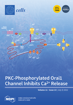
Serine residues S27 and S30 from the Orai1 channel are PKC substrates. O1wt overexpression without STIM1 inhibits store-operated Ca2+ entry but increases Ca2+ influx and inhibits Ca2+ release in response to ATP and thapsigargin (TG).To study the role of these two phosphorylation sites, we overexpressed O1S27/30A and O1S27/30D. The former increases Ca2+ entry in response to ATP and TG, but it cannot inhibit Ca2+ release as O1wt does, while the latter cannot increase Ca2+ entry but inhibits Ca2+ release, even in the presence of PKC inhibitors. ATP and TG can increase O1DD while decreasing O1AA interaction with the IP3R. O1DD, unlike O1AA, shows a clear intracellular location. O1AA increases while O1DD decreases the frequency of ATP-induced [Ca2+]i oscillations. These data suggest that yje PKC-mediated phosphorylation of the O1 channel inhibits Ca2+ release, facilitating ER Ca2+ refilling. View this paper
  • Issues are regarded as officially published after their release is announced to the table of contents alert mailing list.
  • You may sign up for e-mail alerts to receive table of contents of newly released issues.
  • PDF is the official format for papers published in both, html and pdf forms. To view the papers in pdf format, click on the "PDF Full-text" link, and use the free Adobe Reader to open them.
Order results
Result details
Section
Select all
Export citation of selected articles as:
17 pages, 13850 KiB  
Article
ROS Signaling Mediates Directional Cell Elongation and Somatic Cell Fusion in the Red Alga Griffithsia monilis
by Jong-Seok Moon, Chan-Young Hong, Ji-Woong Lee and Gwang-Hoon Kim
Cells 2022, 11(13), 2124; https://doi.org/10.3390/cells11132124 - 05 Jul 2022
Cited by 4 | Viewed by 1631
Abstract
In many filamentous red algae, cells that die from physical damage are replaced through somatic fusion of repair cells formed from adjacent cells. We visualized ROS generation in repair cells of Giriffthsia monilis using DCFH-DA staining and examined the expression of the genes [...] Read more.
In many filamentous red algae, cells that die from physical damage are replaced through somatic fusion of repair cells formed from adjacent cells. We visualized ROS generation in repair cells of Giriffthsia monilis using DCFH-DA staining and examined the expression of the genes involved in wound healing using quantitative PCR. Repair cells elongate along the H2O2 gradient, meet at each other’s tips where the H2O2 concentration is highest, and undergo somatic fusion. No wound response occurred with ascorbic acid treatment. Conversely, H2O2 treatment induced many repair cells, leading to multiple somatic cell fusions. Diphenylene iodonium (DPI) or caffeine treatment reversibly inhibited ROS production in repair cells and blocked the progression of the wound response suggesting that ROS and calcium signaling are involved in the process. Four G. monilis homologues of NADPH-oxidase (GmRBOHs) were identified. The expression of GmRBOHs was upregulated upon injury, peaking 1 h post injury, and decreasing to initial levels when repair cells began to elongate. Our results suggest that ROS generated upon cell injury activates Ca2+ channels and upregulates the expression of GmRBOHs, and that H2O2 generated from repair cells mediates induced repair cell elongation leading to somatic cell fusion and filament repair. Full article
(This article belongs to the Section Plant, Algae and Fungi Cell Biology)
Show Figures

Figure 1

21 pages, 6179 KiB  
Article
Arachidonic Acid Cascade and Eicosanoid Production Are Elevated While LTC4 Synthase Modulates the Lipidomics Profile in the Brain of the HIVgp120-Transgenic Mouse Model of NeuroHIV
by Nina Y. Yuan, Ricky Maung, Ziying Xu, Xianlin Han and Marcus Kaul
Cells 2022, 11(13), 2123; https://doi.org/10.3390/cells11132123 - 05 Jul 2022
Cited by 6 | Viewed by 2397
Abstract
Background: Combination antiretroviral therapy (cART) has transformed HIV infection from a terminal disease to a manageable chronic health condition, extending patients’ life expectancy to that of the general population. However, the incidence of HIV-associated neurocognitive disorders (HANDs) has persisted despite virological suppression. Patients [...] Read more.
Background: Combination antiretroviral therapy (cART) has transformed HIV infection from a terminal disease to a manageable chronic health condition, extending patients’ life expectancy to that of the general population. However, the incidence of HIV-associated neurocognitive disorders (HANDs) has persisted despite virological suppression. Patients with HIV display persistent signs of immune activation and inflammation despite cART. The arachidonic acid (AA) cascade is an important immune response system responsible for both pro- and anti-inflammatory processes. Methods: Lipidomics, mRNA and Western blotting analysis provide valuable insights into the molecular mechanisms surrounding arachidonic acid metabolism and the resulting inflammation caused by perturbations thereof. Results: Here, we report the presence of inflammatory eicosanoids in the brains of a transgenic mouse model of NeuroHIV that expresses soluble HIV-1 envelope glycoprotein in glial cells (HIVgp120tg mice). Additionally, we report that the effect of LTC4S knockout in HIVgp120tg mice resulted in the sexually dimorphic transcription of COX- and 5-LOX-related genes. Furthermore, the absence of LTC4S suppressed ERK1/2 and p38 MAPK signaling activity in female mice only. The mass spectrometry-based lipidomic profiling of these mice reveals beneficial alterations to lipids in the brain. Conclusion: Targeting the AA cascade may hold potential in the treatment of neuroinflammation observed in NeuroHIV and HANDs. Full article
(This article belongs to the Special Issue The Past, Present and Future of NeuroHIV: A Perspective to A Cure)
Show Figures

Figure 1

17 pages, 9743 KiB  
Article
Optimization of Mechanosensitive Cross-Talk between Matrix Stiffness and Protein Density: Independent Matrix Properties Regulate Spreading Dynamics of Myocytes
by Judith Brock, Julia Erhardt, Stephan A. Eisler and Marcel Hörning
Cells 2022, 11(13), 2122; https://doi.org/10.3390/cells11132122 - 05 Jul 2022
Cited by 1 | Viewed by 2128
Abstract
Cells actively sense differences in topology, matrix elasticity and protein composition of the extracellular microenvironment and adapt their function and morphology. In this study, we focus on the cross-talk between matrix stiffness and protein coating density that regulates morphology and proliferation dynamics of [...] Read more.
Cells actively sense differences in topology, matrix elasticity and protein composition of the extracellular microenvironment and adapt their function and morphology. In this study, we focus on the cross-talk between matrix stiffness and protein coating density that regulates morphology and proliferation dynamics of single myocytes. For this, C2C12 myocytes were monitored on L-DOPA functionalized hydrogels of 22 different elasticity and fibronectin density compositions. Static images were recorded and statistically analyzed to determine morphological differences and to identify the optimized extracellular matrix (ECM). Using that information, selected ECMs were used to study the dynamics before and after cell proliferation by statistical comparison of distinct cell states. We observed a fibronectin-density-independent increase of the projected cell area until 12 kPa. Additionally, changes in fibronectin density led to an area that was optimum at about 2.6 μg/cm2, which was confirmed by independent F-actin analysis, revealing a maximum actin-filament-to-cell-area ratio of 7.5%. Proliferation evaluation showed an opposite correlation between cell spreading duration and speed to matrix elasticity and protein density, which did not affect cell-cycle duration. In summary, we identified an optimized ECM composition and found that independent matrix properties regulate distinct cell characteristics. Full article
(This article belongs to the Special Issue Cellular Integrity under Mechanical Stress)
Show Figures

Figure 1

8 pages, 835 KiB  
Brief Report
Association of Clonal Hematopoiesis of Indeterminate Potential with Inflammatory Gene Expression in Patients with COPD
by Stefan Kuhnert, Siavash Mansouri, Michael A. Rieger, Rajkumar Savai, Edibe Avci, Gabriela Díaz-Piña, Manju Padmasekar, Mario Looso, Stefan Hadzic, Till Acker, Stephan Klatt, Jochen Wilhelm, Ingrid Fleming, Natascha Sommer, Norbert Weissmann, Claus Vogelmeier, Robert Bals, Andreas Zeiher, Stefanie Dimmeler, Werner Seeger and Soni S. Pullamsettiadd Show full author list remove Hide full author list
Cells 2022, 11(13), 2121; https://doi.org/10.3390/cells11132121 - 05 Jul 2022
Cited by 5 | Viewed by 2644
Abstract
Chronic obstructive pulmonary disease (COPD) is a disease with an inflammatory phenotype with increasing prevalence in the elderly. Expanded population of mutant blood cells carrying somatic mutations is termed clonal hematopoiesis of indeterminate potential (CHIP). The association between CHIP and COPD and its [...] Read more.
Chronic obstructive pulmonary disease (COPD) is a disease with an inflammatory phenotype with increasing prevalence in the elderly. Expanded population of mutant blood cells carrying somatic mutations is termed clonal hematopoiesis of indeterminate potential (CHIP). The association between CHIP and COPD and its relevant effects on DNA methylation in aging are mainly unknown. Analyzing the deep-targeted amplicon sequencing from 125 COPD patients, we found enhanced incidence of CHIP mutations (~20%) with a predominance of DNMT3A CHIP-mediated hypomethylation of Phospholipase D Family Member 5 (PLD5), which in turn is positively correlated with increased levels of glycerol phosphocholine, pro-inflammatory cytokines, and deteriorating lung function. Full article
Show Figures

Figure 1

17 pages, 3836 KiB  
Article
Effects of Omega-3 and Antioxidant Cocktail Supplement on Prolonged Bed Rest: Results from Serum Proteome and Sphingolipids Analysis
by Pietro Barbacini, Dieter Blottner, Daniele Capitanio, Gabor Trautmann, Katharina Block, Enrica Torretta, Manuela Moriggi, Michele Salanova and Cecilia Gelfi
Cells 2022, 11(13), 2120; https://doi.org/10.3390/cells11132120 - 05 Jul 2022
Cited by 5 | Viewed by 1652
Abstract
Physical inactivity or prolonged bed rest (BR) induces muscle deconditioning in old and young subjects and can increase the cardiovascular disease risk (CVD) with dysregulation of the lipemic profile. Nutritional interventions, combining molecules such as polyphenols, vitamins and essential fatty acids, can influence [...] Read more.
Physical inactivity or prolonged bed rest (BR) induces muscle deconditioning in old and young subjects and can increase the cardiovascular disease risk (CVD) with dysregulation of the lipemic profile. Nutritional interventions, combining molecules such as polyphenols, vitamins and essential fatty acids, can influence some metabolic features associated with physical inactivity and decrease the reactive oxidative and nitrosative stress (RONS). The aim of this study was to detect circulating molecules correlated with BR in serum of healthy male subjects enrolled in a 60-day BR protocol to evaluate a nutritional intervention with an antioxidant cocktail as a disuse countermeasure (Toulouse COCKTAIL study). The serum proteome, sphingolipidome and nitrosoproteome were analyzed adopting different mass spectrometry-based approaches. Results in placebo-treated BR subjects indicated a marked decrease of proteins associated with high-density lipoproteins (HDL) involved in lipemic homeostasis not found in the cocktail-treated BR group. Moreover, long-chain ceramides decreased while sphingomyelin increased in the BR cocktail-treated group. In placebo, the ratio of S-nitrosylated/total protein increased for apolipoprotein D and several proteins were over-nitrosylated. In cocktail-treated BR subjects, the majority of protein showed a pattern of under-nitrosylation, except for ceruloplasmin and hemopexin, which were over-nitrosylated. Collectively, data indicate a positive effect of the cocktail in preserving lipemic and RONS homeostasis in extended disuse conditions. Full article
(This article belongs to the Special Issue Redox Control of Cell Signaling in Cardiac and Skeletal Muscle)
Show Figures

Figure 1

25 pages, 3979 KiB  
Article
Tissue-Based Proteomic Profiling in Patients with Hyperplasia and Endometrial Cancer
by Khalid Akkour, Ibrahim O. Alanazi, Assim A. Alfadda, Hani Alhalal, Afshan Masood, Mohthash Musambil, Anas M. Abdel Rahman, Moudi A. Alwehaibi, Maria Arafah, Ali Bassi and Hicham Benabdelkamel
Cells 2022, 11(13), 2119; https://doi.org/10.3390/cells11132119 - 05 Jul 2022
Cited by 4 | Viewed by 2418
Abstract
Uterine cancers are among the most prevalent gynecological malignancies, and endometrial cancer (EC) is the most common in this group. This study used tissue-based proteomic profiling analysis in patients with endometrial cancer and hyperplasia, and control patients. Conventional 2D gel electrophoresis, followed by [...] Read more.
Uterine cancers are among the most prevalent gynecological malignancies, and endometrial cancer (EC) is the most common in this group. This study used tissue-based proteomic profiling analysis in patients with endometrial cancer and hyperplasia, and control patients. Conventional 2D gel electrophoresis, followed by a mass spectrometry approach with bioinformatics, including a network pathway analysis pipeline, was used to identify differentially expressed proteins and associated metabolic pathways between the study groups. Thirty-six patients (twelve with endometrial cancer, twelve with hyperplasia, and twelve controls) were enrolled in this study. The mean age of the participants was 46–75 years. Eighty-seven proteins were significantly differentially expressed between the study groups, of which fifty-three were significantly differentially regulated (twenty-eight upregulated and twenty-five downregulated) in the tissue samples of EC patients compared to the control (Ctrl). Furthermore, 26 proteins were significantly dysregulated (8 upregulated and 18 downregulated) in tissue samples of hyperplasia (HY) patients compared to Ctrl. Thirty-two proteins (nineteen upregulated and thirteen downregulated) including desmin, peptidyl prolyl cis-trans isomerase A, and zinc finger protein 844 were downregulated in the EC group compared to the HY group. Additionally, fructose bisphosphate aldolase A, alpha enolase, and keratin type 1 cytoskeletal 10 were upregulated in the EC group compared to those in the HY group. The proteins identified in this study were known to regulate cellular processes (36%), followed by biological regulation (16%). Ingenuity pathway analysis found that proteins that are differentially expressed between EC and HY are linked to AKT, ACTA2, and other signaling pathways. The panels of protein markers identified in this study could be used as potential biomarkers for distinguishing between EC and HY and early diagnosis and progression of EC from hyperplasia and normal patients. Full article
(This article belongs to the Special Issue Research on Cancer Proteomics)
Show Figures

Figure 1

15 pages, 2776 KiB  
Article
Elevated NF-κB/SHh/GLI1 Signature Denotes a Worse Prognosis and Represent a Novel Potential Therapeutic Target in Advanced Prostate Cancer
by Davide Vecchiotti, Daniela Verzella, Mauro Di Vito Nolfi, Daniel D’Andrea, Irene Flati, Barbara Di Francesco, Jessica Cornice, Edoardo Alesse, Daria Capece and Francesca Zazzeroni
Cells 2022, 11(13), 2118; https://doi.org/10.3390/cells11132118 - 05 Jul 2022
Cited by 8 | Viewed by 1665
Abstract
Prostate cancer (PCa) is the second most frequent cancer in men worldwide. NF-κB seems to play a key role in cell survival, proliferation and invasion, sustaining the heterogeneous multifocal nature of PCa. In recent years, the Hedgehog (Hh) signaling pathway has attracted attention [...] Read more.
Prostate cancer (PCa) is the second most frequent cancer in men worldwide. NF-κB seems to play a key role in cell survival, proliferation and invasion, sustaining the heterogeneous multifocal nature of PCa. In recent years, the Hedgehog (Hh) signaling pathway has attracted attention as a therapeutic target due to its implication in tumorigenesis and metastasis in several types of cancer, including PCa. Although it is well-known that Sonic Hedgehog (SHh) is a transcriptional target of NF-κB(p65), and that GLI1 is the effector of this crosstalk, the precise role played by this axis in PCa is still not completely clear. Here, we set out to explore the correlation between NF-κB activation and SHh pathways in PCa, investigating if the interplay between NF-κB(p65) and SHh-GLI1 in advanced PCa could be a prospective therapeutic target. Our findings demonstrate that a NF-κB-SHh-GLI1 gene signature is enriched in PCa patients featuring a higher Gleason score. Moreover, elevated levels of this signature are associated with worse prognosis, thus suggesting that this axis could provide a route to treat aggressive PCa. Full article
(This article belongs to the Special Issue Molecular and Cellular Mechanisms of Cancers: Prostate Cancer)
Show Figures

Figure 1

11 pages, 397 KiB  
Article
ProB-Site: Protein Binding Site Prediction Using Local Features
by Sharzil Haris Khan, Hilal Tayara and Kil To Chong
Cells 2022, 11(13), 2117; https://doi.org/10.3390/cells11132117 - 05 Jul 2022
Cited by 5 | Viewed by 4250
Abstract
Protein–protein interactions (PPIs) are responsible for various essential biological processes. This information can help develop a new drug against diseases. Various experimental methods have been employed for this purpose; however, their application is limited by their cost and time consumption. Alternatively, computational methods [...] Read more.
Protein–protein interactions (PPIs) are responsible for various essential biological processes. This information can help develop a new drug against diseases. Various experimental methods have been employed for this purpose; however, their application is limited by their cost and time consumption. Alternatively, computational methods are considered viable means to achieve this crucial task. Various techniques have been explored in the literature using the sequential information of amino acids in a protein sequence, including machine learning and deep learning techniques. The current efficiency of interaction-site prediction still has growth potential. Hence, a deep neural network-based model, ProB-site, is proposed. ProB-site utilizes sequential information of a protein to predict its binding sites. The proposed model uses evolutionary information and predicted structural information extracted from sequential information of proteins, generating three unique feature sets for every amino acid in a protein sequence. Then, these feature sets are fed to their respective sub-CNN architecture to acquire complex features. Finally, the acquired features are concatenated and classified using fully connected layers. This methodology performed better than state-of-the-art techniques because of the selection of the best features and contemplation of local information of each amino acid. Full article
Show Figures

Graphical abstract

17 pages, 2999 KiB  
Article
A miR-9-5p/FOXO1/CPEB3 Feed-Forward Loop Drives the Progression of Hepatocellular Carcinoma
by Hui Hu, Wei Huang, Hong Zhang, Jianye Li, Qiong Zhang, Ya-Ru Miao, Fei-Fei Hu, Lu Gan, Zhenhong Su, Xiangliang Yang and An-Yuan Guo
Cells 2022, 11(13), 2116; https://doi.org/10.3390/cells11132116 - 05 Jul 2022
Cited by 4 | Viewed by 2187
Abstract
Hepatocellular carcinoma (HCC) is the third leading cause of cancer-related death worldwide, but its regulatory mechanism remains unclear and potential clinical biomarkers are still lacking. Co-regulation of TFs and miRNAs in HCC and FFL module studies may help to identify more precise and [...] Read more.
Hepatocellular carcinoma (HCC) is the third leading cause of cancer-related death worldwide, but its regulatory mechanism remains unclear and potential clinical biomarkers are still lacking. Co-regulation of TFs and miRNAs in HCC and FFL module studies may help to identify more precise and critical driver modules in HCC development. Here, we performed a comprehensive gene expression and regulation analysis for HCC in vitro and in vivo. Transcription factor and miRNA co-regulatory networks for differentially expressed genes between tumors and adjacent tissues revealed the critical feed-forward loop (FFL) regulatory module miR-9-5p/FOXO1/CPEB3 in HCC. Gain- and loss-of-function studies demonstrated that miR-9-5p promotes HCC tumor proliferation, while FOXO1 and CPEB3 inhibit hepatocarcinoma growth. Furthermore, by luciferase reporter assay and ChIP-Seq data, CPEB3 was for the first time identified as a direct downstream target of FOXO1, negatively regulated by miR-9-5p. The miR-9-5p/FOXO1/CPEB3 FFL was associated with poor prognosis, and promoted cell growth and tumor progression of HCC in vitro and in vivo. Our study identified for the first time the existence of miR-9-5p/FOXO1/CPEB3 FFL and revealed its regulatory role in HCC progression, which may represent a new potential target for cancer therapy. Full article
(This article belongs to the Section Cell Methods)
Show Figures

Figure 1

17 pages, 3221 KiB  
Review
MFAP4-Mediated Effects in Elastic Fiber Homeostasis, Integrin Signaling and Cancer, and Its Role in Teleost Fish
by Ali Mohammadi, Grith L. Sorensen and Bartosz Pilecki
Cells 2022, 11(13), 2115; https://doi.org/10.3390/cells11132115 - 05 Jul 2022
Cited by 5 | Viewed by 2859
Abstract
Microfibrillar-associated protein 4 (MFAP4) is an extracellular matrix (ECM) protein belonging to the fibrinogen-related domain superfamily. MFAP4 is highly expressed in elastin-rich tissues such as lung, blood vessels and skin. MFAP4 is involved in organization of the ECM, regulating proper elastic fiber assembly. [...] Read more.
Microfibrillar-associated protein 4 (MFAP4) is an extracellular matrix (ECM) protein belonging to the fibrinogen-related domain superfamily. MFAP4 is highly expressed in elastin-rich tissues such as lung, blood vessels and skin. MFAP4 is involved in organization of the ECM, regulating proper elastic fiber assembly. On the other hand, during pathology MFAP4 actively contributes to disease development and progression due to its interactions with RGD-dependent integrin receptors. Both tissue expression and circulating MFAP4 levels are associated with various disorders, including liver fibrosis and cancer. In other experimental models, such as teleost fish, MFAP4 appears to participate in host defense as a macrophage-specific innate immune molecule. The aim of this review is to summarize the accumulating evidence that indicates the importance of MFAP4 in homeostasis as well as pathological conditions, discuss its known biological functions with special focus on elastic fiber assembly, integrin signaling and cancer, as well as describe the reported functions of non-mammalian MFAP4 in fish. Overall, our work provides a comprehensive overview on the role of MFAP4 in health and disease. Full article
(This article belongs to the Section Cell Microenvironment)
Show Figures

Figure 1

17 pages, 2804 KiB  
Article
Suppressing VEGF-A/VEGFR-2 Signaling Contributes to the Anti-Angiogenic Effects of PPE8, a Novel Naphthoquinone-Based Compound
by Ming-Jen Hsu, Han-Kun Chen, Jin-Cherng Lien, Yu-Han Huang and Shiu-Wen Huang
Cells 2022, 11(13), 2114; https://doi.org/10.3390/cells11132114 - 05 Jul 2022
Cited by 5 | Viewed by 2402
Abstract
Natural naphthoquinones and their derivatives exhibit a broad spectrum of pharmacological activities and have thus attracted much attention in modern drug discovery. However, it remains unclear whether naphthoquinones are potential drug candidates for anti-angiogenic agents. The aim of this study was to evaluate [...] Read more.
Natural naphthoquinones and their derivatives exhibit a broad spectrum of pharmacological activities and have thus attracted much attention in modern drug discovery. However, it remains unclear whether naphthoquinones are potential drug candidates for anti-angiogenic agents. The aim of this study was to evaluate the anti-angiogenic properties of a novel naphthoquinone derivative, PPE8, and explore its underlying mechanisms. Determined by various assays including BrdU, migration, invasion, and tube formation analyses, PPE8 treatment resulted in the reduction of VEGF-A-induced proliferation, migration, and invasion, as well as tube formation in human umbilical vein endothelial cells (HUVECs). We also used an aorta ring sprouting assay, Matrigel plug assay, and immunoblotting analysis to examine PPE8’s ex vivo and in vivo anti-angiogenic activities and its actions on VEGF-A signaling. It has been revealed that PPE8 inhibited VEGF-A-induced micro vessel sprouting and was capable of suppressing angiogenesis in in vivo models. In addition, PPE8 inhibited VEGF receptor (VEGFR)-2, Src, FAK, ERK1/2, or AKT phosphorylation in HUVECs exposed to VEGF-A, and it also showed significant decline in xenograft tumor growth in vivo. Taken together, these observations indicated that PPE8 may target VEGF-A–VEGFR-2 signaling to reduce angiogenesis. It also supports the role of PPE8 as a potential drug candidate for the development of therapeutic agents in the treatment of angiogenesis-related diseases including cancer. Full article
Show Figures

Graphical abstract

14 pages, 1649 KiB  
Article
Formation of Lymphoma Hybrid Spheroids and Drug Testing in Real Time with the Use of Fluorescence Optical Tweezers
by Kamila Duś-Szachniewicz, Katarzyna Gdesz-Birula, Emilia Nowosielska, Piotr Ziółkowski and Sławomir Drobczyński
Cells 2022, 11(13), 2113; https://doi.org/10.3390/cells11132113 - 05 Jul 2022
Viewed by 2326
Abstract
Interactions between stromal and lymphoma cells in the bone marrow are closely related to drug resistance and therapy failure. Physiologically relevant pre-clinical three-dimensional (3D) models recapitulating lymphoma microenvironmental complexity do not currently exist. In this study, we proposed a scheme for optically controlled [...] Read more.
Interactions between stromal and lymphoma cells in the bone marrow are closely related to drug resistance and therapy failure. Physiologically relevant pre-clinical three-dimensional (3D) models recapitulating lymphoma microenvironmental complexity do not currently exist. In this study, we proposed a scheme for optically controlled hybrid lymphoma spheroid formation with the use of optical tweezers (OT). Following the preparation of stromal spheroids using agarose hydrogel, two aggressive non-Hodgkin lymphoma B-cell lines, Ri-1 (DLBCL) and Raji (Burkitt lymphoma), were used to conduct multi-cellular spheroid formation driven by in-house-developed fluorescence optical tweezers. Importantly, the newly formed hybrid spheroid preserved the 3D architecture for the next 24 h. Our model was successfully used for the evaluation of the influence of the anticancer agents doxorubicin (DOX), ibrutinib (IBR), and AMD3100 (plerixafor) on the adhesive properties of lymphoma cells. Importantly, our study revealed that a co-treatment of DOX and IBR with AMD3100 affects the adhesion of B-NHL lymphoma cells. Full article
(This article belongs to the Special Issue In Vitro Model for Micro and Nano Technologies)
Show Figures

Figure 1

10 pages, 260 KiB  
Perspective
The Quality Management Ecosystem in Cell Therapy in Catalonia (Spain): An Opportunity for Integrating Standards and Streamlining Quality Compliance
by Joaquim Vives, Maria Glòria Sòria, Eoin McGrath and Mara Magri
Cells 2022, 11(13), 2112; https://doi.org/10.3390/cells11132112 - 05 Jul 2022
Cited by 3 | Viewed by 1852
Abstract
Cell therapies are required to meet with compulsory regulations that co-exist with other optional standards and guidelines that together compose a complex quality management system. Indeed, reliable insights on the mechanisms of action and safety of novel cell-based therapies require adherence to solid [...] Read more.
Cell therapies are required to meet with compulsory regulations that co-exist with other optional standards and guidelines that together compose a complex quality management system. Indeed, reliable insights on the mechanisms of action and safety of novel cell-based therapies require adherence to solid quality management structures in all steps of the value chain, from early research and tissue procurement to clinical trials and biovigilance, thus guaranteeing reproducibility and solid foundations for better science and improved clinical practice. Herein we present the concept of the quality ecosystem as a tool to understand and assist all stakeholders involved in developing and structuring the integration of standards as novel developments are taking place. We conclude that the various quality management initiatives can all be thought about under the umbrella of an ecosystem. Full article
(This article belongs to the Special Issue Cell Biology: State-of-the-Art and Perspectives in Spain II)
21 pages, 13676 KiB  
Article
Activation of Autophagic Flux Maintains Mitochondrial Homeostasis during Cardiac Ischemia/Reperfusion Injury
by Lihao He, Yuxin Chu, Jing Yang, Jin He, Yutao Hua, Yunxi Chen, Gloria Benavides, Glenn C. Rowe, Lufang Zhou, Scott Ballinger, Victor Darley-Usmar, Martin E. Young, Sumanth D. Prabhu, Palaniappan Sethu, Yingling Zhou, Cheng Zhang and Min Xie
Cells 2022, 11(13), 2111; https://doi.org/10.3390/cells11132111 - 04 Jul 2022
Cited by 6 | Viewed by 2394
Abstract
Reperfusion injury after extended ischemia accounts for approximately 50% of myocardial infarct size, and there is no standard therapy. HDAC inhibition reduces infarct size and enhances cardiomyocyte autophagy and PGC1α-mediated mitochondrial biogenesis when administered at the time of reperfusion. Furthermore, a specific autophagy-inducing [...] Read more.
Reperfusion injury after extended ischemia accounts for approximately 50% of myocardial infarct size, and there is no standard therapy. HDAC inhibition reduces infarct size and enhances cardiomyocyte autophagy and PGC1α-mediated mitochondrial biogenesis when administered at the time of reperfusion. Furthermore, a specific autophagy-inducing peptide, Tat-Beclin 1 (TB), reduces infarct size when administered at the time of reperfusion. However, since SAHA affects multiple pathways in addition to inducing autophagy, whether autophagic flux induced by TB maintains mitochondrial homeostasis during ischemia/reperfusion (I/R) injury is unknown. We tested whether the augmentation of autophagic flux by TB has cardioprotection by preserving mitochondrial homeostasis both in vitro and in vivo. Wild-type mice were randomized into two groups: Tat-Scrambled (TS) peptide as the control and TB as the experimental group. Mice were subjected to I/R surgery (45 min coronary ligation, 24 h reperfusion). Autophagic flux, mitochondrial DNA (mtDNA), mitochondrial morphology, and mitochondrial dynamic genes were assayed. Cultured neonatal rat ventricular myocytes (NRVMs) were treated with a simulated I/R injury to verify cardiomyocyte specificity. The essential autophagy gene, ATG7, conditional cardiomyocyte-specific knockout (ATG7 cKO) mice, and isolated adult mouse ventricular myocytes (AMVMs) were used to evaluate the dependency of autophagy in adult cardiomyocytes. In NRVMs subjected to I/R, TB increased autophagic flux, mtDNA content, mitochondrial function, reduced reactive oxygen species (ROS), and mtDNA damage. Similarly, in the infarct border zone of the mouse heart, TB induced autophagy, increased mitochondrial size and mtDNA content, and promoted the expression of PGC1α and mitochondrial dynamic genes. Conversely, loss of ATG7 in AMVMs and in the myocardium of ATG7 cKO mice abolished the beneficial effects of TB on mitochondrial homeostasis. Thus, autophagic flux is a sufficient and essential process to mitigate myocardial reperfusion injury by maintaining mitochondrial homeostasis and partly by inducing PGC1α-mediated mitochondrial biogenesis. Full article
(This article belongs to the Special Issue Mitochondrial Dysfunction in Cardiovascular Disease)
Show Figures

Figure 1

16 pages, 6131 KiB  
Article
Epigenetic Alterations in Cryopreserved Human Spermatozoa: Suspected Potential Functional Defects
by Wanxue Wang, Plamen Todorov, Cheng Pei, Mengying Wang, Evgenia Isachenko, Gohar Rahimi, Peter Mallmann and Vladimir Isachenko
Cells 2022, 11(13), 2110; https://doi.org/10.3390/cells11132110 - 04 Jul 2022
Cited by 2 | Viewed by 2261
Abstract
Background: Gene set enrichment analysis (GSEA) was conducted on raw data, and alternative splicing (AS) events were found after mRNA sequencing of human spermatozoa. In this study, we aimed to compare unknown micro-epigenetics alternations in fresh and cryopreserved spermatozoa to evaluate the effectivity [...] Read more.
Background: Gene set enrichment analysis (GSEA) was conducted on raw data, and alternative splicing (AS) events were found after mRNA sequencing of human spermatozoa. In this study, we aimed to compare unknown micro-epigenetics alternations in fresh and cryopreserved spermatozoa to evaluate the effectivity of cryopreservation protocols. Methods: Spermatozoa were divided into three groups: fresh spermatozoa (group 1), cryoprotectant-free vitrified spermatozoa (group 2), and conventionally frozen spermatozoa (group 3). Nine RNA samples (three replicates in each group) were detected and were used for library preparation with an Illumina compatible kit and sequencing by the Illumina platform. Results: Three Gene Ontology (GO) terms were found to be enriched in vitrified spermatozoa compared with fresh spermatozoa: mitochondrial tRNA aminoacylation, ATP-dependent microtubule motor activity, and male meiotic nuclear division. In alternative splicing analysis, a number of unknown AS events were found, including functional gene exon skipping (SE), alternative 5′ splice sites (A5SS), alternative 3′ splice sites (A3SS), mutually exclusive exon (MXE), and retained intron (RI). Conclusions: Cryopreservation of spermatozoa from some patients can agitate epigenetic instability, including increased alternative splicing events and changes in crucial mitochondrial functional activities. For fertilization of oocytes, for such patients, it is recommended to use fresh spermatozoa whenever possible; cryopreservation of sperm is recommended to be used only in uncontested situations. Full article
(This article belongs to the Special Issue Biobanking of Engineered and Natural Tissues)
Show Figures

Figure 1

10 pages, 3029 KiB  
Review
The Endocrine Adipose Organ: A System Playing a Central Role in COVID-19
by Francesca Cinti and Saverio Cinti
Cells 2022, 11(13), 2109; https://doi.org/10.3390/cells11132109 - 04 Jul 2022
Cited by 6 | Viewed by 2435
Abstract
In the last 30 years the adipose cell has been object of several studies, turning its reputation from an inert cell into the main character involved in the pathophysiology of multiple diseases, including the ongoing COVID-19 pandemic, which has changed the clinical scenario [...] Read more.
In the last 30 years the adipose cell has been object of several studies, turning its reputation from an inert cell into the main character involved in the pathophysiology of multiple diseases, including the ongoing COVID-19 pandemic, which has changed the clinical scenario of the last two years. Composed by two types of tissue (white and brown), with opposite roles, the adipose organ is now classified as a real endocrine organ whose dysfunction is involved in different diseases, mainly obesity and type 2 diabetes. In this mini-review we aim to retrace the adipose organ history from physiology to physiopathology, to provide therapeutic perspectives for the prevention and treatment of its two main related diseases (obesity and type 2 diabetes) and to summarize the most recent discoveries linking adipose tissue to COVID-19. Full article
(This article belongs to the Special Issue The Role of Adipose Tissue in Metabolic Diseases and Beyond)
Show Figures

Figure 1

24 pages, 5945 KiB  
Article
CaMKII Inhibition Attenuates Distinct Gain-of-Function Effects Produced by Mutant Nav1.6 Channels and Reduces Neuronal Excitability
by Agnes S. Zybura, Firoj K. Sahoo, Andy Hudmon and Theodore R. Cummins
Cells 2022, 11(13), 2108; https://doi.org/10.3390/cells11132108 - 04 Jul 2022
Cited by 2 | Viewed by 2285
Abstract
Aberrant Nav1.6 activity can induce hyperexcitability associated with epilepsy. Gain-of-function mutations in the SCN8A gene encoding Nav1.6 are linked to epilepsy development; however, the molecular mechanisms mediating these changes are remarkably heterogeneous and may involve post-translational regulation of Nav1.6. Because calcium/calmodulin-dependent protein kinase [...] Read more.
Aberrant Nav1.6 activity can induce hyperexcitability associated with epilepsy. Gain-of-function mutations in the SCN8A gene encoding Nav1.6 are linked to epilepsy development; however, the molecular mechanisms mediating these changes are remarkably heterogeneous and may involve post-translational regulation of Nav1.6. Because calcium/calmodulin-dependent protein kinase II (CaMKII) is a powerful modulator of Nav1.6 channels, we investigated whether CaMKII modulates disease-linked Nav1.6 mutants. Whole-cell voltage clamp recordings in ND7/23 cells show that CaMKII inhibition of the epilepsy-related mutation R850Q largely recapitulates the effects previously observed for WT Nav1.6. We also characterized a rare missense variant, R639C, located within a regulatory hotspot for CaMKII modulation of Nav1.6. Prediction software algorithms and electrophysiological recordings revealed gain-of-function effects for R639C mutant channel activity, including increased sodium currents and hyperpolarized activation compared to WT Nav1.6. Importantly, the R639C mutation ablates CaMKII phosphorylation at a key regulatory site, T642, and, in contrast to WT and R850Q channels, displays a distinct response to CaMKII inhibition. Computational simulations demonstrate that modeled neurons harboring the R639C or R850Q mutations are hyperexcitable, and simulating the effects of CaMKII inhibition on Nav1.6 activity in modeled neurons differentially reduced hyperexcitability. Acute CaMKII inhibition may represent a promising mechanism to attenuate gain-of-function effects produced by Nav1.6 mutations. Full article
Show Figures

Figure 1

10 pages, 640 KiB  
Review
Lysine-Specific Demethylase 1 (LSD1/KDM1A) Inhibition as a Target for Disease Modification in Myelofibrosis
by Harinder Gill
Cells 2022, 11(13), 2107; https://doi.org/10.3390/cells11132107 - 03 Jul 2022
Cited by 7 | Viewed by 3234 | Correction
Abstract
Myelofibrosis (MF) is the most symptomatic form of myeloproliferative neoplasm and carries the worst outcome. Allogeneic hematopoietic stem cell transplantation is the only therapy with potential for cure at present, but is limited by significant mortality and morbidity. JAK inhibition is the mainstay [...] Read more.
Myelofibrosis (MF) is the most symptomatic form of myeloproliferative neoplasm and carries the worst outcome. Allogeneic hematopoietic stem cell transplantation is the only therapy with potential for cure at present, but is limited by significant mortality and morbidity. JAK inhibition is the mainstay of treatment for intermediate- and high-risk MF. Ruxolitinib is the most widely used JAK1/2 inhibitor and provides durable effects in controlling symptom burden and spleen volumes. Nevertheless, ruxolitinib may not adequately address the underlying disease biology. Its effects on mutant allele burden, bone marrow fibrosis, and the prevention of leukemic transformation are minimal. Multiple small molecules are being tested in multiple phase 2 and 3 studies as either monotherapy or in combination with JAK2 inhibitors. In this review, the role of LSD1/KDM1A inhibition as a potential disease-modification strategy in patients with myelofibrosis is described and discussed. Full article
(This article belongs to the Special Issue The Myeloid Diseases from the Biology to the Clinic)
Show Figures

Graphical abstract

24 pages, 9564 KiB  
Article
Selective Ablation of BCL11A in Epidermal Keratinocytes Alters Skin Homeostasis and Accelerates Excisional Wound Healing In Vivo
by Nilika Bhattacharya, Arup K. Indra and Gitali Ganguli-Indra
Cells 2022, 11(13), 2106; https://doi.org/10.3390/cells11132106 - 03 Jul 2022
Cited by 3 | Viewed by 2707
Abstract
Transcriptional regulator BCL11A plays a crucial role in coordinating a suite of developmental processes including skin morphogenesis, barrier functions and lipid metabolism. There is little or no reports so far documenting the role of BCL11A in postnatal adult skin homeostasis and in the [...] Read more.
Transcriptional regulator BCL11A plays a crucial role in coordinating a suite of developmental processes including skin morphogenesis, barrier functions and lipid metabolism. There is little or no reports so far documenting the role of BCL11A in postnatal adult skin homeostasis and in the physiological process of tissue repair and regeneration. The current study establishes for the first time the In Vivo role of epidermal BCL11A in maintaining adult epidermal homeostasis and as a negative regulator of cutaneous wound healing. Conditional ablation of Bcl11a in skin epidermal keratinocytes (Bcl11aep−/−mice) enhances the keratinocyte proliferation and differentiation program, suggesting its critical role in epidermal homeostasis of adult murine skin. Further, loss of keratinocytic BCL11A promotes rapid closure of excisional wounds both in a cell autonomous manner likely via accelerating wound re-epithelialization and in a non-cell autonomous manner by enhancing angiogenesis. The epidermis specific Bcl11a knockout mouse serves as a prototype to gain mechanistic understanding of various downstream pathways converging towards the manifestation of an accelerated healing phenotype upon its deletion. Full article
(This article belongs to the Special Issue Retinoic Acid and Retinoid X Receptors)
Show Figures

Graphical abstract

16 pages, 725 KiB  
Review
Skeletal Muscle Pathogenesis in Polyglutamine Diseases
by Caterina Marchioretti, Emanuela Zuccaro, Udai Bhan Pandey, Jessica Rosati, Manuela Basso and Maria Pennuto
Cells 2022, 11(13), 2105; https://doi.org/10.3390/cells11132105 - 03 Jul 2022
Cited by 2 | Viewed by 3397
Abstract
Polyglutamine diseases are characterized by selective dysfunction and degeneration of specific types of neurons in the central nervous system. In addition, nonneuronal cells can also be affected as a consequence of primary degeneration or due to neuronal dysfunction. Skeletal muscle is a primary [...] Read more.
Polyglutamine diseases are characterized by selective dysfunction and degeneration of specific types of neurons in the central nervous system. In addition, nonneuronal cells can also be affected as a consequence of primary degeneration or due to neuronal dysfunction. Skeletal muscle is a primary site of toxicity of polyglutamine-expanded androgen receptor, but it is also affected in other polyglutamine diseases, more likely due to neuronal dysfunction and death. Nonetheless, pathological processes occurring in skeletal muscle atrophy impact the entire body metabolism, thus actively contributing to the inexorable progression towards the late and final stages of disease. Skeletal muscle atrophy is well recapitulated in animal models of polyglutamine disease. In this review, we discuss the impact and relevance of skeletal muscle in patients affected by polyglutamine diseases and we review evidence obtained in animal models and patient-derived cells modeling skeletal muscle. Full article
(This article belongs to the Special Issue Skeletal Muscle Atrophy: Mechanisms at a Cellular Level)
Show Figures

Figure 1

14 pages, 12757 KiB  
Article
Extracellular Vesicles from BMSCs Prevent Glucocorticoid-Induced BMECs Injury by Regulating Autophagy via the PI3K/Akt/mTOR Pathway
by Jinhui Ma, Mengran Shen, Debo Yue, Weiguo Wang, Fuqiang Gao and Bailiang Wang
Cells 2022, 11(13), 2104; https://doi.org/10.3390/cells11132104 - 03 Jul 2022
Cited by 9 | Viewed by 2191
Abstract
Osteonecrosis of the femoral head (ONFH) is a common clinical disease with a high disability rate. Injury of bone microvascular endothelial cells (BMECs) caused by glucocorticoid administration is one of the important causes of ONFH, and there is currently a lack of effective [...] Read more.
Osteonecrosis of the femoral head (ONFH) is a common clinical disease with a high disability rate. Injury of bone microvascular endothelial cells (BMECs) caused by glucocorticoid administration is one of the important causes of ONFH, and there is currently a lack of effective clinical treatments. Extracellular vesicles derived from bone stem cells (BMSC-EVs) can prevent ONFH by promoting angiogenesis and can inhibit cell apoptosis by regulating autophagy via the PI3K/Akt/mTOR signaling pathway. The present study aimed to investigate the effect of extracellular vesicles derived from bone marrow stem cells (BMSC) on a glucocorticoid-induced injury of BMECs and possible mechanisms. We found that BMSC-EVs attenuated glucocorticoid-induced viability, angiogenesis capacity injury, and the apoptosis of BMECs. BMSC-EVs increased the LC3 level, but decreased p62 (an autophagy protein receptor) expression, suggesting that BMSC-Exos activated autophagy in glucocorticoid-treated BMECs. The protective effects of BMSC-EVs on the glucocorticoid-induced injury of BMECs was mimicked by a known stimulator of autophagy (rapamycin) and could be enhanced by co-treatment with an autophagy inhibitor (LY294002). BMSC-EVs also suppressed the PI3K/Akt/mTOR signaling pathway, which regulates cell autophagy, in glucocorticoid-treated BMECs. In conclusion, the results indicate that BMSC-EVs prevent the glucocorticoid-induced injury of BMECs by regulating autophagy via the PI3K/Akt/mTOR pathway. Full article
(This article belongs to the Special Issue PI3K/AKT/mTOR Signaling Network in Human Health and Diseases)
Show Figures

Figure 1

20 pages, 17251 KiB  
Article
mTOR-Dependent Autophagy Regulates Slit Diaphragm Density in Podocyte-like Drosophila Nephrocytes
by Dominik Spitz, Maria Comas, Lea Gerstner, Séverine Kayser, Martin Helmstädter, Gerd Walz and Tobias Hermle
Cells 2022, 11(13), 2103; https://doi.org/10.3390/cells11132103 - 02 Jul 2022
Cited by 7 | Viewed by 3115
Abstract
Both mTOR signaling and autophagy are important modulators of podocyte homeostasis, regeneration, and aging and have been implicated in glomerular diseases. However, the mechanistic role of these pathways for the glomerular filtration barrier remains poorly understood. We used Drosophila nephrocytes as an established [...] Read more.
Both mTOR signaling and autophagy are important modulators of podocyte homeostasis, regeneration, and aging and have been implicated in glomerular diseases. However, the mechanistic role of these pathways for the glomerular filtration barrier remains poorly understood. We used Drosophila nephrocytes as an established podocyte model and found that inhibition of mTOR signaling resulted in increased spacing between slit diaphragms. Gain-of-function of mTOR signaling did not affect spacing, suggesting that additional cues limit the maximal slit diaphragm density. Interestingly, both activation and inhibition of mTOR signaling led to decreased nephrocyte function, indicating that a fine balance of signaling activity is needed for proper function. Furthermore, mTOR positively controlled cell size, survival, and the extent of the subcortical actin network. We also showed that basal autophagy in nephrocytes is required for survival and limits the expression of the sns (nephrin) but does not directly affect slit diaphragm formation or endocytic activity. However, using a genetic rescue approach, we demonstrated that excessive, mTOR-dependent autophagy is primarily responsible for slit diaphragm misspacing. In conclusion, we established this invertebrate podocyte model for mechanistic studies on the role of mTOR signaling and autophagy, and we discovered a direct mTOR/autophagy-dependent regulation of the slit diaphragm architecture. Full article
(This article belongs to the Special Issue Model Organisms to Study Autophagy)
Show Figures

Figure 1

15 pages, 2994 KiB  
Article
Angiotensin 1–7 Stimulates Proliferation of Lung Bronchoalveolar Progenitors—Implications for SARS-CoV-2 Infection
by Andrzej K. Ciechanowicz, Wen Xin Lay, Jefte Prado Paulino, Erika Suchocki, Susanne Leszczak, Christian Leszczak and Magdalena Kucia
Cells 2022, 11(13), 2102; https://doi.org/10.3390/cells11132102 - 02 Jul 2022
Cited by 4 | Viewed by 1965
Abstract
SARS-CoV-2 infection leads to severe lung damage due to pneumonia and, in more severe cases, leads to acute respiratory distress syndrome, or ARDS. This affects the viability of bronchoalveolar cells. An important role in the pathogenesis of these complications is the hyperactivation of [...] Read more.
SARS-CoV-2 infection leads to severe lung damage due to pneumonia and, in more severe cases, leads to acute respiratory distress syndrome, or ARDS. This affects the viability of bronchoalveolar cells. An important role in the pathogenesis of these complications is the hyperactivation of the renin-angiotensin-aldosterone (RAA) pathway and induction of cytokine storm that occurs in an Nlrp3 inflammasome-dependent manner. To shed more light on the susceptibility of lung tissue to SARS-CoV-2 infection, we evaluated murine bronchioalveolar stem cells (BASC), alveolar type II cells (AT2), and 3D-derived organoids expression of mRNA encoding genes involved in virus entry into cells, components of RAA, and genes that comprise elements of the Nlrp3 inflammasome pathway. We noticed that all these genes are expressed by lung alveolar stem cells and organoids-derived from these cells. Interestingly, all these cells express a high level of ACE2 that, on the one hand, serves as an entry receptor for SARS-CoV-2 and, on the other, converts angiotensin II into its physiological antagonist, angiotensin 1–7 (Ang 1–7), which has been reported to have a protective role in lung damage. To shed more light on the role of Ang 1–7 on lung tissue, we exposed lung-derived BASC and AT2 cells to this mediator of RAA and noticed that it increases the proliferation of these cells. Based on this, Ang 1–7 could be employed to alleviate the damage to lung alveolar stem/progenitor cells during SARS-CoV-2 infection. Full article
(This article belongs to the Collection Researches on Normal and Cancer Stem Cells)
Show Figures

Figure 1

19 pages, 2010 KiB  
Review
Endogenous Neural Stem Cell Mediated Oligodendrogenesis in the Adult Mammalian Brain
by Daniel Z. Radecki and Jayshree Samanta
Cells 2022, 11(13), 2101; https://doi.org/10.3390/cells11132101 - 02 Jul 2022
Cited by 3 | Viewed by 3502
Abstract
Oligodendrogenesis is essential for replacing worn-out oligodendrocytes, promoting myelin plasticity, and for myelin repair following a demyelinating injury in the adult mammalian brain. Neural stem cells are an important source of oligodendrocytes in the adult brain; however, there are considerable differences in oligodendrogenesis [...] Read more.
Oligodendrogenesis is essential for replacing worn-out oligodendrocytes, promoting myelin plasticity, and for myelin repair following a demyelinating injury in the adult mammalian brain. Neural stem cells are an important source of oligodendrocytes in the adult brain; however, there are considerable differences in oligodendrogenesis from neural stem cells residing in different areas of the adult brain. Amongst the distinct niches containing neural stem cells, the subventricular zone lining the lateral ventricles and the subgranular zone in the dentate gyrus of the hippocampus are considered the principle areas of adult neurogenesis. In addition to these areas, radial glia-like cells, which are the precursors of neural stem cells, are found in the lining of the third ventricle, where they are called tanycytes, and in the cerebellum, where they are called Bergmann glia. In this review, we will describe the contribution and regulation of each of these niches in adult oligodendrogenesis. Full article
Show Figures

Figure 1

17 pages, 4111 KiB  
Article
The Role of the Fibronectin Synergy Site for Skin Wound Healing
by Irene Gimeno-LLuch, María Benito-Jardón, Gemma Guerrero-Barberà, Natalia Burday and Mercedes Costell
Cells 2022, 11(13), 2100; https://doi.org/10.3390/cells11132100 - 02 Jul 2022
Cited by 10 | Viewed by 2788
Abstract
Skin is constantly exposed to injuries that are repaired with different outcomes, either regeneration or scarring. Scars result from fibrotic processes modulated by cellular physical forces transmitted by integrins. Fibronectin (FN) is a major component in the provisional matrix assembled to repair skin [...] Read more.
Skin is constantly exposed to injuries that are repaired with different outcomes, either regeneration or scarring. Scars result from fibrotic processes modulated by cellular physical forces transmitted by integrins. Fibronectin (FN) is a major component in the provisional matrix assembled to repair skin wounds. FN enables cell adhesion binding of α5β1/αIIbβ3 and αv-class integrins to an RGD-motif. An additional linkage for α5/αIIb is the synergy site located in close proximity to the RGD motif. The mutation to impair the FN synergy region (Fn1syn/syn) demonstrated that its absence permits complete development. However, only with the additional engagement to the FN synergy site do cells efficiently resist physical forces. To test how the synergy site-mediated adhesion affects the course of wound healing fibrosis, we used a mouse model of skin injury and in-vitro migration studies with keratinocytes and fibroblasts on FNsyn. The loss of FN synergy site led to normal re-epithelialization caused by two opposing migratory defects of activated keratinocytes and, in the dermis, induced reduced fibrotic responses, with lower contents of myofibroblasts and FN deposition and diminished TGF-β1-mediated cell signalling. We demonstrate that weakened α5β1-mediated traction forces on FNsyn cause reduced TGF-β1 release from its latent complex. Full article
(This article belongs to the Section Intracellular and Plasma Membranes)
Show Figures

Figure 1

21 pages, 4083 KiB  
Article
Increased Resection at DSBs in G2-Phase Is a Unique Phenotype Associated with DNA-PKcs Defects That Is Not Shared by Other Factors of c-NHEJ
by Huaping Xiao, Fanghua Li, Emil Mladenov, Aashish Soni, Veronika Mladenova, Bing Pan, Rositsa Dueva, Martin Stuschke, Beate Timmermann and George Iliakis
Cells 2022, 11(13), 2099; https://doi.org/10.3390/cells11132099 - 02 Jul 2022
Cited by 5 | Viewed by 2024
Abstract
The load of DNA double-strand breaks (DSBs) induced in the genome of higher eukaryotes by different doses of ionizing radiation (IR) is a key determinant of DSB repair pathway choice, with homologous recombination (HR) and ATR substantially gaining ground at doses below 0.5 [...] Read more.
The load of DNA double-strand breaks (DSBs) induced in the genome of higher eukaryotes by different doses of ionizing radiation (IR) is a key determinant of DSB repair pathway choice, with homologous recombination (HR) and ATR substantially gaining ground at doses below 0.5 Gy. Increased resection and HR engagement with decreasing DSB-load generate a conundrum in a classical non-homologous end-joining (c-NHEJ)-dominated cell and suggest a mechanism adaptively facilitating resection. We report that ablation of DNA-PKcs causes hyper-resection, implicating DNA-PK in the underpinning mechanism. However, hyper-resection in DNA-PKcs-deficient cells can also be an indirect consequence of their c-NHEJ defect. Here, we report that all tested DNA-PKcs mutants show hyper-resection, while mutants with defects in all other factors of c-NHEJ fail to do so. This result rules out the model of c-NHEJ versus HR competition and the passive shift from c-NHEJ to HR as the causes of the increased resection and suggests the integration of DNA-PKcs into resection regulation. We develop a model, compatible with the results of others, which integrates DNA-PKcs into resection regulation and HR for a subset of DSBs. For these DSBs, we propose that the kinase remains at the break site, rather than the commonly assumed autophosphorylation-mediated removal from DNA ends. Full article
(This article belongs to the Special Issue Double-Strand DNA Break Repair and Human Disease II)
Show Figures

Figure 1

18 pages, 2966 KiB  
Article
Prevention of Cell Death by Activation of Hydroxycarboxylic Acid Receptor 1 (GPR81) in Retinal Explants
by Rupali Vohra, Berta Sanz-Morello, Anna Luna Mølgaard Tams, Zaynab Ahmad Mouhammad, Kristine Karla Freude, Jens Hannibal, Blanca Irene Aldana, Linda Hildegaard Bergersen and Miriam Kolko
Cells 2022, 11(13), 2098; https://doi.org/10.3390/cells11132098 - 02 Jul 2022
Cited by 4 | Viewed by 2402
Abstract
Background: Progressive retinal ganglion cell (RGC) dysfunction and death are common characteristics of retinal neurodegenerative diseases. Recently, hydroxycarboxylic acid receptor 1 (HCA1R, GPR81) was identified as a key modulator of mitochondrial function and cell survival. Thus, we aimed to test whether [...] Read more.
Background: Progressive retinal ganglion cell (RGC) dysfunction and death are common characteristics of retinal neurodegenerative diseases. Recently, hydroxycarboxylic acid receptor 1 (HCA1R, GPR81) was identified as a key modulator of mitochondrial function and cell survival. Thus, we aimed to test whether activation of HCA1R with 3,5-Dihydroxybenzoic acid (DHBA) also promotes RGC survival and improves energy metabolism in mouse retinas. Methods: Retinal explants were treated with 5 mM of the HCA1R agonist, 3,5-DHBA, for 2, 4, 24, and 72 h. Additionally, explants were also treated with 15 mM of L-glutamate to induce toxicity. Tissue survival was assessed through lactate dehydrogenase (LDH) viability assays. RGC survival was measured through immunohistochemical (IHC) staining. Total ATP levels were quantified through bioluminescence assays. Energy metabolism was investigated through stable isotope labeling and gas chromatography-mass spectrometry (GC-MS). Lactate and nitric oxide levels were measured through colorimetric assays. Results: HCA1R activation with 3,5-DHBAincreased retinal explant survival. During glutamate-induced death, 3,5-DHBA treatment also increased survival. IHC analysis revealed that 3,5-DHBA treatment promoted RGC survival in retinal wholemounts. 3,5-DHBA treatment also enhanced ATP levels in retinal explants, whereas lactate levels decreased. No effects on glucose metabolism were observed, but small changes in lactate metabolism were found. Nitric oxide levels remained unaltered in response to 3,5-DHBA treatment. Conclusion: The present study reveals that activation of HCA1R with 3,5-DHBA treatment has a neuroprotective effect specifically on RGCs and on glutamate-induced retinal degeneration. Hence, HCA1R agonist administration may be a potential new strategy for rescuing RGCs, ultimately preventing visual disability. Full article
Show Figures

Figure 1

19 pages, 4626 KiB  
Review
Hallmarks and Molecular Tools for the Study of Mitophagy in Parkinson’s Disease
by Thomas Goiran, Mohamed A. Eldeeb, Cornelia E. Zorca and Edward A. Fon
Cells 2022, 11(13), 2097; https://doi.org/10.3390/cells11132097 - 02 Jul 2022
Cited by 14 | Viewed by 4225
Abstract
The best-known hallmarks of Parkinson’s disease (PD) are the motor deficits that result from the degeneration of dopaminergic neurons in the substantia nigra. Dopaminergic neurons are thought to be particularly susceptible to mitochondrial dysfunction. As such, for their survival, they rely on the [...] Read more.
The best-known hallmarks of Parkinson’s disease (PD) are the motor deficits that result from the degeneration of dopaminergic neurons in the substantia nigra. Dopaminergic neurons are thought to be particularly susceptible to mitochondrial dysfunction. As such, for their survival, they rely on the elaborate quality control mechanisms that have evolved in mammalian cells to monitor mitochondrial function and eliminate dysfunctional mitochondria. Mitophagy is a specialized type of autophagy that mediates the selective removal of damaged mitochondria from cells, with the net effect of dampening the toxicity arising from these dysfunctional organelles. Despite an increasing understanding of the molecular mechanisms that regulate the removal of damaged mitochondria, the detailed molecular link to PD pathophysiology is still not entirely clear. Herein, we review the fundamental molecular pathways involved in PINK1/Parkin-mediated and receptor-mediated mitophagy, the evidence for the dysfunction of these pathways in PD, and recently-developed state-of-the art assays for measuring mitophagy in vitro and in vivo. Full article
(This article belongs to the Special Issue Hallmarks of Parkinson’s Disease)
Show Figures

Figure 1

31 pages, 12265 KiB  
Review
Mechanisms of Sperm–Egg Interactions: What Ascidian Fertilization Research Has Taught Us
by Hitoshi Sawada and Takako Saito
Cells 2022, 11(13), 2096; https://doi.org/10.3390/cells11132096 - 01 Jul 2022
Cited by 9 | Viewed by 2897
Abstract
Fertilization is an essential process in terrestrial organisms for creating a new organism with genetic diversity. Before gamete fusion, several steps are required to achieve successful fertilization. Animal spermatozoa are first activated and attracted to the eggs by egg-derived chemoattractants. During the sperm [...] Read more.
Fertilization is an essential process in terrestrial organisms for creating a new organism with genetic diversity. Before gamete fusion, several steps are required to achieve successful fertilization. Animal spermatozoa are first activated and attracted to the eggs by egg-derived chemoattractants. During the sperm passage of the egg’s extracellular matrix or upon the sperm binding to the proteinaceous egg coat, the sperm undergoes an acrosome reaction, an exocytosis of acrosome. In hermaphrodites such as ascidians, the self/nonself recognition process occurs when the sperm binds to the egg coat. The activated or acrosome-reacted spermatozoa penetrate through the proteinaceous egg coat. The extracellular ubiquitin–proteasome system, the astacin-like metalloproteases, and the trypsin-like proteases play key roles in this process in ascidians. In the present review, we summarize our current understanding and perspectives on gamete recognition and egg coat lysins in ascidians and consider the general mechanisms of fertilization in animals and plants. Full article
Show Figures

Figure 1

18 pages, 1829 KiB  
Review
Regeneration or Repair? The Role of Alveolar Epithelial Cells in the Pathogenesis of Idiopathic Pulmonary Fibrosis (IPF)
by Paola Confalonieri, Maria Concetta Volpe, Justin Jacob, Serena Maiocchi, Francesco Salton, Barbara Ruaro, Marco Confalonieri and Luca Braga
Cells 2022, 11(13), 2095; https://doi.org/10.3390/cells11132095 - 30 Jun 2022
Cited by 59 | Viewed by 8917
Abstract
Idiopathic pulmonary fibrosis (IPF) is a chronic, progressive interstitial lung disease (ILD) with unknown etiology in which gradual fibrotic scarring of the lungs leads to usual interstitial pneumonia (UIP) and, ultimately, to death. IPF affects three million people worldwide, and the only currently [...] Read more.
Idiopathic pulmonary fibrosis (IPF) is a chronic, progressive interstitial lung disease (ILD) with unknown etiology in which gradual fibrotic scarring of the lungs leads to usual interstitial pneumonia (UIP) and, ultimately, to death. IPF affects three million people worldwide, and the only currently available treatments include the antifibrotic drugs nintedanib and pirfenidone, which effectively reduce fibrosis progression are, unfortunately, not effective in curing the disease. In recent years, the paradigm of IPF pathogenesis has shifted from a fibroblast-driven disease to an epithelium-driven disease, wherein, upon recurrent microinjuries, dysfunctional alveolar type II epithelial cells (ATII) are not only unable to sustain physiological lung regeneration but also promote aberrant epithelial–mesenchymal crosstalk. This creates a drift towards fibrosis rather than regeneration. In the context of this review article, we discuss the most relevant mechanisms involved in IPF pathogenesis with a specific focus on the role of dysfunctional ATII cells in promoting disease progression. In particular, we summarize the main causes of ATII cell dysfunction, such as aging, environmental factors, and genetic determinants. Next, we describe the known mechanisms of physiological lung regeneration by drawing a parallel between embryonic lung development and the known pathways involved in ATII-driven alveolar re-epithelization after injury. Finally, we review the most relevant interventional clinical trials performed in the last 20 years with the aim of underlining the urgency of developing new therapies against IPF that are not only aimed at reducing disease progression by hampering ECM deposition but also boost the physiological processes of ATII-driven alveolar regeneration. Full article
(This article belongs to the Special Issue Epithelial Cells Role in Lung Diseases)
Show Figures

Figure 1

Previous Issue
Next Issue
Back to TopTop