Next Issue
Volume 11, June-2
Previous Issue
Volume 11, May-2


Cells, Volume 11, Issue 11 (June-1 2022) – 135 articles

Cover Story (view full-size image): 
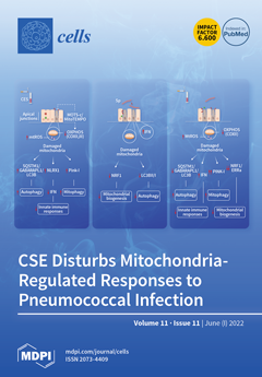
Cigarette smoke extract (CSE)-exposed airway epithelial cells (AECs) showed dysfunctional mitochondria with impaired interferon responses and disrupted airway epithelial barrier function. Nucleotide-binding leucine-rich repeat containing X1 (NLRX1), which negatively regulates interferon responses, increased upon CSE in AECs. Post-treatment with regulators of mitochondrial respiration restored airway epithelial barrier function. Pneumococcal infection induced mitochondrial abnormalities in AECs with a decrease in a regulator of mitochondrial biogenesis, which was accompanied with upregulation of interferon response genes. Pre-exposure of AECs to CSE followed by pneumococcal infection induced a strong increase in key regulators of mitophagy and impairment in mitochondrial biogenesis as well as downregulation of interferon response and airway epithelial barrier genes. View this paper
  • Issues are regarded as officially published after their release is announced to the table of contents alert mailing list.
  • You may sign up for e-mail alerts to receive table of contents of newly released issues.
  • PDF is the official format for papers published in both, html and pdf forms. To view the papers in pdf format, click on the "PDF Full-text" link, and use the free Adobe Reader to open them.
Order results
Result details
Section
Select all
Export citation of selected articles as:
41 pages, 4505 KiB  
Review
p66Shc in Cardiovascular Pathology
by Landon Haslem, Jennifer M. Hays and Franklin A. Hays
Cells 2022, 11(11), 1855; https://doi.org/10.3390/cells11111855 - 06 Jun 2022
Cited by 10 | Viewed by 3034
Abstract
p66Shc is a widely expressed protein that governs a variety of cardiovascular pathologies by generating, and exacerbating, pro-apoptotic ROS signals. Here, we review p66Shc’s connections to reactive oxygen species, expression, localization, and discuss p66Shc signaling and mitochondrial functions. Emphasis is placed on recent [...] Read more.
p66Shc is a widely expressed protein that governs a variety of cardiovascular pathologies by generating, and exacerbating, pro-apoptotic ROS signals. Here, we review p66Shc’s connections to reactive oxygen species, expression, localization, and discuss p66Shc signaling and mitochondrial functions. Emphasis is placed on recent p66Shc mitochondrial function discoveries including structure/function relationships, ROS identity and regulation, mechanistic insights, and how p66Shc-cyt c interactions can influence p66Shc mitochondrial function. Based on recent findings, a new p66Shc mitochondrial function model is also put forth wherein p66Shc acts as a rheostat that can promote or antagonize apoptosis. A discussion of how the revised p66Shc model fits previous findings in p66Shc-mediated cardiovascular pathology follows. Full article
(This article belongs to the Special Issue Redox Control of Cell Signaling in Cardiac and Skeletal Muscle)
Show Figures

Figure 1

19 pages, 2444 KiB  
Review
Immune Checkpoint Proteins, Metabolism and Adhesion Molecules: Overlooked Determinants of CAR T-Cell Migration?
by Luca Simula, Emma Ollivier, Philippe Icard and Emmanuel Donnadieu
Cells 2022, 11(11), 1854; https://doi.org/10.3390/cells11111854 - 06 Jun 2022
Cited by 6 | Viewed by 3347
Abstract
Adoptive transfer of T cells genetically engineered to express chimeric antigen receptors (CAR) has demonstrated striking efficacy for the treatment of several hematological malignancies, including B-cell lymphoma, leukemia, and multiple myeloma. However, many patients still do not respond to this therapy or eventually [...] Read more.
Adoptive transfer of T cells genetically engineered to express chimeric antigen receptors (CAR) has demonstrated striking efficacy for the treatment of several hematological malignancies, including B-cell lymphoma, leukemia, and multiple myeloma. However, many patients still do not respond to this therapy or eventually relapse after an initial remission. In most solid tumors for which CAR T-cell therapy has been tested, efficacy has been very limited. In this context, it is of paramount importance to understand the mechanisms of tumor resistance to CAR T cells. Possible factors contributing to such resistance have been identified, including inherent CAR T-cell dysfunction, the presence of an immunosuppressive tumor microenvironment, and tumor-intrinsic factors. To control tumor growth, CAR T cells have to migrate actively enabling a productive conjugate with their targets. To date, many cells and factors contained within the tumor microenvironment have been reported to negatively control the migration of T cells and their ability to reach cancer cells. Recent evidence suggests that additional determinants, such as immune checkpoint proteins, cellular metabolism, and adhesion molecules, may modulate the motility of CAR T cells in tumors. Here, we review the potential impact of these determinants on CAR T-cell motility, and we discuss possible strategies to restore intratumoral T-cell migration with a special emphasis on approaches targeting these determinants. Full article
(This article belongs to the Special Issue State of the Art in CAR-T Cell Therapy)
Show Figures

Figure 1

17 pages, 664 KiB  
Review
Utility of iPSC-Derived Cells for Disease Modeling, Drug Development, and Cell Therapy
by Martin W. Nicholson, Chien-Yu Ting, Darien Z. H. Chan, Yu-Che Cheng, Yi-Chan Lee, Ching-Chuan Hsu, Ching-Ying Huang and Patrick C. H. Hsieh
Cells 2022, 11(11), 1853; https://doi.org/10.3390/cells11111853 - 06 Jun 2022
Cited by 17 | Viewed by 6915
Abstract
The advent of induced pluripotent stem cells (iPSCs) has advanced our understanding of the molecular mechanisms of human disease, drug discovery, and regenerative medicine. As such, the use of iPSCs in drug development and validation has shown a sharp increase in the past [...] Read more.
The advent of induced pluripotent stem cells (iPSCs) has advanced our understanding of the molecular mechanisms of human disease, drug discovery, and regenerative medicine. As such, the use of iPSCs in drug development and validation has shown a sharp increase in the past 15 years. Furthermore, many labs have been successful in reproducing many disease phenotypes, often difficult or impossible to capture, in commonly used cell lines or animal models. However, there still remain limitations such as the variability between iPSC lines as well as their maturity. Here, we aim to discuss the strategies in generating iPSC-derived cardiomyocytes and neurons for use in disease modeling, drug development and their use in cell therapy. Full article
Show Figures

Figure 1

13 pages, 1437 KiB  
Article
High Expression of a Cancer Stemness-Related Gene, Chromobox 8 (CBX8), in Normal Tissue Adjacent to the Tumor (NAT) Is Associated with Poor Prognosis of Colorectal Cancer Patients
by Lui Ng, Hung-Sing Li, Abraham Tak-Ka Man, Ariel Ka-Man Chow, Dominic Chi-Chung Foo, Oswens Siu-Hung Lo, Roberta Wen-Chi Pang and Wai-Lun Law
Cells 2022, 11(11), 1852; https://doi.org/10.3390/cells11111852 - 06 Jun 2022
Viewed by 2039
Abstract
Background: Several studies have demonstrated that the molecular profile of normal tissue adjacent to the tumor (NAT) is prognostic for recurrence in patients with different cancers. This study investigated the clinical significance of CBX8 gene expression, a cancer stemness-related gene, in tumor and [...] Read more.
Background: Several studies have demonstrated that the molecular profile of normal tissue adjacent to the tumor (NAT) is prognostic for recurrence in patients with different cancers. This study investigated the clinical significance of CBX8 gene expression, a cancer stemness-related gene, in tumor and NAT tissue of colorectal cancer (CRC) patients. Methods: The gene level of CBX8 in paired CRC and NAT specimens from 95 patients was determined by quantitative PCR. CBX8 protein level in CRC and NAT specimens from 66 patients was determined by immunohistochemistry. CBX8 gene and protein levels were correlated with the patients’ clinicopathological parameters and circulatory immune cell profiles. The association between CBX8 and pluripotency-associated genes was analyzed using the TCGA database. Results: NAT CBX8 gene level positively correlated with TNM stage, tumor invasion, lymph node metastasis and distant metastasis, indicating its association with tumor progression and metastasis. There was no correlation between NAT CBX8 protein level and clinicopathological parameters. Moreover, a high level of CBX8 gene and protein in NAT both correlated with poor DFS and OS. There was an inverse correlation between CBX8 gene level and post-operative platelet counts and platelet to lymphocyte level, suggesting its association with systematic inflammation. Finally, TCGA analysis showed that CBX8 level was correlated with a couple of pluripotency-associated genes, supporting its association with cancer stemness. Conclusions: High NAT CBX8 is a poor prognostic factor for tumor progression and survival in CRC patients. Full article
Show Figures

Figure 1

19 pages, 3610 KiB  
Article
Ulcerative Colitis in Response to Fecal Microbiota Transplantation via Modulation of Gut Microbiota and Th17/Treg Cell Balance
by Chunlan Huang, Qixiang Mei, Lihong Lou, Zehua Huang, Yang Fu, Junjie Fan, Jingjing Wang, Nuoming Yin, Yi Zheng, Yingying Lu and Yue Zeng
Cells 2022, 11(11), 1851; https://doi.org/10.3390/cells11111851 - 05 Jun 2022
Cited by 9 | Viewed by 2935
Abstract
Background: Fecal microbiota transplantation (FMT) may contribute to disease remission in ulcerative colitis (UC). We studied the microbiota change and its regulation on T cells after FMT. Methods: Patients with mild to moderately active UC were included to receive FMT. The intestinal histopathological [...] Read more.
Background: Fecal microbiota transplantation (FMT) may contribute to disease remission in ulcerative colitis (UC). We studied the microbiota change and its regulation on T cells after FMT. Methods: Patients with mild to moderately active UC were included to receive FMT. The intestinal histopathological changes and barrier function were evaluated. The fecal samples of donors and patients were analyzed by 16S rRNA gene-based microbiota analysis, and the colon Th17 and Treg cells were assessed. Results: Fifteen patients completed the 8-week-follow-up. A total of 10 patients (66.7%) were in the responders (RE) group and five in the non-responders (NR) group. The Nancy histological index and fecal calprotectin decreased (p < 0.001, p = 0.06, respectively) and Occludin and Claudin1 increased in the RE group. The abundance of Faecalibaterium increased significantly by 2.3-fold in the RE group at week 8 (p = 0.043), but it was suppressed in the NR group. Fecal calprotectin (r = −0.382, p = 0.003) and Nancy index (r = −0.497, p = 0.006) were correlated inversely with the abundance of Faecalibacterium, respectively. In the RE group the relative mRNA expression of RORγt decreased and Foxp3 increased. Significantly decreased CD4+ RORγt+ Th17 and increased CD4+ Foxp3+ Treg were also observed in the RE group. The relative abundance of Faecalibacterium correlated with CD4+ RORγt+ Th17 (r = −0.430, p = 0.018) and CD4+ Foxp3+ Treg (r = 0.571, p = 0.001). Conclusions: The long-term Faecalibaterium colonization following FMT plays a crucial role in UC remission by alleviating intestinal inflammation. This anti-inflammatory effect of Faecalibacterium may be achieved by regulating the imbalance of Th17/Treg levels in UC. Full article
Show Figures

Figure 1

4 pages, 210 KiB  
Editorial
Molecular Biology in Glioblastoma Multiforme Treatment
by Claudia Abbruzzese, Michele Persico, Silvia Matteoni and Marco G. Paggi
Cells 2022, 11(11), 1850; https://doi.org/10.3390/cells11111850 - 05 Jun 2022
Cited by 3 | Viewed by 2266
Abstract
Glioblastoma (GBM, grade IV astrocytoma), the most frequently occurring primary brain tumor, presents unique challenges to therapy due to its location, aggressive biological behavior, and diffuse infiltrative growth, thus contributing to having disproportionately high morbidity and mortality [...] Full article
(This article belongs to the Special Issue Molecular Biology in Glioblastoma Multiforme Treatment)
10 pages, 748 KiB  
Review
Science CommuniCa2+tion Developing Scientific Literacy on Calcium: The Involvement of CRAC Currents in Human Health and Disease
by Christina Humer, Sascha Berlansky, Herwig Grabmayr, Matthias Sallinger, Andreas Bernhard, Marc Fahrner and Irene Frischauf
Cells 2022, 11(11), 1849; https://doi.org/10.3390/cells11111849 - 05 Jun 2022
Cited by 3 | Viewed by 2258
Abstract
All human life starts with a calcium (Ca2+) wave. This ion regulates a plethora of cellular functions ranging from fertilisation and birth to development and cell death. A sophisticated system is responsible for maintaining the essential, tight concentration of calcium within [...] Read more.
All human life starts with a calcium (Ca2+) wave. This ion regulates a plethora of cellular functions ranging from fertilisation and birth to development and cell death. A sophisticated system is responsible for maintaining the essential, tight concentration of calcium within cells. Intricate components of this Ca2+ network are store-operated calcium channels in the cells’ membrane. The best-characterised store-operated channel is the Ca2+ release-activated Ca2+ (CRAC) channel. Currents through CRAC channels are critically dependent on the correct function of two proteins: STIM1 and Orai1. A disruption of the precise mechanism of Ca2+ entry through CRAC channels can lead to defects and in turn to severe impacts on our health. Mutations in either STIM1 or Orai1 proteins can have consequences on our immune cells, the cardiac and nervous system, the hormonal balance, muscle function, and many more. There is solid evidence that altered Ca2+ signalling through CRAC channels is involved in the hallmarks of cancer development: uncontrolled cell growth, resistance to cell death, migration, invasion, and metastasis. In this work we highlight the importance of Ca2+ and its role in human health and disease with focus on CRAC channels. Full article
(This article belongs to the Collection Advance in Ion Channel Signaling in Cancer Cells)
Show Figures

Figure 1

39 pages, 2039 KiB  
Review
Optogenetic Methods to Investigate Brain Alterations in Preclinical Models
by Marco Brondi, Matteo Bruzzone, Claudia Lodovichi and Marco dal Maschio
Cells 2022, 11(11), 1848; https://doi.org/10.3390/cells11111848 - 05 Jun 2022
Cited by 6 | Viewed by 3279
Abstract
Investigating the neuronal dynamics supporting brain functions and understanding how the alterations in these mechanisms result in pathological conditions represents a fundamental challenge. Preclinical research on model organisms allows for a multiscale and multiparametric analysis in vivo of the neuronal mechanisms and holds [...] Read more.
Investigating the neuronal dynamics supporting brain functions and understanding how the alterations in these mechanisms result in pathological conditions represents a fundamental challenge. Preclinical research on model organisms allows for a multiscale and multiparametric analysis in vivo of the neuronal mechanisms and holds the potential for better linking the symptoms of a neurological disorder to the underlying cellular and circuit alterations, eventually leading to the identification of therapeutic/rescue strategies. In recent years, brain research in model organisms has taken advantage, along with other techniques, of the development and continuous refinement of methods that use light and optical approaches to reconstruct the activity of brain circuits at the cellular and system levels, and to probe the impact of the different neuronal components in the observed dynamics. These tools, combining low-invasiveness of optical approaches with the power of genetic engineering, are currently revolutionizing the way, the scale and the perspective of investigating brain diseases. The aim of this review is to describe how brain functions can be investigated with optical approaches currently available and to illustrate how these techniques have been adopted to study pathological alterations of brain physiology. Full article
Show Figures

Figure 1

15 pages, 2443 KiB  
Article
Predicting CRISPR/Cas9 Repair Outcomes by Attention-Based Deep Learning Framework
by Xiuqin Liu, Shuya Wang and Dongmei Ai
Cells 2022, 11(11), 1847; https://doi.org/10.3390/cells11111847 - 05 Jun 2022
Cited by 7 | Viewed by 3231
Abstract
As a simple and programmable nuclease-based genome editing tool, the CRISPR/Cas9 system has been widely used in target-gene repair and gene-expression regulation. The DNA mutation generated by CRISPR/Cas9-mediated double-strand breaks determines its biological and phenotypic effects. Experiments have demonstrated that CRISPR/Cas9-generated cellular-repair outcomes [...] Read more.
As a simple and programmable nuclease-based genome editing tool, the CRISPR/Cas9 system has been widely used in target-gene repair and gene-expression regulation. The DNA mutation generated by CRISPR/Cas9-mediated double-strand breaks determines its biological and phenotypic effects. Experiments have demonstrated that CRISPR/Cas9-generated cellular-repair outcomes depend on local sequence features. Therefore, the repair outcomes after DNA break can be predicted by sequences near the cleavage sites. However, existing prediction methods rely on manually constructed features or insufficiently detailed prediction labels. They cannot satisfy clinical-level-prediction accuracy, which limit the performance of these models to existing knowledge about CRISPR/Cas9 editing. We predict 557 repair labels of DNA, covering the vast majority of Cas9-generated mutational outcomes, and build a deep learning model called Apindel, to predict CRISPR/Cas9 editing outcomes. Apindel, automatically, trains the sequence features of DNA with the GloVe model, introduces location information through Positional Encoding (PE), and embeds the trained-word vector matrixes into a deep learning model, containing BiLSTM and the Attention mechanism. Apindel has better performance and more detailed prediction categories than the most advanced DNA-mutation-predicting models. It, also, reveals that nucleotides at different positions relative to the cleavage sites have different influences on CRISPR/Cas9 editing outcomes. Full article
(This article belongs to the Special Issue Bioinformatics and Cells)
Show Figures

Figure 1

11 pages, 1039 KiB  
Review
Restructuring of Lamina-Associated Domains in Senescence and Cancer
by Aurélie Bellanger, Julia Madsen-Østerbye, Natalia M. Galigniana and Philippe Collas
Cells 2022, 11(11), 1846; https://doi.org/10.3390/cells11111846 - 05 Jun 2022
Cited by 11 | Viewed by 2790
Abstract
Induction of cellular senescence or cancer is associated with a reshaping of the nuclear envelope and a broad reorganization of heterochromatin. At the periphery of mammalian nuclei, heterochromatin is stabilized at the nuclear lamina via lamina-associated domains (LADs). Alterations in the composition of [...] Read more.
Induction of cellular senescence or cancer is associated with a reshaping of the nuclear envelope and a broad reorganization of heterochromatin. At the periphery of mammalian nuclei, heterochromatin is stabilized at the nuclear lamina via lamina-associated domains (LADs). Alterations in the composition of the nuclear lamina during senescence lead to a loss of peripheral heterochromatin, repositioning of LADs, and changes in epigenetic states of LADs. Cancer initiation and progression are also accompanied by a massive reprogramming of the epigenome, particularly in domains coinciding with LADs. Here, we review recent knowledge on alterations in chromatin organization and in the epigenome that affect LADs and related genomic domains in senescence and cancer. Full article
(This article belongs to the Special Issue Heterochromatin and Tumorigenesis)
Show Figures

Figure 1

30 pages, 993 KiB  
Review
Extracellular Vesicles as Drivers of Immunoinflammation in Atherothrombosis
by Rosa Suades, Maria Francesca Greco, Teresa Padró and Lina Badimon
Cells 2022, 11(11), 1845; https://doi.org/10.3390/cells11111845 - 05 Jun 2022
Cited by 17 | Viewed by 3358
Abstract
Atherosclerotic cardiovascular disease is the leading cause of morbidity and mortality all over the world. Extracellular vesicles (EVs), small lipid-bilayer membrane vesicles released by most cellular types, exert pivotal and multifaceted roles in physiology and disease. Emerging evidence emphasizes the importance of EVs [...] Read more.
Atherosclerotic cardiovascular disease is the leading cause of morbidity and mortality all over the world. Extracellular vesicles (EVs), small lipid-bilayer membrane vesicles released by most cellular types, exert pivotal and multifaceted roles in physiology and disease. Emerging evidence emphasizes the importance of EVs in intercellular communication processes with key effects on cell survival, endothelial homeostasis, inflammation, neoangiogenesis, and thrombosis. This review focuses on EVs as effective signaling molecules able to both derail vascular homeostasis and induce vascular dysfunction, inflammation, plaque progression, and thrombus formation as well as drive anti-inflammation, vascular repair, and atheroprotection. We provide a comprehensive and updated summary of the role of EVs in the development or regression of atherosclerotic lesions, highlighting the link between thrombosis and inflammation. Importantly, we also critically describe their potential clinical use as disease biomarkers or therapeutic agents in atherothrombosis. Full article
Show Figures

Figure 1

15 pages, 2085 KiB  
Article
Differential Expression of CD45RO and CD45RA in Bovine T Cells
by Anmol Kandel, Lei Li, Akanksha Hada and Zhengguo Xiao
Cells 2022, 11(11), 1844; https://doi.org/10.3390/cells11111844 - 04 Jun 2022
Cited by 4 | Viewed by 6723
Abstract
Effective vaccination induces immune memory to protect animals upon pathogen re-encounter. Despite contradictory reports, bovine memory T cells are identified based on two isoforms of CD45, expression of CD45RO plus exclusion of CD45RA. In this report, we contrasted CD45RA/RO expression on circulatory T [...] Read more.
Effective vaccination induces immune memory to protect animals upon pathogen re-encounter. Despite contradictory reports, bovine memory T cells are identified based on two isoforms of CD45, expression of CD45RO plus exclusion of CD45RA. In this report, we contrasted CD45RA/RO expression on circulatory T cells with IFNγ and IL4 expression induced by a conventional method. To our surprise, 20% of cattle from an enclosed herd did not express CD45RO on T cells without any significant difference on CD45RA expression and IFNγ or IL4 induction. In CD45RO expressing cattle, CD45RA and CD45RO expressions excluded each other, with dominant CD45RO (>90%) expression on gamma delta (γδ) followed by CD4+ (60%) but significantly higher CD45RA expression on CD8+ T cells (about 80%). Importantly, more than 80% of CD45RO expressing CD4+ and CD8+ T cells failed to produce IFNγ and IL-4; however, within the cytokine inducing cells, CD4+ T cells highly expressed CD45RO but those within CD8+ T cells mostly expressed CD45RA. Hence, CD45RO is not ubiquitously expressed in cattle, and rather than with memory phenotype, CD45RA/RO expression are more associated with distinct T cell subtypes. Full article
(This article belongs to the Section Cellular Immunology)
Show Figures

Figure 1

31 pages, 1173 KiB  
Review
Hematopoietic Stem Cell Gene-Addition/Editing Therapy in Sickle Cell Disease
by Paula Germino-Watnick, Malikiya Hinds, Anh Le, Rebecca Chu, Xiong Liu and Naoya Uchida
Cells 2022, 11(11), 1843; https://doi.org/10.3390/cells11111843 - 04 Jun 2022
Cited by 10 | Viewed by 8232
Abstract
Autologous hematopoietic stem cell (HSC)-targeted gene therapy provides a one-time cure for various genetic diseases including sickle cell disease (SCD) and β-thalassemia. SCD is caused by a point mutation (20A > T) in the β-globin gene. Since SCD is the most common single-gene [...] Read more.
Autologous hematopoietic stem cell (HSC)-targeted gene therapy provides a one-time cure for various genetic diseases including sickle cell disease (SCD) and β-thalassemia. SCD is caused by a point mutation (20A > T) in the β-globin gene. Since SCD is the most common single-gene disorder, curing SCD is a primary goal in HSC gene therapy. β-thalassemia results from either the absence or the reduction of β-globin expression, and it can be cured using similar strategies. In HSC gene-addition therapy, patient CD34+ HSCs are genetically modified by adding a therapeutic β-globin gene with lentiviral transduction, followed by autologous transplantation. Alternatively, novel gene-editing therapies allow for the correction of the mutated β-globin gene, instead of addition. Furthermore, these diseases can be cured by γ-globin induction based on gene addition/editing in HSCs. In this review, we discuss HSC-targeted gene therapy in SCD with gene addition as well as gene editing. Full article
(This article belongs to the Special Issue Novel Approaches in Hematopoiesis Research)
Show Figures

Figure 1

21 pages, 4751 KiB  
Article
Peroxisome Metabolism Contributes to PIEZO2-Mediated Mechanical Allodynia
by Yi Gong, Fiza Laheji, Anna Berenson, April Qian, Sang-O Park, Rene Kok, Martin Selig, Ryan Hahn, Reza Sadjadi, Stephan Kemp and Florian Eichler
Cells 2022, 11(11), 1842; https://doi.org/10.3390/cells11111842 - 04 Jun 2022
Cited by 2 | Viewed by 2774
Abstract
Mutations in the peroxisomal half-transporter ABCD1 cause X-linked adrenoleukodystrophy, resulting in elevated very long-chain fatty acids (VLCFA), progressive neurodegeneration and an associated pain syndrome that is poorly understood. In the nervous system of mice, we found ABCD1 expression to be highest in dorsal [...] Read more.
Mutations in the peroxisomal half-transporter ABCD1 cause X-linked adrenoleukodystrophy, resulting in elevated very long-chain fatty acids (VLCFA), progressive neurodegeneration and an associated pain syndrome that is poorly understood. In the nervous system of mice, we found ABCD1 expression to be highest in dorsal root ganglia (DRG), with satellite glial cells (SGCs) displaying higher expression than neurons. We subsequently examined sensory behavior and DRG pathophysiology in mice deficient in ABCD1 compared to wild-type mice. Beginning at 8 months of age, Abcd1−/y mice developed persistent mechanical allodynia. DRG had a greater number of IB4-positive nociceptive neurons expressing PIEZO2, the mechanosensitive ion channel. Blocking PIEZO2 partially rescued the mechanical allodynia. Beyond affecting neurons, ABCD1 deficiency impacted SGCs, as demonstrated by high levels of VLCFA, increased glial fibrillary acidic protein (GFAP), as well as genes disrupting neuron-SGC connectivity. These findings suggest that lack of the peroxisomal half-transporter ABCD1 leads to PIEZO2-mediated mechanical allodynia as well as SGC dysfunction. Given the known supportive role of SGCs to neurons, this elucidates a novel mechanism underlying pain in X-linked adrenoleukodystrophy. Full article
(This article belongs to the Special Issue Peroxisomal Disorders: Development of Targeted Therapies)
Show Figures

Graphical abstract

16 pages, 2593 KiB  
Article
Inhibition of Adipose Tissue Beiging by HIV Integrase Inhibitors, Dolutegravir and Bictegravir, Is Associated with Adipocyte Hypertrophy, Hypoxia, Elevated Fibrosis, and Insulin Resistance in Simian Adipose Tissue and Human Adipocytes
by Kenza Ngono Ayissi, Jennifer Gorwood, Laura Le Pelletier, Christine Bourgeois, Carine Beaupère, Martine Auclair, Roberta Foresti, Roberto Motterlini, Michael Atlan, Aurélie Barrail-Tran, Roger Le Grand, Delphine Desjardins, Bruno Fève, Olivier Lambotte, Jacqueline Capeau, Véronique Béréziat and Claire Lagathu
Cells 2022, 11(11), 1841; https://doi.org/10.3390/cells11111841 - 04 Jun 2022
Cited by 13 | Viewed by 3212
Abstract
For people living with HIV, treatment with integrase-strand-transfer-inhibitors (INSTIs) can promote adipose tissue (AT) gain. We previously demonstrated that INSTIs can induce hypertrophy and fibrosis in AT of macaques and humans. By promoting energy expenditure, the emergence of beige adipocytes in white AT [...] Read more.
For people living with HIV, treatment with integrase-strand-transfer-inhibitors (INSTIs) can promote adipose tissue (AT) gain. We previously demonstrated that INSTIs can induce hypertrophy and fibrosis in AT of macaques and humans. By promoting energy expenditure, the emergence of beige adipocytes in white AT (beiging) could play an important role by limiting excess lipid storage and associated adipocyte dysfunction. We hypothesized that INSTIs could alter AT via beiging inhibition. Fibrosis and gene expression were measured in subcutaneous (SCAT) and visceral AT (VAT) from SIV-infected, dolutegravir-treated (SIVART) macaques. Beiging capacity was assessed in human adipose stromal cells (ASCs) undergoing differentiation and being exposed to dolutegravir, bictegravir, or raltegravir. Expression of beige markers, such as positive-regulatory-domain-containing-16 (PRDM16), were lower in AT of SIVART as compared to control macaques, whereas fibrosis-related genes were higher. Dolutegravir and bictegravir inhibited beige differentiation in ASCs, as shown by lower expression of beige markers and lower cell respiration. INSTIs also induced a hypertrophic insulin-resistant state associated with a pro-fibrotic phenotype. Our results indicate that adipocyte hypertrophy induced by INSTIs is involved via hypoxia (revealed by a greater hypoxia-inducible-factor-1-alpha gene expression) in fat fibrosis, beiging inhibition, and thus (via positive feedback), probably, further hypertrophy and associated insulin resistance. Full article
(This article belongs to the Special Issue The Role of Adipose Tissue in Metabolic Diseases and Beyond)
Show Figures

Graphical abstract

17 pages, 2841 KiB  
Article
Lysosomal Proteomics Links Disturbances in Lipid Homeostasis and Sphingolipid Metabolism to CLN5 Disease
by Stefano Doccini, Maria Marchese, Federica Morani, Nicola Gammaldi, Serena Mero, Francesco Pezzini, Rabah Soliymani, Melissa Santi, Giovanni Signore, Asahi Ogi, Silvia Rocchiccioli, Katja M. Kanninen, Alessandro Simonati, Maciej M. Lalowski and Filippo M. Santorelli
Cells 2022, 11(11), 1840; https://doi.org/10.3390/cells11111840 - 04 Jun 2022
Cited by 7 | Viewed by 3115
Abstract
CLN5 disease (MIM: 256731) represents a rare late-infantile form of neuronal ceroid lipofuscinosis (NCL), caused by mutations in the CLN5 gene that encodes the CLN5 protein (CLN5p), whose physiological roles stay unanswered. No cure is currently available for CLN5 patients and the opportunities [...] Read more.
CLN5 disease (MIM: 256731) represents a rare late-infantile form of neuronal ceroid lipofuscinosis (NCL), caused by mutations in the CLN5 gene that encodes the CLN5 protein (CLN5p), whose physiological roles stay unanswered. No cure is currently available for CLN5 patients and the opportunities for therapies are lagging. The role of lysosomes in the neuro-pathophysiology of CLN5 disease represents an important topic since lysosomal proteins are directly involved in the primary mechanisms of neuronal injury occurring in various NCL forms. We developed and implemented a lysosome-focused, label-free quantitative proteomics approach, followed by functional validations in both CLN5-knockout neuronal-like cell lines and Cln5−/− mice, to unravel affected pathways and modifying factors involved in this disease scenario. Our results revealed a key role of CLN5p in lipid homeostasis and sphingolipid metabolism and highlighted mutual NCL biomarkers scored with high lysosomal confidence. A newly generated cln5 knockdown zebrafish model recapitulated most of the pathological features seen in NCL disease. To translate the findings from in-vitro and preclinical models to patients, we evaluated whether two FDA-approved drugs promoting autophagy via TFEB activation or inhibition of the glucosylceramide synthase could modulate in-vitro ROS and lipid overproduction, as well as alter the locomotor phenotype in zebrafish. In summary, our data advance the general understanding of disease mechanisms and modifying factors in CLN5 disease, which are recurring in other NCL forms, also stimulating new pharmacological treatments. Full article
Show Figures

Graphical abstract

12 pages, 594 KiB  
Review
Biomarkers in Nephropathic Cystinosis: Current and Future Perspectives
by Francesco Emma, Giovanni Montini, Marco Pennesi, Licia Peruzzi, Enrico Verrina, Bianca Maria Goffredo, Fabrizio Canalini, David Cassiman, Silvia Rossi and Elena Levtchenko
Cells 2022, 11(11), 1839; https://doi.org/10.3390/cells11111839 - 04 Jun 2022
Cited by 2 | Viewed by 2546
Abstract
Early diagnosis and effective therapy are essential for improving the overall prognosis and quality of life of patients with nephropathic cystinosis. The severity of kidney dysfunction and the multi-organ involvement as a consequence of the increased intracellular concentration of cystine highlight the necessity [...] Read more.
Early diagnosis and effective therapy are essential for improving the overall prognosis and quality of life of patients with nephropathic cystinosis. The severity of kidney dysfunction and the multi-organ involvement as a consequence of the increased intracellular concentration of cystine highlight the necessity of accurate monitoring of intracellular cystine to guarantee effective treatment of the disease. Cystine depletion is the only available treatment, which should begin immediately after diagnosis, and not discontinued, to significantly slow progression of renal and extra-renal organ damage. This review aims to discuss the importance of the close monitoring of intracellular cystine concentration to optimize cystine depletion therapy. In addition, the role of new biomarkers in the management of the disease, from timely diagnosis to implementing treatment during follow-up, is overviewed. Full article
(This article belongs to the Special Issue Cellular and Molecular Mechanisms of Nephropathic Cystinosis)
Show Figures

Graphical abstract

32 pages, 8882 KiB  
Review
Forcing the Antitumor Effects of HSPs Using a Modulated Electric Field
by Carrie Anne Minnaar and Andras Szasz
Cells 2022, 11(11), 1838; https://doi.org/10.3390/cells11111838 - 04 Jun 2022
Cited by 10 | Viewed by 2900
Abstract
The role of Heat Shock Proteins (HSPs) is a “double-edged sword” with regards to tumors. The location and interactions of HSPs determine their pro- or antitumor activity. The present review includes an overview of the relevant functions of HSPs, which could improve their [...] Read more.
The role of Heat Shock Proteins (HSPs) is a “double-edged sword” with regards to tumors. The location and interactions of HSPs determine their pro- or antitumor activity. The present review includes an overview of the relevant functions of HSPs, which could improve their antitumor activity. Promoting the antitumor processes could assist in the local and systemic management of cancer. We explore the possibility of achieving this by manipulating the electromagnetic interactions within the tumor microenvironment. An appropriate electric field may select and affect the cancer cells using the electric heterogeneity of the tumor tissue. This review describes the method proposed to effect such changes: amplitude-modulated radiofrequency (amRF) applied with a 13.56 MHz carrier frequency. We summarize the preclinical investigations of the amRF on the HSPs in malignant cells. The preclinical studies show the promotion of the expression of HSP70 on the plasma membrane, participating in the immunogenic cell death (ICD) pathway. The sequence of guided molecular changes triggers innate and adaptive immune reactions. The amRF promotes the secretion of HSP70 also in the extracellular matrix. The extracellular HSP70 accompanied by free HMGB1 and membrane-expressed calreticulin (CRT) form damage-associated molecular patterns encouraging the dendritic cells’ maturing for antigen presentation. The process promotes CD8+ killer T-cells. Clinical results demonstrate the potential of this immune process to trigger a systemic effect. We conclude that the properly applied amRF promotes antitumor HSP activity, and in situ, it could support the tumor-specific immune effects produced locally but acting systemically for disseminated cells and metastatic lesions. Full article
Show Figures

Figure 1

23 pages, 6936 KiB  
Review
‘Come together’—The Regulatory Interaction of Herpesviral Nuclear Egress Proteins Comprises Both Essential and Accessory Functions
by Sigrun Häge and Manfred Marschall
Cells 2022, 11(11), 1837; https://doi.org/10.3390/cells11111837 - 04 Jun 2022
Cited by 8 | Viewed by 1777
Abstract
Herpesviral nuclear egress is a fine-tuned regulatory process that defines the nucleocytoplasmic release of viral capsids. Nuclear capsids are unable to traverse via nuclear pores due to the fact of their large size; therefore, herpesviruses evolved to develop a vesicular transport pathway mediating [...] Read more.
Herpesviral nuclear egress is a fine-tuned regulatory process that defines the nucleocytoplasmic release of viral capsids. Nuclear capsids are unable to traverse via nuclear pores due to the fact of their large size; therefore, herpesviruses evolved to develop a vesicular transport pathway mediating the transition across the two leaflets of the nuclear membrane. The entire process involves a number of regulatory proteins, which support the local distortion of the nuclear envelope. In the case of the prototype species of β-Herpesvirinae, the human cytomegalovirus (HCMV), the nuclear egress complex (NEC) is determined by the core proteins pUL50 and pUL53 that oligomerize, form capsid docking lattices and mediate multicomponent assembly with NEC-associated viral and cellular proteins. The NEC-binding principle is based on the hook-into-groove interaction through an N-terminal hook-like pUL53 protrusion that embraces an α-helical pUL50 binding groove. Thus far, the function and characteristics of herpesviral core NECs have been well studied and point to the groove proteins, such as pUL50, as the multi-interacting, major determinants of NEC formation and egress. This review provides closer insight into (i) sequence and structure conservation of herpesviral core NEC proteins, (ii) experimentation on cross-viral core NEC interactions, (iii) the essential functional roles of hook and groove proteins for viral replication, (iv) an establishment of assay systems for NEC-directed antiviral research and (v) the validation of NEC as putative antiviral drug targets. Finally, this article provides new insights into the conservation, function and antiviral targeting of herpesviral core NEC proteins and, into the complex regulatory role of hook and groove proteins during the assembly, egress and maturation of infectious virus. Full article
(This article belongs to the Section Intracellular and Plasma Membranes)
Show Figures

Figure 1

25 pages, 1253 KiB  
Systematic Review
Clinical Value and Molecular Function of Circulating MicroRNAs in Endometrial Cancer Regulation: A Systematic Review
by Joy Bloomfield, Michèle Sabbah, Mathieu Castela, Céline Mehats, Catherine Uzan and Geoffroy Canlorbe
Cells 2022, 11(11), 1836; https://doi.org/10.3390/cells11111836 - 03 Jun 2022
Cited by 7 | Viewed by 2285
Abstract
This systematic review of literature highlights the different microRNAs circulating in the serum or plasma of endometrial cancer patients and their association with clinical and prognostic characteristics in endometrial cancer. This study also investigates the molecular functions of these circulating microRNAs. According to [...] Read more.
This systematic review of literature highlights the different microRNAs circulating in the serum or plasma of endometrial cancer patients and their association with clinical and prognostic characteristics in endometrial cancer. This study also investigates the molecular functions of these circulating microRNAs. According to this systematic review, a total of 33 individual circulating miRs (-9, -15b, -20b-5p, -21, -27a, -29b, -30a-5p, -92a, -99a, -100, -135b, -141, -142-3p, -143-3p, -146a-5p, -150-5p, -151a-5p, -186, -195-5p, -199b, -200a, -203, -204, -205, -222, -223, -301b, -423-3p, -449, -484, -887-5p, -1228, and -1290) and 6 different panels of miRs (“miR-222/miR-223/miR-186/miR-204”, “miR-142-3p/miR-146a-5p/miR-151a-5p”, “miR-143-3p/miR-195-5p/miR-20b-5p/miR-204-5p/miR-423-3p/miR-484”, “mir-9/miR-1229”, “miR-9/miR-92a”, and “miR-99a/miR-199b”) had a significant expression variation in EC patients compared to healthy patients. Also, seven individual circulating miRs (-9, -21, -27a, -29b, -99a, -142-3p, and -449a) had a significant expression variation according to EC prognostic factors such as the histological type and grade, tumor size, FIGO stage, lymph node involvement, and survival rates. One panel of circulating miRs (“-200b/-200c/-203/-449a”) had a significant expression variation according to EC myometrial invasion. Further studies are needed to better understand their function and circulation. Full article
(This article belongs to the Special Issue Regulatory Roles of Non-coding RNAs in Cancer)
Show Figures

Figure 1

15 pages, 901 KiB  
Review
A Microglial Function for the Nerve Growth Factor: Predictions of the Unpredictable
by Alexia Tiberi, Simona Capsoni and Antonino Cattaneo
Cells 2022, 11(11), 1835; https://doi.org/10.3390/cells11111835 - 03 Jun 2022
Cited by 3 | Viewed by 2600
Abstract
Microglia are the only immune cell population present in the brain parenchyma. Their vantage position in the central nervous system (CNS) enables these myeloid cells to perform the most disparate of tasks: from the classical immune functions of fighting infections and surveilling the [...] Read more.
Microglia are the only immune cell population present in the brain parenchyma. Their vantage position in the central nervous system (CNS) enables these myeloid cells to perform the most disparate of tasks: from the classical immune functions of fighting infections and surveilling the extracellular space for pathogens and damage, to sculpting the neuronal circuitry by pruning unnecessary synapses and assisting neurons in spine formation, aiding in the maintenance of brain homeostasis. The neurotrophin field has always been dominated by the neurocentric view that the primary target of these molecules must be neurons: this holds true even for the Nerve Growth Factor (NGF), which owes its popularity in the neuroscience community to its trophic and tropic activity towards sensory and sympathetic neurons in the peripheral nervous system, and cholinergic neurons in the CNS. The increasing evidence that microglia are an integral part of neuronal computation calls for a closer look as to whether these glial cells are capable of responding directly to NGF. In this review, we will first outline evidence in support of a role for NGF as a molecule mediating neuroimmune communication. Then, we will illustrate some of those non-immune features that have made microglial cells one of the hottest topics of this last decade. In conclusion, we will discuss evidence in support of a microglial function for NGF. Full article
Show Figures

Figure 1

15 pages, 805 KiB  
Review
The Role of Endothelial-to-Mesenchymal Transition in Cardiovascular Disease
by Qianman Peng, Dan Shan, Kui Cui, Kathryn Li, Bo Zhu, Hao Wu, Beibei Wang, Scott Wong, Vikram Norton, Yunzhou Dong, Yao Wei Lu, Changcheng Zhou and Hong Chen
Cells 2022, 11(11), 1834; https://doi.org/10.3390/cells11111834 - 03 Jun 2022
Cited by 17 | Viewed by 4476
Abstract
Endothelial-to-mesenchymal transition (EndoMT) is the process of endothelial cells progressively losing endothelial-specific markers and gaining mesenchymal phenotypes. In the normal physiological condition, EndoMT plays a fundamental role in forming the cardiac valves of the developing heart. However, EndoMT contributes to the development of [...] Read more.
Endothelial-to-mesenchymal transition (EndoMT) is the process of endothelial cells progressively losing endothelial-specific markers and gaining mesenchymal phenotypes. In the normal physiological condition, EndoMT plays a fundamental role in forming the cardiac valves of the developing heart. However, EndoMT contributes to the development of various cardiovascular diseases (CVD), such as atherosclerosis, valve diseases, fibrosis, and pulmonary arterial hypertension (PAH). Therefore, a deeper understanding of the cellular and molecular mechanisms underlying EndoMT in CVD should provide urgently needed insights into reversing this condition. This review summarizes a 30-year span of relevant literature, delineating the EndoMT process in particular, key signaling pathways, and the underlying regulatory networks involved in CVD. Full article
(This article belongs to the Collection Cellular and Molecular Mechanisms of Atherosclerosis)
Show Figures

Figure 1

16 pages, 1073 KiB  
Review
GLIS1-3: Links to Primary Cilium, Reprogramming, Stem Cell Renewal, and Disease
by Anton M. Jetten, David W. Scoville and Hong Soon Kang
Cells 2022, 11(11), 1833; https://doi.org/10.3390/cells11111833 - 03 Jun 2022
Cited by 3 | Viewed by 3391
Abstract
The GLI-Similar 1-3 (GLIS1-3) genes, in addition to encoding GLIS1-3 Krüppel-like zinc finger transcription factors, also generate circular GLIS (circGLIS) RNAs. GLIS1-3 regulate gene transcription by binding to GLIS binding sites in target genes, whereas circGLIS RNAs largely act as miRNA [...] Read more.
The GLI-Similar 1-3 (GLIS1-3) genes, in addition to encoding GLIS1-3 Krüppel-like zinc finger transcription factors, also generate circular GLIS (circGLIS) RNAs. GLIS1-3 regulate gene transcription by binding to GLIS binding sites in target genes, whereas circGLIS RNAs largely act as miRNA sponges. GLIS1-3 play a critical role in the regulation of many biological processes and have been implicated in various pathologies. GLIS protein activities appear to be regulated by primary cilium-dependent and -independent signaling pathways that via post-translational modifications may cause changes in the subcellular localization, proteolytic processing, and protein interactions. These modifications can affect the transcriptional activity of GLIS proteins and, consequently, the biological functions they regulate as well as their roles in disease. Recent studies have implicated GLIS1-3 proteins and circGLIS RNAs in the regulation of stemness, self-renewal, epithelial-mesenchymal transition (EMT), cell reprogramming, lineage determination, and differentiation. These biological processes are interconnected and play a critical role in embryonic development, tissue homeostasis, and cell plasticity. Dysregulation of these processes are part of many pathologies. This review provides an update on our current knowledge of the roles GLIS proteins and circGLIS RNAs in the control of these biological processes in relation to their regulation of normal physiological functions and disease. Full article
(This article belongs to the Special Issue Transcriptional Regulatory Mechanisms in Health and Disease)
Show Figures

Graphical abstract

14 pages, 1584 KiB  
Article
Distinct Motifs in ATAD5 C-Terminal Domain Modulate PCNA Unloading Process
by Eunjin Ryu, Na Young Ha, Woojae Jung, Juyeong Yoo, Kyungjae Myung and Sukhyun Kang
Cells 2022, 11(11), 1832; https://doi.org/10.3390/cells11111832 - 03 Jun 2022
Cited by 2 | Viewed by 2008
Abstract
Proliferating cell nuclear antigen (PCNA) is a DNA clamp that functions in key roles for DNA replication and repair. After the completion of DNA synthesis, PCNA should be unloaded from DNA in a timely way. The ATAD5-RFC-Like Complex (ATAD5-RLC) unloads PCNA from DNA. [...] Read more.
Proliferating cell nuclear antigen (PCNA) is a DNA clamp that functions in key roles for DNA replication and repair. After the completion of DNA synthesis, PCNA should be unloaded from DNA in a timely way. The ATAD5-RFC-Like Complex (ATAD5-RLC) unloads PCNA from DNA. However, the mechanism of the PCNA-unloading process remains unclear. In this study, we determined the minimal PCNA-unloading domain (ULD) of ATAD5. We identified several motifs in the ATAD5 ULD that are essential in the PCNA-unloading process. The C-terminus of ULD is required for the stable association of RFC2-5 for active RLC formation. The N-terminus of ULD participates in the opening of the PCNA ring. ATAD5-RLC was more robustly bound to open-liable PCNA compared to the wild type. These results suggest that distinct motifs of the ATAD5 ULD participate in each step of the PCNA-unloading process. Full article
(This article belongs to the Section Cell Proliferation and Division)
Show Figures

Figure 1

17 pages, 731 KiB  
Review
Extracellular Vesicles from Adipose Tissue Could Promote Metabolic Adaptation through PI3K/Akt/mTOR
by Jaime Delgadillo-Velázquez, Herminia Mendivil-Alvarado, Carlos Daniel Coronado-Alvarado and Humberto Astiazaran-Garcia
Cells 2022, 11(11), 1831; https://doi.org/10.3390/cells11111831 - 03 Jun 2022
Cited by 5 | Viewed by 2691
Abstract
Extracellular vesicles (EVs) are nanoparticles secreted by cells under physiological and pathological conditions, such as metabolic diseases. In this context, EVs are considered potential key mediators in the physiopathology of obesity. It has been reported that EVs derived from adipose tissue (ADEVs) contribute [...] Read more.
Extracellular vesicles (EVs) are nanoparticles secreted by cells under physiological and pathological conditions, such as metabolic diseases. In this context, EVs are considered potential key mediators in the physiopathology of obesity. It has been reported that EVs derived from adipose tissue (ADEVs) contribute to the development of a local inflammatory response that leads to adipose tissue dysfunction. In addition, it has been proposed that EVs are associated with the onset and progression of several obesity-related metabolic diseases such as insulin resistance. In particular, characterizing the molecular fingerprint of obesity-related ADEVs can provide a bigger picture that better reflects metabolic adaptation though PI3K/Akt/mTOR. Hence, in this review we describe the possible crosstalk communication of ADEVs with metabolically active organs and the intracellular response in the insulin signaling pathway. Full article
(This article belongs to the Special Issue Metabolic Inflammation and Cellular Immunity)
Show Figures

Graphical abstract

18 pages, 2599 KiB  
Article
Deep Learning Based Real-Time Semantic Segmentation of Cerebral Vessels and Cranial Nerves in Microvascular Decompression Scenes
by Ruifeng Bai, Xinrui Liu, Shan Jiang and Haijiang Sun
Cells 2022, 11(11), 1830; https://doi.org/10.3390/cells11111830 - 02 Jun 2022
Cited by 2 | Viewed by 2203
Abstract
Automatic extraction of cerebral vessels and cranial nerves has important clinical value in the treatment of trigeminal neuralgia (TGN) and hemifacial spasm (HFS). However, because of the great similarity between different cerebral vessels and between different cranial nerves, it is challenging to segment [...] Read more.
Automatic extraction of cerebral vessels and cranial nerves has important clinical value in the treatment of trigeminal neuralgia (TGN) and hemifacial spasm (HFS). However, because of the great similarity between different cerebral vessels and between different cranial nerves, it is challenging to segment cerebral vessels and cranial nerves in real time on the basis of true-color microvascular decompression (MVD) images. In this paper, we propose a lightweight, fast semantic segmentation Microvascular Decompression Network (MVDNet) for MVD scenarios which achieves a good trade-off between segmentation accuracy and speed. Specifically, we designed a Light Asymmetric Bottleneck (LAB) module in the encoder to encode context features. A Feature Fusion Module (FFM) was introduced into the decoder to effectively combine high-level semantic features and underlying spatial details. The proposed network has no pretrained model, fewer parameters, and a fast inference speed. Specifically, MVDNet achieved 76.59% mIoU on the MVD test set, has 0.72 M parameters, and has a 137 FPS speed using a single GTX 2080Ti card. Full article
(This article belongs to the Collection Computational Imaging for Biophotonics and Biomedicine)
Show Figures

Figure 1

21 pages, 13136 KiB  
Article
Human Mesenchymal Stromal Cells Resolve Lipid Load in High Fat Diet-Induced Non-Alcoholic Steatohepatitis in Mice by Mitochondria Donation
by Sandra Nickel, Madlen Christ, Sandra Schmidt, Joanna Kosacka, Hagen Kühne, Martin Roderfeld, Thomas Longerich, Lysann Tietze, Ina Bosse, Mei-Ju Hsu, Peggy Stock, Elke Roeb and Bruno Christ
Cells 2022, 11(11), 1829; https://doi.org/10.3390/cells11111829 - 02 Jun 2022
Cited by 9 | Viewed by 2805
Abstract
Mesenchymal stromal cells (MSC) increasingly emerge as an option to ameliorate non-alcoholic steatohepatitis (NASH), a serious disease, which untreated may progress to liver cirrhosis and cancer. Before clinical translation, the mode of action of MSC needs to be established. Here, we established NASH [...] Read more.
Mesenchymal stromal cells (MSC) increasingly emerge as an option to ameliorate non-alcoholic steatohepatitis (NASH), a serious disease, which untreated may progress to liver cirrhosis and cancer. Before clinical translation, the mode of action of MSC needs to be established. Here, we established NASH in an immune-deficient mouse model by feeding a high fat diet. Human bone-marrow-derived MSC were delivered to the liver via intrasplenic transplantation. As verified by biochemical and image analyses, human mesenchymal stromal cells improved high-fat-diet-induced NASH in the mouse liver by decreasing hepatic lipid content and inflammation, as well as by restoring tissue homeostasis. MSC-mediated changes in gene expression indicated the switch from lipid storage to lipid utilization. It was obvious that host mouse hepatocytes harbored human mitochondria. Thus, it is feasible that resolution of NASH in mouse livers involved the donation of human mitochondria to the mouse hepatocytes. Therefore, human MSC might provide oxidative capacity for lipid breakdown followed by restoration of metabolic and tissue homeostasis. Full article
(This article belongs to the Collection Progress in Liver Stem Cell Therapy)
Show Figures

Figure 1

23 pages, 1166 KiB  
Review
Breakthroughs and Applications of Organ-on-a-Chip Technology
by Mufeeda C. Koyilot, Priyadarshini Natarajan, Clayton R. Hunt, Sonish Sivarajkumar, Romy Roy, Shreeram Joglekar, Shruti Pandita, Carl W. Tong, Shamsudheen Marakkar, Lakshminarayanan Subramanian, Shalini S. Yadav, Anoop V. Cherian, Tej K. Pandita, Khader Shameer and Kamlesh K. Yadav
Cells 2022, 11(11), 1828; https://doi.org/10.3390/cells11111828 - 02 Jun 2022
Cited by 28 | Viewed by 8531
Abstract
Organ-on-a-chip (OOAC) is an emerging technology based on microfluid platforms and in vitro cell culture that has a promising future in the healthcare industry. The numerous advantages of OOAC over conventional systems make it highly popular. The chip is an innovative combination of [...] Read more.
Organ-on-a-chip (OOAC) is an emerging technology based on microfluid platforms and in vitro cell culture that has a promising future in the healthcare industry. The numerous advantages of OOAC over conventional systems make it highly popular. The chip is an innovative combination of novel technologies, including lab-on-a-chip, microfluidics, biomaterials, and tissue engineering. This paper begins by analyzing the need for the development of OOAC followed by a brief introduction to the technology. Later sections discuss and review the various types of OOACs and the fabrication materials used. The implementation of artificial intelligence in the system makes it more advanced, thereby helping to provide a more accurate diagnosis as well as convenient data management. We introduce selected OOAC projects, including applications to organ/disease modelling, pharmacology, personalized medicine, and dentistry. Finally, we point out certain challenges that need to be surmounted in order to further develop and upgrade the current systems. Full article
(This article belongs to the Special Issue Cell and Tissue Engineering for Functional Analysis)
Show Figures

Graphical abstract

15 pages, 2927 KiB  
Communication
Short O-GlcNAcase Is Targeted to the Mitochondria and Regulates Mitochondrial Reactive Oxygen Species Level
by Patrick Pagesy, Abdelouhab Bouaboud, Zhihao Feng, Philippe Hulin and Tarik Issad
Cells 2022, 11(11), 1827; https://doi.org/10.3390/cells11111827 - 02 Jun 2022
Cited by 10 | Viewed by 2902
Abstract
O-GlcNAcylation is a reversible post-translational modification involved in the regulation of cytosolic, nuclear, and mitochondrial proteins. Only two enzymes, OGT (O-GlcNAc transferase) and OGA (O-GlcNAcase), control the attachment and removal of O-GlcNAc on proteins, respectively. Whereas a variant OGT (mOGT) has been proposed [...] Read more.
O-GlcNAcylation is a reversible post-translational modification involved in the regulation of cytosolic, nuclear, and mitochondrial proteins. Only two enzymes, OGT (O-GlcNAc transferase) and OGA (O-GlcNAcase), control the attachment and removal of O-GlcNAc on proteins, respectively. Whereas a variant OGT (mOGT) has been proposed as the main isoform that O-GlcNAcylates proteins in mitochondria, identification of a mitochondrial OGA has not been performed yet. Two splice variants of OGA (short and long isoforms) have been described previously. In this work, using cell fractionation experiments, we show that short-OGA is preferentially recovered in mitochondria-enriched fractions from HEK-293T cells and RAW 264.7 cells, as well as mouse embryonic fibroblasts. Moreover, fluorescent microscopy imaging confirmed that GFP-tagged short-OGA is addressed to mitochondria. In addition, using a Bioluminescence Resonance Energy Transfer (BRET)-based mitochondrial O-GlcNAcylation biosensor, we show that co-transfection of short-OGA markedly reduced O-GlcNAcylation of the biosensor, whereas long-OGA had no significant effect. Finally, using genetically encoded or chemical fluorescent mitochondrial probes, we show that short-OGA overexpression increases mitochondrial ROS levels, whereas long-OGA has no significant effect. Together, our work reveals that the short-OGA isoform is targeted to the mitochondria where it regulates ROS homoeostasis. Full article
(This article belongs to the Section Cell Signaling)
Show Figures

Graphical abstract

40 pages, 2174 KiB  
Review
Dysfunctional Heteroreceptor Complexes as Novel Targets for the Treatment of Major Depressive and Anxiety Disorders
by Miguel Pérez de la Mora, Dasiel O. Borroto-Escuela, Minerva Crespo-Ramírez, José del Carmen Rejón-Orantes, Daniel Alejandro Palacios-Lagunas, Magda K. Martínez-Mata, Daniela Sánchez-Luna, Emiliano Tesoro-Cruz and Kjell Fuxe
Cells 2022, 11(11), 1826; https://doi.org/10.3390/cells11111826 - 02 Jun 2022
Cited by 5 | Viewed by 3105
Abstract
Among mental diseases, major depressive disorder (MDD) and anxiety deserve a special place due to their high prevalence and their negative impact both on society and patients suffering from these disorders. Consequently, the development of novel strategies designed to treat them quickly and [...] Read more.
Among mental diseases, major depressive disorder (MDD) and anxiety deserve a special place due to their high prevalence and their negative impact both on society and patients suffering from these disorders. Consequently, the development of novel strategies designed to treat them quickly and efficiently, without or at least having limited side effects, is considered a highly important goal. Growing evidence indicates that emerging properties are developed on recognition, trafficking, and signaling of G-protein coupled receptors (GPCRs) upon their heteromerization with other types of GPCRs, receptor tyrosine kinases, and ionotropic receptors such as N-methyl-D-aspartate (NMDA) receptors. Therefore, to develop new treatments for MDD and anxiety, it will be important to identify the most vulnerable heteroreceptor complexes involved in MDD and anxiety. This review focuses on how GPCRs, especially serotonin, dopamine, galanin, and opioid heteroreceptor complexes, modulate synaptic and volume transmission in the limbic networks of the brain. We attempt to provide information showing how these emerging concepts can contribute to finding new ways to treat both MDD and anxiety disorders. Full article
Show Figures

Figure 1

Previous Issue
Next Issue
Back to TopTop