Next Issue
Volume 11, June-1
Previous Issue
Volume 11, May-1


Cells, Volume 11, Issue 10 (May-2 2022) – 129 articles

Cover Story (view full-size image): 
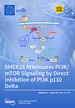
Autophagy can be induced by inhibition of mTORC1, a multiprotein kinase complex. Rapamycin directly disables mTORC1 activity, while the mode of action of the autophagy inducer SMER28 was unknown. In our study, we reveal that SMER28 directly inhibits PI3K, most effectively p110 subunit delta. SMER28 treatment reduces phospho-AKT levels on both phosphorylation sides, threonine-308 and serine-473, indicating upstream inhibition of PDK1 and mTORC2. The autophagy-regulating kinase Ulk1 shows diminished phosphorylation of serine758; consistently, we find increased LC3-positive autophagosome numbers. Moreover, we discover that SMER28 severely retards cell growth and abolishes actin cytoskeleton dynamics leading to dorsal ruffle formation and cell scattering upon RTK activation. We show that cell lines harboring high PI3K delta levels such as B cell lymphoma cells are sensitive to SMER28. View this paper
  • Issues are regarded as officially published after their release is announced to the table of contents alert mailing list.
  • You may sign up for e-mail alerts to receive table of contents of newly released issues.
  • PDF is the official format for papers published in both, html and pdf forms. To view the papers in pdf format, click on the "PDF Full-text" link, and use the free Adobe Reader to open them.
Order results
Result details
Section
Select all
Export citation of selected articles as:
27 pages, 1785 KiB  
Review
Angiogenesis, Lymphangiogenesis, and Inflammation in Chronic Obstructive Pulmonary Disease (COPD): Few Certainties and Many Outstanding Questions
by Remo Poto, Stefania Loffredo, Francesco Palestra, Gianni Marone, Vincenzo Patella and Gilda Varricchi
Cells 2022, 11(10), 1720; https://doi.org/10.3390/cells11101720 - 23 May 2022
Cited by 14 | Viewed by 4428
Abstract
Chronic obstructive pulmonary disease (COPD) is characterized by chronic inflammation, predominantly affecting the lung parenchyma and peripheral airways, that results in progressive and irreversible airflow obstruction. COPD development is promoted by persistent pulmonary inflammation in response to several stimuli (e.g., cigarette smoke, bacterial [...] Read more.
Chronic obstructive pulmonary disease (COPD) is characterized by chronic inflammation, predominantly affecting the lung parenchyma and peripheral airways, that results in progressive and irreversible airflow obstruction. COPD development is promoted by persistent pulmonary inflammation in response to several stimuli (e.g., cigarette smoke, bacterial and viral infections, air pollution, etc.). Angiogenesis, the formation of new blood vessels, and lymphangiogenesis, the formation of new lymphatic vessels, are features of airway inflammation in COPD. There is compelling evidence that effector cells of inflammation (lung-resident macrophages and mast cells and infiltrating neutrophils, eosinophils, basophils, lymphocytes, etc.) are major sources of a vast array of angiogenic (e.g., vascular endothelial growth factor-A (VEGF-A), angiopoietins) and/or lymphangiogenic factors (VEGF-C, -D). Further, structural cells, including bronchial and alveolar epithelial cells, endothelial cells, fibroblasts/myofibroblasts, and airway smooth muscle cells, can contribute to inflammation and angiogenesis in COPD. Although there is evidence that alterations of angiogenesis and, to a lesser extent, lymphangiogenesis, are associated with COPD, there are still many unanswered questions. Full article
(This article belongs to the Special Issue Advances in COPD)
Show Figures

Figure 1

16 pages, 2805 KiB  
Article
Low-Frequency Oscillations of In Vivo Ambient Extracellular Brain Serotonin
by Colby E. Witt, Sergio Mena, Lauren E. Honan, Lauren Batey, Victoria Salem, Yangguang Ou and Parastoo Hashemi
Cells 2022, 11(10), 1719; https://doi.org/10.3390/cells11101719 - 23 May 2022
Cited by 3 | Viewed by 2841
Abstract
Serotonin is an important neurotransmitter that plays a major role in many aspects of neuroscience. Fast-scan cyclic voltammetry measures fast in vivo serotonin dynamics using carbon fiber microelectrodes. More recently, fast-scan controlled-adsorption voltammetry (FSCAV) has been developed to measure slower, minute-to-minute changes in [...] Read more.
Serotonin is an important neurotransmitter that plays a major role in many aspects of neuroscience. Fast-scan cyclic voltammetry measures fast in vivo serotonin dynamics using carbon fiber microelectrodes. More recently, fast-scan controlled-adsorption voltammetry (FSCAV) has been developed to measure slower, minute-to-minute changes in ambient extracellular serotonin. We have previously demonstrated that FSCAV measurements of basal serotonin levels give critical information regarding brain physiology and disease. In this work, we revealed the presence of low-periodicity fluctuations in serotonin levels in mouse hippocampi, measured in vivo with FSCAV. Using correlation analyses, we found robust evidence of oscillations in the basal serotonin levels, which had a period of 10 min and were not present in vitro. Under control conditions, the oscillations did not differ between male and female mice, nor do they differ between mice that underwent a chronic stress paradigm and those in the control group. After the acute administration of a selective serotonin reuptake inhibitor, we observed a shift in the frequency of the oscillations, leading us to hypothesize that the newly observed fluctuations were transporter regulated. Finally, we optimized the experimental parameters of the FSCAV to measure at a higher temporal resolution and found more pronounced shifts in the oscillation frequency, along with a decreased oscillation amplitude. We postulate that this work may serve as a potential bridge for studying serotonin/endocrine interactions that occur on the same time scale. Full article
(This article belongs to the Special Issue Neurotransmitter Transporters in Health and Disease)
Show Figures

Figure 1

17 pages, 3859 KiB  
Article
Effect of Thrombin on the Metabolism and Function of Murine Macrophages
by Ürün Ukan, Fredy Delgado Lagos, Sebastian Kempf, Stefan Günther, Mauro Siragusa, Beate Fisslthaler and Ingrid Fleming
Cells 2022, 11(10), 1718; https://doi.org/10.3390/cells11101718 - 23 May 2022
Cited by 5 | Viewed by 2623
Abstract
Macrophages are plastic and heterogeneous immune cells that adapt pro- or anti-inflammatory phenotypes upon exposure to different stimuli. Even though there has been evidence supporting a crosstalk between coagulation and innate immunity, the way in which protein components of the hemostasis pathway influence [...] Read more.
Macrophages are plastic and heterogeneous immune cells that adapt pro- or anti-inflammatory phenotypes upon exposure to different stimuli. Even though there has been evidence supporting a crosstalk between coagulation and innate immunity, the way in which protein components of the hemostasis pathway influence macrophages remains unclear. We investigated the effect of thrombin on macrophage polarization. On the basis of gene expression and cytokine secretion, our results suggest that polarization with thrombin induces an anti-inflammatory, M2-like phenotype. In functional studies, thrombin polarization promoted oxLDL phagocytosis by macrophages, and conditioned medium from the same cells increased endothelial cell proliferation. There were, however, clear differences between the classical M2a polarization and the effects of thrombin on gene expression. Finally, the deletion and inactivation of secreted modular Ca2+-binding protein 1 (SMOC1) attenuated phagocytosis by thrombin-stimulated macrophages, a phenomenon revered by the addition of recombinant SMOC1. Manipulation of SMOC1 levels also had a pronounced impact on the expression of TGF-β-signaling-related genes. Taken together, our results show that thrombin induces an anti-inflammatory macrophage phenotype with similarities as well as differences to the classical alternatively activated M2 polarization states, highlighting the importance of tissue levels of SMOC1 in modifying thrombin-induced macrophage polarization. Full article
(This article belongs to the Special Issue State-of-Art in Innate Immunity)
Show Figures

Figure 1

14 pages, 1209 KiB  
Review
Pyroptosis and Its Role in SARS-CoV-2 Infection
by Zsofia Agnes Bittner, Markus Schrader, Shilpa Elizabeth George and Ralf Amann
Cells 2022, 11(10), 1717; https://doi.org/10.3390/cells11101717 - 23 May 2022
Cited by 16 | Viewed by 4241
Abstract
The pore-forming inflammatory cell death pathway, pyroptosis, was first described in the early 1990s and its role in health and disease has been intensively studied since. The effector molecule GSDMD is cleaved by activated caspases, mainly Caspase 1 or 11 (Caspase 4/5 in [...] Read more.
The pore-forming inflammatory cell death pathway, pyroptosis, was first described in the early 1990s and its role in health and disease has been intensively studied since. The effector molecule GSDMD is cleaved by activated caspases, mainly Caspase 1 or 11 (Caspase 4/5 in humans), downstream of inflammasome formation. In this review, we describe the molecular events related to GSDMD-mediated pore formation. Furthermore, we summarize the so far elucidated ways of SARS-CoV-2 induced NLRP3 inflammasome formation leading to pyroptosis, which strongly contributes to COVID-19 pathology. We also explore the potential of NLRP3 and GSDMD inhibitors as therapeutics to counter excessive inflammation. Full article
(This article belongs to the Special Issue Factsheets of Cell Death)
Show Figures

Figure 1

19 pages, 1308 KiB  
Review
Exosome-Associated circRNAs as Key Regulators of EMT in Cancer
by Laura Amicone, Alessandra Marchetti and Carla Cicchini
Cells 2022, 11(10), 1716; https://doi.org/10.3390/cells11101716 - 23 May 2022
Cited by 9 | Viewed by 2618
Abstract
Epithelial-to-mesenchymal transition (EMT) is a dynamic program of cell plasticity aberrantly reactivated in cancer. The crosstalk between tumor cells and the tumoral microenvironment (TME) has a pivotal importance for the induction of the EMT and the progression toward a malignant phenotype. Notably, exosomes [...] Read more.
Epithelial-to-mesenchymal transition (EMT) is a dynamic program of cell plasticity aberrantly reactivated in cancer. The crosstalk between tumor cells and the tumoral microenvironment (TME) has a pivotal importance for the induction of the EMT and the progression toward a malignant phenotype. Notably, exosomes are key mediators of this crosstalk as vehicles of specific molecular signals that include the class of circular RNAs (circRNAs). This review specifically focuses on the role of exosome-associated circRNAs as key regulators of EMT in cancer. The relevance of these molecules in regulating the intercellular communication in TME and tumor progression is highlighted. Moreover, the here-presented evidence indicates that exosome-associated circRNA modulation should be taken in account for cancer diagnostic and therapeutic approaches. Full article
(This article belongs to the Special Issue Extracellular Vesicle-Associated Non-Coding RNAs)
Show Figures

Figure 1

16 pages, 2202 KiB  
Article
Extracellular Vesicular Transmission of miR-423-5p from HepG2 Cells Inhibits the Differentiation of Hepatic Stellate Cells
by Michal Safran, Rula Masoud, Maya Sultan, Irena Tachlytski, Chofit Chai Gadot, Ron Pery, Nora Balint-Lahat, Orit Pappo, Nahum Buzaglo and Ziv Ben-Ari
Cells 2022, 11(10), 1715; https://doi.org/10.3390/cells11101715 - 23 May 2022
Cited by 9 | Viewed by 2719
Abstract
Liver fibrosis (LF) is a major cause of morbidity and mortality worldwide. Hepatic stellate cells (HSCs) are the primary source of extracellular matrix in the liver and their activation is a central event in LF development. Extracellular vesicles (EVs) are intercellular communication agents, [...] Read more.
Liver fibrosis (LF) is a major cause of morbidity and mortality worldwide. Hepatic stellate cells (HSCs) are the primary source of extracellular matrix in the liver and their activation is a central event in LF development. Extracellular vesicles (EVs) are intercellular communication agents, which play important roles in physiological processes in chronic liver diseases. The aim of this study was to examine the crosstalk between hepatocytes and HSCs mediated by hepatocyte-secreted EVs. EVs were purified from primary mouse hepatocytes, HepG2 cell lines, under normal or stressed conditions. The effect of EVs on primary HSCs (pHSCs) differentiation was evaluated by measuring of differentiation markers. In addition, their impact on the carbon tetrachloride (CCl4)-induced fibrosis mouse model was evaluated. The results demonstrated that HepG2-EVs regulate HSC differentiation and that under stress conditions, promoted pHSCs differentiation into the myofibroblast phenotype. The evaluation of miRNA sequences in the HepG2 secreted EVs demonstrated high levels of miR-423-5p. The examination of EV cargo following stress conditions identified a significant reduction of miR-423-5p in HepG2-EVs relative to HepG2-EVs under normal conditions. In addition, pHSCs transfected with miR-423-5p mimic and exhibit lower mRNA levels of alpha smooth muscle actin and Collagen type 1 alpha, and the mRNA expression level of genes targeted the family with sequence-similarity-3 (FAM3) and Monoacylglycerol lipase (Mgll). This study strengthened the hypothesis that EVs are involved in LF and that their cargo changes in stress conditions. In addition, miR-423-5p was shown to be involved in HSCs differentiation and hence, fibrosis development. Full article
Show Figures

Figure 1

16 pages, 2836 KiB  
Review
Yeast Chronological Lifespan: Longevity Regulatory Genes and Mechanisms
by Mario G. Mirisola and Valter D. Longo
Cells 2022, 11(10), 1714; https://doi.org/10.3390/cells11101714 - 23 May 2022
Cited by 15 | Viewed by 4982
Abstract
S. cerevisiae plays a pivotal role as a model system in understanding the biochemistry and molecular biology of mammals including humans. A considerable portion of our knowledge on the genes and pathways involved in cellular growth, resistance to toxic agents, and death has [...] Read more.
S. cerevisiae plays a pivotal role as a model system in understanding the biochemistry and molecular biology of mammals including humans. A considerable portion of our knowledge on the genes and pathways involved in cellular growth, resistance to toxic agents, and death has in fact been generated using this model organism. The yeast chronological lifespan (CLS) is a paradigm to study age-dependent damage and longevity. In combination with powerful genetic screening and high throughput technologies, the CLS has allowed the identification of longevity genes and pathways but has also introduced a unicellular “test tube” model system to identify and study macromolecular and cellular damage leading to diseases. In addition, it has played an important role in studying the nutrients and dietary regimens capable of affecting stress resistance and longevity and allowing the characterization of aging regulatory networks. The parallel description of the pro-aging roles of homologs of RAS, S6 kinase, adenylate cyclase, and Tor in yeast and in higher eukaryotes in S. cerevisiae chronological survival studies is valuable to understand human aging and disease. Here we review work on the S. cerevisiae chronological lifespan with a focus on the genes regulating age-dependent macromolecular damage and longevity extension. Full article
(This article belongs to the Special Issue Yeast as a Model in Aging Research)
Show Figures

Figure 1

19 pages, 4540 KiB  
Article
Mitochondrial Membrane Potential Identifies a Subpopulation of Mesenchymal Progenitor Cells to Promote Angiogenesis and Myocardial Repair
by Xiuchun Li, Xiaoliang Wang, Pan He, Edward Bennett, Erin Haggard, Jianjie Ma and Chuanxi Cai
Cells 2022, 11(10), 1713; https://doi.org/10.3390/cells11101713 - 22 May 2022
Cited by 3 | Viewed by 2287
Abstract
Identifying effective donor cells is one of obstacles that limits cell therapy for heart disease. In this study, we sorted a subpopulation of human mesenchymal progenitor cells (hMPCs) from the right atrial appendage using the low mitochondrial membrane potential. Compared to the non-sorted [...] Read more.
Identifying effective donor cells is one of obstacles that limits cell therapy for heart disease. In this study, we sorted a subpopulation of human mesenchymal progenitor cells (hMPCs) from the right atrial appendage using the low mitochondrial membrane potential. Compared to the non-sorted cells, hMPCs hold the capacity for stemness and enrich mesenchymal stem cell markers. The hMPCs display better ability for survival, faster proliferation, less production of reactive oxygen species (ROS), and greater release of cytoprotective cytokines. The hMPCs exhibit decreased expression of senescence genes and increased expression of anti-apoptotic and antioxidant genes. Intramyocardial injection of hMPCs into the infarcted heart resulted in increased left ventricular ejection fraction and reduced cardiac remodeling and infarct size in the group of animals receiving hMPCs. Both in vitro and in vivo studies indicated hMPCs have the potential to differentiate into endothelial cells and smooth muscle cells. Immunohistochemistry staining showed that cell therapy with hMPCs enhances cardiac vascular regeneration and cardiac proliferation, and decreases cardiac cell apoptosis, which is associated with the increased secretion of cytoprotective and pro-angiogenic cytokines. Overall, we discovered a subpopulation of human mesenchymal progenitor cells via their low mitochondrial membrane potential, which might provide an alternative donor cell source for cellular therapy for ischemic heart disease. Full article
(This article belongs to the Special Issue Cardiovascular Disease and Vascular Engineering)
Show Figures

Figure 1

17 pages, 1818 KiB  
Review
Colonic Fluid and Electrolyte Transport 2022: An Update
by Abel B. Negussie, Annika C. Dell, Bruce A. Davis and John P. Geibel
Cells 2022, 11(10), 1712; https://doi.org/10.3390/cells11101712 - 22 May 2022
Cited by 13 | Viewed by 5813
Abstract
Colonic epithelial cells are responsible for maintaining a delicate balance between luminal secretion and the absorption of fluids and ions. This review aims to discuss and update the model of colonic electrolyte secretion and absorption via the cystic fibrosis transmembrane regulator (CFTR), epithelial [...] Read more.
Colonic epithelial cells are responsible for maintaining a delicate balance between luminal secretion and the absorption of fluids and ions. This review aims to discuss and update the model of colonic electrolyte secretion and absorption via the cystic fibrosis transmembrane regulator (CFTR), epithelial sodium channel (ENaC), Na-K-Cl cotransporters (NKCC1 and 2), Na-H exchangers (NHE1–4), colonic H,KATPase, and several other key components involved in multi-level transepithelial ion transport. Developments in our understanding of the activity, regulation, localization, and relationships of these ion transporters and their interactions have helped forge a more robust understanding of colonic ion movement that accounts for the colonic epithelium’s role in mucosal pH modulation, the setting of osmotic gradients pivotal for fluid retention and secretion, and cell death regulation. Deviations from homeostatic ion transport cause diarrhea, constipation, and epithelial cell death and contribute to cystic fibrosis, irritable bowel syndrome (IBS), ulcerative colitis, and cancer pathologies. Signal transduction pathways that regulate electrolyte movement and the regulatory relationships between various sensors and transporters (CFTR as a target of CaSR regulation and as a regulator of ENaC and DRA, for example) are imperative aspects of a dynamic and comprehensive model of colonic ion homeostasis. Full article
Show Figures

Figure 1

29 pages, 21230 KiB  
Review
Infertility in Men: Advances towards a Comprehensive and Integrative Strategy for Precision Theranostics
by Mourad Assidi
Cells 2022, 11(10), 1711; https://doi.org/10.3390/cells11101711 - 22 May 2022
Cited by 12 | Viewed by 9029
Abstract
Male infertility is an increasing and serious medical concern, though the mechanism remains poorly understood. Impaired male reproductive function affects approximately half of infertile couples worldwide. Multiple factors related to the environment, genetics, age, and comorbidities have been associated with impaired sperm function. [...] Read more.
Male infertility is an increasing and serious medical concern, though the mechanism remains poorly understood. Impaired male reproductive function affects approximately half of infertile couples worldwide. Multiple factors related to the environment, genetics, age, and comorbidities have been associated with impaired sperm function. Present-day clinicians rely primarily on standard semen analysis to diagnose male reproductive potential and develop treatment strategies. To address sperm quality assessment bias and enhance analysis accuracy, the World Health Organization (WHO) has recommended standardized sperm testing; however, conventional diagnostic and therapeutic options for male infertility, including physical examination and semen standard analysis, remain ineffective in relieving the associated social burden. Instead, assisted reproductive techniques are becoming the primary therapeutic approach. In the post-genomic era, multiomics technologies that deeply interrogate the genome, transcriptome, proteome, and/or the epigenome, even at single-cell level, besides the breakthroughs in robotic surgery, stem cell therapy, and big data, offer promises towards solving semen quality deterioration and male factor infertility. This review highlights the complex etiology of male infertility, especially the roles of lifestyle and environmental factors, and discusses advanced technologies/methodologies used in characterizing its pathophysiology. A comprehensive combination of these innovative approaches in a global and multi-centric setting and fulfilling the suitable ethical consent could ensure optimal reproductive and developmental outcomes. These combinatorial approaches should allow for the development of diagnostic markers, molecular stratification classes, and personalized treatment strategies. Since lifestyle choices and environmental factors influence male fertility, their integration in any comprehensive approach is required for safe, proactive, cost-effective, and noninvasive precision male infertility theranostics that are affordable, accessible, and facilitate couples realizing their procreation dream. Full article
(This article belongs to the Section Intracellular and Plasma Membranes)
Show Figures

Figure 1

17 pages, 4209 KiB  
Article
FBP2—A New Player in Regulation of Motility of Mitochondria and Stability of Microtubules in Cardiomyocytes
by Łukasz Pietras, Ewa Stefanik, Dariusz Rakus and Agnieszka Gizak
Cells 2022, 11(10), 1710; https://doi.org/10.3390/cells11101710 - 21 May 2022
Cited by 3 | Viewed by 2663
Abstract
Recently, we have shown that the physiological roles of a multifunctional protein fructose 1,6-bisphosphatase 2 (FBP2, also called muscle FBP) depend on the oligomeric state of the protein. Here, we present several lines of evidence that in HL-1 cardiomyocytes, a forced, chemically induced [...] Read more.
Recently, we have shown that the physiological roles of a multifunctional protein fructose 1,6-bisphosphatase 2 (FBP2, also called muscle FBP) depend on the oligomeric state of the protein. Here, we present several lines of evidence that in HL-1 cardiomyocytes, a forced, chemically induced reduction in the FBP2 dimer-tetramer ratio that imitates AMP and NAD+ action and restricts FBP2-mitochondria interaction, results in an increase in Tau phosphorylation, augmentation of FBP2-Tau and FBP2-MAP1B interactions, disturbance of tubulin network, marked reduction in the speed of mitochondrial trafficking and increase in mitophagy. These results not only highlight the significance of oligomerization for the regulation of FBP2 physiological role in the cell, but they also demonstrate a novel, important cellular function of this multitasking protein—a function that might be crucial for processes that take place during physiological and pathological cardiac remodeling, and during the onset of diseases which are rooted in the destabilization of MT and/or mitochondrial network dynamics. Full article
(This article belongs to the Special Issue Multitasking Proteins and Their Involvement in Pathogenesis)
Show Figures

Figure 1

19 pages, 4640 KiB  
Article
The Dually Localized EF-Hand Domain-Containing Protein TgEFP1 Regulates the Lytic Cycle of Toxoplasma gondii
by Noopur Dave, Kaice LaFavers and Gustavo Arrizabalaga
Cells 2022, 11(10), 1709; https://doi.org/10.3390/cells11101709 - 21 May 2022
Cited by 5 | Viewed by 2048
Abstract
The propagation of the obligate intracellular parasite Toxoplasma gondii is tightly regulated by calcium signaling. However, the mechanisms by which calcium homeostasis and fluxes are regulated in this human pathogen are not fully understood. To identify Toxoplasma’s calcium homeostasis network, we have characterized [...] Read more.
The propagation of the obligate intracellular parasite Toxoplasma gondii is tightly regulated by calcium signaling. However, the mechanisms by which calcium homeostasis and fluxes are regulated in this human pathogen are not fully understood. To identify Toxoplasma’s calcium homeostasis network, we have characterized a novel EF-hand domain-containing protein, which we have named TgEFP1. We have determined that TgEFP1 localizes to a previously described compartment known as the plant-like vacuole or the endosomal-like compartment (PLV/ELC), which harbors several proteins related to ionic regulation. Interestingly, partial permeabilization techniques showed that TgEFP1 is also secreted into the parasitophorous vacuole (PV), within which the parasite divides. Ultrastructure expansion microscopy confirmed the unusual dual localization of TgEFP1 at the PLV/ELC and the PV. Furthermore, we determined that the localization of TgEFP1 to the PV, but not to the PLV/ELC, is affected by disruption of Golgi-dependent transport with Brefeldin A. Knockout of TgEFP1 results in faster propagation in tissue culture, hypersensitivity to calcium ionophore-induced egress, and premature natural egress. Thus, our work has revealed an interplay between the PV and the PLV/ELC and a role for TgEFP1 in the regulation of calcium-dependent events. Full article
(This article belongs to the Section Cell Signaling)
Show Figures

Figure 1

27 pages, 2302 KiB  
Review
Current Therapeutic Landscape and Safety Roadmap for Targeting the Aryl Hydrocarbon Receptor in Inflammatory Gastrointestinal Indications
by Samantha C. Faber, Tejas S. Lahoti, Ewan R. Taylor, Lauren Lewis, Jessica M. Sapiro, Vicencia Toledo Sales, Yvonne P. Dragan and Brandon D. Jeffy
Cells 2022, 11(10), 1708; https://doi.org/10.3390/cells11101708 - 21 May 2022
Cited by 3 | Viewed by 3365
Abstract
Target modulation of the AhR for inflammatory gastrointestinal (GI) conditions holds great promise but also the potential for safety liabilities both within and beyond the GI tract. The ubiquitous expression of the AhR across mammalian tissues coupled with its role in diverse signaling [...] Read more.
Target modulation of the AhR for inflammatory gastrointestinal (GI) conditions holds great promise but also the potential for safety liabilities both within and beyond the GI tract. The ubiquitous expression of the AhR across mammalian tissues coupled with its role in diverse signaling pathways makes development of a “clean” AhR therapeutically challenging. Ligand promiscuity and diversity in context-specific AhR activation further complicates targeting the AhR for drug development due to limitations surrounding clinical translatability. Despite these concerns, several approaches to target the AhR have been explored such as small molecules, microbials, PROTACs, and oligonucleotide-based approaches. These various chemical modalities are not without safety liabilities and require unique de-risking strategies to parse out toxicities. Collectively, these programs can benefit from in silico and in vitro methodologies that investigate specific AhR pathway activation and have the potential to implement thresholding parameters to categorize AhR ligands as “high” or “low” risk for sustained AhR activation. Exploration into transcriptomic signatures for AhR safety assessment, incorporation of physiologically-relevant in vitro model systems, and investigation into chronic activation of the AhR by structurally diverse ligands will help address gaps in our understanding regarding AhR-dependent toxicities. Here, we review the role of the AhR within the GI tract, novel therapeutic modality approaches to target the AhR, key AhR-dependent safety liabilities, and relevant strategies that can be implemented to address drug safety concerns. Together, this review discusses the emerging therapeutic landscape of modalities targeting the AhR for inflammatory GI indications and offers a safety roadmap for AhR drug development. Full article
Show Figures

Graphical abstract

30 pages, 2088 KiB  
Review
Central Nervous System Pericytes Contribute to Health and Disease
by Francesco Girolamo, Mariella Errede, Antonella Bizzoca, Daniela Virgintino and Domenico Ribatti
Cells 2022, 11(10), 1707; https://doi.org/10.3390/cells11101707 - 20 May 2022
Cited by 9 | Viewed by 4651
Abstract
Successful neuroprotection is only possible with contemporary microvascular protection. The prevention of disease-induced vascular modifications that accelerate brain damage remains largely elusive. An improved understanding of pericyte (PC) signalling could provide important insight into the function of the neurovascular unit (NVU), and into [...] Read more.
Successful neuroprotection is only possible with contemporary microvascular protection. The prevention of disease-induced vascular modifications that accelerate brain damage remains largely elusive. An improved understanding of pericyte (PC) signalling could provide important insight into the function of the neurovascular unit (NVU), and into the injury-provoked responses that modify cell–cell interactions and crosstalk. Due to sharing the same basement membrane with endothelial cells, PCs have a crucial role in the control of endothelial, astrocyte, and oligodendrocyte precursor functions and hence blood–brain barrier stability. Both cerebrovascular and neurodegenerative diseases impair oxygen delivery and functionally impair the NVU. In this review, the role of PCs in central nervous system health and disease is discussed, considering their origin, multipotency, functions and also dysfunction, focusing on new possible avenues to modulate neuroprotection. Dysfunctional PC signalling could also be considered as a potential biomarker of NVU pathology, allowing us to individualize therapeutic interventions, monitor responses, or predict outcomes. Full article
(This article belongs to the Special Issue Biomarkers in Inflammation and Angiogenesis)
Show Figures

Figure 1

16 pages, 6091 KiB  
Article
Exercise Enhances Branched-Chain Amino Acid Catabolism and Decreases Cardiac Vulnerability to Myocardial Ischemic Injury
by Guiling Wu, Yanjie Guo, Min Li, Chenhan Li, Yanzhen Tan, Yueyang Li, Jia Li, Li Wang, Xing Zhang and Feng Gao
Cells 2022, 11(10), 1706; https://doi.org/10.3390/cells11101706 - 20 May 2022
Cited by 8 | Viewed by 2514
Abstract
Long-term exercise-induced metabolic adaptations occupy a central position in exercise-afforded cardiac benefits. Emerging evidence suggests that branched-chain amino acid (BCAA) catabolic defect contributes to cardiac dysfunction in multiple cardiometabolic diseases. However, the role of BCAA catabolism in exercise-afforded cardiac benefits remains unknown. Here, [...] Read more.
Long-term exercise-induced metabolic adaptations occupy a central position in exercise-afforded cardiac benefits. Emerging evidence suggests that branched-chain amino acid (BCAA) catabolic defect contributes to cardiac dysfunction in multiple cardiometabolic diseases. However, the role of BCAA catabolism in exercise-afforded cardiac benefits remains unknown. Here, we show that exercise improves BCAA catabolism and thus reduce cardiac vulnerability to myocardial ischemic injury. Exercise increased circulating BCAA levels in both humans (male adolescent athletes) and mice (following an 8-week swimming intervention). It increased the expression of mitochondrial localized 2C-type serine-threonine protein phosphatase (PP2Cm), a key enzyme in regulating BCAA catabolism, and decreased BCAA accumulation in mouse hearts, indicating an increase in BCAA catabolism. Pharmacological promotion of BCAA catabolism protected the mouse heart against myocardial infarction (MI) induced by permanent ligation of the left descending coronary artery. Although cardiac-specific PP2Cm knockout showed no significant effects on cardiac structural and functional adaptations to exercise, it blunted the cardioprotective effects of exercise against MI. Mechanistically, exercise alleviated BCAA accumulation and subsequently inactivated the mammalian target of rapamycin in MI hearts. These results showed that exercise elevated BCAA catabolism and protected the heart against myocardial ischemic injury, reinforcing the role of exercise in the promotion of cardiac health. Full article
(This article belongs to the Special Issue Mitochondrial Dysfunction in Aging and Metabolic Diseases)
Show Figures

Graphical abstract

22 pages, 8910 KiB  
Article
Interleukin-6 Signaling in Triple Negative Breast Cancer Cells Elicits the Annexin A1/Formyl Peptide Receptor 1 Axis and Affects the Tumor Microenvironment
by Lara Vecchi, Sara Teixeira Soares Mota, Mariana Alves Pereira Zóia, Isabella Castro Martins, Jessica Brito de Souza, Tiago Góss Santos, Adriano de Oliveira Beserra, Victor Piana de Andrade, Luiz Ricardo Goulart and Thaise Gonçalves Araújo
Cells 2022, 11(10), 1705; https://doi.org/10.3390/cells11101705 - 20 May 2022
Cited by 10 | Viewed by 2417
Abstract
Annexin A1 (AnxA1) is a pleiotropic protein that exerts essential roles in breast cancer (BC) growth and aggressiveness. In our previous work, we described the autocrine signaling of AnxA1 through formyl peptide receptor 1 (FPR1) in the triple-negative (TN) BC cell line, MDA-MB-231. [...] Read more.
Annexin A1 (AnxA1) is a pleiotropic protein that exerts essential roles in breast cancer (BC) growth and aggressiveness. In our previous work, we described the autocrine signaling of AnxA1 through formyl peptide receptor 1 (FPR1) in the triple-negative (TN) BC cell line, MDA-MB-231. Here, we aimed to describe the interaction between the AnxA1/FPR1 and the Interleukin-6 (IL-6) signaling pathways and their role in the tumor microenvironment (TME). First, we demonstrated that AnxA1 and IL-6 expression levels are correlated in BC tissue samples. In three TNBC cell lines, overexpression of both AnxA1 and IL-6 was also identified. Next, we inhibited FPR1, the IL-6 receptor and STAT3 in both MDA-MB-231 and MDA-MB-157 cells. The FPR1 inhibition led to increased levels of IL-6 and secreted AnxA1 in both cell lines. On the other side, inhibition of the IL-6 receptor or STAT3 led to the impairment of AnxA1 secretion, suggesting the essential role of the IL-6 signaling cascade in the activation of the AnxA1/FPR1 autocrine axis. Finally, we described the interaction between IL-6 and the AnxA1/FPR1 pathways and their role on the TME by analyzing the effect of supernatants derived from MDA-MB-231 and MDA-MB-157 cells under the inhibition of FPR1 or IL-6 signaling on fibroblast cell motility. Full article
(This article belongs to the Special Issue Role of Annexin A1 in the Tumor Microenvironment)
Show Figures

Figure 1

18 pages, 2944 KiB  
Article
Epigallocatechine-3-gallate Inhibits the Adipogenesis of Human Mesenchymal Stem Cells via the Regulation of Protein Phosphatase-2A and Myosin Phosphatase
by Bálint Bécsi, Zoltán Kónya, Anita Boratkó, Katalin Kovács and Ferenc Erdődi
Cells 2022, 11(10), 1704; https://doi.org/10.3390/cells11101704 - 20 May 2022
Cited by 2 | Viewed by 2117
Abstract
Epigallocatechin-3-gallate (EGCG) has widespread effects on adipocyte development. However, the molecular mechanisms of EGCG are not fully understood. We investigate the adipogenic differentiation of human-derived mesenchymal stem cells, including lipid deposition and changes in the expression and phosphorylation of key transcription factors, myosin, [...] Read more.
Epigallocatechin-3-gallate (EGCG) has widespread effects on adipocyte development. However, the molecular mechanisms of EGCG are not fully understood. We investigate the adipogenic differentiation of human-derived mesenchymal stem cells, including lipid deposition and changes in the expression and phosphorylation of key transcription factors, myosin, protein phosphatase-2A (PP2A), and myosin phosphatase (MP). On day 6 of adipogenic differentiation, EGCG (1–20 µM) suppressed lipid droplet formation, which was counteracted by an EGCG-binding peptide for the 67 kDa laminin receptor (67LR), suggesting that EGCG acts via 67LR. EGCG decreased the phosphorylation of CCAAT-enhancer-binding protein beta via the activation of PP2A in a protein kinase A (PKA)-dependent manner, leading to the partial suppression of peroxisome proliferator-activated receptor gamma (PPARγ) and adiponectin expression. Differentiated cells exhibited a rounded shape, cortical actin filaments, and lipid accumulation. The EGCG treatment induced cell elongation, stress fiber formation, and less lipid accumulation. These effects were accompanied by the degradation of the MP target subunit-1 and increased the phosphorylation of the 20 kDa myosin light chain. Our results suggest that EGCG acts as an agonist of 67LR to inhibit adipogenesis via the activation of PP2A and suppression of MP. These events are coupled with the decreased phosphorylation and expression levels of adipogenic transcription factors and changes in cell shape, culminating in curtailed adipogenesis. Full article
Show Figures

Figure 1

9 pages, 795 KiB  
Review
Unveiling the Function of the Mitochondrial Filament-Forming Protein LACTB in Lipid Metabolism and Cancer
by Annunziata Cascone, Maciej Lalowski, Dan Lindholm and Ove Eriksson
Cells 2022, 11(10), 1703; https://doi.org/10.3390/cells11101703 - 20 May 2022
Cited by 4 | Viewed by 2429
Abstract
LACTB is a relatively unknown mitochondrial protein structurally related to the bacterial penicillin-binding and beta-lactamase superfamily of serine proteases. LACTB has recently gained an increased interest due to its potential role in lipid metabolism and tumorigenesis. To date, around ninety studies pertaining to [...] Read more.
LACTB is a relatively unknown mitochondrial protein structurally related to the bacterial penicillin-binding and beta-lactamase superfamily of serine proteases. LACTB has recently gained an increased interest due to its potential role in lipid metabolism and tumorigenesis. To date, around ninety studies pertaining to LACTB have been published, but the exact biochemical and cell biological function of LACTB still remain elusive. In this review, we summarise the current knowledge about LACTB with particular attention to the implications of the recently published study on the cryo-electron microscopy structure of the filamentous form of LACTB. From this and other studies, several specific properties of LACTB emerge, suggesting that the protein has distinct functions in different physiological settings. Resolving these issues by further research may ultimately lead to a unified model of LACTB’s function in cell and organismal physiology. LACTB is the only member of its protein family in higher animals and LACTB may, therefore, be of particular interest for future drug targeting initiatives. Full article
(This article belongs to the Special Issue 10th Anniversary of Cells—Advances in Organelle Function)
Show Figures

Figure 1

6 pages, 26031 KiB  
Editorial
Claude Bernard, the Founder of Modern Medicine
by René Habert
Cells 2022, 11(10), 1702; https://doi.org/10.3390/cells11101702 - 20 May 2022
Cited by 2 | Viewed by 2847
Abstract
Claude Bernard is the first and one of the very few French scientists to have been honored with a national funeral [...] Full article
Show Figures

Figure 1

15 pages, 5182 KiB  
Article
An In-Vitro Study of the Expansion and Transcriptomics of CD4+ and CD8+ Naïve and Memory T Cells Stimulated by IL-2, IL-7 and IL-15
by Brooks Hopkins, Justin Fisher, Meiping Chang, Xiaoyan Tang, Zhimei Du, William J. Kelly and Zuyi Huang
Cells 2022, 11(10), 1701; https://doi.org/10.3390/cells11101701 - 20 May 2022
Cited by 3 | Viewed by 2072
Abstract
The growth of T cells ex vivo for the purpose of T cell therapies is a rate-limiting step in the overall process for cancer patients to achieve remission. Growing T cells is a fiscally-, time-, and resource-intensive process. Cytokines have been shown to [...] Read more.
The growth of T cells ex vivo for the purpose of T cell therapies is a rate-limiting step in the overall process for cancer patients to achieve remission. Growing T cells is a fiscally-, time-, and resource-intensive process. Cytokines have been shown to accelerate the growth of T cells, specifically IL-2, IL-7, and IL-15. Here a design of experiments was conducted to optimize the growth rate of different naïve and memory T cell subsets using combinations of cytokines. Mathematical models were developed to study the impact of IL-2, IL-7, and IL-15 on the growth of T cells. The results show that CD4+ and CD8+ naïve T cells grew effectively using moderate IL-2 and IL-7 in combination, and IL-7, respectively. CD4+ and CD8+ memory cells favored moderate IL-2 and IL-15 in combination and moderate IL-7 and IL-15 in combination, respectively. A statistically significant interaction was observed between IL-2 and IL-7 in the growth data of CD4+ naïve T cells, while the interaction between IL-7 and IL-15 was found for CD8+ naïve T cells. The important genes and related signaling pathways and metabolic reactions were identified from the RNA sequencing data for each of the four subsets stimulated by each of the three cytokines. This systematic investigation lays the groundwork for studying other T cell subsets. Full article
(This article belongs to the Collection Trends and Advances in Tumor Immunology)
Show Figures

Figure 1

2 pages, 1812 KiB  
Correction
Correction: Zakrzewicz et al. Stabilization of Keratinocyte Monolayer Integrity in the Presence of Anti-Desmoglein-3 Antibodies through FcRn Blockade with Efgartigimod: Novel Treatment Paradigm for Pemphigus? Cells 2022, 11, 942
by Anna Zakrzewicz, Celina Würth, Benedikt Beckert, Simon Feldhoff, Katrien Vanderheyden, Stian Foss, Jan Terje Andersen, Hans de Haard, Peter Verheesen, Vladimir Bobkov and Ritva Tikkanen
Cells 2022, 11(10), 1700; https://doi.org/10.3390/cells11101700 - 20 May 2022
Cited by 3 | Viewed by 1169
Abstract
The authors wish to make the following changes to their paper [...] Full article
(This article belongs to the Special Issue Autoimmune Diseases: Molecular Mechanisms and Therapies)
16 pages, 1608 KiB  
Review
Microfluidics for 3D Cell and Tissue Cultures: Microfabricative and Ethical Aspects Updates
by Tania Limongi, Francesco Guzzi, Elvira Parrotta, Patrizio Candeloro, Stefania Scalise, Valeria Lucchino, Francesco Gentile, Luca Tirinato, Maria Laura Coluccio, Bruno Torre, Marco Allione, Monica Marini, Francesca Susa, Enzo Di Fabrizio, Giovanni Cuda and Gerardo Perozziello
Cells 2022, 11(10), 1699; https://doi.org/10.3390/cells11101699 - 20 May 2022
Cited by 6 | Viewed by 3728
Abstract
The necessity to improve in vitro cell screening assays is becoming ever more important. Pharmaceutical companies, research laboratories and hospitals require technologies that help to speed up conventional screening and therapeutic procedures to produce more data in a short time in a realistic [...] Read more.
The necessity to improve in vitro cell screening assays is becoming ever more important. Pharmaceutical companies, research laboratories and hospitals require technologies that help to speed up conventional screening and therapeutic procedures to produce more data in a short time in a realistic and reliable manner. The design of new solutions for test biomaterials and active molecules is one of the urgent problems of preclinical screening and the limited correlation between in vitro and in vivo data remains one of the major issues. The establishment of the most suitable in vitro model provides reduction in times, costs and, last but not least, in the number of animal experiments as recommended by the 3Rs (replace, reduce, refine) ethical guiding principles for testing involving animals. Although two-dimensional (2D) traditional cell screening assays are generally cheap and practical to manage, they have strong limitations, as cells, within the transition from the three-dimensional (3D) in vivo to the 2D in vitro growth conditions, do not properly mimic the real morphologies and physiology of their native tissues. In the study of human pathologies, especially, animal experiments provide data closer to what happens in the target organ or apparatus, but they imply slow and costly procedures and they generally do not fully accomplish the 3Rs recommendations, i.e., the amount of laboratory animals and the stress that they undergo must be minimized. Microfluidic devices seem to offer different advantages in relation to the mentioned issues. This review aims to describe the critical issues connected with the conventional cells culture and screening procedures, showing what happens in the in vivo physiological micro and nano environment also from a physical point of view. During the discussion, some microfluidic tools and their components are described to explain how these devices can circumvent the actual limitations described in the introduction. Full article
(This article belongs to the Special Issue In Vitro Model for Micro and Nano Technologies)
Show Figures

Graphical abstract

14 pages, 1872 KiB  
Review
Redox Control of Signalling Responses to Contractile Activity and Ageing in Skeletal Muscle
by Malcolm J. Jackson, Natalie Pollock, Caroline Staunton, Samantha Jones and Anne McArdle
Cells 2022, 11(10), 1698; https://doi.org/10.3390/cells11101698 - 20 May 2022
Cited by 8 | Viewed by 2376
Abstract
Research over almost 40 years has established that reactive oxygen species are generated at different sites in skeletal muscle and that the generation of these species is increased by various forms of exercise. Initially, this was thought to be potentially deleterious to skeletal [...] Read more.
Research over almost 40 years has established that reactive oxygen species are generated at different sites in skeletal muscle and that the generation of these species is increased by various forms of exercise. Initially, this was thought to be potentially deleterious to skeletal muscle and other tissues, but more recent data have identified key roles of these species in muscle adaptations to exercise. The aim of this review is to summarise our current understanding of these redox signalling roles of reactive oxygen species in mediating responses of muscle to contractile activity, with a particular focus on the effects of ageing on these processes. In addition, we provide evidence that disruption of the redox status of muscle mitochondria resulting from age-associated denervation of muscle fibres may be an important factor leading to an attenuation of some muscle responses to contractile activity, and we speculate on potential mechanisms involved. Full article
(This article belongs to the Special Issue Redox Control of Cell Signaling in Cardiac and Skeletal Muscle)
Show Figures

Figure 1

15 pages, 4025 KiB  
Article
Interaction of Cationic Carbosilane Dendrimers and Their siRNA Complexes with MCF-7 Cells Cultured in 3D Spheroids
by Kamila Białkowska, Piotr Komorowski, Rafael Gomez-Ramirez, Francisco Javier de la Mata, Maria Bryszewska and Katarzyna Miłowska
Cells 2022, 11(10), 1697; https://doi.org/10.3390/cells11101697 - 19 May 2022
Cited by 1 | Viewed by 1617
Abstract
Cationic dendrimers are effective carriers for the delivery of siRNA into cells; they can penetrate cell membranes and protect nucleic acids against RNase degradation. Two types of dendrimers (CBD-1 and CBD-2) and their complexes with pro-apoptotic siRNA (Mcl-1 and Bcl-2) were tested on [...] Read more.
Cationic dendrimers are effective carriers for the delivery of siRNA into cells; they can penetrate cell membranes and protect nucleic acids against RNase degradation. Two types of dendrimers (CBD-1 and CBD-2) and their complexes with pro-apoptotic siRNA (Mcl-1 and Bcl-2) were tested on MCF-7 cells cultured as spheroids. Cytotoxicity of dendrimers and dendriplexes was measured using the live–dead test and Annexin V-FITC Apoptosis Detection Kit (flow cytometry). Uptake of dendriplexes was examined using flow cytometry and confocal microscopy. The live–dead test showed that for cells in 3D, CBD-2 is more toxic than CBD-1, contrasting with the data for 2D cultures. Attaching siRNA to a dendrimer molecule did not lead to increased cytotoxic effect in cells, either after 24 or 48 h. Measurements of apoptosis did not show a high increase in the level of the apoptosis marker after 24 h exposure of spheroids to CBD-2 and its dendriplexes. Measurements of the internalization of dendriplexes and microscopy images confirmed that the dendriplexes were transported into cells of the spheroids. Flow cytometry analysis of internalization indicated that CBD-2 transported siRNAs more effectively than CBD-1. Cytotoxic effects were visible after incubation with 3 doses of complexes for CBD-1 and both siRNAs. Full article
(This article belongs to the Section Cell and Gene Therapy)
Show Figures

Graphical abstract

19 pages, 3884 KiB  
Article
Orobanche crenata Forssk. Extract Affects Human Breast Cancer Cell MCF-7 Survival and Viral Replication
by Carlo Genovese, Adriana Garozzo, Floriana D’Angeli, Giuseppe Antonio Malfa, Francesco Bellia, Barbara Tomasello, Daria Nicolosi, Roberta Malaguarnera, Simone Ronsisvalle, Fiorella Guadagni and Rosaria Acquaviva
Cells 2022, 11(10), 1696; https://doi.org/10.3390/cells11101696 - 19 May 2022
Cited by 4 | Viewed by 2431
Abstract
Background: Breast cancer (BC) is the leading cause of death worldwide. The severity of BC strictly depends on the molecular subtype. The less aggressive hormone-positive subtype is treated with adjuvant endocrine therapy (AET), which causes both physical and psychological side effects. This condition [...] Read more.
Background: Breast cancer (BC) is the leading cause of death worldwide. The severity of BC strictly depends on the molecular subtype. The less aggressive hormone-positive subtype is treated with adjuvant endocrine therapy (AET), which causes both physical and psychological side effects. This condition strongly impacts the adherence and persistence of AET among oncologic patients. Moreover, viral infections also constitute a serious problem for public health. Despite their efficacy, antiviral agents present several therapeutic limits. Accordingly, in the present work, we investigated the antitumor and antiviral activities of Orobanche crenata Forssk. (O. crenata), a parasitic plant, endemic to the Mediterranean basin, traditionally known for its beneficial properties for human health. Methods: The MTT assay was carried out to evaluate the cytotoxic effect of O. crenata leaf extract (OCLE) on human breast cancer cells (MCF-7 and MDA-MB-231) and the primary HFF-1 cell line. The lactic dehydrogenase (LDH) assay was performed on MCF-7 cells to analyze necrotic cell death. The antioxidant effect of OCLE was evaluated by intracellular determination of the reactive oxygen species and thiol groups, by DPPH and ABTS assays. The antiviral activity of OCLE was determined against Poliovirus 1, Echovirus 9, Human respiratory syncytial virus, Adenovirus type 2 and type 5, Coxsackievirus B1 (CoxB1) and B3 (CoxB3), Herpes simplex type 1 (HSV-1) and type 2 (HSV-2), and β-Coronavirus by the plaque reduction assay. Results: The extract, after 24 h of incubation, did not affect MDA-MB-231 and HFF-1 cell viability. However, at the same time point, it showed a dose-dependent inhibitory effect on MCF-7 cells, with an increase in LDH release. OCLE exhibited free radical scavenging activity and significantly increased non-protein thiol levels in MCF-7 cells. OCLE effectively inhibited HSV-1, HSV-2, CoxB1, and CoxB3 replication. Conclusions: The overall results showed an interesting inhibitory effect of OCLE on both MCF-7 cell survival and viral replication. Full article
(This article belongs to the Special Issue Advances in Plants-Derived Bioactives for Cancer Treatment)
Show Figures

Graphical abstract

16 pages, 2926 KiB  
Article
Hypoxia Induces Autophagy in Human Dendritic Cells: Involvement of Class III PI3K/Vps34
by Sara Monaci, Federica Coppola, Daniela Rossi, Gaia Giuntini, Irene Filippi, Giuseppe Marotta, Silvano Sozzani, Fabio Carraro and Antonella Naldini
Cells 2022, 11(10), 1695; https://doi.org/10.3390/cells11101695 - 19 May 2022
Cited by 7 | Viewed by 1945
Abstract
Hypoxia is a component of both physiological and pathological conditions, including inflammation, solid tumors, and lymphoid tissues, where O2 demand is not balanced by O2 supply. During their lifespan, dendritic cells (DCs) are exposed to different pO2 and activate different [...] Read more.
Hypoxia is a component of both physiological and pathological conditions, including inflammation, solid tumors, and lymphoid tissues, where O2 demand is not balanced by O2 supply. During their lifespan, dendritic cells (DCs) are exposed to different pO2 and activate different adaptive responses, including autophagy, to preserve their viability and functions. Autophagy plays multiple roles in DC physiology. Very recently, we demonstrated that hypoxia shapes autophagy in DCs upon their differentiation state. Here, we proposed a role for PI3Ks, and especially class III PI3K/Vps34, that could be relevant in hypoxia-induced autophagy, in either immature or mature DCs. Hypoxia inhibited mTOR phosphorylation and activated a pro-autophagic program. By using different pharmacological inhibitors, we demonstrated that hypoxia-induced autophagy was mediated by PI3Ks, especially by Vps34. Furthermore, Vps34 expression was enhanced by LPS, a TLR4 ligand, along with the promotion of autophagy under hypoxia. The Vps34 inhibitor, SAR405, abolished hypoxia-induced autophagy, inhibited pro-survival signaling and viability, and increased the expression of proinflammatory cytokines. Our results underlined the impact of autophagy in the maintenance of DC homeostasis at both cell survival and inflammatory response levels, therefore, contributing to a better understanding of the significance of autophagy in DC physiology and pathology. Full article
(This article belongs to the Special Issue The Autophagic Process in Human Physiology and Pathogenesis)
Show Figures

Graphical abstract

19 pages, 3359 KiB  
Article
Mediating EGFR-TKI Resistance by VEGF/VEGFR Autocrine Pathway in Non-Small Cell Lung Cancer
by Chike Osude, Leo Lin, Meet Patel, Adam Eckburg, Joseph Berei, Adijan Kuckovic, Namrata Dube, Aayush Rastogi, Shruti Gautam, Thomas J. Smith, Shylendra B. Sreenivassappa and Neelu Puri
Cells 2022, 11(10), 1694; https://doi.org/10.3390/cells11101694 - 19 May 2022
Cited by 18 | Viewed by 2698
Abstract
NSCLC treatment includes targeting of EGFR with tyrosine kinase inhibitors (TKIs) such as Erlotinib; however, resistance to TKIs is commonly acquired through T790M EGFR mutations or overexpression of vascular endothelial growth factor receptor-2 (VEGFR-2). We investigated the mechanisms of EGFR-TKI resistance in NSCLC [...] Read more.
NSCLC treatment includes targeting of EGFR with tyrosine kinase inhibitors (TKIs) such as Erlotinib; however, resistance to TKIs is commonly acquired through T790M EGFR mutations or overexpression of vascular endothelial growth factor receptor-2 (VEGFR-2). We investigated the mechanisms of EGFR-TKI resistance in NSCLC cell lines with EGFR mutations or acquired resistance to Erlotinib. These studies showed upregulated gene and protein expression of VEGF, VEGFR-2, and a VEGF co-receptor neuropilin-1 (NP-1) in Erlotinib-resistant (1.4–5.3-fold) and EGFR double-mutant (L858R and T790M; 4.1–8.3-fold) NSCLC cells compared to parental and EGFR single-mutant (L858R) NSCLC cell lines, respectively. Immunofluorescence and FACS analysis revealed increased expression of VEGFR-2 and NP-1 in EGFR-TKI-resistant cell lines compared to TKI-sensitive cell lines. Cell proliferation assays showed that treatment with a VEGFR-2 inhibitor combined with Erlotinib lowered cell survival in EGFR double-mutant NSCLC cells to 9% compared to 72% after treatment with Erlotinib alone. Furthermore, Kaplan–Meier analysis revealed shorter median survival in late-stage NSCLC patients with high vs. low VEGFR-2 expression (14 mos vs. 21 mos). The results indicate that VEGFR-2 may play a key role in EGFR-TKI resistance and that combined treatment of Erlotinib with a VEGFR-2 inhibitor may serve as an effective therapy in NSCLC patients with EGFR mutations. Full article
(This article belongs to the Special Issue Emerging Targets and Therapeutic Strategies in Cancer)
Show Figures

Figure 1

18 pages, 2428 KiB  
Article
Slack Potassium Channels Modulate TRPA1-Mediated Nociception in Sensory Neurons
by Fangyuan Zhou, Katharina Metzner, Patrick Engel, Annika Balzulat, Marco Sisignano, Peter Ruth, Robert Lukowski, Achim Schmidtko and Ruirui Lu
Cells 2022, 11(10), 1693; https://doi.org/10.3390/cells11101693 - 19 May 2022
Cited by 3 | Viewed by 2209
Abstract
The transient receptor potential (TRP) ankyrin type 1 (TRPA1) channel is highly expressed in a subset of sensory neurons where it acts as an essential detector of painful stimuli. However, the mechanisms that control the activity of sensory neurons upon TRPA1 activation remain [...] Read more.
The transient receptor potential (TRP) ankyrin type 1 (TRPA1) channel is highly expressed in a subset of sensory neurons where it acts as an essential detector of painful stimuli. However, the mechanisms that control the activity of sensory neurons upon TRPA1 activation remain poorly understood. Here, using in situ hybridization and immunostaining, we found TRPA1 to be extensively co-localized with the potassium channel Slack (KNa1.1, Slo2.2, or Kcnt1) in sensory neurons. Mice lacking Slack globally (Slack−/−) or conditionally in sensory neurons (SNS-Slack−/−) demonstrated increased pain behavior after intraplantar injection of the TRPA1 activator allyl isothiocyanate. By contrast, pain behavior induced by the TRP vanilloid 1 (TRPV1) activator capsaicin was normal in Slack-deficient mice. Patch-clamp recordings in sensory neurons and in a HEK cell line transfected with TRPA1 and Slack revealed that Slack-dependent potassium currents (IKS) are modulated in a TRPA1-dependent manner. Taken together, our findings highlight Slack as a modulator of TRPA1-mediated, but not TRPV1-mediated, activation of sensory neurons. Full article
Show Figures

Graphical abstract

19 pages, 2109 KiB  
Review
Role of Transcriptional and Epigenetic Regulation in Lymphatic Endothelial Cell Development
by Hyeonwoo La, Hyunjin Yoo, Young Bin Park, Nguyen Xuan Thang, Chanhyeok Park, Seonho Yoo, Hyeonji Lee, Youngsok Choi, Hyuk Song, Jeong Tae Do and Kwonho Hong
Cells 2022, 11(10), 1692; https://doi.org/10.3390/cells11101692 - 19 May 2022
Cited by 2 | Viewed by 2739
Abstract
The lymphatic system is critical for maintaining the homeostasis of lipids and interstitial fluid and regulating the immune cell development and functions. Developmental anomaly-induced lymphatic dysfunction is associated with various pathological conditions, including lymphedema, inflammation, and cancer. Most lymphatic endothelial cells (LECs) are [...] Read more.
The lymphatic system is critical for maintaining the homeostasis of lipids and interstitial fluid and regulating the immune cell development and functions. Developmental anomaly-induced lymphatic dysfunction is associated with various pathological conditions, including lymphedema, inflammation, and cancer. Most lymphatic endothelial cells (LECs) are derived from a subset of endothelial cells in the cardinal vein. However, recent studies have reported that the developmental origin of LECs is heterogeneous. Multiple regulatory mechanisms, including those mediated by signaling pathways, transcription factors, and epigenetic pathways, are involved in lymphatic development and functions. Recent studies have demonstrated that the epigenetic regulation of transcription is critical for embryonic LEC development and functions. In addition to the chromatin structures, epigenetic modifications may modulate transcriptional signatures during the development or differentiation of LECs. Therefore, the understanding of the epigenetic mechanisms involved in the development and function of the lymphatic system can aid in the management of various congenital or acquired lymphatic disorders. Future studies must determine the role of other epigenetic factors and changes in mammalian lymphatic development and function. Here, the recent findings on key factors involved in the development of the lymphatic system and their epigenetic regulation, LEC origins from different organs, and lymphatic diseases are reviewed. Full article
Show Figures

Figure 1

23 pages, 4390 KiB  
Article
Dopamine Reduces SARS-CoV-2 Replication In Vitro through Downregulation of D2 Receptors and Upregulation of Type-I Interferons
by Fiona Limanaqi, Silvia Zecchini, Borana Dino, Sergio Strizzi, Gioia Cappelletti, Olga Utyro, Claudia Vanetti, Micaela Garziano, Irma Saulle, Mario Clerici and Mara Biasin
Cells 2022, 11(10), 1691; https://doi.org/10.3390/cells11101691 - 19 May 2022
Cited by 9 | Viewed by 2341
Abstract
Recent evidence suggests that SARS-CoV-2 hinders immune responses via dopamine (DA)-related mechanisms. Nonetheless, studies addressing the specific role of DA in the frame of SARS-CoV-2 infection are still missing. In the present study, we investigate the role of DA in SARS-CoV-2 replication along [...] Read more.
Recent evidence suggests that SARS-CoV-2 hinders immune responses via dopamine (DA)-related mechanisms. Nonetheless, studies addressing the specific role of DA in the frame of SARS-CoV-2 infection are still missing. In the present study, we investigate the role of DA in SARS-CoV-2 replication along with potential links with innate immune pathways in CaLu-3 human epithelial lung cells. We document here for the first time that, besides DA synthetic pathways, SARS-CoV-2 alters the expression of D1 and D2 DA receptors (D1DR, D2DR), while DA administration reduces viral replication. Such an effect occurs at non-toxic, micromolar-range DA doses, which are known to induce receptor desensitization and downregulation. Indeed, the antiviral effects of DA were associated with a robust downregulation of D2DRs both at mRNA and protein levels, while the amount of D1DRs was not significantly affected. While halting SARS-CoV-2 replication, DA, similar to the D2DR agonist quinpirole, upregulates the expression of ISGs and Type-I IFNs, which goes along with the downregulation of various pro-inflammatory mediators. In turn, administration of Type-I IFNs, while dramatically reducing SARS-CoV-2 replication, converges in downregulating D2DRs expression. Besides configuring the CaLu-3 cell line as a suitable model to study SARS-CoV-2-induced alterations at the level of the DA system in the periphery, our findings disclose a previously unappreciated correlation between DA pathways and Type-I IFN response, which may be disrupted by SARS-CoV-2 for host cell invasion and replication. Full article
(This article belongs to the Special Issue Role of Neurotransmitter and Neuromodulators in Viral Infections)
Show Figures

Graphical abstract

Previous Issue
Next Issue
Back to TopTop