Next Issue
Volume 12, June-2
Previous Issue
Volume 12, May-2


Cells, Volume 12, Issue 11 (June-1 2023) – 108 articles

Cover Story (view full-size image): 
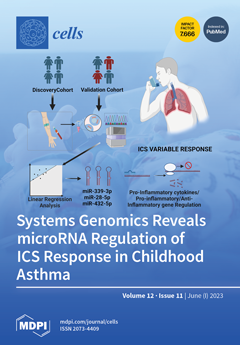
Asthmatic patients' responses to inhaled corticosteroids (ICSs) vary. We previously defined a measure called CASTER to quantify ICS responses. This study aimed to find associations between circulating miRNAs and ICS responses in childhood asthma. Small RNA sequencing was carried out on 580 children on ICSs from the GACRS and CAMP cohorts. Thirty-six miRNAs were associated with ICS responses in the GACRS cohort, with three miRNAs (miR-28-5p, miR-339-3p, and miR-432-5p) showing consistent effects in the CAMP replication cohort. The analysis of lymphoblastoid cell lines revealed genes associated with these miRNAs and steroid response. miR-339-3p was linked to immune dysregulation and a poor ICS response. This study highlights the role of specific miRNAs in ICS responses in childhood asthma. View this paper
  • Issues are regarded as officially published after their release is announced to the table of contents alert mailing list.
  • You may sign up for e-mail alerts to receive table of contents of newly released issues.
  • PDF is the official format for papers published in both, html and pdf forms. To view the papers in pdf format, click on the "PDF Full-text" link, and use the free Adobe Reader to open them.
Order results
Result details
Section
Select all
Export citation of selected articles as:
24 pages, 875 KiB  
Review
Generation of Red Blood Cells from Human Pluripotent Stem Cells—An Update
by Shin-Jeong Lee, Cholomi Jung, Jee Eun Oh, Sangsung Kim, Sangho Lee, Ji Yoon Lee and Young-sup Yoon
Cells 2023, 12(11), 1554; https://doi.org/10.3390/cells12111554 - 05 Jun 2023
Cited by 2 | Viewed by 4094
Abstract
Red blood cell (RBC) transfusion is a lifesaving medical procedure that can treat patients with anemia and hemoglobin disorders. However, the shortage of blood supply and risks of transfusion-transmitted infection and immune incompatibility present a challenge for transfusion. The in vitro generation of [...] Read more.
Red blood cell (RBC) transfusion is a lifesaving medical procedure that can treat patients with anemia and hemoglobin disorders. However, the shortage of blood supply and risks of transfusion-transmitted infection and immune incompatibility present a challenge for transfusion. The in vitro generation of RBCs or erythrocytes holds great promise for transfusion medicine and novel cell-based therapies. While hematopoietic stem cells and progenitors derived from peripheral blood, cord blood, and bone marrow can give rise to erythrocytes, the use of human pluripotent stem cells (hPSCs) has also provided an important opportunity to obtain erythrocytes. These hPSCs include both human embryonic stem cells (hESCs) and human induced pluripotent stem cells (hiPSCs). As hESCs carry ethical and political controversies, hiPSCs can be a more universal source for RBC generation. In this review, we first discuss the key concepts and mechanisms of erythropoiesis. Thereafter, we summarize different methodologies to differentiate hPSCs into erythrocytes with an emphasis on the key features of human definitive erythroid lineage cells. Finally, we address the current limitations and future directions of clinical applications using hiPSC-derived erythrocytes. Full article
(This article belongs to the Special Issue Gene and Cell Therapy in Regenerative Medicine)
Show Figures

Figure 1

28 pages, 1979 KiB  
Review
Advances in Understanding the Links between Metabolism and Autophagy in Acute Myeloid Leukemia: From Biology to Therapeutic Targeting
by Ernestina Saulle, Isabella Spinello, Maria Teresa Quaranta and Catherine Labbaye
Cells 2023, 12(11), 1553; https://doi.org/10.3390/cells12111553 - 05 Jun 2023
Cited by 1 | Viewed by 1784
Abstract
Autophagy is a highly conserved cellular degradation process that regulates cellular metabolism and homeostasis under normal and pathophysiological conditions. Autophagy and metabolism are linked in the hematopoietic system, playing a fundamental role in the self-renewal, survival, and differentiation of hematopoietic stem and progenitor [...] Read more.
Autophagy is a highly conserved cellular degradation process that regulates cellular metabolism and homeostasis under normal and pathophysiological conditions. Autophagy and metabolism are linked in the hematopoietic system, playing a fundamental role in the self-renewal, survival, and differentiation of hematopoietic stem and progenitor cells, and in cell death, particularly affecting the cellular fate of the hematopoietic stem cell pool. In leukemia, autophagy sustains leukemic cell growth, contributes to survival of leukemic stem cells and chemotherapy resistance. The high frequency of disease relapse caused by relapse-initiating leukemic cells resistant to therapy occurs in acute myeloid leukemia (AML), and depends on the AML subtypes and treatments used. Targeting autophagy may represent a promising strategy to overcome therapeutic resistance in AML, for which prognosis remains poor. In this review, we illustrate the role of autophagy and the impact of its deregulation on the metabolism of normal and leukemic hematopoietic cells. We report updates on the contribution of autophagy to AML development and relapse, and the latest evidence indicating autophagy-related genes as potential prognostic predictors and drivers of AML. We review the recent advances in autophagy manipulation, combined with various anti-leukemia therapies, for an effective autophagy-targeted therapy for AML. Full article
Show Figures

Figure 1

16 pages, 6875 KiB  
Article
Performance of the Photosynthetic Apparatus under Glass with a Luminophore Modifying Red-To-Far-Red-Light Ratio—A Case Study
by Krzysztof M. Tokarz, Wojciech Makowski, Barbara Tokarz, Ewa Muszyńska, Zbigniew Gajewski, Stanisław Mazur, Edward Kunicki, Olgierd Jeremiasz, Piotr Sobik, Paweł Nowak, Karolina Miernicka, Kinga Mrzygłód and Piotr Rozpądek
Cells 2023, 12(11), 1552; https://doi.org/10.3390/cells12111552 - 05 Jun 2023
Viewed by 1298
Abstract
The aim of this study was to examine the effect of the modified light spectrum of glass containing red luminophore on the performance of the photosynthetic apparatus of two types of lettuce cultivated in soil in a greenhouse. Butterhead and iceberg lettuce were [...] Read more.
The aim of this study was to examine the effect of the modified light spectrum of glass containing red luminophore on the performance of the photosynthetic apparatus of two types of lettuce cultivated in soil in a greenhouse. Butterhead and iceberg lettuce were cultivated in two types of greenhouses: (1) covered with transparent glass (control) and (2) covered with glass containing red luminophore (red). After 4 weeks of culture, structural and functional changes in the photosynthetic apparatus were examined. The presented study indicated that the red luminophore used changed the sunlight spectrum, providing an adequate blue:red light ratio, while decreasing the red:far-red radiation ratio. In such light conditions, changes in the efficiency parameters of the photosynthetic apparatus, modifications in the chloroplast ultrastructure, and altered proportions of structural proteins forming the photosynthetic apparatus were observed. These changes led to a decrease of CO2 carboxylation efficiency in both examined lettuce types. Full article
(This article belongs to the Special Issue Research on Photosynthesis under Stress)
Show Figures

Figure 1

9 pages, 1625 KiB  
Communication
Collagen VI Is a Gi-Biased Ligand of the Adhesion GPCR GPR126/ADGRG6
by Caroline Wilde, Paulomi Mehta Chaudhry, Rong Luo, Kay-Uwe Simon, Xianhua Piao and Ines Liebscher
Cells 2023, 12(11), 1551; https://doi.org/10.3390/cells12111551 - 05 Jun 2023
Cited by 2 | Viewed by 2391
Abstract
GPR126/ADGRG6, a member of the adhesion G-protein-coupled receptor family, balances cell differentiation and proliferation through fine-tuning of intracellular cAMP levels, which is achieved through coupling to Gs and Gi proteins. While GPR126-mediated cAMP increase has been proven to be essential for differentiation of [...] Read more.
GPR126/ADGRG6, a member of the adhesion G-protein-coupled receptor family, balances cell differentiation and proliferation through fine-tuning of intracellular cAMP levels, which is achieved through coupling to Gs and Gi proteins. While GPR126-mediated cAMP increase has been proven to be essential for differentiation of Schwann cells, adipocytes and osteoblasts, Gi-signaling of the receptor was found to propagate breast cancer cell proliferation. Extracellular ligands or mechanical forces can modulate GPR126 activity but require an intact encrypted agonist sequence, coined the Stachel. Even though coupling to Gi can be seen for constitutively active truncated receptor versions of GPR126 as well as with a peptide agonist derived from the Stachel sequence, all known N-terminal modulators have so far only been shown to modulate Gs coupling. Here, we identified collagen VI as the first extracellular matrix ligand of GPR126 that induces Gi signaling at the receptor, which shows that N-terminal binding partners can mediate selective G protein signaling cascades that are masked by fully active truncated receptor variants. Full article
Show Figures

Figure 1

22 pages, 4227 KiB  
Article
Systematic Approaches to Study Eclipsed Targeting of Proteins Uncover a New Family of Mitochondrial Proteins
by Maayan Mark, Ofir Klein, Yu Zhang, Koyeli Das, Adi Elbaz, Reut Noa Hazan, Michal Lichtenstein, Norbert Lehming, Maya Schuldiner and Ophry Pines
Cells 2023, 12(11), 1550; https://doi.org/10.3390/cells12111550 - 05 Jun 2023
Cited by 1 | Viewed by 1297
Abstract
Dual localization or dual targeting refers to the phenomenon by which identical, or almost identical, proteins are localized to two (or more) separate compartments of the cell. From previous work in the field, we had estimated that a third of the mitochondrial proteome [...] Read more.
Dual localization or dual targeting refers to the phenomenon by which identical, or almost identical, proteins are localized to two (or more) separate compartments of the cell. From previous work in the field, we had estimated that a third of the mitochondrial proteome is dual-targeted to extra-mitochondrial locations and suggested that this abundant dual targeting presents an evolutionary advantage. Here, we set out to study how many additional proteins whose main activity is outside mitochondria are also localized, albeit at low levels, to mitochondria (eclipsed). To do this, we employed two complementary approaches utilizing the α-complementation assay in yeast to uncover the extent of such an eclipsed distribution: one systematic and unbiased and the other based on mitochondrial targeting signal (MTS) predictions. Using these approaches, we suggest 280 new eclipsed distributed protein candidates. Interestingly, these proteins are enriched for distinctive properties compared to their exclusively mitochondrial-targeted counterparts. We focus on one unexpected eclipsed protein family of the Triose-phosphate DeHydrogenases (TDH) and prove that, indeed, their eclipsed distribution in mitochondria is important for mitochondrial activity. Our work provides a paradigm of deliberate eclipsed mitochondrial localization, targeting and function, and should expand our understanding of mitochondrial function in health and disease. Full article
(This article belongs to the Collection The Pathomechanism of Mitochondrial Diseases)
Show Figures

Graphical abstract

17 pages, 2783 KiB  
Article
TREM2 Agonism with a Monoclonal Antibody Attenuates Tau Pathology and Neurodegeneration
by Michael Fassler, Clara Benaim and Jacob George
Cells 2023, 12(11), 1549; https://doi.org/10.3390/cells12111549 - 05 Jun 2023
Viewed by 1984
Abstract
TREM2 is a membrane receptor expressed on microglia that plays a pivotal role in the organization and function of these innate immune cell components within the neurodegenerated brain. Whereas TREM2 deletion has been studied extensively in experimental beta-amyloid and Tau-based models of Alzheimer’s [...] Read more.
TREM2 is a membrane receptor expressed on microglia that plays a pivotal role in the organization and function of these innate immune cell components within the neurodegenerated brain. Whereas TREM2 deletion has been studied extensively in experimental beta-amyloid and Tau-based models of Alzheimer’s disease, its engagement, and subsequent agonism have not been tested in the context of Tau pathology. Herein, we explored the effects of Ab-T1, an agonistic TREM2 monoclonal antibody on Tau uptake, phosphorylation, seeding, and spreading as well as its therapeutic efficacy in a Tauopathy model. Ab-T1 enhanced the uptake of misfolded Tau to microglia and induced a non-cell autonomous attenuation of spontaneous Tau seeding and phosphorylation in primary neurons from human Tau transgenic mice. Ex vivo, incubation with Ab-T1 led to a significant reduction in the seeding of Tau pathology in the hTau murine organoid brain system. Systemic administration of Ab-T1 resulted in reduced Tau pathology and propagation when hTau was stereotactically injected into the hemispheres of hTau mice. Intraperitoneal treatment with Ab-T1 lead to attenuation of cognitive decline in the hTau mice that was associated with reduced neurodegeneration and synaptic preservation with amelioration of the global neuroinflammatory program. Collectively, these observations show that TREM2 engagement with an agonistic antibody result in reduced Tau burden concomitant with attenuated neurodegeneration ascribed to the education of resident microglia. These results may suggest that despite the opposing results with regard to the effect of TREM2 knockout in experimental Tau-based model systems, receptor engagement and activation by Ab-T1 appears to possess beneficial effects with respect to the various mechanisms mediating Tau-driven neurodegeneration. Full article
Show Figures

Figure 1

23 pages, 4445 KiB  
Article
Multi-Drug Cocktail Therapy Improves Survival and Neurological Function after Asphyxial Cardiac Arrest in Rodents
by Rishabh C. Choudhary, Muhammad Shoaib, Kei Hayashida, Tai Yin, Santiago J. Miyara, Cristina d’Abramo, William G. Heuser, Koichiro Shinozaki, Nancy Kim, Ryosuke Takegawa, Mitsuaki Nishikimi, Timmy Li, Casey Owens, Ernesto P. Molmenti, Mingzhu He, Sonya Vanpatten, Yousef Al-Abed, Junhwan Kim and Lance B. Becker
Cells 2023, 12(11), 1548; https://doi.org/10.3390/cells12111548 - 05 Jun 2023
Viewed by 2126
Abstract
Background: Cardiac arrest (CA) can lead to neuronal degeneration and death through various pathways, including oxidative, inflammatory, and metabolic stress. However, current neuroprotective drug therapies will typically target only one of these pathways, and most single drug attempts to correct the multiple dysregulated [...] Read more.
Background: Cardiac arrest (CA) can lead to neuronal degeneration and death through various pathways, including oxidative, inflammatory, and metabolic stress. However, current neuroprotective drug therapies will typically target only one of these pathways, and most single drug attempts to correct the multiple dysregulated metabolic pathways elicited following cardiac arrest have failed to demonstrate clear benefit. Many scientists have opined on the need for novel, multidimensional approaches to the multiple metabolic disturbances after cardiac arrest. In the current study, we have developed a therapeutic cocktail that includes ten drugs capable of targeting multiple pathways of ischemia–reperfusion injury after CA. We then evaluated its effectiveness in improving neurologically favorable survival through a randomized, blind, and placebo-controlled study in rats subjected to 12 min of asphyxial CA, a severe injury model. Results: 14 rats were given the cocktail and 14 received the vehicle after resuscitation. At 72 h post-resuscitation, the survival rate was 78.6% among cocktail-treated rats, which was significantly higher than the 28.6% survival rate among vehicle-treated rats (log-rank test; p = 0.006). Moreover, in cocktail-treated rats, neurological deficit scores were also improved. These survival and neurological function data suggest that our multi-drug cocktail may be a potential post-CA therapy that deserves clinical translation. Conclusions: Our findings demonstrate that, with its ability to target multiple damaging pathways, a multi-drug therapeutic cocktail offers promise both as a conceptual advance and as a specific multi-drug formulation capable of combatting neuronal degeneration and death following cardiac arrest. Clinical implementation of this therapy may improve neurologically favorable survival rates and neurological deficits in patients suffering from cardiac arrest. Full article
Show Figures

Figure 1

16 pages, 2810 KiB  
Review
SNARE Protein Snc1 Is Essential for Vesicle Trafficking, Membrane Fusion and Protein Secretion in Fungi
by Muhammad Adnan, Waqar Islam, Abdul Waheed, Quaid Hussain, Ling Shen, Juan Wang and Gang Liu
Cells 2023, 12(11), 1547; https://doi.org/10.3390/cells12111547 - 05 Jun 2023
Cited by 2 | Viewed by 2249
Abstract
Fungi are an important group of microorganisms that play crucial roles in a variety of ecological and biotechnological processes. Fungi depend on intracellular protein trafficking, which involves moving proteins from their site of synthesis to the final destination within or outside the cell. [...] Read more.
Fungi are an important group of microorganisms that play crucial roles in a variety of ecological and biotechnological processes. Fungi depend on intracellular protein trafficking, which involves moving proteins from their site of synthesis to the final destination within or outside the cell. The soluble N-ethylmaleimide-sensitive factor attachment protein receptors (SNARE) proteins are vital components of vesicle trafficking and membrane fusion, ultimately leading to the release of cargos to the target destination. The v-SNARE (vesicle-associated SNARE) Snc1 is responsible for anterograde and retrograde vesicle trafficking between the plasma membrane (PM) and Golgi. It allows for the fusion of exocytic vesicles to the PM and the subsequent recycling of Golgi-localized proteins back to the Golgi via three distinct and parallel recycling pathways. This recycling process requires several components, including a phospholipid flippase (Drs2-Cdc50), an F-box protein (Rcy1), a sorting nexin (Snx4-Atg20), a retromer submit, and the COPI coat complex. Snc1 interacts with exocytic SNAREs (Sso1/2, Sec9) and the exocytic complex to complete the process of exocytosis. It also interacts with endocytic SNAREs (Tlg1 and Tlg2) during endocytic trafficking. Snc1 has been extensively investigated in fungi and has been found to play crucial roles in various aspects of intracellular protein trafficking. When Snc1 is overexpressed alone or in combination with some key secretory components, it results in enhanced protein production. This article will cover the role of Snc1 in the anterograde and retrograde trafficking of fungi and its interactions with other proteins for efficient cellular transportation. Full article
Show Figures

Figure 1

18 pages, 1877 KiB  
Review
Hypoxic-Ischemic Brain Injury in ECMO: Pathophysiology, Neuromonitoring, and Therapeutic Opportunities
by Shivalika Khanduja, Jiah Kim, Jin Kook Kang, Cheng-Yuan Feng, Melissa Ann Vogelsong, Romergryko G. Geocadin, Glenn Whitman and Sung-Min Cho
Cells 2023, 12(11), 1546; https://doi.org/10.3390/cells12111546 - 05 Jun 2023
Cited by 2 | Viewed by 3065
Abstract
Extracorporeal membrane oxygenation (ECMO), in conjunction with its life-saving benefits, carries a significant risk of acute brain injury (ABI). Hypoxic-ischemic brain injury (HIBI) is one of the most common types of ABI in ECMO patients. Various risk factors, such as history of hypertension, [...] Read more.
Extracorporeal membrane oxygenation (ECMO), in conjunction with its life-saving benefits, carries a significant risk of acute brain injury (ABI). Hypoxic-ischemic brain injury (HIBI) is one of the most common types of ABI in ECMO patients. Various risk factors, such as history of hypertension, high day 1 lactate level, low pH, cannulation technique, large peri-cannulation PaCO2 drop (∆PaCO2), and early low pulse pressure, have been associated with the development of HIBI in ECMO patients. The pathogenic mechanisms of HIBI in ECMO are complex and multifactorial, attributing to the underlying pathology requiring initiation of ECMO and the risk of HIBI associated with ECMO itself. HIBI is likely to occur in the peri-cannulation or peri-decannulation time secondary to underlying refractory cardiopulmonary failure before or after ECMO. Current therapeutics target pathological mechanisms, cerebral hypoxia and ischemia, by employing targeted temperature management in the case of extracorporeal cardiopulmonary resuscitation (eCPR), and optimizing cerebral O2 saturations and cerebral perfusion. This review describes the pathophysiology, neuromonitoring, and therapeutic techniques to improve neurological outcomes in ECMO patients in order to prevent and minimize the morbidity of HIBI. Further studies aimed at standardizing the most relevant neuromonitoring techniques, optimizing cerebral perfusion, and minimizing the severity of HIBI once it occurs will improve long-term neurological outcomes in ECMO patients. Full article
Show Figures

Figure 1

19 pages, 1259 KiB  
Review
Modulation of NRF2/KEAP1 Signaling in Preeclampsia
by Giovanni Tossetta, Sonia Fantone, Federica Piani, Caterina Crescimanno, Andrea Ciavattini, Stefano Raffaele Giannubilo and Daniela Marzioni
Cells 2023, 12(11), 1545; https://doi.org/10.3390/cells12111545 - 04 Jun 2023
Cited by 23 | Viewed by 2261
Abstract
Placentation is a key and tightly regulated process that ensures the normal development of the placenta and fetal growth. Preeclampsia (PE) is a hypertensive pregnancy-related disorder involving about 5–8% of all pregnancies and clinically characterized by de novo maternal hypertension and proteinuria. In [...] Read more.
Placentation is a key and tightly regulated process that ensures the normal development of the placenta and fetal growth. Preeclampsia (PE) is a hypertensive pregnancy-related disorder involving about 5–8% of all pregnancies and clinically characterized by de novo maternal hypertension and proteinuria. In addition, PE pregnancies are also characterized by increased oxidative stress and inflammation. The NRF2/KEAP1 signaling pathway plays an important role in protecting cells against oxidative damage due to increased reactive oxygen species (ROS) levels. ROS activate NRF2, allowing its binding to the antioxidant response element (ARE) region present in the promoter of several antioxidant genes such as heme oxygenase, catalase, glutathione peroxidase and superoxide dismutase that neutralize ROS, protecting cells against oxidative stress damages. In this review, we analyze the current literature regarding the role of the NRF2/KEAP1 pathway in preeclamptic pregnancies, discussing the main cellular modulators of this pathway. Moreover, we also discuss the main natural and synthetic compounds that can regulate this pathway in in vivo and in vitro models. Full article
(This article belongs to the Special Issue Signaling Pathways in Pregnancy)
Show Figures

Figure 1

20 pages, 1736 KiB  
Review
Regulators of the Asexual Life Cycle of Aspergillus nidulans
by Ye-Eun Son, Jae-Hyuk Yu and Hee-Soo Park
Cells 2023, 12(11), 1544; https://doi.org/10.3390/cells12111544 - 04 Jun 2023
Cited by 4 | Viewed by 1868
Abstract
The genus Aspergillus, one of the most abundant airborne fungi, is classified into hundreds of species that affect humans, animals, and plants. Among these, Aspergillus nidulans, as a key model organism, has been extensively studied to understand the mechanisms governing growth [...] Read more.
The genus Aspergillus, one of the most abundant airborne fungi, is classified into hundreds of species that affect humans, animals, and plants. Among these, Aspergillus nidulans, as a key model organism, has been extensively studied to understand the mechanisms governing growth and development, physiology, and gene regulation in fungi. A. nidulans primarily reproduces by forming millions of asexual spores known as conidia. The asexual life cycle of A. nidulans can be simply divided into growth and asexual development (conidiation). After a certain period of vegetative growth, some vegetative cells (hyphae) develop into specialized asexual structures called conidiophores. Each A. nidulans conidiophore is composed of a foot cell, stalk, vesicle, metulae, phialides, and 12,000 conidia. This vegetative-to-developmental transition requires the activity of various regulators including FLB proteins, BrlA, and AbaA. Asymmetric repetitive mitotic cell division of phialides results in the formation of immature conidia. Subsequent conidial maturation requires multiple regulators such as WetA, VosA, and VelB. Matured conidia maintain cellular integrity and long-term viability against various stresses and desiccation. Under appropriate conditions, the resting conidia germinate and form new colonies, and this process is governed by a myriad of regulators, such as CreA and SocA. To date, a plethora of regulators for each asexual developmental stage have been identified and investigated. This review summarizes our current understanding of the regulators of conidial formation, maturation, dormancy, and germination in A. nidulans. Full article
Show Figures

Figure 1

13 pages, 2185 KiB  
Article
CRISPR/Cas9 Knock-Out in Primary Neonatal and Adult Cardiomyocytes Reveals Distinct cAMP Dynamics Regulation by Various PDE2A and PDE3A Isoforms
by Egor B. Skryabin, Kirstie A. De Jong, Hariharan Subramanian, Nadja I. Bork, Alexander Froese, Boris V. Skryabin and Viacheslav O. Nikolaev
Cells 2023, 12(11), 1543; https://doi.org/10.3390/cells12111543 - 04 Jun 2023
Viewed by 1552
Abstract
Cyclic nucleotide phosphodiesterases 2A (PDE2A) and PDE3A play an important role in the regulation of cyclic adenosine monophosphate (cAMP) and cyclic guanosine monophosphate (cGMP)-to-cAMP crosstalk. Each of these PDEs has up to three distinct isoforms. However, their specific contributions to cAMP dynamics are [...] Read more.
Cyclic nucleotide phosphodiesterases 2A (PDE2A) and PDE3A play an important role in the regulation of cyclic adenosine monophosphate (cAMP) and cyclic guanosine monophosphate (cGMP)-to-cAMP crosstalk. Each of these PDEs has up to three distinct isoforms. However, their specific contributions to cAMP dynamics are difficult to explore because it has been challenging to generate isoform-specific knock-out mice or cells using conventional methods. Here, we studied whether the CRISPR/Cas9 approach for precise genome editing can be used to knock out Pde2a and Pde3a genes and their distinct isoforms using adenoviral gene transfer in neonatal and adult rat cardiomyocytes. Cas9 and several specific gRNA constructs were cloned and introduced into adenoviral vectors. Primary adult and neonatal rat ventricular cardiomyocytes were transduced with different amounts of Cas9 adenovirus in combination with PDE2A or PDE3A gRNA constructs and cultured for up to 6 (adult) or 14 (neonatal) days to analyze PDE expression and live cell cAMP dynamics. A decline in mRNA expression for PDE2A (~80%) and PDE3A (~45%) was detected as soon as 3 days post transduction, with both PDEs being reduced at the protein level by >50–60% in neonatal cardiomyocytes (after 14 days) and >95% in adult cardiomyocytes (after 6 days). This correlated with the abrogated effects of selective PDE inhibitors in the live cell imaging experiments based on using cAMP biosensor measurements. Reverse transcription PCR analysis revealed that only the PDE2A2 isoform was expressed in neonatal myocytes, while adult cardiomyocytes expressed all three PDE2A isoforms (A1, A2, and A3) which contributed to the regulation of cAMP dynamics as detected by live cell imaging. In conclusion, CRISPR/Cas9 is an effective tool for the in vitro knock-out of PDEs and their specific isoforms in primary somatic cells. This novel approach suggests distinct regulation of live cell cAMP dynamics by various PDE2A and PDE3A isoforms in neonatal vs. adult cardiomyocytes. Full article
(This article belongs to the Collection Compartmentilisation of Cellular Signaling)
Show Figures

Figure 1

26 pages, 4770 KiB  
Article
A Rapid Alkalinization Factor-like Peptide EaF82 Impairs Tapetum Degeneration during Pollen Development through Induced ATP Deficiency
by Chiu-Yueh Hung, Farooqahmed S. Kittur, Keely N. Wharton, Makendra L. Umstead, D’Shawna B. Burwell, Martinique Thomas, Qi Qi, Jianhui Zhang, Carla E. Oldham, Kent O. Burkey, Jianjun Chen and Jiahua Xie
Cells 2023, 12(11), 1542; https://doi.org/10.3390/cells12111542 - 04 Jun 2023
Cited by 1 | Viewed by 1472
Abstract
In plants, the timely degeneration of tapetal cells is essential for providing nutrients and other substances to support pollen development. Rapid alkalinization factors (RALFs) are small, cysteine-rich peptides known to be involved in various aspects of plant development and growth, as well as [...] Read more.
In plants, the timely degeneration of tapetal cells is essential for providing nutrients and other substances to support pollen development. Rapid alkalinization factors (RALFs) are small, cysteine-rich peptides known to be involved in various aspects of plant development and growth, as well as defense against biotic and abiotic stresses. However, the functions of most of them remain unknown, while no RALF has been reported to involve tapetum degeneration. In this study, we demonstrated that a novel cysteine-rich peptide, EaF82, isolated from shy-flowering ‘Golden Pothos’ (Epipremnum aureum) plants, is a RALF-like peptide and displays alkalinizing activity. Its heterologous expression in Arabidopsis delayed tapetum degeneration and reduced pollen production and seed yields. RNAseq, RT-qPCR, and biochemical analyses showed that overexpression of EaF82 downregulated a group of genes involved in pH changes, cell wall modifications, tapetum degeneration, and pollen maturation, as well as seven endogenous Arabidopsis RALF genes, and decreased proteasome activity and ATP levels. Yeast two-hybrid screening identified AKIN10, a subunit of energy-sensing SnRK1 kinase, as its interacting partner. Our study reveals a possible regulatory role for RALF peptide in tapetum degeneration and suggests that EaF82 action may be mediated through AKIN10 leading to the alteration of transcriptome and energy metabolism, thereby causing ATP deficiency and impairing pollen development. Full article
Show Figures

Figure 1

24 pages, 5413 KiB  
Article
Metronomic Photodynamic Therapy with Conjugated Polymer Nanoparticles in Glioblastoma Tumor Microenvironment
by Matías Daniel Caverzán, Paula Martina Oliveda, Lucía Beaugé, Rodrigo Emiliano Palacios, Carlos Alberto Chesta and Luis Exequiel Ibarra
Cells 2023, 12(11), 1541; https://doi.org/10.3390/cells12111541 - 04 Jun 2023
Cited by 9 | Viewed by 1654
Abstract
Alternative therapies such as photodynamic therapy (PDT) that combine light, oxygen and photosensitizers (PSs) have been proposed for glioblastoma (GBM) management to overcome conventional treatment issues. An important disadvantage of PDT using a high light irradiance (fluence rate) (cPDT) is the abrupt oxygen [...] Read more.
Alternative therapies such as photodynamic therapy (PDT) that combine light, oxygen and photosensitizers (PSs) have been proposed for glioblastoma (GBM) management to overcome conventional treatment issues. An important disadvantage of PDT using a high light irradiance (fluence rate) (cPDT) is the abrupt oxygen consumption that leads to resistance to the treatment. PDT metronomic regimens (mPDT) involving administering light at a low irradiation intensity over a relatively long period of time could be an alternative to circumvent the limitations of conventional PDT protocols. The main objective of the present work was to compare the effectiveness of PDT with an advanced PS based on conjugated polymer nanoparticles (CPN) developed by our group in two irradiation modalities: cPDT and mPDT. The in vitro evaluation was carried out based on cell viability, the impact on the macrophage population of the tumor microenvironment in co-culture conditions and the modulation of HIF-1α as an indirect indicator of oxygen consumption. mPDT regimens with CPNs resulted in more effective cell death, a lower activation of molecular pathways of therapeutic resistance and macrophage polarization towards an antitumoral phenotype. Additionally, mPDT was tested in a GBM heterotopic mouse model, confirming its good performance with promising tumor growth inhibition and apoptotic cell death induction. Full article
(This article belongs to the Special Issue Cell Death Mechanisms and Therapeutic Opportunities in Glioblastoma)
Show Figures

Figure 1

16 pages, 2310 KiB  
Article
Pharmacometabolic Effects of Pteryxin and Valproate on Pentylenetetrazole-Induced Seizures in Zebrafish Reveal Vagus Nerve Stimulation
by Adrianna Skiba, Daniele Pellegata, Veronika Morozova, Ewelina Kozioł, Barbara Budzyńska, Simon Ming-Yuen Lee, Jürg Gertsch and Krystyna Skalicka-Woźniak
Cells 2023, 12(11), 1540; https://doi.org/10.3390/cells12111540 - 04 Jun 2023
Cited by 2 | Viewed by 1789
Abstract
Zebrafish (Danio rerio) assays provide a versatile pharmacological platform to test compounds on a wide range of behaviors in a whole organism. A major challenge lies in the lack of knowledge about the bioavailability and pharmacodynamic effects of bioactive compounds in this model [...] Read more.
Zebrafish (Danio rerio) assays provide a versatile pharmacological platform to test compounds on a wide range of behaviors in a whole organism. A major challenge lies in the lack of knowledge about the bioavailability and pharmacodynamic effects of bioactive compounds in this model organism. Here, we employed a combined methodology of LC-ESI-MS/MS analytics and targeted metabolomics with behavioral experiments to evaluate the anticonvulsant and potentially toxic effects of the angular dihydropyranocoumarin pteryxin (PTX) in comparison to the antiepileptic drug sodium valproate (VPN) in zebrafish larvae. PTX occurs in different Apiaceae plants traditionally used in Europe to treat epilepsy but has not been investigated so far. To compare potency and efficacy, the uptake of PTX and VPN into zebrafish larvae was quantified as larvae whole-body concentrations together with amino acids and neurotransmitters as proxy pharmacodynamic readout. The convulsant agent pentylenetetrazole (PTZ) acutely reduced the levels of most metabolites, including acetylcholine and serotonin. Conversely, PTX strongly reduced neutral essential amino acids in a LAT1 (SLCA5)-independent manner, but, similarly to VPN specifically increased the levels of serotonin, acetylcholine, and choline, but also ethanolamine. PTX dose and time-dependent manner inhibited PTZ-induced seizure-like movements resulting in a ~70% efficacy after 1 h at 20 µM (the equivalent of 4.28 ± 0.28 µg/g in larvae whole-body). VPN treated for 1 h with 5 mM (the equivalent of 18.17 ± 0.40 µg/g in larvae whole-body) showed a ~80% efficacy. Unexpectedly, PTX (1–20 µM) showed significantly higher bioavailability than VPN (0.1–5 mM) in immersed zebrafish larvae, possibly because VPN in the medium dissociated partially to the readily bioavailable valproic acid. The anticonvulsive effect of PTX was confirmed by local field potential (LFP) recordings. Noteworthy, both substances specifically increased and restored whole-body acetylcholine, choline, and serotonin levels in control and PTZ-treated zebrafish larvae, indicative of vagus nerve stimulation (VNS), which is an adjunctive therapeutic strategy to treat refractory epilepsy in humans. Our study demonstrates the utility of targeted metabolomics in zebrafish assays and shows that VPN and PTX pharmacologically act on the autonomous nervous system by activating parasympathetic neurotransmitters. Full article
Show Figures

Figure 1

14 pages, 1926 KiB  
Review
Role of A-Kinase Anchoring Protein 1 in Retinal Ganglion Cells: Neurodegeneration and Neuroprotection
by Tonking Bastola, Guy A. Perkins, Keun-Young Kim, Seunghwan Choi, Jin-Woo Kwon, Ziyao Shen, Stefan Strack and Won-Kyu Ju
Cells 2023, 12(11), 1539; https://doi.org/10.3390/cells12111539 - 03 Jun 2023
Cited by 2 | Viewed by 1717
Abstract
A-Kinase anchoring protein 1 (AKAP1) is a multifunctional mitochondrial scaffold protein that regulates mitochondrial dynamics, bioenergetics, and calcium homeostasis by anchoring several proteins, including protein kinase A, to the outer mitochondrial membrane. Glaucoma is a complex, multifactorial disease characterized by a slow and [...] Read more.
A-Kinase anchoring protein 1 (AKAP1) is a multifunctional mitochondrial scaffold protein that regulates mitochondrial dynamics, bioenergetics, and calcium homeostasis by anchoring several proteins, including protein kinase A, to the outer mitochondrial membrane. Glaucoma is a complex, multifactorial disease characterized by a slow and progressive degeneration of the optic nerve and retinal ganglion cells (RGCs), ultimately resulting in vision loss. Impairment of the mitochondrial network and function is linked to glaucomatous neurodegeneration. Loss of AKAP1 induces dynamin-related protein 1 dephosphorylation-mediated mitochondrial fragmentation and loss of RGCs. Elevated intraocular pressure triggers a significant reduction in AKAP1 protein expression in the glaucomatous retina. Amplification of AKAP1 expression protects RGCs from oxidative stress. Hence, modulation of AKAP1 could be considered a potential therapeutic target for neuroprotective intervention in glaucoma and other mitochondria-associated optic neuropathies. This review covers the current research on the role of AKAP1 in the maintenance of mitochondrial dynamics, bioenergetics, and mitophagy in RGCs and provides a scientific basis to identify and develop new therapeutic strategies that could protect RGCs and their axons in glaucoma. Full article
Show Figures

Figure 1

19 pages, 4647 KiB  
Article
RANKL Inhibition Reduces Cardiac Hypertrophy in mdx Mice and Possibly in Children with Duchenne Muscular Dystrophy
by Laetitia Marcadet, Emma Sara Juracic, Nasrin Khan, Zineb Bouredji, Hideo Yagita, Leanne M. Ward, A. Russell Tupling, Anteneh Argaw and Jérôme Frenette
Cells 2023, 12(11), 1538; https://doi.org/10.3390/cells12111538 - 03 Jun 2023
Cited by 1 | Viewed by 2122
Abstract
Cardiomyopathy has become one of the leading causes of death in patients with Duchenne muscular dystrophy (DMD). We recently reported that the inhibition of the interaction between the receptor activator of nuclear factor κB ligand (RANKL) and receptor activator of nuclear factor κB [...] Read more.
Cardiomyopathy has become one of the leading causes of death in patients with Duchenne muscular dystrophy (DMD). We recently reported that the inhibition of the interaction between the receptor activator of nuclear factor κB ligand (RANKL) and receptor activator of nuclear factor κB (RANK) significantly improves muscle and bone functions in dystrophin-deficient mdx mice. RANKL and RANK are also expressed in cardiac muscle. Here, we investigate whether anti-RANKL treatment prevents cardiac hypertrophy and dysfunction in dystrophic mdx mice. Anti-RANKL treatment significantly reduced LV hypertrophy and heart mass, and maintained cardiac function in mdx mice. Anti-RANKL treatment also inhibited NFκB and PI3K, two mediators implicated in cardiac hypertrophy. Furthermore, anti-RANKL treatment increased SERCA activity and the expression of RyR, FKBP12, and SERCA2a, leading possibly to an improved Ca2+ homeostasis in dystrophic hearts. Interestingly, preliminary post hoc analyses suggest that denosumab, a human anti-RANKL, reduced left ventricular hypertrophy in two patients with DMD. Taken together, our results indicate that anti-RANKL treatment prevents the worsening of cardiac hypertrophy in mdx mice and could potentially maintain cardiac function in teenage or adult patients with DMD. Full article
Show Figures

Graphical abstract

15 pages, 4015 KiB  
Article
Short-Term Exposure to Bisphenol A Does Not Impact Gonadal Cell Steroidogenesis In Vitro
by Neena Roy, Clara Lazzaretti, Elia Paradiso, Chiara Capponi, Tommaso Ferrari, Francesca Reggianini, Samantha Sperduti, Lara Baschieri, Elisa Mascolo, Carmela Perri, Manuela Varani, Giulia Canu, Tommaso Trenti, Alessia Nicoli, Daria Morini, Francesca Iannotti, Maria Teresa Villani, Elena Vicini, Manuela Simoni and Livio Casarini
Cells 2023, 12(11), 1537; https://doi.org/10.3390/cells12111537 - 02 Jun 2023
Cited by 1 | Viewed by 1506
Abstract
Bisphenol A (BPA) is a ubiquitous, synthetic chemical proven to induce reproductive disorders in both men and women. The available studies investigated the effects of BPA on male and female steroidogenesis following long-term exposure to the compound at relatively high environmental concentrations. However, [...] Read more.
Bisphenol A (BPA) is a ubiquitous, synthetic chemical proven to induce reproductive disorders in both men and women. The available studies investigated the effects of BPA on male and female steroidogenesis following long-term exposure to the compound at relatively high environmental concentrations. However, the impact of short-term exposure to BPA on reproduction is poorly studied. We evaluated if 8 and 24 h exposure to 1 nM and 1 µM BPA perturbs luteinizing hormone/choriogonadotropin (LH/hCG)-mediated signalling in two steroidogenic cell models, i.e., the mouse tumour Leydig cell line mLTC1, and human primary granulosa lutein cells (hGLC). Cell signalling studies were performed using a homogeneous time-resolved fluorescence (HTRF) assay and Western blotting, while gene expression analysis was carried out using real-time PCR. Immunostainings and an immunoassay were used for intracellular protein expression and steroidogenesis analyses, respectively. The presence of BPA leads to no significant changes in gonadotropin-induced cAMP accumulation, alongside phosphorylation of downstream molecules, such as ERK1/2, CREB and p38 MAPK, in both the cell models. BPA did not impact STARD1, CYP11A1 and CYP19A1 gene expression in hGLC, nor Stard1 and Cyp17a1 expression in mLTC1 treated with LH/hCG. Additionally, the StAR protein expression was unchanged upon exposure to BPA. Progesterone and oestradiol levels in the culture medium, measured by hGLC, as well as the testosterone and progesterone levels in the culture medium, measured by mLTC1, did not change in the presence of BPA combined with LH/hCG. These data suggest that short-term exposure to environmental concentrations of BPA does not compromise the LH/hCG-induced steroidogenic potential of either human granulosa or mouse Leydig cells. Full article
Show Figures

Figure 1

15 pages, 784 KiB  
Review
Altered Metabolism in Motor Neuron Diseases: Mechanism and Potential Therapeutic Target
by Cassandra Barone and Xin Qi
Cells 2023, 12(11), 1536; https://doi.org/10.3390/cells12111536 - 02 Jun 2023
Cited by 1 | Viewed by 1866
Abstract
Motor Neuron Diseases (MND) are neurological disorders characterized by a loss of varying motor neurons resulting in decreased physical capabilities. Current research is focused on hindering disease progression by determining causes of motor neuron death. Metabolic malfunction has been proposed as a promising [...] Read more.
Motor Neuron Diseases (MND) are neurological disorders characterized by a loss of varying motor neurons resulting in decreased physical capabilities. Current research is focused on hindering disease progression by determining causes of motor neuron death. Metabolic malfunction has been proposed as a promising topic when targeting motor neuron loss. Alterations in metabolism have also been noted at the neuromuscular junction (NMJ) and skeletal muscle tissue, emphasizing the importance of a cohesive system. Finding metabolism changes consistent throughout both neurons and skeletal muscle tissue could pose as a target for therapeutic intervention. This review will focus on metabolic deficits reported in MNDs and propose potential therapeutic targets for future intervention. Full article
(This article belongs to the Special Issue Mitochondria at the Crossroad of Health and Disease)
Show Figures

Figure 1

9 pages, 2097 KiB  
Communication
Adenoviral Transfer of Human Aquaporin-8 Gene to Mouse Liver Improves Ammonia-Derived Ureagenesis
by Alejo M. Capiglioni, María C. Capitani, Julieta Marrone and Raúl A. Marinelli
Cells 2023, 12(11), 1535; https://doi.org/10.3390/cells12111535 - 02 Jun 2023
Cited by 1 | Viewed by 927
Abstract
We previously reported that, in cultured hepatocytes, mitochondrial aquaporin-8 (AQP8) channels facilitate the conversion of ammonia to urea and that the expression of human AQP8 (hAQP8) enhances ammonia-derived ureagenesis. In this study, we evaluated whether hepatic gene transfer of hAQP8 improves detoxification of [...] Read more.
We previously reported that, in cultured hepatocytes, mitochondrial aquaporin-8 (AQP8) channels facilitate the conversion of ammonia to urea and that the expression of human AQP8 (hAQP8) enhances ammonia-derived ureagenesis. In this study, we evaluated whether hepatic gene transfer of hAQP8 improves detoxification of ammonia to urea in normal mice as well as in mice with impaired hepatocyte ammonia metabolism. A recombinant adenoviral (Ad) vector encoding hAQP8, AdhAQP8, or a control Ad vector was administered via retrograde infusion into the bile duct of the mice. Hepatocyte mitochondrial expression of hAQP8 was confirmed using confocal immunofluorescence and immunoblotting. The normal hAQP8-transduced mice showed decreased plasma ammonia and increased liver urea. Enhanced ureagenesis was confirmed via the NMR studies assessing the synthesis of 15N-labeled urea from 15N-labeled ammonia. In separate experiments, we made use of the model hepatotoxic agent, thioacetamide, to induce defective hepatic metabolism of ammonia in mice. The adenovirus-mediated mitochondrial expression of hAQP8 was able to restore normal ammonemia and ureagenesis in the liver of the mice. Our data suggest that hAQP8 gene transfer to mouse liver improves detoxification of ammonia to urea. This finding could help better understand and treat disorders with defective hepatic ammonia metabolism. Full article
(This article belongs to the Special Issue Advances in Aquaporins II)
Show Figures

Figure 1

26 pages, 2319 KiB  
Review
CAR-Based Therapy for Autoimmune Diseases: A Novel Powerful Option
by Györgyi Műzes and Ferenc Sipos
Cells 2023, 12(11), 1534; https://doi.org/10.3390/cells12111534 - 02 Jun 2023
Cited by 3 | Viewed by 4530
Abstract
The pervasive application of chimeric antigen receptor (CAR)-based cellular therapies in the treatment of oncological diseases has long been recognized. However, CAR T cells can target and eliminate autoreactive cells in autoimmune and immune-mediated diseases. By doing so, they can contribute to an [...] Read more.
The pervasive application of chimeric antigen receptor (CAR)-based cellular therapies in the treatment of oncological diseases has long been recognized. However, CAR T cells can target and eliminate autoreactive cells in autoimmune and immune-mediated diseases. By doing so, they can contribute to an effective and relatively long-lasting remission. In turn, CAR Treg interventions may have a highly effective and durable immunomodulatory effect via a direct or bystander effect, which may have a positive impact on the course and prognosis of autoimmune diseases. CAR-based cellular techniques have a complex theoretical foundation and are difficult to implement in practice, but they have a remarkable capacity to suppress the destructive functions of the immune system. This article provides an overview of the numerous CAR-based therapeutic options developed for the treatment of immune-mediated and autoimmune diseases. We believe that well-designed, rigorously tested cellular therapies could provide a promising new personalized treatment strategy for a significant number of patients with immune-mediated disorders. Full article
Show Figures

Graphical abstract

15 pages, 4022 KiB  
Article
Mustard Gas Exposure Actuates SMAD2/3 Signaling to Promote Myofibroblast Generation in the Cornea
by Nishant R. Sinha, Ratnakar Tripathi, Praveen K. Balne, Laila Suleiman, Katherine Simkins, Shyam S. Chaurasia and Rajiv R. Mohan
Cells 2023, 12(11), 1533; https://doi.org/10.3390/cells12111533 - 02 Jun 2023
Cited by 1 | Viewed by 1444
Abstract
Sulfur mustard gas (SM) is a vesicating and alkylating agent used as a chemical weapon in many mass-casualty incidents since World War I. Ocular injuries were reported in >90% of exposed victims. The mechanisms underlying SM-induced blindness remain elusive. This study tested the [...] Read more.
Sulfur mustard gas (SM) is a vesicating and alkylating agent used as a chemical weapon in many mass-casualty incidents since World War I. Ocular injuries were reported in >90% of exposed victims. The mechanisms underlying SM-induced blindness remain elusive. This study tested the hypothesis that SM-induced corneal fibrosis occurs due to the generation of myofibroblasts from resident fibroblasts via the SMAD2/3 signaling pathway in rabbit eyes in vivo and primary human corneal fibroblasts (hCSFs) isolated from donor corneas in vitro. Fifty-four New Zealand White Rabbits were divided into three groups (Naïve, Vehicle, SM-Vapor treated). The SM-Vapor group was exposed to SM at 200 mg-min/m3 for 8 min at the MRI Global facility. Rabbit corneas were collected on day 3, day 7, and day 14 for immunohistochemistry, RNA, and protein lysates. SM caused a significant increase in SMAD2/3, pSMAD, and ɑSMA expression on day 3, day 7, and day 14 in rabbit corneas. For mechanistic studies, hCSFs were treated with nitrogen mustard (NM) or NM + SIS3 (SMAD3-specific inhibitor) and collected at 30 m, 8 h, 24 h, 48 h, and 72 h. NM significantly increased TGFβ, pSMAD3, and SMAD2/3 levels. On the contrary, inhibition of SMAD2/3 signaling by SIS3 treatment significantly reduced SMAD2/3, pSMAD3, and ɑSMA expression in hCSFs. We conclude that SMAD2/3 signaling appears to play a vital role in myofibroblast formation in the cornea following mustard gas exposure. Full article
(This article belongs to the Special Issue Cell Biology of the Cornea and Ocular Surface)
Show Figures

Figure 1

1 pages, 173 KiB  
Correction
Correction: Hui et al. Wrangling Actin Assemblies: Actin Ring Dynamics during Cell Wound Repair. Cells 2022, 11, 2777
by Justin Hui, Viktor Stjepić, Mitsutoshi Nakamura and Susan M. Parkhurst
Cells 2023, 12(11), 1532; https://doi.org/10.3390/cells12111532 - 02 Jun 2023
Viewed by 580
Abstract
In the original publication [...] Full article
(This article belongs to the Special Issue Toward Understanding Wound Repair Mechanism)
18 pages, 40741 KiB  
Article
Establishment of an In Vitro Model to Study Viral Infections of the Fish Intestinal Epithelium
by Guro Løkka, Amr A. A. Gamil, Øystein Evensen and Trond M. Kortner
Cells 2023, 12(11), 1531; https://doi.org/10.3390/cells12111531 - 01 Jun 2023
Cited by 4 | Viewed by 2165
Abstract
Viral infections are still a major concern for the aquaculture industry. For salmonid fish, even though breeding strategies and vaccine development have reduced disease outbreaks, viral diseases remain among the main challenges having a negative impact on the welfare of fish and causing [...] Read more.
Viral infections are still a major concern for the aquaculture industry. For salmonid fish, even though breeding strategies and vaccine development have reduced disease outbreaks, viral diseases remain among the main challenges having a negative impact on the welfare of fish and causing massive economic losses for the industry. The main entry port for viruses into the fish is through mucosal surfaces including that of the gastrointestinal tract. The contradictory functions of this surface, both creating a barrier towards the external environment and at the same time being responsible for the uptake of nutrients and ion/water regulation make it particularly vulnerable. The connection between dietary components and viral infections in fish has been poorly investigated and until now, a fish intestinal in vitro model to investigate virus–host interactions has been lacking. Here, we established the permissiveness of the rainbow trout intestinal cell line RTgutGC towards the important salmonid viruses—infectious pancreatic necrosis virus (IPNV), salmonid alphavirus (subtype 3, SAV3) and infectious salmon anemia virus (ISAV)—and explored the infection mechanisms of the three different viruses in these cells at different virus to cell ratios. Cytopathic effect (CPE), virus replication in the RTgutGC cells, antiviral cell responses and viral effects on the barrier permeability of polarized cells were investigated. We found that all virus species infected and replicated in RTgutGC cells, although with different replication kinetics and ability to induce CPE and host responses. The onset and progression of CPE was more rapid at high multiplicity of infection (MOI) for IPNV and SAV3 while the opposite was true of ISAV. A positive correlation between the MOI used and the induction of antiviral responses was observed for IPNV while a negative correlation was detected for SAV3. Viral infections compromised barrier integrity at early time points prior to observations of CPE microscopically. Further, the replication of IPNV and ISAV had a more pronounced effect on barrier function than SAV3. The in vitro infection model established herein can thus provide a novel tool to generate knowledge about the infection pathways and mechanisms used to surpass the intestinal epithelium in salmonid fish, and to study how a virus can potentially compromise gut epithelial barrier functions. Full article
Show Figures

Graphical abstract

20 pages, 3046 KiB  
Article
Low CDK Activity and Enhanced Degradation by APC/CCDH1 Abolishes CtIP Activity and Alt-EJ in Quiescent Cells
by Fanghua Li, Emil Mladenov, Yanjie Sun, Aashish Soni, Martin Stuschke, Beate Timmermann and George Iliakis
Cells 2023, 12(11), 1530; https://doi.org/10.3390/cells12111530 - 01 Jun 2023
Cited by 3 | Viewed by 1278
Abstract
Alt-EJ is an error-prone DNA double-strand break (DSBs) repair pathway coming to the fore when first-line repair pathways, c-NHEJ and HR, are defective or fail. It is thought to benefit from DNA end-resection—a process whereby 3′ single-stranded DNA-tails are generated—initiated by the CtIP/MRE11-RAD50-NBS1 [...] Read more.
Alt-EJ is an error-prone DNA double-strand break (DSBs) repair pathway coming to the fore when first-line repair pathways, c-NHEJ and HR, are defective or fail. It is thought to benefit from DNA end-resection—a process whereby 3′ single-stranded DNA-tails are generated—initiated by the CtIP/MRE11-RAD50-NBS1 (MRN) complex and extended by EXO1 or the BLM/DNA2 complex. The connection between alt-EJ and resection remains incompletely characterized. Alt-EJ depends on the cell cycle phase, is at maximum in G2-phase, substantially reduced in G1-phase and almost undetectable in quiescent, G0-phase cells. The mechanism underpinning this regulation remains uncharacterized. Here, we compare alt-EJ in G1- and G0-phase cells exposed to ionizing radiation (IR) and identify CtIP-dependent resection as the key regulator. Low levels of CtIP in G1-phase cells allow modest resection and alt-EJ, as compared to G2-phase cells. Strikingly, CtIP is undetectable in G0-phase cells owing to APC/C-mediated degradation. The suppression of CtIP degradation with bortezomib or CDH1-depletion rescues CtIP and alt-EJ in G0-phase cells. CtIP activation in G0-phase cells also requires CDK-dependent phosphorylation by any available CDK but is restricted to CDK4/6 at the early stages of the normal cell cycle. We suggest that suppression of mutagenic alt-EJ in G0-phase is a mechanism by which cells of higher eukaryotes maintain genomic stability in a large fraction of non-cycling cells in their organisms. Full article
(This article belongs to the Special Issue Double-Strand DNA Break Repair and Human Disease II)
Show Figures

Figure 1

15 pages, 6564 KiB  
Article
Effect of Cell Age and Membrane Rigidity on Red Blood Cell Shape in Capillary Flow
by Mohammed Nouaman, Alexis Darras, Thomas John, Greta Simionato, Minke A. E. Rab, Richard van Wijk, Matthias W. Laschke, Lars Kaestner, Christian Wagner and Steffen M. Recktenwald
Cells 2023, 12(11), 1529; https://doi.org/10.3390/cells12111529 - 01 Jun 2023
Cited by 3 | Viewed by 1759
Abstract
Blood flow in the microcirculatory system is crucially affected by intrinsic red blood cell (RBC) properties, such as their deformability. In the smallest vessels of this network, RBCs adapt their shapes to the flow conditions. Although it is known that the age of [...] Read more.
Blood flow in the microcirculatory system is crucially affected by intrinsic red blood cell (RBC) properties, such as their deformability. In the smallest vessels of this network, RBCs adapt their shapes to the flow conditions. Although it is known that the age of RBCs modifies their physical properties, such as increased cytosol viscosity and altered viscoelastic membrane properties, the evolution of their shape-adapting abilities during senescence remains unclear. In this study, we investigated the effect of RBC properties on the microcapillary in vitro flow behavior and their characteristic shapes in microfluidic channels. For this, we fractioned RBCs from healthy donors according to their age. Moreover, the membranes of fresh RBCs were chemically rigidified using diamide to study the effect of isolated graded-membrane rigidity. Our results show that a fraction of stable, asymmetric, off-centered slipper-like cells at high velocities decreases with increasing age or diamide concentration. However, while old cells form an enhanced number of stable symmetric croissants at the channel centerline, this shape class is suppressed for purely rigidified cells with diamide. Our study provides further knowledge about the distinct effects of age-related changes of intrinsic cell properties on the single-cell flow behavior of RBCs in confined flows due to inter-cellular age-related cell heterogeneity. Full article
(This article belongs to the Topic Biochemical and Biophysical Properties of Red Blood Cells)
Show Figures

Figure 1

17 pages, 13240 KiB  
Article
Corneal Edema in Inducible Slc4a11 Knockout Is Initiated by Mitochondrial Superoxide Induced Src Kinase Activation
by Diego G. Ogando, Edward T. Kim, Shimin Li and Joseph A. Bonanno
Cells 2023, 12(11), 1528; https://doi.org/10.3390/cells12111528 - 01 Jun 2023
Cited by 1 | Viewed by 1497
Abstract
Purpose: Inducible Slc4a11 KO leads to corneal edema by disruption of the pump and barrier functions of the corneal endothelium (CE). The loss of Slc4a11 NH3-activated mitochondrial uncoupling leads to mitochondrial membrane potential hyperpolarization-induced oxidative stress. The goal of this study [...] Read more.
Purpose: Inducible Slc4a11 KO leads to corneal edema by disruption of the pump and barrier functions of the corneal endothelium (CE). The loss of Slc4a11 NH3-activated mitochondrial uncoupling leads to mitochondrial membrane potential hyperpolarization-induced oxidative stress. The goal of this study was to investigate the link between oxidative stress and the failure of pump and barrier functions and to test different approaches to revert the process. Methods: Mice which were homozygous for Slc4a11 Flox and Estrogen receptor –Cre Recombinase fusion protein alleles at 8 weeks of age were fed Tamoxifen (Tm)-enriched chow (0.4 g/Kg) for 2 weeks, and controls were fed normal chow. During the initial 14 days, Slc4a11 expression, corneal thickness (CT), stromal [lactate], Na+-K+ ATPase activity, mitochondrial superoxide levels, expression of lactate transporters, and activity of key kinases were assessed. In addition, barrier function was assessed by fluorescein permeability, ZO-1 tight junction integrity, and cortical cytoskeleton F-actin morphology. Results: Tm induced a rapid decay in Slc4a11 expression that was 84% complete at 7 days and 96% complete at 14 days of treatment. Superoxide levels increased significantly by day 7; CT and fluorescein permeability by day 14. Tight junction ZO-1 distribution and the cortical cytoskeleton were disrupted at day 14, concomitant with decreased expression of Cldn1, yet with increased tyrosine phosphorylation. Stromal lactate increased by 60%, Na+-K+ ATPase activity decreased by 40%, and expression of lactate transporters MCT2 and MCT4 significantly decreased, but MCT1 was unchanged at 14 days. Src kinase was activated, but not Rock, PKCα, JNK, or P38Mapk. Mitochondrial antioxidant Visomitin (SkQ1, mitochondrial targeted antioxidant) and Src kinase inhibitor eCF506 significantly slowed the increase in CT, with concomitant decreased stromal lactate retention, improved barrier function, reduced Src activation and Cldn1 phosphorylation, and rescued MCT2 and MCT4 expression. Conclusions: Slc4a11 KO-induced CE oxidative stress triggered increased Src kinase activity that resulted in perturbation of the pump components and barrier function of the CE. Full article
(This article belongs to the Special Issue Cell Biology of the Cornea and Ocular Surface)
Show Figures

Graphical abstract

19 pages, 8501 KiB  
Article
Role of Pellino-1 in Inflammation and Cardioprotection following Severe Sepsis: A Novel Mechanism in a Murine Severe Sepsis Model
by Mahesh Thirunavukkarasu, Santosh Swaminathan, Andrew Kemerley, Seetur R. Pradeep, Sue Ting Lim, Diego Accorsi, Rickesha Wilson, Jacob Campbell, Ibnalwalid Saad, Siu-Pok Yee, J. Alexander Palesty, David W. McFadden and Nilanjana Maulik
Cells 2023, 12(11), 1527; https://doi.org/10.3390/cells12111527 - 01 Jun 2023
Viewed by 2351
Abstract
Objectives: Intra-abdominal sepsis is commonly diagnosed in the surgical population and remains the second most common cause of sepsis overall. Sepsis-related mortality remains a significant burden in the intensive care unit despite advances in critical care. Nearly a quarter of the deaths in [...] Read more.
Objectives: Intra-abdominal sepsis is commonly diagnosed in the surgical population and remains the second most common cause of sepsis overall. Sepsis-related mortality remains a significant burden in the intensive care unit despite advances in critical care. Nearly a quarter of the deaths in people with heart failure are caused by sepsis. We have observed that overexpression of mammalian Pellino-1 (Peli1), an E3 ubiquitin ligase, causes inhibition of apoptosis, oxidative stress, and preservation of cardiac function in a myocardial infarction model. Given these manifold applications, we investigated the role of Peli1 in sepsis using transgenic and knockout mouse models specific to this protein. Therefore, we aimed to explore further the myocardial dysfunction seen in sepsis through its relation to the Peli 1 protein by using the loss of function and gain-of-function strategy. Methods: A series of genetic animals were created to understand the role of Peli1 in sepsis and the preservation of heart function. Wild-type, global Peli1 knock out (Peli1−/−), cardiomyocyte-specific Peli1 deletion (CP1KO), and cardiomyocyte-specific Peli1 overexpressing (alpha MHC (αMHC) Peli1; AMPEL1Tg/+) animals were divided into sham and cecal ligation and puncture (CLP) surgical procedure groups. Cardiac function was determined by two-dimensional echocardiography pre-surgery and at 6- and 24-h post-surgery. Serum IL-6 and TNF-alpha levels (ELISA) (6 h), cardiac apoptosis (TUNEL assay), and Bax expression (24 h) post-surgery were measured. Results are expressed as mean ± S.E.M. Results: AMPEL1Tg/+ prevents sepsis-induced cardiac dysfunction assessed by echocardiographic analysis, whereas global and cardiomyocyte-specific deletion of Peli1 shows significant deterioration of cardiac functions. Cardiac function was similar across the sham groups in all three genetically modified mice. ELISA assay displayed how Peli 1 overexpression decreased cardo-suppressive circulating inflammatory cytokines (TNF-alpha, IL-6) compared to both the knockout groups. The proportion of TUNEL-positive cells varied according to Peli1 expression, with overexpression (AMPEL1Tg/+) leading to a significant reduction and Peli1 gene knockout (Peli1−/− and CP1KO) leading to a significant increase in their presence. A similar trend was also observed with Bax protein expression. The improved cellular survival associated with Peli1 overexpression was again shown with the reduction of oxidative stress marker 4-Hydroxy-2-Nonenal (4-HNE). Conclusion: Our results indicate that overexpression of Peli1 is a novel approach that not only preserved cardiac function but reduced inflammatory markers and apoptosis following severe sepsis in a murine genetic model. Full article
Show Figures

Figure 1

18 pages, 6618 KiB  
Article
Hepatoprotective Effects of Hyaluronic Acid-Preconditioned Bone Marrow Mesenchymal Stem Cells against Liver Toxicity via the Inhibition of Apoptosis and the Wnt/β-Catenin Signaling Pathway
by Amira Awadalla, Eman T. Hamam, Sally Abdallah Mostafa, Seham Ahmed Mahmoud, Khalid Mohamed Elazab, Ahmed Mohamed El Nakib, Mamdouh Eldesoqui, Mohamed El-Sherbiny, Omar A. Ammar, Rasha Hamed Al-Serwi, Mohamed A. Saleh, Amira Sarhan and Mohamed Ali
Cells 2023, 12(11), 1526; https://doi.org/10.3390/cells12111526 - 01 Jun 2023
Viewed by 1703
Abstract
Background: Doxorubicin (DOX) is widely used to treat a variety of malignancies in both adults and children, including those of the bladder, breast, stomach, and ovaries. Despite this, it has been reported to cause hepatotoxicity. The recent discovery of bone marrow-derived mesenchymal stem [...] Read more.
Background: Doxorubicin (DOX) is widely used to treat a variety of malignancies in both adults and children, including those of the bladder, breast, stomach, and ovaries. Despite this, it has been reported to cause hepatotoxicity. The recent discovery of bone marrow-derived mesenchymal stem cells’ (BMSCs) therapeutic effects in the context of liver diseases suggests that their administration plays a part in the mitigation and rehabilitation of drug-induced toxicities. Objectives: This study investigated whether bone BMSCs could reduce DOX-induced liver damage by blocking the Wnt/β-catenin pathway that causes fibrotic liver. Materials and methods: BMSCs were isolated and treated with hyaluronic acid (HA) for 14 days before injection. Thirty-five mature male SD rats were categorized into four groups; group one (control) rats were supplemented with saline 0.9% for 28 days, group two (DOX) rats were injected with DOX (20 mg/kg), group three (DOX + BMSCs) rats were injected with 2 × 10⁶ BMSCs after 4 days of DOX injection, group four (DOX + BMSCs + HA) rats were injected with 0.1 mL BMSCs pretreated with HA after 4 days of DOX. After 28 days the rats were sacrificed, and blood and liver tissue samples were subjected to biochemical and molecular analysis. Morphological and immunohistochemical observations were also carried out. Results: In terms of liver function and antioxidant findings, cells treated with HA showed considerable improvement compared to the DOX group (p < 0.05). Moreover, the expression of inflammatory markers (TGFβ1, iNos), apoptotic markers (Bax, Bcl2), cell tracking markers (SDF1α), fibrotic markers (β-catenin, Wnt7b, FN1, VEGF, and Col-1), and ROS markers (Nrf2, HO-1) was improved in BMSCs conditioned with HA in contrast to BMSCs alone (p < 0.05). Conclusion: Our findings proved that BMSCs treated with HA exert their paracrine therapeutic effects via their secretome, suggesting that cell-based regenerative therapies conditioned with HA may be a viable alternative to reduce hepatotoxicity. Full article
(This article belongs to the Collection Stem Cell-Based Therapy, Disease Modeling and Drug Discovery)
Show Figures

Figure 1

19 pages, 3572 KiB  
Article
Intermittent Theta Burst Stimulation Improves Motor and Behavioral Dysfunction through Modulation of NMDA Receptor Subunit Composition in Experimental Model of Parkinson’s Disease
by Milica Zeljkovic Jovanovic, Jelena Stanojevic, Ivana Stevanovic, Andjela Stekic, Samuel J. Bolland, Nebojsa Jasnic, Milica Ninkovic, Marina Zaric Kontic, Tihomir V. Ilic, Jennifer Rodger, Nadezda Nedeljkovic and Milorad Dragic
Cells 2023, 12(11), 1525; https://doi.org/10.3390/cells12111525 - 01 Jun 2023
Cited by 3 | Viewed by 1947
Abstract
Parkinson’s disease (PD) is the second most common neurodegenerative disorder characterized by the progressive degeneration of the dopaminergic system, leading to a variety of motor and nonmotor symptoms. The currently available symptomatic therapy loses efficacy over time, indicating the need for new therapeutic [...] Read more.
Parkinson’s disease (PD) is the second most common neurodegenerative disorder characterized by the progressive degeneration of the dopaminergic system, leading to a variety of motor and nonmotor symptoms. The currently available symptomatic therapy loses efficacy over time, indicating the need for new therapeutic approaches. Repetitive transcranial magnetic stimulation (rTMS) has emerged as one of the potential candidates for PD therapy. Intermittent theta burst stimulation (iTBS), an excitatory protocol of rTMS, has been shown to be beneficial in several animal models of neurodegeneration, including PD. The aim of this study was to investigate the effects of prolonged iTBS on motor performance and behavior and the possible association with changes in the NMDAR subunit composition in the 6-hydroxydopamine (6-OHDA)-induced experimental model of PD. Two-month-old male Wistar rats were divided into four groups: controls, 6-OHDA rats, 6-OHDA + iTBS protocol (two times/day/three weeks) and the sham group. The therapeutic effect of iTBS was evaluated by examining motor coordination, balance, spontaneous forelimb use, exploratory behavior, anxiety-like, depressive/anhedonic-like behavior and short-term memory, histopathological changes and changes at the molecular level. We demonstrated the positive effects of iTBS at both motor and behavioral levels. In addition, the beneficial effects were reflected in reduced degeneration of dopaminergic neurons and a subsequent increase in the level of DA in the caudoputamen. Finally, iTBS altered protein expression and NMDAR subunit composition, suggesting a sustained effect. Applied early in the disease course, the iTBS protocol may be a promising candidate for early-stage PD therapy, affecting motor and nonmotor deficits. Full article
Show Figures

Figure 1

Previous Issue
Next Issue
Back to TopTop