Cell Death Mechanisms and Therapeutic Opportunities in Glioblastoma

A special issue of Cells (ISSN 2073-4409). This special issue belongs to the section "Cells of the Nervous System".

Deadline for manuscript submissions: closed (15 April 2024) | Viewed by 24828

Special Issue Editor


E-Mail Website
Guest Editor
University of South Carolina School of Medicine, Columbia, SC, USA
Interests: apoptosis; angiogenesis; autophagy; chemotherapy; CRISPR technology; epigenetics; glioblastoma; immunotherapy; miRNAs; photodynamic therapy; RNA interference technology

Special Issue Information

Dear Colleagues,

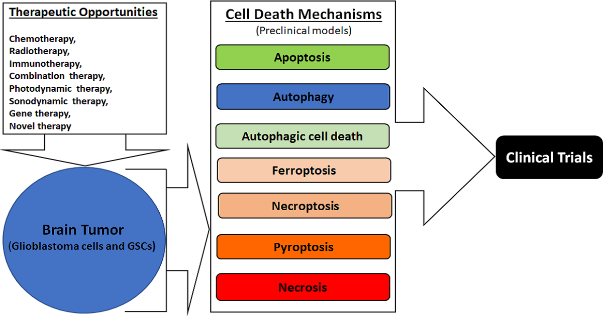
Cell death mechanisms, either induction or inhibition by specific therapeutic strategies, provide great opportunities for controlling the growth of glioblastoma multiforme, often simply called glioblastoma, the most malignant brain tumor in humans. Although there is an ever-increasing list of cell death mechanisms, about a half dozen of them (apoptosis, autophagy, ferroptosis, necroptosis, pyroptosis, and necrosis) are currently considered to be important for discovering new therapeutic opportunities in glioblastoma, which harbors heterogeneity, increasing its ability to adapt to the ever-changing adverse tumor microenvironment. Apoptosis and autophagy maintain cell-membrane integrity and cause no inflammatory response, while all other cell death mechanisms in this list are associated with an ascending order of cell-membrane disruption and thus an increasing order of inflammatory response (mostly an undesirable outcome) after therapy. All of these except necrosis are known to be regulated cell death (RCD) mechanisms. All five RCD mechanisms in this list are now being extensively studied with the aim of modulating their signaling mechanisms using specific therapeutic modalities (chemotherapy, immunotherapy, gene therapy, radiotherapy, photodynamic therapy, sonodynamic therapy, combination therapy, etc.) for preventing the growth of glioblastoma cells and glioblastoma stem cells (GSCs) in preclinical (cell culture and animal) models. Apoptosis (type 1 cell death), autophagic cell death (type 2 cell death), and necrosis (type 3 cell death) are conventionally known to be the most desirable, second-most desirable, and least desirable cell death mechanisms, respectably, in glioblastoma following treatment with a therapeutic modality.

The induction of apoptosis is conventionally the gold standard and the main intention of using a therapy in glioblastoma or in any other cancer. However, all cancers essentially acquire the mechanism of resistance to avoid the induction of apoptosis via alterations in different actors or factors in its complex signaling mechanism in course of the therapy. Tumor microenvironment (e.g., hypoxia, nutrient deficiency, low pH) and some conventional therapies (e.g., chemotherapy, radiotherapy, targeted therapy) promote autophagy, which is basically a mechanism for the bulk degradation of the dysfunctional cellular components (e.g., proteins, nucleic acids, carbohydrates) and the damaged mitochondria and other organelles for producing and recycling the building blocks (i.e., amino acids, nucleotides, sugars), for cellular homeostasis and survival of the tumor cells. Autophagy is a double-edge sword in glioblastoma because it can act as a cell-survival mechanism or a cell-death mechanism depending on the therapeutic strategy used. Studies show that the induction of autophagy inhibits senescence (cessation of cell division) in GSCs, maintaining stemness and promoting senescence in other glioblastoma cells, causing inhibition of their apoptosis, ultimately relapsing the growth of glioblastoma. Therefore, most of the recent therapeutic strategies aim at the induction of apoptosis and inhibition of autophagy, while some of them aim at the induction of autophagic cell death, and a few finding no other viable alternatives aim at the promotion of inflammatory RCD mechanisms in different glioblastoma cells and GSCs.

The non-inflammatory or inflammatory RCD mechanisms encourage the exploration of the efficacy of multiple therapeutic opportunities that  need a strong prospect of success not only in preclinical models of glioblastoma but also in clinical trials in glioblastoma patients.

Original research articles of preclinical models and contemporary review articles that directly or indirectly relate to this exciting topic of “Cell Death Mechanisms and Therapeutic Opportunities in Glioblastoma” are cordially invited for submission.

We look forward to receiving your article contributions and working with you for their publication in this Special Issue of Cells.  

Prof. Dr. Swapan K. Ray
Guest Editor

Manuscript Submission Information

Manuscripts should be submitted online at www.mdpi.com by registering and logging in to this website. Once you are registered, click here to go to the submission form. Manuscripts can be submitted until the deadline. All submissions that pass pre-check are peer-reviewed. Accepted papers will be published continuously in the journal (as soon as accepted) and will be listed together on the special issue website. Research articles, review articles as well as short communications are invited. For planned papers, a title and short abstract (about 100 words) can be sent to the Editorial Office for announcement on this website.

Submitted manuscripts should not have been published previously, nor be under consideration for publication elsewhere (except conference proceedings papers). All manuscripts are thoroughly refereed through a single-blind peer-review process. A guide for authors and other relevant information for submission of manuscripts is available on the Instructions for Authors page. Cells is an international peer-reviewed open access semimonthly journal published by MDPI.

Please visit the Instructions for Authors page before submitting a manuscript. The Article Processing Charge (APC) for publication in this open access journal is 2700 CHF (Swiss Francs). Submitted papers should be well formatted and use good English. Authors may use MDPI's English editing service prior to publication or during author revisions.

Keywords

  • apoptosis
  • autophagy
  • cell death mechanisms
  • cell signaling
  • glioblastoma
  • therapeutic opportunities

Published Papers (15 papers)

Order results
Result details
Select all
Export citation of selected articles as:

Research

Jump to: Review

26 pages, 9175 KiB  
Article
A Novel Approach for Glioblastoma Treatment by Combining Apoptosis Inducers (TMZ, MTX, and Cytarabine) with E.V.A. (Eltanexor, Venetoclax, and A1210477) Inhibiting XPO1, Bcl-2, and Mcl-1
by Kai Zhao, Madita Braun, Leonie Meyer, Katharina Otte, Hartmann Raifer, Frederik Helmprobst, Vincent Möschl, Axel Pagenstecher, Hans Urban, Michael W. Ronellenfitsch, Joachim P. Steinbach, Jelena Pesek, Bernhard Watzer, Wolfgang A. Nockher, R. Verena Taudte, Andreas Neubauer, Christopher Nimsky, Jörg W. Bartsch and Tillmann Rusch
Cells 2024, 13(7), 632; https://doi.org/10.3390/cells13070632 - 04 Apr 2024
Viewed by 441
Abstract
Adjuvant treatment for Glioblastoma Grade 4 with Temozolomide (TMZ) inevitably fails due to therapeutic resistance, necessitating new approaches. Apoptosis induction in GB cells is inefficient, due to an excess of anti-apoptotic XPO1/Bcl-2-family proteins. We assessed TMZ, Methotrexate (MTX), and Cytarabine (Ara-C) (apoptosis inducers) [...] Read more.
Adjuvant treatment for Glioblastoma Grade 4 with Temozolomide (TMZ) inevitably fails due to therapeutic resistance, necessitating new approaches. Apoptosis induction in GB cells is inefficient, due to an excess of anti-apoptotic XPO1/Bcl-2-family proteins. We assessed TMZ, Methotrexate (MTX), and Cytarabine (Ara-C) (apoptosis inducers) combined with XPO1/Bcl-2/Mcl-1-inhibitors (apoptosis rescue) in GB cell lines and primary GB stem-like cells (GSCs). Using CellTiter-Glo® and Caspase-3 activity assays, we generated dose–response curves and analyzed the gene and protein regulation of anti-apoptotic proteins via PCR and Western blots. Optimal drug combinations were examined for their impact on the cell cycle and apoptosis induction via FACS analysis, paralleled by the assessment of potential toxicity in healthy mouse brain slices. Ara-C and MTX proved to be 150- to 10,000-fold more potent in inducing apoptosis than TMZ. In response to inhibitors Eltanexor (XPO1; E), Venetoclax (Bcl-2; V), and A1210477 (Mcl-1; A), genes encoding for the corresponding proteins were upregulated in a compensatory manner. TMZ, MTX, and Ara-C combined with E, V, and A evidenced highly lethal effects when combined. As no significant cell death induction in mouse brain slices was observed, we conclude that this drug combination is effective in vitro and expected to have low side effects in vivo. Full article
(This article belongs to the Special Issue Cell Death Mechanisms and Therapeutic Opportunities in Glioblastoma)
Show Figures

Figure 1

19 pages, 5076 KiB  
Article
Exploring Regorafenib Responsiveness and Uncovering Molecular Mechanisms in Recurrent Glioblastoma Tumors through Longitudinal In Vitro Sampling
by Mariangela Morelli, Francesca Lessi, Sara Franceschi, Gianmarco Ferri, Manuel Giacomarra, Michele Menicagli, Carlo Gambacciani, Francesco Pieri, Francesco Pasqualetti, Nicola Montemurro, Paolo Aretini, Orazio Santo Santonocito, Anna Luisa Di Stefano and Chiara Maria Mazzanti
Cells 2024, 13(6), 487; https://doi.org/10.3390/cells13060487 - 11 Mar 2024
Viewed by 701
Abstract
Glioblastoma, a deadly brain tumor, shows limited response to standard therapies like temozolomide (TMZ). Recent findings from the REGOMA trial underscore a significant survival improvement offered by Regorafenib (REGO) in recurrent glioblastoma. Our study aimed to propose a 3D ex vivo drug response [...] Read more.
Glioblastoma, a deadly brain tumor, shows limited response to standard therapies like temozolomide (TMZ). Recent findings from the REGOMA trial underscore a significant survival improvement offered by Regorafenib (REGO) in recurrent glioblastoma. Our study aimed to propose a 3D ex vivo drug response precision medicine approach to investigate recurrent glioblastoma sensitivity to REGO and elucidate the underlying molecular mechanisms involved in tumor resistance or responsiveness to treatment. Three-dimensional glioblastoma organoids (GB-EXPs) obtained from 18 patients’ resected recurrent glioblastoma tumors were treated with TMZ and REGO. Drug responses were evaluated using NAD(P)H FLIM, stratifying tumors as responders (Resp) or non-responders (NRs). Whole-exome sequencing was performed on 16 tissue samples, and whole-transcriptome analysis on 13 GB-EXPs treated and untreated. We found 35% (n = 9) and 77% (n = 20) of tumors responded to TMZ and REGO, respectively, with no instances of TMZ-Resp being REGO-NRs. Exome analysis revealed a unique mutational profile in REGO-Resp tumors compared to NR tumors. Transcriptome analysis identified distinct expression patterns in Resp and NR tumors, impacting Rho GTPase and NOTCH signaling, known to be involved in drug response. In conclusion, recurrent glioblastoma tumors were more responsive to REGO compared to TMZ treatment. Importantly, our approach enables a comprehensive longitudinal exploration of the molecular changes induced by treatment, unveiling promising biomarkers indicative of drug response. Full article
(This article belongs to the Special Issue Cell Death Mechanisms and Therapeutic Opportunities in Glioblastoma)
Show Figures

Figure 1

17 pages, 5900 KiB  
Article
In Vitro Glioblastoma Model on a Plate for Localized Drug Release Study from a 3D-Printed Drug-Eluted Hydrogel Mesh
by Behnad Chehri, Kaiwen Liu, Golnaz Vaseghi, Amir Seyfoori and Mohsen Akbari
Cells 2024, 13(4), 363; https://doi.org/10.3390/cells13040363 - 19 Feb 2024
Viewed by 1098
Abstract
Glioblastoma multiforme (GBM) is an aggressive type of brain tumor that has limited treatment options. Current standard therapies, including surgery followed by radiotherapy and chemotherapy, are not very effective due to the rapid progression and recurrence of the tumor. Therefore, there is an [...] Read more.
Glioblastoma multiforme (GBM) is an aggressive type of brain tumor that has limited treatment options. Current standard therapies, including surgery followed by radiotherapy and chemotherapy, are not very effective due to the rapid progression and recurrence of the tumor. Therefore, there is an urgent need for more effective treatments, such as combination therapy and localized drug delivery systems that can reduce systemic side effects. Recently, a handheld printer was developed that can deliver drugs directly to the tumor site. In this study, the feasibility of using this technology for localized co-delivery of temozolomide (TMZ) and deferiprone (DFP) to treat glioblastoma is showcased. A flexible drug-loaded mesh (GlioMesh) loaded with poly (lactic-co-glycolic acid) (PLGA) microparticles is printed, which shows the sustained release of both drugs for up to a month. The effectiveness of the printed drug-eluting mesh in terms of tumor toxicity and invasion inhibition is evaluated using a 3D micro-physiological system on a plate and the formation of GBM tumoroids within the microenvironment. The proposed in vitro model can identify the effective combination doses of TMZ and DFP in a sustained drug delivery platform. Additionally, our approach shows promise in GB therapy by enabling localized delivery of multiple drugs, preventing off-target cytotoxic effects. Full article
(This article belongs to the Special Issue Cell Death Mechanisms and Therapeutic Opportunities in Glioblastoma)
Show Figures

Figure 1

14 pages, 2878 KiB  
Article
Conoidin A, a Covalent Inhibitor of Peroxiredoxin 2, Reduces Growth of Glioblastoma Cells by Triggering ROS Production
by Monika Szeliga and Radosław Rola
Cells 2023, 12(15), 1934; https://doi.org/10.3390/cells12151934 - 26 Jul 2023
Viewed by 1229
Abstract
Compounds that cause oxidative stress have recently gained considerable interest as potential anticancer treatment modalities. Nevertheless, their efficiency may be diminished by the antioxidant systems often upregulated in cancer cells. Peroxiredoxins (PRDXs) are antioxidant enzymes that scavenge peroxides and contribute to redox homeostasis. [...] Read more.
Compounds that cause oxidative stress have recently gained considerable interest as potential anticancer treatment modalities. Nevertheless, their efficiency may be diminished by the antioxidant systems often upregulated in cancer cells. Peroxiredoxins (PRDXs) are antioxidant enzymes that scavenge peroxides and contribute to redox homeostasis. They play a role in carcinogenesis and are upregulated in several cancer types. Here, we assessed the expression pattern of PRDX1 and PRDX2 in glioblastoma (GBM) and examined the efficacy of their inhibitors in GBM cell lines and patient-derived GBM cells. Both PRDX1 and PRDX2 were upregulated in GBM compared to non-tumor brain tissues and their considerable amounts were observed in GBM cells. Adenanthin, a compound inhibiting PRDX1 activity, slightly decreased GBM cell viability, while conoidin A (CONA), a covalent PRDX2 inhibitor, displayed high toxicity in GBM cells. CONA elevated the intracellular reactive oxygen species (ROS) level. Pre-treatment with an ROS scavenger protected cells from CONA-induced death, indicating that ROS accumulation plays a crucial role in this phenomenon. Menadione or celecoxib, both of which are ROS-inducing agents, potentiated the anticancer activity of CONA. Collectively, our results unveil PRDX1 and PRDX2 as potential targets for GBM therapy, and substantiate the further exploration of their inhibitors. Full article
(This article belongs to the Special Issue Cell Death Mechanisms and Therapeutic Opportunities in Glioblastoma)
Show Figures

Graphical abstract

21 pages, 4147 KiB  
Article
Propofol Inhibits Glioma Stem Cell Growth and Migration and Their Interaction with Microglia via BDNF-AS and Extracellular Vesicles
by Rephael Nizar, Simona Cazacu, Cunli Xiang, Matan Krasner, Efrat Barbiro-Michaely, Doron Gerber, Jonathan Schwartz, Iris Fried, Shira Yuval, Aharon Brodie, Gila Kazimirsky, Naama Amos, Ron Unger, Stephen Brown, Lisa Rogers, Donald H. Penning and Chaya Brodie
Cells 2023, 12(15), 1921; https://doi.org/10.3390/cells12151921 - 25 Jul 2023
Cited by 4 | Viewed by 1645
Abstract
Glioblastoma (GBM) is the most common and aggressive primary brain tumor. GBM contains a small subpopulation of glioma stem cells (GSCs) that are implicated in treatment resistance, tumor infiltration, and recurrence, and are thereby considered important therapeutic targets. Recent clinical studies have suggested [...] Read more.
Glioblastoma (GBM) is the most common and aggressive primary brain tumor. GBM contains a small subpopulation of glioma stem cells (GSCs) that are implicated in treatment resistance, tumor infiltration, and recurrence, and are thereby considered important therapeutic targets. Recent clinical studies have suggested that the choice of general anesthetic (GA), particularly propofol, during tumor resection, affects subsequent tumor response to treatments and patient prognosis. In this study, we investigated the molecular mechanisms underlying propofol’s anti-tumor effects on GSCs and their interaction with microglia cells. Propofol exerted a dose-dependent inhibitory effect on the self-renewal, expression of mesenchymal markers, and migration of GSCs and sensitized them to both temozolomide (TMZ) and radiation. At higher concentrations, propofol induced a large degree of cell death, as demonstrated using microfluid chip technology. Propofol increased the expression of the lncRNA BDNF-AS, which acts as a tumor suppressor in GBM, and silencing of this lncRNA partially abrogated propofol’s effects. Propofol also inhibited the pro-tumorigenic GSC-microglia crosstalk via extracellular vesicles (EVs) and delivery of BDNF-AS. In conclusion, propofol exerted anti-tumor effects on GSCs, sensitized these cells to radiation and TMZ, and inhibited their pro-tumorigenic interactions with microglia via transfer of BDNF-AS by EVs. Full article
(This article belongs to the Special Issue Cell Death Mechanisms and Therapeutic Opportunities in Glioblastoma)
Show Figures

Figure 1

21 pages, 5688 KiB  
Article
Autophagy Inhibition with Chloroquine Increased Pro-Apoptotic Potential of New Aziridine-Hydrazide Hydrazone Derivatives against Glioblastoma Cells
by Monika Witusik-Perkowska, Pola Głowacka, Adam M. Pieczonka, Ewa Świderska, Agnieszka Pudlarz, Michał Rachwalski, Julia Szymańska, Magdalena Zakrzewska, Dariusz J. Jaskólski and Janusz Szemraj
Cells 2023, 12(14), 1906; https://doi.org/10.3390/cells12141906 - 21 Jul 2023
Cited by 2 | Viewed by 1200
Abstract
Tumor therapy escape due to undesired side effects induced by treatment, such as prosurvival autophagy or cellular senescence, is one of the key mechanisms of resistance that eventually leads to tumor dormancy and recurrence. Glioblastoma is the most frequent and practically incurable neoplasm [...] Read more.
Tumor therapy escape due to undesired side effects induced by treatment, such as prosurvival autophagy or cellular senescence, is one of the key mechanisms of resistance that eventually leads to tumor dormancy and recurrence. Glioblastoma is the most frequent and practically incurable neoplasm of the central nervous system; thus, new treatment modalities have been investigated to find a solution more effective than the currently applied standards based on temozolomide. The present study examined the newly synthesized compounds of aziridine–hydrazide hydrazone derivatives to determine their antineoplastic potential against glioblastoma cells in vitro. Although the output of our investigation clearly demonstrates their proapoptotic activity, the cytotoxic effect appeared to be blocked by treatment-induced autophagy, the phenomenon also detected in the case of temozolomide action. The addition of an autophagy inhibitor, chloroquine, resulted in a significant increase in apoptosis triggered by the tested compounds, as well as temozolomide. The new aziridine–hydrazide hydrazone derivatives, which present cytotoxic potential against glioblastoma cells comparable to or even higher than that of temozolomide, show promising results and, thus, should be further investigated as antineoplastic agents. Moreover, our findings suggest that the combination of an apoptosis inducer with an autophagy inhibitor could optimize chemotherapeutic efficiency, and the addition of an autophagy inhibitor should be considered as an optional adjunctive therapy minimizing the risk of tumor escape from treatment. Full article
(This article belongs to the Special Issue Cell Death Mechanisms and Therapeutic Opportunities in Glioblastoma)
Show Figures

Figure 1

24 pages, 5413 KiB  
Article
Metronomic Photodynamic Therapy with Conjugated Polymer Nanoparticles in Glioblastoma Tumor Microenvironment
by Matías Daniel Caverzán, Paula Martina Oliveda, Lucía Beaugé, Rodrigo Emiliano Palacios, Carlos Alberto Chesta and Luis Exequiel Ibarra
Cells 2023, 12(11), 1541; https://doi.org/10.3390/cells12111541 - 04 Jun 2023
Cited by 9 | Viewed by 1590
Abstract
Alternative therapies such as photodynamic therapy (PDT) that combine light, oxygen and photosensitizers (PSs) have been proposed for glioblastoma (GBM) management to overcome conventional treatment issues. An important disadvantage of PDT using a high light irradiance (fluence rate) (cPDT) is the abrupt oxygen [...] Read more.
Alternative therapies such as photodynamic therapy (PDT) that combine light, oxygen and photosensitizers (PSs) have been proposed for glioblastoma (GBM) management to overcome conventional treatment issues. An important disadvantage of PDT using a high light irradiance (fluence rate) (cPDT) is the abrupt oxygen consumption that leads to resistance to the treatment. PDT metronomic regimens (mPDT) involving administering light at a low irradiation intensity over a relatively long period of time could be an alternative to circumvent the limitations of conventional PDT protocols. The main objective of the present work was to compare the effectiveness of PDT with an advanced PS based on conjugated polymer nanoparticles (CPN) developed by our group in two irradiation modalities: cPDT and mPDT. The in vitro evaluation was carried out based on cell viability, the impact on the macrophage population of the tumor microenvironment in co-culture conditions and the modulation of HIF-1α as an indirect indicator of oxygen consumption. mPDT regimens with CPNs resulted in more effective cell death, a lower activation of molecular pathways of therapeutic resistance and macrophage polarization towards an antitumoral phenotype. Additionally, mPDT was tested in a GBM heterotopic mouse model, confirming its good performance with promising tumor growth inhibition and apoptotic cell death induction. Full article
(This article belongs to the Special Issue Cell Death Mechanisms and Therapeutic Opportunities in Glioblastoma)
Show Figures

Figure 1

25 pages, 6674 KiB  
Article
Concurrent Activation of Both Survival-Promoting and Death-Inducing Signaling by Chloroquine in Glioblastoma Stem Cells: Implications for Potential Risks and Benefits of Using Chloroquine as Radiosensitizer
by Andreas Müller, Patrick Weyerhäuser, Nancy Berte, Fitriasari Jonin, Bogdan Lyubarskyy, Bettina Sprang, Sven Rainer Kantelhardt, Gabriela Salinas, Lennart Opitz, Walter Schulz-Schaeffer, Alf Giese and Ella L. Kim
Cells 2023, 12(9), 1290; https://doi.org/10.3390/cells12091290 - 30 Apr 2023
Cited by 3 | Viewed by 1643
Abstract
Lysosomotropic agent chloroquine was shown to sensitize non-stem glioblastoma cells to radiation in vitro with p53-dependent apoptosis implicated as one of the underlying mechanisms. The in vivo outcomes of chloroquine or its effects on glioblastoma stem cells have not been previously addressed. This [...] Read more.
Lysosomotropic agent chloroquine was shown to sensitize non-stem glioblastoma cells to radiation in vitro with p53-dependent apoptosis implicated as one of the underlying mechanisms. The in vivo outcomes of chloroquine or its effects on glioblastoma stem cells have not been previously addressed. This study undertakes a combinatorial approach encompassing in vitro, in vivo and in silico investigations to address the relationship between chloroquine-mediated radiosensitization and p53 status in glioblastoma stem cells. Our findings reveal that chloroquine elicits antagonistic impacts on signaling pathways involved in the regulation of cell fate via both transcription-dependent and transcription-independent mechanisms. Evidence is provided that transcriptional impacts of chloroquine are primarily determined by p53 with chloroquine-mediated activation of pro-survival mevalonate and p21-DREAM pathways being the dominant response in the background of wild type p53. Non-transcriptional effects of chloroquine are conserved and converge on key cell fate regulators ATM, HIPK2 and AKT in glioblastoma stem cells irrespective of their p53 status. Our findings indicate that pro-survival responses elicited by chloroquine predominate in the context of wild type p53 and are diminished in cells with transcriptionally impaired p53. We conclude that p53 is an important determinant of the balance between pro-survival and pro-death impacts of chloroquine and propose that p53 functional status should be taken into consideration when evaluating the efficacy of glioblastoma radiosensitization by chloroquine. Full article
(This article belongs to the Special Issue Cell Death Mechanisms and Therapeutic Opportunities in Glioblastoma)
Show Figures

Figure 1

14 pages, 8664 KiB  
Article
Blockage of Autophagy Increases Timosaponin AIII-Induced Apoptosis of Glioma Cells In Vitro and In Vivo
by Chu-Che Lee, Jen-Pi Tsai, Hsiang-Lin Lee, Yung-Jen Chen, Yong-Syuan Chen, Yi-Hsien Hsieh and Jin-Cherng Chen
Cells 2023, 12(1), 168; https://doi.org/10.3390/cells12010168 - 30 Dec 2022
Cited by 6 | Viewed by 1341
Abstract
Timosaponin AIII (TSAIII), a saponin isolated from Anemarrhena asphodeloides and used in traditional Chinese medicine, exerts antitumor, anti-inflammatory, anti-angiogenesis, and pro-apoptotic activity on a variety of tumor cells. This study investigated the antitumor effects of TSAIII and the underlying mechanisms in human glioma [...] Read more.
Timosaponin AIII (TSAIII), a saponin isolated from Anemarrhena asphodeloides and used in traditional Chinese medicine, exerts antitumor, anti-inflammatory, anti-angiogenesis, and pro-apoptotic activity on a variety of tumor cells. This study investigated the antitumor effects of TSAIII and the underlying mechanisms in human glioma cells in vitro and in vivo. TSAIII significantly inhibited glioma cell viability in a dose- and time-dependent manner but did not affect the growth of normal astrocytes. We also observed that in both glioma cell lines, TSAIII induces cell death and mitochondrial dysfunction, consistent with observed increases in the protein expression of cleaved-caspase-3, cleaved-caspase-9, cleaved-PARP, cytochrome c, and Mcl-1. TSAIII also activated autophagy, as indicated by increased accumulation of the autophagosome markers p62 and LC3-II and the autolysosome marker LAMP1. LC3 silencing, as well as TSAIII combined with the autophagy inhibitor 3-methyladenine (3MA), increased apoptosis in GBM8401 cells. TSAIII inhibited tumor growth in xenografts and in an orthotopic GBM8401 mice model in vivo. These results demonstrate that TSAIII exhibits antitumor effects and may hold potential as a therapy for glioma. Full article
(This article belongs to the Special Issue Cell Death Mechanisms and Therapeutic Opportunities in Glioblastoma)
Show Figures

Figure 1

10 pages, 2399 KiB  
Article
ALDH1-Mediated Autophagy Sensitizes Glioblastoma Cells to Ferroptosis
by Yang Wu, Helena Kram, Jens Gempt, Friederike Liesche-Starnecker, Wei Wu and Jürgen Schlegel
Cells 2022, 11(24), 4015; https://doi.org/10.3390/cells11244015 - 12 Dec 2022
Cited by 8 | Viewed by 2151
Abstract
The fatal clinical course of human glioblastoma (GBM) despite aggressive adjuvant therapies is due to high rates of recurrent tumor growth driven by tumor cells with stem-cell characteristics (glioma stem cells, GSCs). The aldehyde dehydrogenase 1 (ALDH1) family of enzymes has been shown [...] Read more.
The fatal clinical course of human glioblastoma (GBM) despite aggressive adjuvant therapies is due to high rates of recurrent tumor growth driven by tumor cells with stem-cell characteristics (glioma stem cells, GSCs). The aldehyde dehydrogenase 1 (ALDH1) family of enzymes has been shown to be a biomarker for GSCs, and ALDH1 seems to be involved in the biological processes causing therapy resistance. Ferroptosis is a recently discovered cell death mechanism, that depends on iron overload and lipid peroxidation, and it could, therefore, be a potential therapeutic target in various cancer types. Since both ALDH1 and ferroptosis interact with lipid peroxidation (LPO), we aimed to investigate a possible connection between ALDH1 and ferroptosis. Here, we show that RSL3-induced LPO and ferroptotic cell death revealed RSL3-sensitive and -resistant malignant glioma cell lines. Most interestingly, RSL3 sensitivity correlates with ALDH1a3 expression; only high ALDH1a3-expressing cells seem to be sensitive to ferroptosis induction. In accordance, inhibition of ALDH1a3 enzymatic activity by chemical inhibition or genetic knockout protects tumor cells from RSL3-induced ferroptotic cell death. Both RSL-3-dependent binding of ALDH1a3 to LC3B and autophagic downregulation of ferritin could be completely blocked by ALDH inhibition. Therefore, ALDH1a3 seems to be involved in ferroptosis through the essential release of iron by ferritinophagy. Our results also indicate that ferroptosis induction might be a particularly interesting clinical approach for targeting the highly aggressive cell population of GSC. Full article
(This article belongs to the Special Issue Cell Death Mechanisms and Therapeutic Opportunities in Glioblastoma)
Show Figures

Figure 1

17 pages, 5732 KiB  
Article
Abrogation of Cellular Senescence Induced by Temozolomide in Glioblastoma Cells: Search for Senolytics
by Lea Beltzig, Markus Christmann and Bernd Kaina
Cells 2022, 11(16), 2588; https://doi.org/10.3390/cells11162588 - 19 Aug 2022
Cited by 12 | Viewed by 3238
Abstract
A first-line therapeutic for high-grade glioma, notably glioblastoma (GBM), is the DNA methylating drug temozolomide (TMZ). Previously, we showed that TMZ induces not only apoptosis and autophagy, but also cellular senescence (CSEN). We presented the hypothesis that GBM cells may escape from CSEN, [...] Read more.
A first-line therapeutic for high-grade glioma, notably glioblastoma (GBM), is the DNA methylating drug temozolomide (TMZ). Previously, we showed that TMZ induces not only apoptosis and autophagy, but also cellular senescence (CSEN). We presented the hypothesis that GBM cells may escape from CSEN, giving rise to recurrent tumors. Furthermore, the inflammatory phenotype associated with CSEN may attenuate chemotherapy and drive tumor progression. Therefore, treatments that specifically target senescent cells, i.e., senolytic drugs, may lead to a better outcome of GBM therapy by preventing recurrences and tumor inflammation. Here, we tested Bcl-2 targeting drugs including ABT-737, ABT-263 (navitoclax), several natural substances such as artesunate, fisetin and curcumin as well as lomustine (CCNU) and ionizing radiation (IR) for their senolytic capacity in GBM cells. Additionally, several proteins involved in the DNA damage response (DDR), ATM, ATR, Chk1/2, p53, p21, NF-kB, Rad51, PARP, IAPs and autophagy, a pathway involved in CSEN induction, were tested for their impact in maintaining CSEN. Treatment of GBM cells with a low dose of TMZ for 8–10 days resulted in >80% CSEN, confirming CSEN to be the major trait induced by TMZ. To identify senolytics, we treated the senescent population with the compounds of interest and found that ABT-737, navitoclax, chloroquine, ATMi, ATRi, BV-6, PX-866 and the natural compounds fisetin and artesunate exhibit senolytic activity, inducing death in senescent cells more efficiently than in proliferating cells. Curcumin showed the opposite effect. No specific effect on CSEN cells was observed by inhibition of Chk1/Chk2, p21, NF-kB, Rad51 and PARP. We conclude that these factors neither play a critical role in maintaining TMZ-induced CSEN nor can their inhibitors be considered as senolytics. Since IR and CCNU did not exhibit senolytic activity, radio- and chemotherapy with alkylating drugs is not designed to eliminate TMZ-induced senescent cancer cells. Full article
(This article belongs to the Special Issue Cell Death Mechanisms and Therapeutic Opportunities in Glioblastoma)
Show Figures

Figure 1

Review

Jump to: Research

16 pages, 975 KiB  
Review
Mesenchymal-Stem-Cell-Based Therapy against Gliomas
by Sisa M. Santillán-Guaján, Mehdi H. Shahi and Javier S. Castresana
Cells 2024, 13(7), 617; https://doi.org/10.3390/cells13070617 - 02 Apr 2024
Viewed by 605
Abstract
Glioblastoma is the most aggressive, malignant, and lethal brain tumor of the central nervous system. Its poor prognosis lies in its inefficient response to currently available treatments that consist of surgical resection, radiotherapy, and chemotherapy. Recently, the use of mesenchymal stem cells (MSCs) [...] Read more.
Glioblastoma is the most aggressive, malignant, and lethal brain tumor of the central nervous system. Its poor prognosis lies in its inefficient response to currently available treatments that consist of surgical resection, radiotherapy, and chemotherapy. Recently, the use of mesenchymal stem cells (MSCs) as a possible kind of cell therapy against glioblastoma is gaining great interest due to their immunomodulatory properties, tumor tropism, and differentiation into other cell types. However, MSCs seem to present both antitumor and pro-tumor properties depending on the tissue from which they come. In this work, the possibility of using MSCs to deliver therapeutic genes, oncolytic viruses, and miRNA is presented, as well as strategies that can improve their therapeutic efficacy against glioblastoma, such as CAR-T cells, nanoparticles, and exosomes. Full article
(This article belongs to the Special Issue Cell Death Mechanisms and Therapeutic Opportunities in Glioblastoma)
Show Figures

Figure 1

20 pages, 807 KiB  
Review
Chimeric Antigen Receptor T Cells in Glioblastoma—Current Concepts and Promising Future
by Rebecca Kringel, Katrin Lamszus and Malte Mohme
Cells 2023, 12(13), 1770; https://doi.org/10.3390/cells12131770 - 03 Jul 2023
Cited by 2 | Viewed by 1667
Abstract
Glioblastoma (GBM) is a highly aggressive primary brain tumor that is largely refractory to treatment and, therefore, invariably relapses. GBM patients have a median overall survival of 15 months and, given this devastating prognosis, there is a high need for therapy improvement. One [...] Read more.
Glioblastoma (GBM) is a highly aggressive primary brain tumor that is largely refractory to treatment and, therefore, invariably relapses. GBM patients have a median overall survival of 15 months and, given this devastating prognosis, there is a high need for therapy improvement. One of the therapeutic approaches currently tested in GBM is chimeric antigen receptor (CAR)-T cell therapy. CAR-T cells are genetically altered T cells that are redirected to eliminate tumor cells in a highly specific manner. There are several challenges to CAR-T cell therapy in solid tumors such as GBM, including restricted trafficking and penetration of tumor tissue, a highly immunosuppressive tumor microenvironment (TME), as well as heterogeneous antigen expression and antigen loss. In addition, CAR-T cells have limitations concerning safety, toxicity, and the manufacturing process. To date, CAR-T cells directed against several target antigens in GBM including interleukin-13 receptor alpha 2 (IL-13Rα2), epidermal growth factor receptor variant III (EGFRvIII), human epidermal growth factor receptor 2 (HER2), and ephrin type-A receptor 2 (EphA2) have been tested in preclinical and clinical studies. These studies demonstrated that CAR-T cell therapy is a feasible option in GBM with at least transient responses and acceptable adverse effects. Further improvements in CAR-T cells regarding their efficacy, flexibility, and safety could render them a promising therapy option in GBM. Full article
(This article belongs to the Special Issue Cell Death Mechanisms and Therapeutic Opportunities in Glioblastoma)
Show Figures

Figure 1

25 pages, 2411 KiB  
Review
Advanced Bioinformatics Analysis and Genetic Technologies for Targeting Autophagy in Glioblastoma Multiforme
by Amanda J. Manea and Swapan K. Ray
Cells 2023, 12(6), 897; https://doi.org/10.3390/cells12060897 - 15 Mar 2023
Cited by 2 | Viewed by 2249
Abstract
As the most malignant primary brain tumor in adults, a diagnosis of glioblastoma multiforme (GBM) continues to carry a poor prognosis. GBM is characterized by cytoprotective homeostatic processes such as the activation of autophagy, capability to confer therapeutic resistance, evasion of apoptosis, and [...] Read more.
As the most malignant primary brain tumor in adults, a diagnosis of glioblastoma multiforme (GBM) continues to carry a poor prognosis. GBM is characterized by cytoprotective homeostatic processes such as the activation of autophagy, capability to confer therapeutic resistance, evasion of apoptosis, and survival strategy even in the hypoxic and nutrient-deprived tumor microenvironment. The current gold standard of therapy, which involves radiotherapy and concomitant and adjuvant chemotherapy with temozolomide (TMZ), has been a game-changer for patients with GBM, relatively improving both overall survival (OS) and progression-free survival (PFS); however, TMZ is now well-known to upregulate undesirable cytoprotective autophagy, limiting its therapeutic efficacy for induction of apoptosis in GBM cells. The identification of targets utilizing bioinformatics-driven approaches, advancement of modern molecular biology technologies such as clustered regularly interspaced short palindromic repeats (CRISPR)—CRISPR-associated protein (Cas9) or CRISPR-Cas9 genome editing, and usage of microRNA (miRNA)-mediated regulation of gene expression led to the selection of many novel targets for new therapeutic development and the creation of promising combination therapies. This review explores the current state of advanced bioinformatics analysis and genetic technologies and their utilization for synergistic combination with TMZ in the context of inhibition of autophagy for controlling the growth of GBM. Full article
(This article belongs to the Special Issue Cell Death Mechanisms and Therapeutic Opportunities in Glioblastoma)
Show Figures

Graphical abstract

23 pages, 2707 KiB  
Review
Intersections of Ubiquitin-Proteosome System and Autophagy in Promoting Growth of Glioblastoma Multiforme: Challenges and Opportunities
by Rhett Visintin and Swapan K. Ray
Cells 2022, 11(24), 4063; https://doi.org/10.3390/cells11244063 - 15 Dec 2022
Cited by 2 | Viewed by 1785
Abstract
Glioblastoma multiforme (GBM) is a brain tumor notorious for its propensity to recur after the standard treatments of surgical resection, ionizing radiation (IR), and temozolomide (TMZ). Combined with the acquired resistance to standard treatments and recurrence, GBM is an especially deadly malignancy with [...] Read more.
Glioblastoma multiforme (GBM) is a brain tumor notorious for its propensity to recur after the standard treatments of surgical resection, ionizing radiation (IR), and temozolomide (TMZ). Combined with the acquired resistance to standard treatments and recurrence, GBM is an especially deadly malignancy with hardly any worthwhile treatment options. The treatment resistance of GBM is influenced, in large part, by the contributions from two main degradative pathways in eukaryotic cells: ubiquitin-proteasome system (UPS) and autophagy. These two systems influence GBM cell survival by removing and recycling cellular components that have been damaged by treatments, as well as by modulating metabolism and selective degradation of components of cell survival or cell death pathways. There has recently been a large amount of interest in potential cancer therapies involving modulation of UPS or autophagy pathways. There is significant crosstalk between the two systems that pose therapeutic challenges, including utilization of ubiquitin signaling, the degradation of components of one system by the other, and compensatory activation of autophagy in the case of proteasome inhibition for GBM cell survival and proliferation. There are several important regulatory nodes which have functions affecting both systems. There are various molecular components at the intersections of UPS and autophagy pathways that pose challenges but also show some new therapeutic opportunities for GBM. This review article aims to provide an overview of the recent advancements in research regarding the intersections of UPS and autophagy with relevance to finding novel GBM treatment opportunities, especially for combating GBM treatment resistance. Full article
(This article belongs to the Special Issue Cell Death Mechanisms and Therapeutic Opportunities in Glioblastoma)
Show Figures

Graphical abstract

Back to TopTop