Next Issue
Volume 12, June-1
Previous Issue
Volume 12, May-1


Cells, Volume 12, Issue 10 (May-2 2023) – 102 articles

Cover Story (view full-size image): 
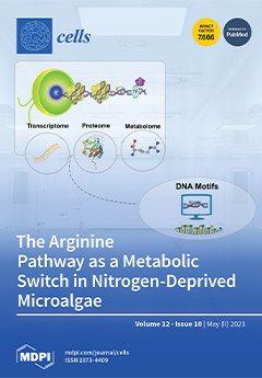
The reanalysis of metabolome, proteome, and transcriptome datasets revealed that the priming of Arginine (Arg) metabolism pathways occurs during nitrogen (N) deprivation, and it plays a significant role in inducing the accumulation of triacylglycerols (TAGs), as a metabolic switch. The findings suggest the presence of regulatory modules that integrate the transcriptional regulation of TAGs accumulation with N assimilation via the upregulation of enzymes involved in Arg metabolism. The analysis of gene promoter motifs further supports the existence of a regulatory network integrating these processes, suggesting the involvement of phosphorylation-, nitric oxide-, and hydrogen peroxide-mediated signaling, providing valuable insights into the regulatory and signaling mechanisms underlying N deprivation responses in Chlamydomonas reinhardtii. View this paper
  • Issues are regarded as officially published after their release is announced to the table of contents alert mailing list.
  • You may sign up for e-mail alerts to receive table of contents of newly released issues.
  • PDF is the official format for papers published in both, html and pdf forms. To view the papers in pdf format, click on the "PDF Full-text" link, and use the free Adobe Reader to open them.
Order results
Result details
Section
Select all
Export citation of selected articles as:
13 pages, 2301 KiB  
Article
Double Positive CD4+CD8+ (DP) T-Cells Display Distinct Exhaustion Phenotype in Chronic Hepatitis C
by Anna Maria Kochanowicz, Sylwia Osuch, Hanna Berak, Aleksandra Kumorek and Kamila Caraballo Cortés
Cells 2023, 12(10), 1446; https://doi.org/10.3390/cells12101446 - 22 May 2023
Viewed by 1486
Abstract
In chronic hepatitis C (CHC), characterized by exhaustion of T-cell function, increased frequencies of double-positive (DP) (CD4+CD8+) cells are present in peripheral blood. We compared the exhaustion phenotype between DP and single positive (SP) T-cells, including HCV-specific cells, and [...] Read more.
In chronic hepatitis C (CHC), characterized by exhaustion of T-cell function, increased frequencies of double-positive (DP) (CD4+CD8+) cells are present in peripheral blood. We compared the exhaustion phenotype between DP and single positive (SP) T-cells, including HCV-specific cells, and assessed the effect of successful HCV treatment on inhibitory receptors expression. Blood samples from 97 CHC patients were collected before and six months post-treatment. PD-1 (programmed cell death protein 1) and Tim-3 (T-cell immunoglobulin and mucin domain-containing molecule-3) expression was assessed by flow cytometry. DP T-cells displayed significantly higher PD-1 expression, lower Tim-3 expression than CD8+ SP T-cells and lower percentages of PD-1Tim-3 cells than CD4+ SP T-cells, both before and after treatment. PD-1+Tim-3+ DP T-cells decreased following treatment. HCV-specific cells were more frequent among DP than SP T-cells, both before and after treatment. HCV-specific DP T-cells were characterized by lower PD-1 expression, higher PD-1 and Tim-3 co-expression, and lower percentages of PD-1Tim-3 cells (both before and after treatment) and higher post-treatment Tim-3 than HCV-specific SP T-cells. Their percentages decreased following treatment, but the exhaustion phenotype remained unchanged. DP T-cells in CHC exhibit a distinct exhaustion phenotype from SP T-cells, and these changes mostly persist following successful treatment. Full article
(This article belongs to the Collection Feature Papers in ‘Cellular Immunology’)
Show Figures

Figure 1

17 pages, 4267 KiB  
Article
LRP1 Deficiency Promotes Mitostasis in Response to Oxidative Stress: Implications for Mitochondrial Targeting after Traumatic Brain Injury
by Gopal V. Velmurugan, W. Brad Hubbard, Paresh Prajapati, Hemendra J. Vekaria, Samir P. Patel, Alexander G. Rabchevsky and Patrick G. Sullivan
Cells 2023, 12(10), 1445; https://doi.org/10.3390/cells12101445 - 22 May 2023
Cited by 1 | Viewed by 1529
Abstract
The brain undergoes oxidative stress and mitochondrial dysfunction following physiological insults such as Traumatic brain injury (TBI), ischemia-reperfusion, and stroke. Pharmacotherapeutics targeting mitochondria (mitoceuticals) against oxidative stress include antioxidants, mild uncouplers, and enhancers of mitochondrial biogenesis, which have been shown to improve pathophysiological [...] Read more.
The brain undergoes oxidative stress and mitochondrial dysfunction following physiological insults such as Traumatic brain injury (TBI), ischemia-reperfusion, and stroke. Pharmacotherapeutics targeting mitochondria (mitoceuticals) against oxidative stress include antioxidants, mild uncouplers, and enhancers of mitochondrial biogenesis, which have been shown to improve pathophysiological outcomes after TBI. However, to date, there is no effective treatment for TBI. Studies have suggested that the deletion of LDL receptor-related protein 1 (LRP1) in adult neurons or glial cells could be beneficial and promote neuronal health. In this study, we used WT and LRP1 knockout (LKO) mouse embryonic fibroblast cells to examine mitochondrial outcomes following exogenous oxidative stress. Furthermore, we developed a novel technique to measure mitochondrial morphometric dynamics using transgenic mitochondrial reporter mice mtD2g (mitochondrial-specific Dendra2 green) in a TBI model. We found that oxidative stress increased the quantity of fragmented and spherical-shaped mitochondria in the injury core of the ipsilateral cortex following TBI, whereas rod-like elongated mitochondria were seen in the corresponding contralateral cortex. Critically, LRP1 deficiency significantly decreased mitochondrial fragmentation, preserving mitochondrial function and cell growth following exogenous oxidative stress. Collectively, our results show that targeting LRP1 to improve mitochondrial function is a potential pharmacotherapeutic strategy against oxidative damage in TBI and other neurodegenerative diseases. Full article
(This article belongs to the Special Issue Mitochondria at the Crossroad of Health and Disease)
Show Figures

Figure 1

28 pages, 2443 KiB  
Article
An Insight into Survivin in Relevance to Hematological, Biochemical and Genetic Characteristics in Tobacco Chewers with Oral Squamous Cell Carcinoma
by Susanna Theophilus Yesupatham, C. D. Dayanand, S. M. Azeem Mohiyuddin and M. L. Harendra Kumar
Cells 2023, 12(10), 1444; https://doi.org/10.3390/cells12101444 - 22 May 2023
Cited by 3 | Viewed by 1869
Abstract
Background: Survivin is an inhibitor of apoptosis protein (IAP), encoded by the Baculoviral IAP Repeat Containing 5 (BIRC5) gene located on q arm (25.3) on chromosome 17. It is expressed in various human cancers and involved in tumor resistance to radiation [...] Read more.
Background: Survivin is an inhibitor of apoptosis protein (IAP), encoded by the Baculoviral IAP Repeat Containing 5 (BIRC5) gene located on q arm (25.3) on chromosome 17. It is expressed in various human cancers and involved in tumor resistance to radiation and chemotherapy. The genetic analysis of the BIRC5 gene and its protein survivin levels in buccal tissue related to oral squamous cell carcinoma (OSCC) in South Indian tobacco chewers has not been studied. Hence, the study was designed to quantify survivin in buccal tissue and its association with pretreatment hematological parameters and to analyze the BIRC5 gene sequence. Method: In a single centric case control study, buccal tissue survivin levels were measured by ELISA. A total of 189 study subjects were categorized into Group 1 (n = 63) habitual tobacco chewers with OSCC, Group 2 (n = 63) habitual tobacco chewers without OSCC, and Group 3 (n = 63) healthy subjects as control. Retrospective hematological data were collected from Group 1 subjects and statistically analyzed. The BIRC5 gene was sequenced and data were analyzed using a bioinformatics tool. Results: Survivin protein mean ± SD in Group 1 was (1670.9 ± 796.21 pg/mL), in Group 2 it was (1096.02 ± 346.17 pg/mL), and in Group 3 it was (397.5 ± 96.1 pg/mL) with significance (p < 0.001). Survivin levels showed significance with cut-off levels of absolute monocyte count (AMC), neutrophil/lymphocyte ratio (NLR), and lymphocyte/monocyte ratio (LMR) at (p = 0.001). The unique variants found only in OSCC patients were T → G in the promoter region, G → C in exon 3, C → A, A → G, G → T, T → G, A → C, G → A in exon 4, C → A, G → T, G → C in the exon 5 region. Conclusions: The tissue survivin level increased in OSCC patients compared to controls; pretreatment AMC, LMR, and NLR may serve as add-on markers along with survivin to measure the progression of OSCC. Unique mutations in the promoter and exons 3–5 were observed in sequence analysis and were associated with survivin concentrations. Full article
Show Figures

Figure 1

19 pages, 2191 KiB  
Review
Roles of Aging, Circular RNAs, and RNA Editing in the Pathogenesis of Amyotrophic Lateral Sclerosis: Potential Biomarkers and Therapeutic Targets
by Takashi Hosaka, Hiroshi Tsuji and Shin Kwak
Cells 2023, 12(10), 1443; https://doi.org/10.3390/cells12101443 - 22 May 2023
Cited by 2 | Viewed by 2103
Abstract
Amyotrophic lateral sclerosis (ALS) is an incurable motor neuron disease caused by upper and lower motor neuron death. Despite advances in our understanding of ALS pathogenesis, effective treatment for this fatal disease remains elusive. As aging is a major risk factor for ALS, [...] Read more.
Amyotrophic lateral sclerosis (ALS) is an incurable motor neuron disease caused by upper and lower motor neuron death. Despite advances in our understanding of ALS pathogenesis, effective treatment for this fatal disease remains elusive. As aging is a major risk factor for ALS, age-related molecular changes may provide clues for the development of new therapeutic strategies. Dysregulation of age-dependent RNA metabolism plays a pivotal role in the pathogenesis of ALS. In addition, failure of RNA editing at the glutamine/arginine (Q/R) site of GluA2 mRNA causes excitotoxicity due to excessive Ca2+ influx through Ca2+-permeable α-amino-3-hydroxy-5-methyl-4-isoxazole propionic acid receptors, which is recognized as an underlying mechanism of motor neuron death in ALS. Circular RNAs (circRNAs), a circular form of cognate RNA generated by back-splicing, are abundant in the brain and accumulate with age. Hence, they are assumed to play a role in neurodegeneration. Emerging evidence has demonstrated that age-related dysregulation of RNA editing and changes in circRNA expression are involved in ALS pathogenesis. Herein, we review the potential associations between age-dependent changes in circRNAs and RNA editing, and discuss the possibility of developing new therapies and biomarkers for ALS based on age-related changes in circRNAs and dysregulation of RNA editing. Full article
Show Figures

Graphical abstract

22 pages, 2259 KiB  
Review
Transcriptome-Powered Pluripotent Stem Cell Differentiation for Regenerative Medicine
by Derek A. Ogi and Sha Jin
Cells 2023, 12(10), 1442; https://doi.org/10.3390/cells12101442 - 22 May 2023
Cited by 2 | Viewed by 2002
Abstract
Pluripotent stem cells are endless sources for in vitro engineering human tissues for regenerative medicine. Extensive studies have demonstrated that transcription factors are the key to stem cell lineage commitment and differentiation efficacy. As the transcription factor profile varies depending on the cell [...] Read more.
Pluripotent stem cells are endless sources for in vitro engineering human tissues for regenerative medicine. Extensive studies have demonstrated that transcription factors are the key to stem cell lineage commitment and differentiation efficacy. As the transcription factor profile varies depending on the cell type, global transcriptome analysis through RNA sequencing (RNAseq) has been a powerful tool for measuring and characterizing the success of stem cell differentiation. RNAseq has been utilized to comprehend how gene expression changes as cells differentiate and provide a guide to inducing cellular differentiation based on promoting the expression of specific genes. It has also been utilized to determine the specific cell type. This review highlights RNAseq techniques, tools for RNAseq data interpretation, RNAseq data analytic methods and their utilities, and transcriptomics-enabled human stem cell differentiation. In addition, the review outlines the potential benefits of the transcriptomics-aided discovery of intrinsic factors influencing stem cell lineage commitment, transcriptomics applied to disease physiology studies using patients’ induced pluripotent stem cell (iPSC)-derived cells for regenerative medicine, and the future outlook on the technology and its implementation. Full article
(This article belongs to the Special Issue Gene and Cell Therapy in Regenerative Medicine)
Show Figures

Figure 1

16 pages, 2726 KiB  
Article
Effect of Photobiomodulation on Protein Kinase Cδ, Cytochrome C, and Mitochondria in U87 MG Cells
by Viktória Pevná, Georges Wagnières, Daniel Jancura and Veronika Huntošová
Cells 2023, 12(10), 1441; https://doi.org/10.3390/cells12101441 - 22 May 2023
Cited by 3 | Viewed by 1397
Abstract
Photobiomodulation (PBM) therapy is a relatively new modality for the combined treatment of cancer. Pre-treatment of certain types of cancer cells with PBM potentiates the treatment efficacy of photodynamic therapy (PDT). The mechanism of action of this synergetic effect is not yet fully [...] Read more.
Photobiomodulation (PBM) therapy is a relatively new modality for the combined treatment of cancer. Pre-treatment of certain types of cancer cells with PBM potentiates the treatment efficacy of photodynamic therapy (PDT). The mechanism of action of this synergetic effect is not yet fully understood. In the present study, we focused on protein kinase Cδ (PKCδ) as a proapoptotic agent that is highly expressed in U87MG cells. The distribution of PKCδ in the cytoplasm was changed and its concentration was increased by PBM using radiation at 808 nm (15 mW/cm2, 120 s). This process was accompanied by the organelle specific phosphorylation of PKCδ amino acids (serine/tyrosine). Enhanced phosphorylation of serine 645 in the catalytic domain of PKCδ was found in the cytoplasm, whereas the phosphorylation of tyrosine 311 was mainly localized in the mitochondria. Despite a local increase in the level of oxidative stress, only a small amount of cytochrome c was released from the mitochondria to cytosol. Although a partial inhibition of mitochondrial metabolic activity was induced in PBM-exposed cells, apoptosis was not observed. We hypothesized that PBM-induced photodamage of organelles was neutralized by autophagy maintained in these cells. However, photodynamic therapy may effectively exploit this behaviour to generate apoptosis in cancer treatment, which may increase the treatment efficacy and open up prospects for further applications. Full article
(This article belongs to the Section Mitochondria)
Show Figures

Graphical abstract

15 pages, 62803 KiB  
Article
Urothelial Oxidative Stress and ERK Activation Mediate HMGB1-Induced Bladder Pain
by Shaojing Ye, Dlovan F. D. Mahmood, Fei Ma, Lin Leng, Richard Bucala and Pedro L. Vera
Cells 2023, 12(10), 1440; https://doi.org/10.3390/cells12101440 - 22 May 2023
Cited by 1 | Viewed by 1196
Abstract
Activation of intravesical protease activated receptors-4 (PAR4) results in bladder pain through the release of urothelial macrophage migration inhibitory factor (MIF) and high mobility group box-1 (HMGB1). We aimed to identify HMGB1 downstream signaling events at the bladder that mediate HMGB1-induced bladder pain [...] Read more.
Activation of intravesical protease activated receptors-4 (PAR4) results in bladder pain through the release of urothelial macrophage migration inhibitory factor (MIF) and high mobility group box-1 (HMGB1). We aimed to identify HMGB1 downstream signaling events at the bladder that mediate HMGB1-induced bladder pain in MIF-deficient mice to exclude any MIF-related effects. We studied whether oxidative stress and ERK activation are involved by examining bladder tissue in mice treated with intravesical disulfide HMGB1 for 1 h and analyzed with Western blot and immunohistochemistry. HMGB1 intravesical treatment increased urothelium 4HNE and phospho-ERK1/2 staining, suggesting that HMGB1 increased urothelial oxidative stress and ERK activation. Furthermore, we examined the functional roles of these events. We evaluated lower abdominal mechanical thresholds (an index of bladder pain) before and 24 h after intravesical PAR4 or disulfide HMGB1. Intravesical pre-treatments (10 min prior) included: N-acetylcysteine amide (NACA, reactive oxygen species scavenger) and FR180204 (FR, selective ERK1/2 inhibitor). Awake micturition parameters (voided volume; frequency) were assessed at 24 h after treatment. Bladders were collected for histology at the end of the experiment. Pre-treatment with NACA or FR significantly prevented HMGB1-induced bladder pain. No significant effects were noted on micturition volume, frequency, inflammation, or edema. Thus, HMGB1 activates downstream urothelial oxidative stress production and ERK1/2 activation to mediate bladder pain. Further dissection of HMGB1 downstream signaling pathway may lead to novel potential therapeutic strategies to treat bladder pain. Full article
(This article belongs to the Special Issue High Mobility Group Box-1 (HMGB1) in a Neuroimmune Crosstalk)
Show Figures

Figure 1

16 pages, 2657 KiB  
Article
Mast Cell Tryptase Promotes Airway Remodeling by Inducing Anti-Apoptotic and Cell Growth Properties in Human Alveolar and Bronchial Epithelial Cells
by Frida Berlin, Sofia Mogren, Camilla Ly, Sangeetha Ramu, Morten Hvidtfeldt, Lena Uller, Celeste Porsbjerg and Cecilia K. Andersson
Cells 2023, 12(10), 1439; https://doi.org/10.3390/cells12101439 - 22 May 2023
Cited by 2 | Viewed by 1426
Abstract
Bronchial and alveolar remodeling and impaired epithelial function are characteristics of chronic respiratory diseases. In these patients, an increased number of mast cells (MCs) positive for serine proteases, tryptase and chymase, infiltrate the epithelium and alveolar parenchyma. However, little is known regarding the [...] Read more.
Bronchial and alveolar remodeling and impaired epithelial function are characteristics of chronic respiratory diseases. In these patients, an increased number of mast cells (MCs) positive for serine proteases, tryptase and chymase, infiltrate the epithelium and alveolar parenchyma. However, little is known regarding the implication of intraepithelial MCs on the local environment, such as epithelial cell function and properties. In this study, we investigated whether MC tryptase is involved in bronchial and alveolar remodeling and the mechanisms of regulation during inflammation. Using novel holographic live cell imaging, we found that MC tryptase enhanced human bronchial and alveolar epithelial cell growth and shortened the cell division intervals. The elevated cell growth induced by tryptase remained in a pro-inflammatory state. Tryptase also increased the expression of the anti-apoptotic protein BIRC3, as well as growth factor release in epithelial cells. Thus, our data imply that the intraepithelial and alveolar MC release of tryptase may play a critical role in disturbing bronchial epithelial and alveolar homeostasis by altering cell growth–death regulation. Full article
(This article belongs to the Special Issue Mast Cells in Immunity and Inflammation)
Show Figures

Figure 1

21 pages, 9537 KiB  
Article
Lactiplantibacillus plantarum Lac16 Attenuates Enterohemorrhagic Escherichia coli O157:H7 Infection by Inhibiting Virulence Traits and Improving Intestinal Epithelial Barrier Function
by Baikui Wang, Yuanhao Zhou, Qi Wang, Shujie Xu, Fei Wang, Min Yue, Zhonghua Zeng and Weifen Li
Cells 2023, 12(10), 1438; https://doi.org/10.3390/cells12101438 - 21 May 2023
Viewed by 4670
Abstract
Large-scale use of antimicrobials in agriculture and medicine contributes to antibiotic residues in raw foods, the spread of antimicrobial resistance (AMR) and drug pollution, which seriously threatens human health and imposes significant economic burdens on society, suggesting the need for novel therapeutic options [...] Read more.
Large-scale use of antimicrobials in agriculture and medicine contributes to antibiotic residues in raw foods, the spread of antimicrobial resistance (AMR) and drug pollution, which seriously threatens human health and imposes significant economic burdens on society, suggesting the need for novel therapeutic options that prevent or control zoonoses. In this study, four probiotics were selected to assess their capability to alleviate pathogen-induced damage. Results showed that a simulated gastrointestinal juice and bile tolerated L. plantarum Lac16 with high lactic acid secretion can significantly inhibit the growth of multiple zoonotic pathogens. Lac16 also significantly inhibited the biofilm formation and mRNA expression of virulence traits (genes related to virulence, toxins, flagella biogenesis and motility, antibiotic resistance, biofilm formation and AI-2 quorum sensing) of enterohemorrhagic E. coli O157:H7 (EHEC). Furthermore, Lac16 and Lac26 significantly protected C. elegans against zoonotic pathogen-induced (EHEC, S. typhimurium, C. perfringens) deaths. Moreover, Lac16 significantly promoted epithelial repair and ameliorated lipopolysaccharide (LPS)-induced intestinal epithelial apoptosis and barrier dysfunction by activating the Wnt/β-catenin signaling pathway, and markedly reduced LPS-induced inflammatory responses by inhibiting the TLR4/MyD88 signaling pathway. The present results indicate that Lac16 attenuates enterohemorrhagic E. coli infection-induced damage by inhibiting key virulence traits of E. coli, promoting epithelial repair and improving intestinal epithelial barrier function, which may be mediated by the activated Wnt/β-catenin signaling pathway and the inhibited TLR4/MyD88 signaling pathway of the intestinal epithelium. Full article
Show Figures

Figure 1

16 pages, 2257 KiB  
Article
Genetic and Protein Network Underlying the Convergence of Rett-Syndrome-like (RTT-L) Phenotype in Neurodevelopmental Disorders
by Eric Frankel, Avijit Podder, Megan Sharifi, Roshan Pillai, Newell Belnap, Keri Ramsey, Julius Dodson, Pooja Venugopal, Molly Brzezinski, Lorida Llaci, Brittany Gerald, Gabrielle Mills, Meredith Sanchez-Castillo, Chris D. Balak, Szabolcs Szelinger, Wayne M. Jepsen, Ashley L. Siniard, Ryan Richholt, Marcus Naymik, Isabelle Schrauwen, David W. Craig, Ignazio S. Piras, Matthew J. Huentelman, Nicholas J. Schork, Vinodh Narayanan and Sampathkumar Rangasamyadd Show full author list remove Hide full author list
Cells 2023, 12(10), 1437; https://doi.org/10.3390/cells12101437 - 21 May 2023
Cited by 1 | Viewed by 2168
Abstract
Mutations of the X-linked gene encoding methyl-CpG-binding protein 2 (MECP2) cause classical forms of Rett syndrome (RTT) in girls. A subset of patients who are recognized to have an overlapping neurological phenotype with RTT but are lacking a mutation in a [...] Read more.
Mutations of the X-linked gene encoding methyl-CpG-binding protein 2 (MECP2) cause classical forms of Rett syndrome (RTT) in girls. A subset of patients who are recognized to have an overlapping neurological phenotype with RTT but are lacking a mutation in a gene that causes classical or atypical RTT can be described as having a ‘Rett-syndrome-like phenotype (RTT-L). Here, we report eight patients from our cohort diagnosed as having RTT-L who carry mutations in genes unrelated to RTT. We annotated the list of genes associated with RTT-L from our patient cohort, considered them in the light of peer-reviewed articles on the genetics of RTT-L, and constructed an integrated protein–protein interaction network (PPIN) consisting of 2871 interactions connecting 2192 neighboring proteins among RTT- and RTT-L-associated genes. Functional enrichment analysis of RTT and RTT-L genes identified a number of intuitive biological processes. We also identified transcription factors (TFs) whose binding sites are common across the set of RTT and RTT-L genes and appear as important regulatory motifs for them. Investigation of the most significant over-represented pathway analysis suggests that HDAC1 and CHD4 likely play a central role in the interactome between RTT and RTT-L genes. Full article
Show Figures

Figure 1

17 pages, 20776 KiB  
Article
Characterization of the Zebrafish Elastin a (elnasa12235) Mutant: A New Model of Elastinopathy Leading to Heart Valve Defects
by Marie Hoareau, Naïma El Kholti, Romain Debret and Elise Lambert
Cells 2023, 12(10), 1436; https://doi.org/10.3390/cells12101436 - 21 May 2023
Cited by 1 | Viewed by 1895
Abstract
Elastic fibers are extracellular macromolecules that provide resilience and elastic recoil to elastic tissues and organs in vertebrates. They are composed of an elastin core surrounded by a mantle of fibrillin-rich microfibrils and are essentially produced during a relatively short period around birth [...] Read more.
Elastic fibers are extracellular macromolecules that provide resilience and elastic recoil to elastic tissues and organs in vertebrates. They are composed of an elastin core surrounded by a mantle of fibrillin-rich microfibrils and are essentially produced during a relatively short period around birth in mammals. Thus, elastic fibers have to resist many physical, chemical, and enzymatic constraints occurring throughout their lives, and their high stability can be attributed to the elastin protein. Various pathologies, called elastinopathies, are linked to an elastin deficiency, such as non-syndromic supravalvular aortic stenosis (SVAS), Williams–Beuren syndrome (WBS), and autosomal dominant cutis laxa (ADCL). To understand these diseases, as well as the aging process related to elastic fiber degradation, and to test potential therapeutic molecules in order to compensate for elastin impairments, different animal models have been proposed. Considering the many advantages of using zebrafish, we here characterize a zebrafish mutant for the elastin a paralog (elnasa12235) with a specific focus on the cardiovascular system and highlight premature heart valve defects at the adult stage. Full article
(This article belongs to the Special Issue Advances in Zebrafish Cardiac Disease Models)
Show Figures

Graphical abstract

37 pages, 9392 KiB  
Article
The First Transcriptomic Atlas of the Adult Lacrimal Gland Reveals Epithelial Complexity and Identifies Novel Progenitor Cells in Mice
by Vanessa Delcroix, Olivier Mauduit, Hyun Soo Lee, Anastasiia Ivanova, Takeshi Umazume, Sarah M. Knox, Cintia S. de Paiva, Darlene A. Dartt and Helen P. Makarenkova
Cells 2023, 12(10), 1435; https://doi.org/10.3390/cells12101435 - 21 May 2023
Cited by 3 | Viewed by 2213
Abstract
The lacrimal gland (LG) secretes aqueous tears. Previous studies have provided insights into the cell lineage relationships during tissue morphogenesis. However, little is known about the cell types composing the adult LG and their progenitors. Using scRNAseq, we established the first comprehensive cell [...] Read more.
The lacrimal gland (LG) secretes aqueous tears. Previous studies have provided insights into the cell lineage relationships during tissue morphogenesis. However, little is known about the cell types composing the adult LG and their progenitors. Using scRNAseq, we established the first comprehensive cell atlas of the adult mouse LG to investigate the cell hierarchy, its secretory repertoire, and the sex differences. Our analysis uncovered the complexity of the stromal landscape. Epithelium subclustering revealed myoepithelial cells, acinar subsets, and two novel acinar subpopulations: Tfrchi and Car6hi cells. The ductal compartment contained Wfdc2+ multilayered ducts and an Ltf+ cluster formed by luminal and intercalated duct cells. Kit+ progenitors were identified as: Krt14+ basal ductal cells, Aldh1a1+ cells of Ltf+ ducts, and Sox10+ cells of the Car6hi acinar and Ltf+ epithelial clusters. Lineage tracing experiments revealed that the Sox10+ adult populations contribute to the myoepithelial, acinar, and ductal lineages. Using scRNAseq data, we found that the postnatally developing LG epithelium harbored key features of putative adult progenitors. Finally, we showed that acinar cells produce most of the sex-biased lipocalins and secretoglobins detected in mouse tears. Our study provides a wealth of new data on LG maintenance and identifies the cellular origin of sex-biased tear components. Full article
(This article belongs to the Special Issue Cell Biology of the Cornea and Ocular Surface)
Show Figures

Figure 1

22 pages, 1332 KiB  
Review
Mitochondrial Cholesterol Metabolites in a Bile Acid Synthetic Pathway Drive Nonalcoholic Fatty Liver Disease: A Revised “Two-Hit” Hypothesis
by Genta Kakiyama, Daniel Rodriguez-Agudo and William M. Pandak
Cells 2023, 12(10), 1434; https://doi.org/10.3390/cells12101434 - 20 May 2023
Cited by 2 | Viewed by 2758
Abstract
The rising prevalence of nonalcoholic fatty liver disease (NAFLD)-related cirrhosis highlights the need for a better understanding of the molecular mechanisms responsible for driving the transition of hepatic steatosis (fatty liver; NAFL) to steatohepatitis (NASH) and fibrosis/cirrhosis. Obesity-related insulin resistance (IR) is a [...] Read more.
The rising prevalence of nonalcoholic fatty liver disease (NAFLD)-related cirrhosis highlights the need for a better understanding of the molecular mechanisms responsible for driving the transition of hepatic steatosis (fatty liver; NAFL) to steatohepatitis (NASH) and fibrosis/cirrhosis. Obesity-related insulin resistance (IR) is a well-known hallmark of early NAFLD progression, yet the mechanism linking aberrant insulin signaling to hepatocyte inflammation has remained unclear. Recently, as a function of more distinctly defining the regulation of mechanistic pathways, hepatocyte toxicity as mediated by hepatic free cholesterol and its metabolites has emerged as fundamental to the subsequent necroinflammation/fibrosis characteristics of NASH. More specifically, aberrant hepatocyte insulin signaling, as found with IR, leads to dysregulation in bile acid biosynthetic pathways with the subsequent intracellular accumulation of mitochondrial CYP27A1-derived cholesterol metabolites, (25R)26-hydroxycholesterol and 3β-Hydroxy-5-cholesten-(25R)26-oic acid, which appear to be responsible for driving hepatocyte toxicity. These findings bring forth a “two-hit” interpretation as to how NAFL progresses to NAFLD: abnormal hepatocyte insulin signaling, as occurs with IR, develops as a “first hit” that sequentially drives the accumulation of toxic CYP27A1-driven cholesterol metabolites as the “second hit”. In the following review, we examine the mechanistic pathway by which mitochondria-derived cholesterol metabolites drive the development of NASH. Insights into mechanistic approaches for effective NASH intervention are provided. Full article
(This article belongs to the Special Issue The Molecular Mechanisms of Bile Acids in Diseases)
Show Figures

Figure 1

15 pages, 2138 KiB  
Article
Regulation of IDO2 by the Aryl Hydrocarbon Receptor (AhR) in Breast Cancer
by Sarah Y. Kado, Keith Bein, Alejandro R. Castaneda, Arshia A. Pouraryan, Nicole Garrity, Yasuhiro Ishihara, Andrea Rossi, Thomas Haarmann-Stemmann, Colleen A. Sweeney and Christoph F. A. Vogel
Cells 2023, 12(10), 1433; https://doi.org/10.3390/cells12101433 - 20 May 2023
Cited by 2 | Viewed by 2120
Abstract
Indoleamine 2,3-dioxygenase 2 (IDO2) is a tryptophan-catabolizing enzyme and a homolog of IDO1 with a distinct expression pattern compared with IDO1. In dendritic cells (DCs), IDO activity and the resulting changes in tryptophan level regulate T-cell differentiation and promote immune tolerance. Recent studies [...] Read more.
Indoleamine 2,3-dioxygenase 2 (IDO2) is a tryptophan-catabolizing enzyme and a homolog of IDO1 with a distinct expression pattern compared with IDO1. In dendritic cells (DCs), IDO activity and the resulting changes in tryptophan level regulate T-cell differentiation and promote immune tolerance. Recent studies indicate that IDO2 exerts an additional, non-enzymatic function and pro-inflammatory activity, which may play an important role in diseases such as autoimmunity and cancer. Here, we investigated the impact of aryl hydrocarbon receptor (AhR) activation by endogenous compounds and environmental pollutants on the expression of IDO2. Treatment with AhR ligands induced IDO2 in MCF-7 wildtype cells but not in CRISPR-cas9 AhR-knockout MCF-7 cells. Promoter analysis with IDO2 reporter constructs revealed that the AhR-dependent induction of IDO2 involves a short-tandem repeat containing four core sequences of a xenobiotic response element (XRE) upstream of the start site of the human ido2 gene. The analysis of breast cancer datasets revealed that IDO2 expression increased in breast cancer compared with normal samples. Our findings suggest that the AhR-mediated expression of IDO2 in breast cancer could contribute to a pro-tumorigenic microenvironment in breast cancer. Full article
Show Figures

Figure 1

31 pages, 1713 KiB  
Review
Pharmacological Cardioprotection against Ischemia Reperfusion Injury—The Search for a Clinical Effective Therapy
by Qian Wang, Coert J. Zuurbier, Ragnar Huhn, Carolin Torregroza, Markus W. Hollmann, Benedikt Preckel, Charissa E. van den Brom and Nina C. Weber
Cells 2023, 12(10), 1432; https://doi.org/10.3390/cells12101432 - 20 May 2023
Cited by 9 | Viewed by 2066
Abstract
Pharmacological conditioning aims to protect the heart from myocardial ischemia-reperfusion injury (IRI). Despite extensive research in this area, today, a significant gap remains between experimental findings and clinical practice. This review provides an update on recent developments in pharmacological conditioning in the experimental [...] Read more.
Pharmacological conditioning aims to protect the heart from myocardial ischemia-reperfusion injury (IRI). Despite extensive research in this area, today, a significant gap remains between experimental findings and clinical practice. This review provides an update on recent developments in pharmacological conditioning in the experimental setting and summarizes the clinical evidence of these cardioprotective strategies in the perioperative setting. We start describing the crucial cellular processes during ischemia and reperfusion that drive acute IRI through changes in critical compounds (∆GATP, Na+, Ca2+, pH, glycogen, succinate, glucose-6-phosphate, mitoHKII, acylcarnitines, BH4, and NAD+). These compounds all precipitate common end-effector mechanisms of IRI, such as reactive oxygen species (ROS) generation, Ca2+ overload, and mitochondrial permeability transition pore opening (mPTP). We further discuss novel promising interventions targeting these processes, with emphasis on cardiomyocytes and the endothelium. The limited translatability from basic research to clinical practice is likely due to the lack of comorbidities, comedications, and peri-operative treatments in preclinical animal models, employing only monotherapy/monointervention, and the use of no-flow (always in preclinical models) versus low-flow ischemia (often in humans). Future research should focus on improved matching between preclinical models and clinical reality, and on aligning multitarget therapy with optimized dosing and timing towards the human condition. Full article
Show Figures

Graphical abstract

19 pages, 2259 KiB  
Article
Genome-Wide Analysis of MYB Transcription Factors in the Wheat Genome and Their Roles in Salt Stress Response
by Selvakumar Sukumaran, Johanna Lethin, Xin Liu, Justyna Pelc, Peng Zeng, Sameer Hassan and Henrik Aronsson
Cells 2023, 12(10), 1431; https://doi.org/10.3390/cells12101431 - 20 May 2023
Cited by 4 | Viewed by 1932
Abstract
Large and rapidly increasing areas of salt-affected soils are posing major challenges for the agricultural sector. Most fields used for the important food crop Triticum aestivum (wheat) are expected to be salt-affected within 50 years. To counter the associated problems, it is essential [...] Read more.
Large and rapidly increasing areas of salt-affected soils are posing major challenges for the agricultural sector. Most fields used for the important food crop Triticum aestivum (wheat) are expected to be salt-affected within 50 years. To counter the associated problems, it is essential to understand the molecular mechanisms involved in salt stress responses and tolerance, thereby enabling their exploitation in the development of salt-tolerant varieties. The myeloblastosis (MYB) family of transcription factors are key regulators of responses to both biotic and abiotic stress, including salt stress. Thus, we used the Chinese spring wheat genome assembled by the International Wheat Genome Sequencing Consortium to identify putative MYB proteins (719 in total). Protein families (PFAM) analysis of the MYB sequences identified 28 combinations of 16 domains in the encoded proteins. The most common consisted of MYB_DNA-binding and MYB-DNA-bind_6 domains, and five highly conserved tryptophans were located in the aligned MYB protein sequence. Interestingly, we found and characterized a novel 5R-MYB group in the wheat genome. In silico studies showed that MYB transcription factors MYB3, MYB4, MYB13 and MYB59 are involved in salt stress responses. qPCR analysis confirmed upregulation of the expression of all these MYBs in both roots and shoots of the wheat variety BARI Gom-25 (except MYB4, which was downregulated in roots) under salt stress. Moreover, we identified nine target genes involved in salt stress that are regulated by the four MYB proteins, most of which have cellular locations and are involved in catalytic and binding activities associated with various cellular and metabolic processes. Full article
Show Figures

Figure 1

15 pages, 2275 KiB  
Article
Reconsidering Dogmas about the Growth of Bacterial Populations
by Bettina Ughy, Sarolta Nagyapati, Dezi B. Lajko, Tamas Letoha, Adam Prohaszka, Dima Deeb, Andras Der, Aladar Pettko-Szandtner and Laszlo Szilak
Cells 2023, 12(10), 1430; https://doi.org/10.3390/cells12101430 - 19 May 2023
Viewed by 3225
Abstract
The growth of bacterial populations has been described as a dynamic process of continuous reproduction and cell death. However, this is far from the reality. In a well fed, growing bacterial population, the stationary phase inevitably occurs, and it is not due to [...] Read more.
The growth of bacterial populations has been described as a dynamic process of continuous reproduction and cell death. However, this is far from the reality. In a well fed, growing bacterial population, the stationary phase inevitably occurs, and it is not due to accumulated toxins or cell death. A population spends the most time in the stationary phase, where the phenotype of the cells alters from the proliferating ones, and only the colony forming unit (CFU) decreases after a while, not the total cell concentration. A bacterial population can be considered as a virtual tissue as a result of a specific differentiation process, in which the exponential-phase cells develop to stationary-phase cells and eventually reach the unculturable form. The richness of the nutrient had no effect on growth rate or on stationary cell density. The generation time seems not to be a constant value, but it depended on the concentration of the starter cultures. Inoculations with serial dilutions of stationary populations reveal a so-called minimal stationary cell concentration (MSCC) point, up to which the cell concentrations remain constant upon dilutions; that seems to be universal among unicellular organisms. Full article
(This article belongs to the Section Plant, Algae and Fungi Cell Biology)
Show Figures

Figure 1

20 pages, 4831 KiB  
Review
Multicellular Liver Organoids: Generation and Importance of Diverse Specialized Cellular Components
by Giuseppe Ietto, Valentina Iori, Mattia Gritti, Davide Inversini, Angelita Costantino, Sofia Izunza Barba, Z. Gordon Jiang, Giulio Carcano, Daniela Dalla Gasperina and Giuseppe Pettinato
Cells 2023, 12(10), 1429; https://doi.org/10.3390/cells12101429 - 19 May 2023
Viewed by 2151
Abstract
Over 40,000 patients in the United States are estimated to suffer from end-stage liver disease and acute hepatic failure, for which liver transplantation is the only available therapy. Human primary hepatocytes (HPH) have not been employed as a therapeutic tool due to the [...] Read more.
Over 40,000 patients in the United States are estimated to suffer from end-stage liver disease and acute hepatic failure, for which liver transplantation is the only available therapy. Human primary hepatocytes (HPH) have not been employed as a therapeutic tool due to the difficulty in growing and expanding them in vitro, their sensitivity to cold temperatures, and tendency to dedifferentiate following two-dimensional culture. The differentiation of human-induced pluripotent stem cells (hiPSCs) into liver organoids (LO) has emerged as a potential alternative to orthotropic liver transplantation (OLT). However, several factors limit the efficiency of liver differentiation from hiPSCs, including a low proportion of differentiated cells capable of reaching a mature phenotype, the poor reproducibility of existing differentiation protocols, and insufficient long-term viability in vitro and in vivo. This review will analyze various methodologies being developed to improve hepatic differentiation from hiPSCs into liver organoids, paying particular attention to the use of endothelial cells as supportive cells for their further maturation. Here, we demonstrate why differentiated liver organoids can be used as a research tool for drug testing and disease modeling, or employed as a bridge for liver transplantation following liver failure. Full article
(This article belongs to the Collection Emerging Topics in Vascular Endothelial Cell Biology)
Show Figures

Figure 1

13 pages, 3889 KiB  
Article
SIRT3 Deficiency Enhances Ferroptosis and Promotes Cardiac Fibrosis via p53 Acetylation
by Han Su, Aubrey C. Cantrell, Jian-Xiong Chen, Wei Gu and Heng Zeng
Cells 2023, 12(10), 1428; https://doi.org/10.3390/cells12101428 - 19 May 2023
Cited by 10 | Viewed by 2461
Abstract
Cardiac fibrosis plays an essential role in the development of diastolic dysfunction and contributes to heart failure with preserved ejection fraction (HFpEF). Our previous studies suggested Sirtuin 3 (SIRT3) as a potential target for cardiac fibrosis and heart failure. In the present study, [...] Read more.
Cardiac fibrosis plays an essential role in the development of diastolic dysfunction and contributes to heart failure with preserved ejection fraction (HFpEF). Our previous studies suggested Sirtuin 3 (SIRT3) as a potential target for cardiac fibrosis and heart failure. In the present study, we explored the role of SIRT3 in cardiac ferroptosis and its contribution to cardiac fibrosis. Our data showed that knockout of SIRT3 resulted in a significant increase in ferroptosis, with increased levels of 4-hydroxynonenal (4-HNE) and downregulation of glutathione peroxidase 4 (GPX-4) in the mouse hearts. Overexpression of SIRT3 significantly blunted ferroptosis in response to erastin, a known ferroptosis inducer, in H9c2 myofibroblasts. Knockout of SIRT3 resulted in a significant increase in p53 acetylation. Inhibition of p53 acetylation by C646 significantly alleviated ferroptosis in H9c2 myofibroblasts. To further explore the involvement of p53 acetylation in SIRT3-mediated ferroptosis, we crossed acetylated p53 mutant (p534KR) mice, which cannot activate ferroptosis, with SIRT3KO mice. SIRT3KO/p534KR mice exhibited a significant reduction in ferroptosis and less cardiac fibrosis compared to SIRT3KO mice. Furthermore, cardiomyocyte-specific knockout of SIRT3 (SIRT3-cKO) in mice resulted in a significant increase in ferroptosis and cardiac fibrosis. Treatment of SIRT3-cKO mice with the ferroptosis inhibitor ferrostatin-1 (Fer-1) led to a significant reduction in ferroptosis and cardiac fibrosis. We concluded that SIRT3-mediated cardiac fibrosis was partly through a mechanism involving p53 acetylation-induced ferroptosis in myofibroblasts. Full article
Show Figures

Figure 1

20 pages, 8786 KiB  
Article
Optimizing THP-1 Macrophage Culture for an Immune-Responsive Human Intestinal Model
by Pornwipa Phuangbubpha, Sanya Thara, Patsawee Sriboonaied, Puretat Saetan, Wanwiwa Tumnoi and Adisri Charoenpanich
Cells 2023, 12(10), 1427; https://doi.org/10.3390/cells12101427 - 19 May 2023
Cited by 2 | Viewed by 5689
Abstract
Previously established immune-responsive co-culture models with macrophages have limitations due to the dedifferentiation of macrophages in long-term cultures. This study is the first report of a long-term (21-day) triple co-culture of THP-1 macrophages (THP-1m) with Caco-2 intestinal epithelial cells and HT-29-methotrexate (MTX) goblet [...] Read more.
Previously established immune-responsive co-culture models with macrophages have limitations due to the dedifferentiation of macrophages in long-term cultures. This study is the first report of a long-term (21-day) triple co-culture of THP-1 macrophages (THP-1m) with Caco-2 intestinal epithelial cells and HT-29-methotrexate (MTX) goblet cells. We demonstrated that high-density seeded THP-1 cells treated with 100 ng/mL phorbol 12-myristate 13-acetate for 48 h differentiated stably and could be cultured for up to 21 days. THP-1m were identified by their adherent morphology and lysosome expansion. In the triple co-culture immune-responsive model, cytokine secretions during lipopolysaccharide-induced inflammation were confirmed. Tumor necrosis factor-alpha and interleukin 6 levels were elevated in the inflamed state, reaching 824.7 ± 130.0 pg/mL and 609.7 ± 139.5 pg/mL, respectively. Intestinal membrane integrity was maintained with a transepithelial electrical resistance value of 336.4 ± 18.0 Ω·cm2. Overall, our findings suggest that THP-1m can be effectively employed in models of long-term immune responses in both normal and chronic inflammatory states of the intestinal epithelium, making them a valuable tool for future research on the association between the immune system and gut health. Full article
(This article belongs to the Section Cell Methods)
Show Figures

Figure 1

14 pages, 3795 KiB  
Article
Cold Shock Domain Protein DbpA Orchestrates Tubular Cell Damage and Interstitial Fibrosis in Inflammatory Kidney Disease
by Jonathan A. Lindquist, Anja Bernhardt, Charlotte Reichardt, Eva Sauter, Sabine Brandt, Rajiv Rana, Maja T. Lindenmeyer, Lars Philipsen, Berend Isermann, Cheng Zhu and Peter R. Mertens
Cells 2023, 12(10), 1426; https://doi.org/10.3390/cells12101426 - 19 May 2023
Cited by 1 | Viewed by 1364
Abstract
DNA-binding protein A (DbpA) belongs to the Y-box family of cold shock domain proteins that exert transcriptional and translational activities in the cell via their ability to bind and regulate mRNA. To investigate the role of DbpA in kidney disease, we utilized the [...] Read more.
DNA-binding protein A (DbpA) belongs to the Y-box family of cold shock domain proteins that exert transcriptional and translational activities in the cell via their ability to bind and regulate mRNA. To investigate the role of DbpA in kidney disease, we utilized the murine unilateral ureter obstruction (UUO) model, which recapitulates many features of obstructive nephropathy seen in humans. We observed that DbpA protein expression is induced within the renal interstitium following disease induction. Compared with wild-type animals, obstructed kidneys from Ybx3-deficient mice are protected from tissue injury, with a significant reduction in the number of infiltrating immune cells as well as in extracellular matrix deposition. RNAseq data from UUO kidneys show that Ybx3 is expressed by activated fibroblasts, which reside within the renal interstitium. Our data support a role for DbpA in orchestrating renal fibrosis and suggest that strategies targeting DbpA may be a therapeutic option to slow disease progression. Full article
Show Figures

Figure 1

21 pages, 2523 KiB  
Article
Beyond Pattern Recognition: TLR2 Promotes Chemotaxis, Cell Adhesion, and Migration in THP-1 Cells
by Katrin Colleselli, Marie Ebeyer-Masotta, Benjamin Neuditschko, Anna Stierschneider, Christopher Pollhammer, Mia Potocnjak, Harald Hundsberger, Franz Herzog and Christoph Wiesner
Cells 2023, 12(10), 1425; https://doi.org/10.3390/cells12101425 - 19 May 2023
Cited by 3 | Viewed by 2236
Abstract
The interaction between monocytes and endothelial cells in inflammation is central to chemoattraction, adhesion, and transendothelial migration. Key players, such as selectins and their ligands, integrins, and other adhesion molecules, and their functions in these processes are well studied. Toll-like receptor 2 (TLR2), [...] Read more.
The interaction between monocytes and endothelial cells in inflammation is central to chemoattraction, adhesion, and transendothelial migration. Key players, such as selectins and their ligands, integrins, and other adhesion molecules, and their functions in these processes are well studied. Toll-like receptor 2 (TLR2), expressed in monocytes, is critical for sensing invading pathogens and initiating a rapid and effective immune response. However, the extended role of TLR2 in monocyte adhesion and migration has only been partially elucidated. To address this question, we performed several functional cell-based assays using monocyte-like wild type (WT), TLR2 knock-out (KO), and TLR2 knock-in (KI) THP-1 cells. We found that TLR2 promotes the faster and stronger adhesion of monocytes to the endothelium and a more intense endothelial barrier disruption after endothelial activation. In addition, we performed quantitative mass spectrometry, STRING protein analysis, and RT-qPCR, which not only revealed the association of TLR2 with specific integrins but also uncovered novel proteins affected by TLR2. In conclusion, we show that unstimulated TLR2 influences cell adhesion, endothelial barrier disruption, migration, and actin polymerization. Full article
Show Figures

Figure 1

15 pages, 3242 KiB  
Article
PPARγ Acetylation in Adipocytes Exacerbates BAT Whitening and Worsens Age-Associated Metabolic Dysfunction
by Ying He, Ruotong Zhang, Lexiang Yu, Tarik Zahr, Xueming Li, Tae-Wan Kim and Li Qiang
Cells 2023, 12(10), 1424; https://doi.org/10.3390/cells12101424 - 18 May 2023
Cited by 3 | Viewed by 1891
Abstract
Aging and obesity are the two prominent driving forces of metabolic dysfunction, yet the common underlying mechanisms remain elusive. PPARγ, a central metabolic regulator and primary drug target combatting insulin resistance, is hyperacetylated in both aging and obesity. By employing a unique adipocyte-specific [...] Read more.
Aging and obesity are the two prominent driving forces of metabolic dysfunction, yet the common underlying mechanisms remain elusive. PPARγ, a central metabolic regulator and primary drug target combatting insulin resistance, is hyperacetylated in both aging and obesity. By employing a unique adipocyte-specific PPARγ acetylation-mimetic mutant knock-in mouse model, namely aKQ, we demonstrate that these mice develop worsened obesity, insulin resistance, dyslipidemia, and glucose intolerance as they age, and these metabolic deregulations are resistant to intervention by intermittent fasting. Interestingly, aKQ mice show a whitening phenotype of brown adipose tissue (BAT) manifested in lipid filling and suppressed BAT markers. Diet-induced obese aKQ mice retain an expected response to thiazolidinedione (TZD) treatment, while BAT function remains impaired. This BAT whitening phenotype persists even with the activation of SirT1 through resveratrol treatment. Moreover, the adverse effect of TZDs on bone loss is exacerbated in aKQ mice and is potentially mediated by their increased Adipsin levels. Our results collectively suggest pathogenic implications of adipocyte PPARγ acetylation, contributing to metabolic dysfunction in aging and thus posing as a potential therapeutic target. Full article
Show Figures

Figure 1

18 pages, 762 KiB  
Review
Acute and Chronic Ethanol Effects during Adolescence on Neuroimmune Responses: Consequences and Potential Pharmacologic Interventions
by Kala N. Nwachukwu, Hassan E. Mohammed, DaQuan R. Mebane, Andrew W. Barber, H. Scott Swartzwelder and S. Alex Marshall
Cells 2023, 12(10), 1423; https://doi.org/10.3390/cells12101423 - 18 May 2023
Cited by 2 | Viewed by 1719
Abstract
Heavy ethanol consumption during adolescence has been linked to neuroimmune response dysregulation and cognitive deficits in the developing adolescent brain. During adolescence, the brain is particularly susceptible to the pharmacological effects of ethanol that are induced by acute and chronic bouts of exposure. [...] Read more.
Heavy ethanol consumption during adolescence has been linked to neuroimmune response dysregulation and cognitive deficits in the developing adolescent brain. During adolescence, the brain is particularly susceptible to the pharmacological effects of ethanol that are induced by acute and chronic bouts of exposure. Numerous preclinical rodent model studies have used different ethanol administration techniques, such as intragastric gavage, self-administration, vapor, intraperitoneal, and free access, and while most models indicated proinflammatory neuroimmune responses in the adolescent brain, there are various factors that appear to influence this observation. This review synthesizes the most recent findings of the effects of adolescent alcohol use on toll-like receptors, cytokines, and chemokines, as well as the activation of astrocytes and microglia with an emphasis on differences associated with the duration of ethanol exposure (acute vs. chronic), the amount of exposure (e.g., dose or blood ethanol concentrations), sex differences, and the timing of the neuroimmune observation (immediate vs. persistent). Finally, this review discusses new therapeutics and interventions that may ameliorate the dysregulation of neuroimmune maladaptations after ethanol exposure. Full article
(This article belongs to the Special Issue Alcohol and Neuroimmunology)
Show Figures

Figure 1

18 pages, 3875 KiB  
Article
Organotypic Hippocampal Slice Cultures from Adult Tauopathy Mice and Theragnostic Evaluation of Nanomaterial Phospho-TAU Antibody-Conjugates
by Susanna Kemppainen, Nadine Huber, Roosa-Maria Willman, Ana Zamora, Petra Mäkinen, Henna Martiskainen, Mari Takalo, Annakaisa Haapasalo, Tomás Sobrino, Manuel Antonio González Gómez, Yolanda Piñeiro, José Rivas, Uwe Himmelreich and Mikko Hiltunen
Cells 2023, 12(10), 1422; https://doi.org/10.3390/cells12101422 - 18 May 2023
Viewed by 1893
Abstract
Organotypic slice culture models surpass conventional in vitro methods in many aspects. They retain all tissue-resident cell types and tissue hierarchy. For studying multifactorial neurodegenerative diseases such as tauopathies, it is crucial to maintain cellular crosstalk in an accessible model system. Organotypic slice [...] Read more.
Organotypic slice culture models surpass conventional in vitro methods in many aspects. They retain all tissue-resident cell types and tissue hierarchy. For studying multifactorial neurodegenerative diseases such as tauopathies, it is crucial to maintain cellular crosstalk in an accessible model system. Organotypic slice cultures from postnatal tissue are an established research tool, but adult tissue-originating systems are missing, yet necessary, as young tissue-originating systems cannot fully model adult or senescent brains. To establish an adult-originating slice culture system for tauopathy studies, we made hippocampal slice cultures from transgenic 5-month-old hTau.P301S mice. In addition to the comprehensive characterization, we set out to test a novel antibody for hyperphosphorylated TAU (pTAU, B6), with and without a nanomaterial conjugate. Adult hippocampal slices retained intact hippocampal layers, astrocytes, and functional microglia during culturing. The P301S-slice neurons expressed pTAU throughout the granular cell layer and secreted pTAU to the culture medium, whereas the wildtype slices did not. Additionally, cytotoxicity and inflammation-related determinants were increased in the P301S slices. Using fluorescence microscopy, we showed target engagement of the B6 antibody to pTAU-expressing neurons and a subtle but consistent decrease in intracellular pTAU with the B6 treatment. Collectively, this tauopathy slice culture model enables measuring the extracellular and intracellular effects of different mechanistic or therapeutic manipulations on TAU pathology in adult tissue without the hindrance of the blood–brain barrier. Full article
Show Figures

Figure 1

25 pages, 1323 KiB  
Review
Therapeutic Perspectives for Inflammation and Senescence in Osteoarthritis Using Mesenchymal Stem Cells, Mesenchymal Stem Cell-Derived Extracellular Vesicles and Senolytic Agents
by Michael G. Rizzo, Thomas M. Best, Johnny Huard, Marc Philippon, Francis Hornicek, Zhenfeng Duan, Anthony J. Griswold, Lee D. Kaplan, Joshua M. Hare and Dimitrios Kouroupis
Cells 2023, 12(10), 1421; https://doi.org/10.3390/cells12101421 - 18 May 2023
Cited by 9 | Viewed by 3583
Abstract
Osteoarthritis (OA) is the most common cause of disability worldwide among the elderly. Alarmingly, the incidence of OA in individuals less than 40 years of age is rising, likely due to the increase in obesity and post-traumatic osteoarthritis (PTOA). In recent years, due [...] Read more.
Osteoarthritis (OA) is the most common cause of disability worldwide among the elderly. Alarmingly, the incidence of OA in individuals less than 40 years of age is rising, likely due to the increase in obesity and post-traumatic osteoarthritis (PTOA). In recent years, due to a better understanding of the underlying pathophysiology of OA, several potential therapeutic approaches targeting specific molecular pathways have been identified. In particular, the role of inflammation and the immune system has been increasingly recognized as important in a variety of musculoskeletal diseases, including OA. Similarly, higher levels of host cellular senescence, characterized by cessation of cell division and the secretion of a senescence-associated secretory phenotype (SASP) within the local tissue microenvironments, have also been linked to OA and its progression. New advances in the field, including stem cell therapies and senolytics, are emerging with the goal of slowing disease progression. Mesenchymal stem/stromal cells (MSCs) are a subset of multipotent adult stem cells that have demonstrated the potential to modulate unchecked inflammation, reverse fibrosis, attenuate pain, and potentially treat patients with OA. Numerous studies have demonstrated the potential of MSC extracellular vesicles (EVs) as cell-free treatments that comply with FDA regulations. EVs, including exosomes and microvesicles, are released by numerous cell types and are increasingly recognized as playing a critical role in cell–cell communication in age-related diseases, including OA. Treatment strategies for OA are being developed that target senescent cells and the paracrine and autocrine secretions of SASP. This article highlights the encouraging potential for MSC or MSC-derived products alone or in combination with senolytics to control patient symptoms and potentially mitigate the progression of OA. We will also explore the application of genomic principles to the study of OA and the potential for the discovery of OA phenotypes that can motivate more precise patient-driven treatments. Full article
Show Figures

Figure 1

15 pages, 2687 KiB  
Article
Fibroblast Activation Protein-Targeting Minibody-IRDye700DX for Ablation of the Cancer-Associated Fibroblast with Photodynamic Therapy
by Esther M. M. Smeets, Daphne N. Dorst, Gerben M. Franssen, Merijn S. van Essen, Cathelijne Frielink, Martijn W. J. Stommel, Marija Trajkovic-Arsic, Phyllis F. Cheung, Jens T. Siveke, Ian Wilson, Alessandro Mascioni, Erik H. J. G. Aarntzen and Sanne A. M. van Lith
Cells 2023, 12(10), 1420; https://doi.org/10.3390/cells12101420 - 18 May 2023
Cited by 2 | Viewed by 2335
Abstract
Fibroblast activation protein (FAP), expressed on cancer-associated fibroblasts, is a target for diagnosis and therapy in multiple tumour types. Strategies to systemically deplete FAP-expressing cells show efficacy; however, these induce toxicities, as FAP-expressing cells are found in normal tissues. FAP-targeted photodynamic therapy offers [...] Read more.
Fibroblast activation protein (FAP), expressed on cancer-associated fibroblasts, is a target for diagnosis and therapy in multiple tumour types. Strategies to systemically deplete FAP-expressing cells show efficacy; however, these induce toxicities, as FAP-expressing cells are found in normal tissues. FAP-targeted photodynamic therapy offers a solution, as it acts only locally and upon activation. Here, a FAP-binding minibody was conjugated to the chelator diethylenetriaminepentaacetic acid (DTPA) and the photosensitizer IRDye700DX (DTPA-700DX-MB). DTPA-700DX-MB showed efficient binding to FAP-overexpressing 3T3 murine fibroblasts (3T3-FAP) and induced the protein’s dose-dependent cytotoxicity upon light exposure. Biodistribution of DTPA-700DX-MB in mice carrying either subcutaneous or orthotopic tumours of murine pancreatic ductal adenocarcinoma cells (PDAC299) showed maximal tumour uptake of 111In-labelled DTPA-700DX-MB at 24 h post injection. Co-injection with an excess DTPA-700DX-MB reduced uptake, and autoradiography correlated with FAP expression in the stromal tumour region. Finally, in vivo therapeutic efficacy was determined in two simultaneous subcutaneous PDAC299 tumours; only one was treated with 690 nm light. Upregulation of an apoptosis marker was only observed in the treated tumours. In conclusion, DTPA-700DX-MB binds to FAP-expressing cells and targets PDAC299 tumours in mice with good signal-to-background ratios. Furthermore, the induced apoptosis indicates the feasibility of targeted depletion of FAP-expressing cells with photodynamic therapy. Full article
(This article belongs to the Special Issue Photodynamic Therapy for Cancers: Advances and Perspectives)
Show Figures

Graphical abstract

20 pages, 2475 KiB  
Review
Cannabinoid Signaling in Kidney Disease
by Liana Arceri, Thanh Khoa Nguyen, Shannon Gibson, Sophia Baker and Rebecca A. Wingert
Cells 2023, 12(10), 1419; https://doi.org/10.3390/cells12101419 - 18 May 2023
Cited by 1 | Viewed by 2185
Abstract
Endocannabinoid signaling plays crucial roles in human physiology in the function of multiple systems. The two cannabinoid receptors, CB1 and CB2, are cell membrane proteins that interact with both exogenous and endogenous bioactive lipid ligands, or endocannabinoids. Recent evidence has established that endocannabinoid [...] Read more.
Endocannabinoid signaling plays crucial roles in human physiology in the function of multiple systems. The two cannabinoid receptors, CB1 and CB2, are cell membrane proteins that interact with both exogenous and endogenous bioactive lipid ligands, or endocannabinoids. Recent evidence has established that endocannabinoid signaling operates within the human kidney, as well as suggests the important role it plays in multiple renal pathologies. CB1, specifically, has been identified as the more prominent ECS receptor within the kidney, allowing us to place emphasis on this receptor. The activity of CB1 has been repeatedly shown to contribute to both diabetic and non-diabetic chronic kidney disease (CKD). Interestingly, recent reports of acute kidney injury (AKI) have been attributed to synthetic cannabinoid use. Therefore, the exploration of the ECS, its receptors, and its ligands can help provide better insight into new methods of treatment for a range of renal diseases. This review explores the endocannabinoid system, with a focus on its impacts within the healthy and diseased kidney. Full article
Show Figures

Figure 1

17 pages, 3605 KiB  
Article
LPS-Induced Systemic Inflammation Affects the Dynamic Interactions of Astrocytes and Microglia with the Vasculature of the Mouse Brain Cortex
by Evangelia Xingi, Paraskevi N. Koutsoudaki, Irini Thanou, Minh-Son Phan, Maria Margariti, Anja Scheller, Jean-Yves Tinevez, Frank Kirchhoff and Dimitra Thomaidou
Cells 2023, 12(10), 1418; https://doi.org/10.3390/cells12101418 - 17 May 2023
Cited by 5 | Viewed by 3034
Abstract
The Neurovascular Unit (NVU), composed of glia (astrocytes, oligodendrocytes, microglia), neurons, pericytes and endothelial cells, is a dynamic interface ensuring the physiological functioning of the central nervous system (CNS), which gets affected and contributes to the pathology of several neurodegenerative diseases. Neuroinflammation is [...] Read more.
The Neurovascular Unit (NVU), composed of glia (astrocytes, oligodendrocytes, microglia), neurons, pericytes and endothelial cells, is a dynamic interface ensuring the physiological functioning of the central nervous system (CNS), which gets affected and contributes to the pathology of several neurodegenerative diseases. Neuroinflammation is a common feature of neurodegenerative diseases and is primarily related to the activation state of perivascular microglia and astrocytes, which constitute two of its major cellular components. Our studies focus on monitoring in real time the morphological changes of perivascular astrocytes and microglia, as well as their dynamic interactions with the brain vasculature, under physiological conditions and following systemic neuroinflammation triggering both microgliosis and astrogliosis. To this end, we performed 2-photon laser scanning microscopy (2P-LSM) for intravital imaging of the cortex of transgenic mice visualizing the dynamics of microglia and astroglia following neuroinflammation induced by systemic administration of the endotoxin lipopolysaccharide (LPS). Our results indicate that following neuroinflammation the endfeet of activated perivascular astrocytes lose their close proximity and physiological cross-talk with vasculature, an event that most possibly contributes to a loss of blood–brain barrier (BBB) integrity. At the same time, microglial cells become activated and exhibit a higher extent of physical contact with the blood vessels. These dynamic responses of perivascular astrocytes and microglia are peaking at 4 days following LPS administration; however, they still persist at a lower level at 8 days after LPS injection, revealing incomplete reversal of inflammation affecting the glial properties and interactions within the NVU. Full article
(This article belongs to the Section Cells of the Nervous System)
Show Figures

Figure 1

24 pages, 8204 KiB  
Article
Immunomodulatory Macrophages Enable E-MNC Therapy for Radiation-Induced Salivary Gland Hypofunction
by Ryo Honma, Takashi I, Makoto Seki, Mayumi Iwatake, Takunori Ogaeri, Kayo Hasegawa, Seigo Ohba, Simon D. Tran, Izumi Asahina and Yoshinori Sumita
Cells 2023, 12(10), 1417; https://doi.org/10.3390/cells12101417 - 17 May 2023
Cited by 1 | Viewed by 1790
Abstract
A newly developed therapy using effective-mononuclear cells (E-MNCs) is reportedly effective against radiation-damaged salivary glands (SGs) due to anti-inflammatory and revascularization effects. However, the cellular working mechanism of E-MNC therapy in SGs remains to be elucidated. In this study, E-MNCs were induced from [...] Read more.
A newly developed therapy using effective-mononuclear cells (E-MNCs) is reportedly effective against radiation-damaged salivary glands (SGs) due to anti-inflammatory and revascularization effects. However, the cellular working mechanism of E-MNC therapy in SGs remains to be elucidated. In this study, E-MNCs were induced from peripheral blood mononuclear cells (PBMNCs) by culture for 5–7 days in medium supplemented with five specific recombinant proteins (5G-culture). We analyzed the anti-inflammatory characteristics of macrophage fraction of E-MNCs using a co-culture model with CD3/CD28-stimulated PBMNCs. To test therapeutic efficacy in vivo, either E-MNCs or E-MNCs depleted of CD11b-positive cells were transplanted intraglandularly into mice with radiation-damaged SGs. Following transplantation, SG function recovery and immunohistochemical analyses of harvested SGs were assessed to determine if CD11b-positive macrophages contributed to tissue regeneration. The results indicated that CD11b/CD206-positive (M2-like) macrophages were specifically induced in E-MNCs during 5G-culture, and Msr1- and galectin3-positive cells (immunomodulatory macrophages) were predominant. CD11b-positive fraction of E-MNCs significantly inhibited the expression of inflammation-related genes in CD3/CD28-stimulated PBMNCs. Transplanted E-MNCs exhibited a therapeutic effect on saliva secretion and reduced tissue fibrosis in radiation-damaged SGs, whereas E-MNCs depleted of CD11b-positive cells and radiated controls did not. Immunohistochemical analyses revealed HMGB1 phagocytosis and IGF1 secretion by CD11b/Msr1-positive macrophages from both transplanted E-MNCs and host M2-macrophages. Thus, the anti-inflammatory and tissue-regenerative effects observed in E-MNC therapy against radiation-damaged SGs can be partly explained by the immunomodulatory effect of M2-dominant macrophage fraction. Full article
Show Figures

Figure 1

Previous Issue
Next Issue
Back to TopTop