Next Issue
Volume 12, February-1
Previous Issue
Volume 12, January-1


Cells, Volume 12, Issue 2 (January-2 2023) – 137 articles

Cover Story (view full-size image): 
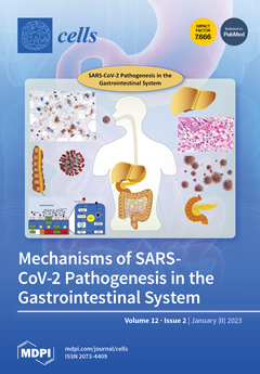
The novel coronavirus SARS-CoV-2 rapidly spread worldwide, causing a global pandemic. Although the respiratory system is the primary site of infection, growing evidence indicates the involvement of the gastrointestinal tract. ACE2 receptors are particularly abundant throughout the GI tract, making intestinal epithelial cells, liver, and pancreas extrapulmonary sites of infection and reservoir sites. Once inside these cells, the mechanisms of pathogenesis involve cytotoxic effects due to viral invasion and replication, tissue necrosis, and cell damage secondary to cytokine storm, dysregulation of autophagy, transport via extracellular vesicles/exosomes, increase in myeloid-derived suppressor cells, which inhibit the normal function of T-lymphocytes, dysbiosis, and ischemia secondary to thrombosis. View this paper
  • Issues are regarded as officially published after their release is announced to the table of contents alert mailing list.
  • You may sign up for e-mail alerts to receive table of contents of newly released issues.
  • PDF is the official format for papers published in both, html and pdf forms. To view the papers in pdf format, click on the "PDF Full-text" link, and use the free Adobe Reader to open them.
Order results
Result details
Section
Select all
Export citation of selected articles as:
27 pages, 8117 KiB  
Article
Multiomics Study of a Novel Naturally Derived Small Molecule, NSC772864, as a Potential Inhibitor of Proto-Oncogenes Regulating Cell Cycle Progression in Colorectal Cancer
by Ntlotlang Mokgautsi, Yu-Cheng Kuo, Chien-Hsin Chen, Yan-Jiun Huang, Alexander T. H. Wu and Hsu-Shan Huang
Cells 2023, 12(2), 340; https://doi.org/10.3390/cells12020340 - 16 Jan 2023
Cited by 1 | Viewed by 2302
Abstract
Colorectal cancer (CRC) is one of the most prevalent malignant tumors, and it contributes to high numbers of deaths globally. Although advances in understanding CRC molecular mechanisms have shed significant light on its pathogenicity, current treatment options, including combined chemotherapy and molecular-targeted agents, [...] Read more.
Colorectal cancer (CRC) is one of the most prevalent malignant tumors, and it contributes to high numbers of deaths globally. Although advances in understanding CRC molecular mechanisms have shed significant light on its pathogenicity, current treatment options, including combined chemotherapy and molecular-targeted agents, are still limited due to resistance, with almost 25% of patients developing distant metastasis. Therefore, identifying novel biomarkers for early diagnosis is crucial, as they will also influence strategies for new targeted therapies. The proto-oncogene, c-Met, a tyrosine kinase that promotes cell proliferation, motility, and invasion; c-MYC, a transcription factor associated with the modulation of the cell cycle, proliferation, apoptosis; and cyclin D1 (CCND1), an essential regulatory protein in the cell cycle, all play crucial roles in cancer progression. In the present study, we explored computational simulations through bioinformatics analysis and identified the overexpression of c-Met/GSK3β/MYC/CCND1 oncogenic signatures that were associated with cancer progression, drug resistance, metastasis, and poor clinical outcomes in CRC. We further demonstrated the anticancer activities of our newly synthesized quinoline-derived compound, NSC772864, against panels of the National Cancer Institute’s human CRC cell lines. The compound exhibited cytotoxic activities against various CRC cell lines. Using target prediction tools, we found that c-Met/GSK3β/MYC/CCND1 were target genes for the NSC772864 compound. Subsequently, we performed in silico molecular docking to investigate protein–ligand interactions and discovered that NSC772864 exhibited higher binding affinities with these oncogenes compared to FDA-approved drugs. These findings strongly suggest that NSC772864 is a novel and potential antiCRC agent. Full article
(This article belongs to the Section Cell Proliferation and Division)
Show Figures

Figure 1

14 pages, 4104 KiB  
Article
The Generation of Human iPSC Lines from Three Individuals with Dravet Syndrome and Characterization of Neural Differentiation Markers in iPSC-Derived Ventral Forebrain Organoid Model
by Valery Zayat, Zuzanna Kuczynska, Michal Liput, Erkan Metin, Sylwia Rzonca-Niewczas, Marta Smyk, Tomasz Mazurczak, Alicja Goszczanska-Ciuchta, Pawel Leszczynski, Dorota Hoffman-Zacharska and Leonora Buzanska
Cells 2023, 12(2), 339; https://doi.org/10.3390/cells12020339 - 16 Jan 2023
Cited by 2 | Viewed by 2467
Abstract
Dravet syndrome (DRVT) is a rare form of neurodevelopmental disorder with a high risk of sudden unexpected death in epilepsy (SUDEP), caused mainly (>80% cases) by mutations in the SCN1A gene, coding the Nav1.1 protein (alfa-subunit of voltage-sensitive sodium channel). Mutations in SCN1A [...] Read more.
Dravet syndrome (DRVT) is a rare form of neurodevelopmental disorder with a high risk of sudden unexpected death in epilepsy (SUDEP), caused mainly (>80% cases) by mutations in the SCN1A gene, coding the Nav1.1 protein (alfa-subunit of voltage-sensitive sodium channel). Mutations in SCN1A are linked to heterogenous epileptic phenotypes of various types, severity, and patient prognosis. Here we generated iPSC lines from fibroblasts obtained from three individuals affected with DRVT carrying distinct mutations in the SCN1A gene (nonsense mutation p.Ser1516*, missense mutation p.Arg1596His, and splicing mutation c.2589+2dupT). The iPSC lines, generated with the non-integrative approach, retained the distinct SCN1A gene mutation of the donor fibroblasts and were characterized by confirming the expression of the pluripotency markers, the three-germ layer differentiation potential, the absence of exogenous vector expression, and a normal karyotype. The generated iPSC lines were used to establish ventral forebrain organoids, the most affected type of neurons in the pathology of DRVT. The DRVT organoid model will provide an additional resource for deciphering the pathology behind Nav1.1 haploinsufficiency and drug screening to remediate the functional deficits associated with the disease. Full article
(This article belongs to the Special Issue Neural Stem Cells: Developmental Mechanisms and Disease Modelling)
Show Figures

Figure 1

15 pages, 867 KiB  
Review
Experimentally Created Magnetic Force in Microbiological Space and On-Earth Studies: Perspectives and Restrictions
by Svetlana A. Ermolaeva, Vladislav A. Parfenov, Pavel A. Karalkin, Yusef D. Khesuani and Pavel A. Domnin
Cells 2023, 12(2), 338; https://doi.org/10.3390/cells12020338 - 16 Jan 2023
Cited by 1 | Viewed by 1662
Abstract
Magnetic force and gravity are two fundamental forces affecting all living organisms, including bacteria. On Earth, experimentally created magnetic force can be used to counterbalance gravity and place living organisms in conditions of magnetic levitation. Under conditions of microgravity, magnetic force becomes the [...] Read more.
Magnetic force and gravity are two fundamental forces affecting all living organisms, including bacteria. On Earth, experimentally created magnetic force can be used to counterbalance gravity and place living organisms in conditions of magnetic levitation. Under conditions of microgravity, magnetic force becomes the only force that moves bacteria, providing an acceleration towards areas of the lowest magnetic field and locking cells in this area. In this review, we consider basic principles and experimental systems used to create a magnetic force strong enough to balance gravity. Further, we describe how magnetic levitation is applied in on-Earth microbiological studies. Next, we consider bacterial behavior under combined conditions of microgravity and magnetic force onboard a spacecraft. At last, we discuss restrictions on applications of magnetic force in microbiological studies and the impact of these restrictions on biotechnological applications under space and on-Earth conditions. Full article
(This article belongs to the Special Issue New Insights into Microgravity and Space Biology)
Show Figures

Figure 1

27 pages, 5570 KiB  
Article
Abnormal Cellular Phenotypes Induced by Three TMPO/LAP2 Variants Identified in Men with Cardiomyopathies
by Nathalie Vadrot, Flavie Ader, Maryline Moulin, Marie Merlant, Françoise Chapon, Estelle Gandjbakhch, Fabien Labombarda, Pascale Maragnes, Patricia Réant, Caroline Rooryck, Vincent Probst, Erwan Donal, Pascale Richard, Ana Ferreiro and Brigitte Buendia
Cells 2023, 12(2), 337; https://doi.org/10.3390/cells12020337 - 16 Jan 2023
Viewed by 1814
Abstract
A single missense variant of the TMPO/LAP2α gene, encoding LAP2 proteins, has been associated with cardiomyopathy in two brothers. To further evaluate its role in cardiac muscle, we included TMPO in our cardiomyopathy diagnostic gene panel. A screening of ~5000 patients revealed [...] Read more.
A single missense variant of the TMPO/LAP2α gene, encoding LAP2 proteins, has been associated with cardiomyopathy in two brothers. To further evaluate its role in cardiac muscle, we included TMPO in our cardiomyopathy diagnostic gene panel. A screening of ~5000 patients revealed three novel rare TMPO heterozygous variants in six males diagnosed with hypertrophic or dilated cardiomypathy. We identified in different cellular models that (1) the frameshift variant LAP2α p.(Gly395Glufs*11) induced haploinsufficiency, impeding cell proliferation and/or producing a truncated protein mislocalized in the cytoplasm; (2) the C-ter missense variant LAP2α p.(Ala240Thr) led to a reduced proximity events between LAP2α and the nucleosome binding protein HMGN5; and (3) the LEM-domain missense variant p.(Leu124Phe) decreased both associations of LAP2α/β with the chromatin-associated protein BAF and inhibition of the E2F1 transcription factor activity which is known to be dependent on Rb, partner of LAP2α. Additionally, the LAP2α expression was lower in the left ventricles of male mice compared to females. In conclusion, our study reveals distinct altered properties of LAP2 induced by these TMPO/LAP2 variants, leading to altered cell proliferation, chromatin structure or gene expression-regulation pathways, and suggests a potential sex-dependent role of LAP2 in myocardial function and disease. Full article
(This article belongs to the Collection Lamins and Laminopathies)
Show Figures

Figure 1

19 pages, 6627 KiB  
Article
Erk1/2-Dependent HNSCC Cell Susceptibility to Erastin-Induced Ferroptosis
by Dragana Savic, Teresa Bernadette Steinbichler, Julia Ingruber, Giulia Negro, Bertram Aschenbrenner, Herbert Riechelmann, Ute Ganswindt, Sergej Skvortsov, József Dudás and Ira-Ida Skvortsova
Cells 2023, 12(2), 336; https://doi.org/10.3390/cells12020336 - 16 Jan 2023
Cited by 4 | Viewed by 3289
Abstract
Unfavorable clinical outcomes mean that cancer researchers must attempt to develop novel therapeutic strategies to overcome therapeutic resistance in patients with HNSCC. Recently, ferroptosis was shown to be a promising pathway possessing druggable targets, such as xCT (SLC7A11). Unfortunately, little is known about [...] Read more.
Unfavorable clinical outcomes mean that cancer researchers must attempt to develop novel therapeutic strategies to overcome therapeutic resistance in patients with HNSCC. Recently, ferroptosis was shown to be a promising pathway possessing druggable targets, such as xCT (SLC7A11). Unfortunately, little is known about the molecular mechanisms underlying the susceptibility of HNSCC cells to ferroptosis. The goal of this study was to determine whether HNSCC cells with activated Erk1/2 are vulnerable to ferroptosis induction. Our results have shown that xCT (SLC7A11) was overexpressed in malignant tissues obtained from the patients with HNSCC, whereas normal mucosa demonstrated weak expression of the protein. In order to investigate the role of Erk1/2 in the decrease in cell viability caused by erastin, xCT-overexpressing FaDu and SCC25 HNSCC cells were used. The ravoxertinib-dependent inhibition of Erk1/2 signaling led to the decrease in erastin efficacy due to the effect on ROS production and the upregulation of ROS scavengers SOD1 and SOD2, resulting in repressed lipid peroxidation. Therefore, it was concluded that the erastin-dependent activation of ferroptosis seems to be a promising approach which can be further developed as an additional strategy for the treatment of HNSCC. As ferroptosis induction via erastin is strongly dependent on the expression of Erk1/2, this MAP kinase can be considered as a predictor for cancer cells’ response to erastin. Full article
(This article belongs to the Special Issue Novel Mechanisms and Therapeutic Opportunities of Ferroptosis)
Show Figures

Figure 1

20 pages, 6625 KiB  
Article
Vitamin D Receptor Expression Limits the Angiogenic and Inflammatory Properties of Retinal Endothelial Cells
by Yong-Seok Song, Nasim Jamali, Christine M. Sorenson and Nader Sheibani
Cells 2023, 12(2), 335; https://doi.org/10.3390/cells12020335 - 16 Jan 2023
Cited by 3 | Viewed by 2335
Abstract
The integrity of retinal endothelial cell (EC) is essential for establishing and maintaining the retinal blood barrier to ensure proper vision. Vitamin D is a hormone with known protective roles in EC function. The majority of vitamin D action is mediated through the [...] Read more.
The integrity of retinal endothelial cell (EC) is essential for establishing and maintaining the retinal blood barrier to ensure proper vision. Vitamin D is a hormone with known protective roles in EC function. The majority of vitamin D action is mediated through the vitamin D receptor (VDR). VDR is a nuclear receptor whose engagement by vitamin D impacts the expression of many genes with important roles in regulation of angiogenesis and inflammation. Although many studies have investigated vitamin D-VDR action in cardiovascular protection and tumor angiogenesis, its impact on retinal EC function and regulation of ocular angiogenesis and inflammation is exceedingly limited. We previously showed calcitriol, the active form of vitamin D, is a potent inhibitor of retinal neovascularization in vivo and retinal EC capillary morphogenesis in vitro. Here, using retinal EC prepared from wild-type (Vdr+/+) and VDR-deficient (Vdr−/−) mice, we show that retinal EC express VDR and its expression is induced by calcitriol. The lack of VDR expression had a significant impact on endothelial cell–cell and cell–matrix interactions. Vdr−/− retinal EC proliferated at a slower rate and were more adherent and less migratory. They also exhibited increased expression levels of inflammatory markers driven in part by sustained activation of STAT1 and NF-κB pathways and were more sensitive to oxidative challenge. These changes were attributed, in part, to down-regulation of endothelial nitric oxide synthetase, enhanced hepcidin expression, and increased intracellular iron levels. Taken together, our results indicate that VDR expression plays a fundamental role in maintaining the proper angiogenic and inflammatory state of retinal EC. Full article
(This article belongs to the Collection Functions of Nuclear Receptors)
Show Figures

Figure 1

16 pages, 3105 KiB  
Article
Stability and Requirement for Thiamin in a Cell Culture Feed Used to Produce New Biological Entities
by Alisa Schnellbächer and Aline Zimmer
Cells 2023, 12(2), 334; https://doi.org/10.3390/cells12020334 - 16 Jan 2023
Viewed by 1711
Abstract
Thiamin is susceptible to heat and oxidation, which is a concern for the development of concentrated and room temperature stable feeds used to produce recombinant proteins. Hence, it is critical to understand the reactivity and necessity of the vitamin in liquid feeds to [...] Read more.
Thiamin is susceptible to heat and oxidation, which is a concern for the development of concentrated and room temperature stable feeds used to produce recombinant proteins. Hence, it is critical to understand the reactivity and necessity of the vitamin in liquid feeds to be able to either develop mitigation strategies to stabilize the vitamin or to remove thiamin from formulations if it is unnecessary. LC-MS/MS was used to investigate thiamin stability in different liquid feed formulations and to identify thiamin degradation products. Results indicate oxidation of thiamin and interaction with amino acids, keto acids, and sulfur containing components. Thiamin necessity in feed was assessed during a fed batch experiment, focusing on cell performance and critical quality attributes of the produced recombinant proteins. The impact of thiamin depletion in the feed on the intra- and extracellular metabolome was investigated using untargeted LC-MS/MS. Results indicate that thiamin can be removed from the feed without affecting the performance or the intra- and extracellular metabolome of the tested cell lines. Overall, profound insights on thiamin reactivity and necessity are presented in this study, suggesting the removal of the dispensable and instable vitamin as a simple means for the development of next generation feeds used to produce therapeutic biological entities. Full article
(This article belongs to the Special Issue The Molecular Application of Mass Spectrometry in Health and Disease)
Show Figures

Figure 1

19 pages, 2397 KiB  
Review
The Multitasker Protein: A Look at the Multiple Capabilities of NUMB
by Sara M. Ortega-Campos and José Manuel García-Heredia
Cells 2023, 12(2), 333; https://doi.org/10.3390/cells12020333 - 15 Jan 2023
Cited by 3 | Viewed by 2834
Abstract
NUMB, a plasma membrane-associated protein originally described in Drosophila, is involved in determining cell function and fate during early stages of development. It is secreted asymmetrically in dividing cells, with one daughter cell inheriting NUMB and the other inheriting its antagonist, NOTCH. [...] Read more.
NUMB, a plasma membrane-associated protein originally described in Drosophila, is involved in determining cell function and fate during early stages of development. It is secreted asymmetrically in dividing cells, with one daughter cell inheriting NUMB and the other inheriting its antagonist, NOTCH. NUMB has been proposed as a polarizing agent and has multiple functions, including endocytosis and serving as an adaptor in various cellular pathways such as NOTCH, Hedgehog, and the P53-MDM2 axis. Due to its role in maintaining cellular homeostasis, it has been suggested that NUMB may be involved in various human pathologies such as cancer and Alzheimer’s disease. Further research on NUMB could aid in understanding disease mechanisms and advancing the field of personalized medicine and the development of new therapies. Full article
(This article belongs to the Special Issue Cancers: Genetics and Cellular Perspective)
Show Figures

Figure 1

16 pages, 10683 KiB  
Article
Effect of Proton Therapy on Tumor Cell Killing and Immune Microenvironment for Hepatocellular Carcinoma
by Miao-Fen Chen, Ping-Tsung Chen, Ching-Chuan Hsieh and Chih-Chi Wang
Cells 2023, 12(2), 332; https://doi.org/10.3390/cells12020332 - 15 Jan 2023
Cited by 5 | Viewed by 2471
Abstract
Radiotherapy with proton therapy (PT) has dosimetric advantages over photon therapy, which helps to enlarge the therapeutic window of radiotherapy for hepatocellular carcinoma (HCC). We evaluated the response of HCC to PT and examined the underlying mechanisms. The human liver cancer cell lines [...] Read more.
Radiotherapy with proton therapy (PT) has dosimetric advantages over photon therapy, which helps to enlarge the therapeutic window of radiotherapy for hepatocellular carcinoma (HCC). We evaluated the response of HCC to PT and examined the underlying mechanisms. The human liver cancer cell lines HepG2 and HuH7 and the murine liver cancer cell line Hepa1–6 were selected for cell and animal experiments to examine the response induced by PT irradiation. Biological changes and the immunological response following PT irradiation were examined. In vitro experiments showed no significant difference in cell survival following PT compared with photon radiotherapy. In a murine tumor model, the tumors were obviously smaller in size 12 days after PT irradiation. The underlying changes included increased DNA damage, upregulated IL-6 levels, and a regulated immune tumor microenvironment. Protein analysis in vitro and in vivo showed that PT increased the level of programmed cell death ligand 1 (PD-L1) expressed in tumor cells and recruited myeloid-derived suppressor cells (MDSCs). The increase in PD-L1 was positively correlated with the irradiation dose. In Hepa1–6 syngeneic mouse models, the combination of PT with anti-PD-L1 increased tumor growth delay compared with PT alone, which was associated with increased tumor-infiltrating T cells and attenuated MDSC recruitment in the microenvironment. Furthermore, when PT was applied to the primary HCC tumor, anti-PD-L1 antibody-treated mice showed smaller synchronous unirradiated tumors. In conclusion, the response of HCC to PT was determined by tumor cell killing and the immunological response in the tumor microenvironment. The combination with the anti-PD-L1 antibody to enhance antitumor immunity was responsible for the therapeutic synergism for HCC treated with PT. Based on our results, we suggest that PT combined with anti-PD-L1 may be a promising therapeutic policy for HCC. Full article
(This article belongs to the Section Cell Microenvironment)
Show Figures

Figure 1

17 pages, 6224 KiB  
Article
Induced Remodelling of Astrocytes In Vitro and In Vivo by Manipulation of Astrocytic RhoA Activity
by Cátia Domingos, Franziska E. Müller, Stefan Passlick, Dagmar Wachten, Evgeni Ponimaskin, Martin K. Schwarz, Susanne Schoch, André Zeug and Christian Henneberger
Cells 2023, 12(2), 331; https://doi.org/10.3390/cells12020331 - 15 Jan 2023
Cited by 1 | Viewed by 2450
Abstract
Structural changes of astrocytes and their perisynaptic processes occur in response to various physiological and pathophysiological stimuli. They are thought to profoundly affect synaptic signalling and neuron-astrocyte communication. Understanding the causal relationship between astrocyte morphology changes and their functional consequences requires experimental tools [...] Read more.
Structural changes of astrocytes and their perisynaptic processes occur in response to various physiological and pathophysiological stimuli. They are thought to profoundly affect synaptic signalling and neuron-astrocyte communication. Understanding the causal relationship between astrocyte morphology changes and their functional consequences requires experimental tools to selectively manipulate astrocyte morphology. Previous studies indicate that RhoA-related signalling can play a major role in controlling astrocyte morphology, but the direct effect of increased RhoA activity has not been documented in vitro and in vivo. Therefore, we established a viral approach to manipulate astrocytic RhoA activity. We tested if and how overexpression of wild-type RhoA, of a constitutively active RhoA mutant (RhoA-CA), and of a dominant-negative RhoA variant changes the morphology of cultured astrocytes. We found that astrocytic expression of RhoA-CA induced robust cytoskeletal changes and a withdrawal of processes in cultured astrocytes. In contrast, overexpression of other RhoA variants led to more variable changes of astrocyte morphology. These induced morphology changes were reproduced in astrocytes of the hippocampus in vivo. Importantly, astrocytic overexpression of RhoA-CA did not alter the branching pattern of larger GFAP-positive processes of astrocytes. This indicates that a prolonged increase of astrocytic RhoA activity leads to a distinct morphological phenotype in vitro and in vivo, which is characterized by an isolated reduction of fine peripheral astrocyte processes in vivo. At the same time, we identified a promising experimental approach for investigating the functional consequences of astrocyte morphology changes. Full article
(This article belongs to the Special Issue Astroglial (Patho)Physiology)
Show Figures

Figure 1

19 pages, 3118 KiB  
Article
Synovial Fluid Derived from Human Knee Osteoarthritis Increases the Viability of Human Adipose-Derived Stem Cells through Upregulation of FOSL1
by Hironori Kitajima, Takuya Sakamoto, Tetsuhiro Horie, Ayane Kuwano, Atsushi Fuku, Yasuhiko Taki, Yuka Nakamura, Ikuhiro Tanida, Hiroshi Sunami, Hiroaki Hirata, Yoshiyuki Tachi, Naoki Yamamoto, Yasuo Iida, Yasuhito Ishigaki, Sohsuke Yamada, Shigetaka Shimodaira, Yusuke Shimizu, Toru Ichiseki, Ayumi Kaneuji, Satoshi Osawa and Norio Kawaharaadd Show full author list remove Hide full author list
Cells 2023, 12(2), 330; https://doi.org/10.3390/cells12020330 - 15 Jan 2023
Cited by 1 | Viewed by 3409
Abstract
Knee osteoarthritis (Knee OA) is an irreversible condition that causes bone deformity and degeneration of the articular cartilage that comprises the joints, resulting in chronic pain and movement disorders. The administration of cultured adipose-derived stem cells (ADSCs) into the knee joint cavity improves [...] Read more.
Knee osteoarthritis (Knee OA) is an irreversible condition that causes bone deformity and degeneration of the articular cartilage that comprises the joints, resulting in chronic pain and movement disorders. The administration of cultured adipose-derived stem cells (ADSCs) into the knee joint cavity improves the clinical symptoms of Knee OA; however, the effect of synovial fluid (SF) filling the joint cavity on the injected ADSCs remains unclear. In this study, we investigated the effect of adding SF from Knee OA patients to cultured ADSCs prepared for therapeutic use in an environment that mimics the joint cavity. An increase in the viability of ADSCs was observed following the addition of SF. Gene expression profiling of SF-treated ADSCs using DNA microarrays revealed changes in several genes involved in cell survival. Of these genes, we focused on FOSL1, which is involved in the therapeutic effect of ADSCs and the survival and proliferation of cancer stem cells. We confirmed the upregulation of FOSL1 mRNA and protein expression using RT-PCR and western blot analysis, respectively. Next, we knocked down FOSL1 in ADSCs using siRNA and observed a decrease in cell viability, indicating the involvement of FOSL1 in the survival of ADSCs. Interestingly, in the knockdown cells, ADSC viability was also decreased by SF exposure. These results suggest that SF enhances cell viability by upregulating FOSL1 expression in ADSCs. For therapy using cultured ADSCs, the therapeutic effect of ADSCs may be further enhanced if an environment more conducive to the upregulation of FOSL1 expression in ADSCs can be established. Full article
(This article belongs to the Special Issue Gene and Cell Therapy in Regenerative Medicine)
Show Figures

Figure 1

9 pages, 1371 KiB  
Communication
Non-Steroidal Drug Interferences in a Quantitative Multisteroid LC-MS/MS Assay
by Valentin Braun, Hermann Stuppner and Christoph Seger
Cells 2023, 12(2), 329; https://doi.org/10.3390/cells12020329 - 15 Jan 2023
Cited by 1 | Viewed by 1423
Abstract
Screening for possible interferences from steroidal compounds other than the target analytes (endogenous or exogenous) is well established in LC-MS/MS assay development for steroid quantification in a routine clinical setting. However, interferences from non-steroidal substances have, hitherto, not been explored. After screening more [...] Read more.
Screening for possible interferences from steroidal compounds other than the target analytes (endogenous or exogenous) is well established in LC-MS/MS assay development for steroid quantification in a routine clinical setting. However, interferences from non-steroidal substances have, hitherto, not been explored. After screening more than 150 pharmaceuticals and their metabolites by analyzing commercial quality control samples from TDM analysis kits (Recipe, Chromsystems) with a multisteroid LC-MS/MS assay (protein precipitation followed by HybridSPE filtration, biphenyl column, methanol–water gradient with NH4F additive), we can report the finding of two newly discovered potential interferences from non-steroidal drugs. Antidepressant paroxetine (PX) was identified as an interference to 17-hydroxyprogesterone (17P), and α-hydroxytriazolam (α-OH-TZM)—a major metabolite of benzodiazepine triazolam (TZM)—was identified as an interference to aldosterone (ALDO). Despite different elemental and structural compositions and nominal masses, the M+1 isotopologues of PX and α-OH-TZM produced overlapping signals in ion traces monitored for the respective analytes (m/z 331 → 109/97 and 361→315/343, respectively). PX and TZM are frequently prescribed drugs, and their therapeutic ranges are far exceeding the reference ranges of 17P or ALDO (µmol vs nmol); therefore, these interferences should be considered clinically relevant. Striving for faster multi-analyte methods with high sample turnover, especially in the field of steroid quantification, can limit assay selectivity and specificity. Therefore, supported by the findings of this study, screening for potential interferences in multi-analyte LC-MS/MS method development should not cover only substances of the same class but also include a set of common drugs. Full article
(This article belongs to the Special Issue The Molecular Application of Mass Spectrometry in Health and Disease)
Show Figures

Figure 1

10 pages, 2957 KiB  
Article
Sex-Specific Whole-Transcriptome Analysis in the Cerebral Cortex of FAE Offspring
by Nitish K. Mishra, Pulastya Shrinath, Radhakrishna Rao and Pradeep K. Shukla
Cells 2023, 12(2), 328; https://doi.org/10.3390/cells12020328 - 15 Jan 2023
Cited by 2 | Viewed by 2346
Abstract
Fetal alcohol spectrum disorders (FASDs) are associated with systemic inflammation and neurodevelopmental abnormalities. Several candidate genes were found to be associated with fetal alcohol exposure (FAE)-associated behaviors, but a sex-specific complete transcriptomic analysis was not performed at the adult stage. Recent studies have [...] Read more.
Fetal alcohol spectrum disorders (FASDs) are associated with systemic inflammation and neurodevelopmental abnormalities. Several candidate genes were found to be associated with fetal alcohol exposure (FAE)-associated behaviors, but a sex-specific complete transcriptomic analysis was not performed at the adult stage. Recent studies have shown that they are regulated at the developmental stage. However, the sex-specific role of RNA in FAE offspring brain development and function has not been studied yet. Here, we carried out the first systematic RNA profiling by utilizing a high-throughput transcriptomic (RNA-seq) approach in response to FAE in the brain cortex of male and female offspring at adulthood (P60). Our RNA-seq data analysis suggests that the changes in RNA expression in response to FAE are marked sex-specific. We show that the genes Muc3a, Pttg1, Rec8, Clcnka, Capn11, and pnp2 exhibit significantly higher expression in the male offspring than in the female offspring at P60. FAE female mouse brain sequencing data also show an increased expression of Eno1, Tpm3, and Pcdhb2 compared to male offspring. We performed a pathway analysis using a commercial software package (Ingenuity Pathway Analysis). We found that the sex-specific top regulator genes (Rictor, Gaba, Fmri, Mlxipl) are highly associated with eIF2 (translation initiation), synaptogenesis (the formation of synapses between neurons in the nervous system), sirtuin (metabolic regulation), and estrogen receptor (involved in obesity, aging, and cancer) signaling. Taken together, our transcriptomic results demonstrate that FAE differentially alters RNA expression in the adult brain in a sex-specific manner. Full article
(This article belongs to the Special Issue Signaling Pathways in Alcohol Induced Inflammation)
Show Figures

Figure 1

13 pages, 1801 KiB  
Article
Increased Galectin-9 Levels Correlate with Disease Activity in Patients with DMARD-Naïve Rheumatoid Arthritis and Modulate the Secretion of MCP-1 and IL-6 from Synovial Fibroblasts
by Morten A. Nielsen, Ditte Køster, Akul Y. Mehta, Kristian Stengaard-Pedersen, Pierre Busson, Peter Junker, Kim Hørslev-Petersen, Merete Lund Hetland, Mikkel Østergaard, Malene Hvid, Hakon Leffler, Tue W. Kragstrup, Richard D. Cummings and Bent Deleuran
Cells 2023, 12(2), 327; https://doi.org/10.3390/cells12020327 - 15 Jan 2023
Cited by 2 | Viewed by 2269
Abstract
Background: Fibroblast-like synoviocytes (FLSs) are essential mediators in the expansive growth and invasiveness of rheumatoid synovitis, and patients with a fibroblastic-rich pauci-immune pathotype respond poorly to currently approved antirheumatic drugs. Galectin-9 (Gal-9) has been reported to directly modulate rheumatoid arthritis (RA) FLSs [...] Read more.
Background: Fibroblast-like synoviocytes (FLSs) are essential mediators in the expansive growth and invasiveness of rheumatoid synovitis, and patients with a fibroblastic-rich pauci-immune pathotype respond poorly to currently approved antirheumatic drugs. Galectin-9 (Gal-9) has been reported to directly modulate rheumatoid arthritis (RA) FLSs and to hold both pro- and anti-inflammatory properties. The objective of this study was to evaluate clinical and pathogenic aspects of Gal-9 in RA, combining national patient cohorts and cellular models. Methods: Soluble Gal-9 was measured in plasma from patients with newly diagnosed, treatment-naïve RA (n = 98). The disease activity score 28-joint count C-reactive protein (DAS28CRP) and total Sharp score were used to evaluate the disease course serially over a two-year period. Plasma and synovial fluid samples were examined for soluble Gal-9 in patients with established RA (n = 18). A protein array was established to identify Gal-9 binding partners in the extracellular matrix (ECM). Synovial fluid mononuclear cells (SFMCs), harvested from RA patients, were used to obtain synovial-fluid derived FLSs (SF-FLSs) (n = 7). FLSs from patients suffering from knee Osteoarthritis (OA) were collected from patients when undergoing joint replacement surgery (n = 5). Monocultures of SF-FLSs (n = 6) and autologous co-cultures of SF-FLSs and peripheral blood mononuclear cells (PBMCs) were cultured with and without a neutralizing anti-Gal-9 antibody (n = 7). The mono- and co-cultures were subsequently analyzed by flow cytometry, MTT assay, and ELISA. Results: Patients with early and established RA had persistently increased plasma levels of Gal-9 compared with healthy controls (HC). The plasma levels of Gal-9 were associated with disease activity and remained unaffected when adding a TNF-inhibitor to their standard treatment. Gal-9 levels were elevated in the synovial fluid of established RA patients with advanced disease, compared with corresponding plasma samples. Gal-9 adhered to fibronectin, laminin and thrombospondin, while not to interstitial collagens in the ECM protein array. In vitro, a neutralizing Gal-9 antibody decreased MCP-1 and IL-6 production from both RA FLSs and OA FLSs. In co-cultures of autologous RA FLSs and PBMCs, the neutralization of Gal-9 also decreased MCP-1 and IL-6 production, without affecting the proportion of inflammatory FLSs. Conclusions: In RA, pretreatment plasma Gal-9 levels in early RA were increased and correlated with clinical disease activity. Gal-9 levels remained increased despite a significant reduction in the disease activity score in patients with early RA. The in vitro neutralization of Gal-9 decreased both MCP-1 and IL-6 production in an inflammatory subset of RA FLSs. Collectively these findings indicate that the persistent overexpression of Gal-9 in RA may modulate synovial FLS activities and could be involved in the maintenance of subclinical disease activity in RA. Full article
(This article belongs to the Special Issue Galectins in Health and Disease)
Show Figures

Graphical abstract

13 pages, 3115 KiB  
Article
Reduced Cell–ECM Interactions in the EpiSC Colony Center Cause Heterogeneous Differentiation
by Kshitij Amar, Sanjoy Saha, Avishek Debnath, Chun Hung Weng, Arpan Roy, Kyu Young Han and Farhan Chowdhury
Cells 2023, 12(2), 326; https://doi.org/10.3390/cells12020326 - 15 Jan 2023
Viewed by 2486
Abstract
Mechanoregulation of cell–extracellular matrix (ECM) interactions are crucial for dictating pluripotent stem cell differentiation. However, not all pluripotent cells respond homogeneously which results in heterogeneous cell populations. When cells, such as mouse epiblast stem cells (EpiSCs), are cultured in clusters, the heterogeneity effect [...] Read more.
Mechanoregulation of cell–extracellular matrix (ECM) interactions are crucial for dictating pluripotent stem cell differentiation. However, not all pluripotent cells respond homogeneously which results in heterogeneous cell populations. When cells, such as mouse epiblast stem cells (EpiSCs), are cultured in clusters, the heterogeneity effect during differentiation is even more pronounced. While past studies implicated variations in signaling pathways to be the root cause of heterogeneity, the biophysical aspects of differentiation have not been thoroughly considered. Here, we demonstrate that the heterogeneity of EpiSC differentiation arises from differences in the colony size and varying degrees of interactions between cells within the colonies and the ECM. Confocal imaging demonstrates that cells in the colony periphery established good contact with the surface while the cells in the colony center were separated by an average of 1–2 µm from the surface. Traction force measurements of the cells within the EpiSC colonies show that peripheral cells generate large tractions while the colony center cells do not. A finite element modeling of EpiSC colonies shows that tractions generated by the cells at the colony periphery lift off the colony center preventing the colony center from undergoing differentiation. Together, our results demonstrate a biophysical regulation of heterogeneous EpiSC colony differentiation. Full article
(This article belongs to the Special Issue Mechanics of Stem Cells in Regenerative Medicine)
Show Figures

Figure 1

18 pages, 2399 KiB  
Article
Characterization of Hepatoma-Derived Growth Factor-Related Protein 2 Interactions with Heterochromatin
by Sarah C. Wistner, Ian A. MacDonald, Karly A. Stanley and Nathaniel A. Hathaway
Cells 2023, 12(2), 325; https://doi.org/10.3390/cells12020325 - 14 Jan 2023
Viewed by 1824
Abstract
The expression of genetic information is tightly controlled by chromatin regulatory proteins, including those in the heterochromatin gene repression family. Many of these regulatory proteins work together on the chromatin substrate to precisely regulate gene expression during mammalian development, giving rise to many [...] Read more.
The expression of genetic information is tightly controlled by chromatin regulatory proteins, including those in the heterochromatin gene repression family. Many of these regulatory proteins work together on the chromatin substrate to precisely regulate gene expression during mammalian development, giving rise to many different tissues in higher organisms from a fixed genomic template. Here we identify and characterize the interactions of two related heterochromatin regulatory proteins, heterochromatin protein 1 alpha (HP1α) and M-phase phosphoprotein 8 (MPP8), with hepatoma-derived growth factor-related protein 2 (HRP2). We find in biochemical experiments that HRP2 copurifies and co-sediments with heterochromatin-associated proteins, including HP1α and MPP8. Using the Chromatin in vivo Assay in multiple cell types, we demonstrate that HP1α-mediated gene repression dynamics are altered by the presence of HRP2. Furthermore, the knockout of HRP2 in MDA-MB-231 cells results in significant changes to chromatin structure and stability, which alter gene expression patterns. Here, we detail a mechanism by which HRP2 contributes to epigenetic transcriptional regulation through engagement with heterochromatin-associated proteins to stabilize the chromatin landscape and influence gene expression. Full article
(This article belongs to the Special Issue Heterochromatin and Tumorigenesis II)
Show Figures

Figure 1

44 pages, 6654 KiB  
Review
Molecular Modeling Insights into the Structure and Behavior of Integrins: A Review
by Igor Tvaroška, Stanislav Kozmon and Juraj Kóňa
Cells 2023, 12(2), 324; https://doi.org/10.3390/cells12020324 - 14 Jan 2023
Cited by 10 | Viewed by 3140
Abstract
Integrins are heterodimeric glycoproteins crucial to the physiology and pathology of many biological functions. As adhesion molecules, they mediate immune cell trafficking, migration, and immunological synapse formation during inflammation and cancer. The recognition of the vital roles of integrins in various diseases revealed [...] Read more.
Integrins are heterodimeric glycoproteins crucial to the physiology and pathology of many biological functions. As adhesion molecules, they mediate immune cell trafficking, migration, and immunological synapse formation during inflammation and cancer. The recognition of the vital roles of integrins in various diseases revealed their therapeutic potential. Despite the great effort in the last thirty years, up to now, only seven integrin-based drugs have entered the market. Recent progress in deciphering integrin functions, signaling, and interactions with ligands, along with advancement in rational drug design strategies, provide an opportunity to exploit their therapeutic potential and discover novel agents. This review will discuss the molecular modeling methods used in determining integrins’ dynamic properties and in providing information toward understanding their properties and function at the atomic level. Then, we will survey the relevant contributions and the current understanding of integrin structure, activation, the binding of essential ligands, and the role of molecular modeling methods in the rational design of antagonists. We will emphasize the role played by molecular modeling methods in progress in these areas and the designing of integrin antagonists. Full article
(This article belongs to the Special Issue PTM Rewired Protein Network in Diseases)
Show Figures

Graphical abstract

6 pages, 238 KiB  
Editorial
Circular RNAs: Non-Canonical Observations on Non-Canonical RNAs
by Brett W. Stringer, Laura Gantley and Simon J. Conn
Cells 2023, 12(2), 323; https://doi.org/10.3390/cells12020323 - 14 Jan 2023
Cited by 1 | Viewed by 1413
Abstract
The existence of circular RNA (circRNA) research in mainstream science can be attributed to the contemporary synergism of big data and keen attention to detail by several research groups worldwide. Since the re-emergence of these non-canonical RNA transcripts, seminal advances have been made [...] Read more.
The existence of circular RNA (circRNA) research in mainstream science can be attributed to the contemporary synergism of big data and keen attention to detail by several research groups worldwide. Since the re-emergence of these non-canonical RNA transcripts, seminal advances have been made in understanding their biogenesis, interactome, and functions in diverse fields and a myriad of human diseases. However, most research outputs to date have focused on the ability of highly stable circRNAs to interact with, and impact signalling through, microRNAs. This is likely to be the result of seminal papers in the field ascribing a few remarkable circRNAs as “miRNA sponges”. However, the stoichiometric ratio between the (often-lowly-expressed) circRNA and their (commonly-more-abundant) target is rarely in favour of a biologically relevant and functional consequence of these interactions. It is time for yet another revolution in circRNA research to uncover functions beyond their documented ability to bind miRNAs. This Special Issue aims to highlight non-canonical functions for this non-canonical family of RNA molecules. Full article
(This article belongs to the Special Issue Circular RNAs: Non-canonical Observations on Non-canonical RNAs)
24 pages, 1456 KiB  
Review
Novel Insights into the Role of Kras in Myeloid Differentiation: Engaging with Wnt/β-Catenin Signaling
by Noriko Yokoyama, Hitoshi Nakayama and Kazuhisa Iwabuchi
Cells 2023, 12(2), 322; https://doi.org/10.3390/cells12020322 - 14 Jan 2023
Cited by 2 | Viewed by 1939
Abstract
Cells of the HL-60 myeloid leukemia cell line can be differentiated into neutrophil-like cells by treatment with dimethyl sulfoxide (DMSO). The molecular mechanisms involved in this differentiation process, however, remain unclear. This review focuses on the differentiation of HL-60 cells. Although the Ras [...] Read more.
Cells of the HL-60 myeloid leukemia cell line can be differentiated into neutrophil-like cells by treatment with dimethyl sulfoxide (DMSO). The molecular mechanisms involved in this differentiation process, however, remain unclear. This review focuses on the differentiation of HL-60 cells. Although the Ras proteins, a group of small GTP-binding proteins, are ubiquitously expressed and highly homologous, each has specific molecular functions. Kras was shown to be essential for normal mouse development, whereas Hras and Nras are not. Kras knockout mice develop profound hematopoietic defects, indicating that Kras is required for hematopoiesis in adults. The Wnt/β-catenin signaling pathway plays a crucial role in regulating the homeostasis of hematopoietic cells. The protein β-catenin is a key player in the Wnt/β-catenin signaling pathway. A great deal of evidence shows that the Wnt/β-catenin signaling pathway is deregulated in malignant tumors, including hematological malignancies. Wild-type Kras acts as a tumor suppressor during DMSO-induced differentiation of HL-60 cells. Upon DMSO treatment, Kras translocates to the plasma membrane, and its activity is enhanced. Inhibition of Kras attenuates CD11b expression. DMSO also elevates levels of GSK3β phosphorylation, resulting in the release of unphosphorylated β-catenin from the β-catenin destruction complex and its accumulation in the cytoplasm. The accumulated β-catenin subsequently translocates into the nucleus. Inhibition of Kras attenuates Lef/Tcf-sensitive transcription activity. Thus, upon treatment of HL-60 cells with DMSO, wild-type Kras reacts with the Wnt/β-catenin pathway, thereby regulating the granulocytic differentiation of HL-60 cells. Wild-type Kras and the Wnt/β-catenin signaling pathway are activated sequentially, increasing the levels of expression of C/EBPα, C/EBPε, and granulocyte colony-stimulating factor (G-CSF) receptor. Full article
(This article belongs to the Special Issue Wnt Signaling in Development and Aging)
Show Figures

Figure 1

30 pages, 1747 KiB  
Review
De Novo Generation of Human Hematopoietic Stem Cells from Pluripotent Stem Cells for Cellular Therapy
by Jianyi Ding, Yongqin Li and Andre Larochelle
Cells 2023, 12(2), 321; https://doi.org/10.3390/cells12020321 - 14 Jan 2023
Cited by 8 | Viewed by 3355
Abstract
The ability to manufacture human hematopoietic stem cells (HSCs) in the laboratory holds enormous promise for cellular therapy of human blood diseases. Several differentiation protocols have been developed to facilitate the emergence of HSCs from human pluripotent stem cells (PSCs). Most approaches employ [...] Read more.
The ability to manufacture human hematopoietic stem cells (HSCs) in the laboratory holds enormous promise for cellular therapy of human blood diseases. Several differentiation protocols have been developed to facilitate the emergence of HSCs from human pluripotent stem cells (PSCs). Most approaches employ a stepwise addition of cytokines and morphogens to recapitulate the natural developmental process. However, these protocols globally lack clinical relevance and uniformly induce PSCs to produce hematopoietic progenitors with embryonic features and limited engraftment and differentiation capabilities. This review examines how key intrinsic cues and extrinsic environmental inputs have been integrated within human PSC differentiation protocols to enhance the emergence of definitive hematopoiesis and how advances in genomics set the stage for imminent breakthroughs in this field. Full article
Show Figures

Figure 1

10 pages, 2345 KiB  
Article
A Combination of Taurine and Caffeine in Stallion Semen Extender Positively Affects the Spermatozoa Parameters
by Marko Halo, Jr., Filip Tirpák, Tomáš Slanina, Katarína Tokárová, Martin Massányi, Lucia Dianová, Eva Mlyneková, Agnieszka Greń, Marko Halo and Peter Massányi
Cells 2023, 12(2), 320; https://doi.org/10.3390/cells12020320 - 14 Jan 2023
Viewed by 1541
Abstract
This study was aimed to determine the impact of different taurine and caffeine combinations on the motility, viability, and oxidative markers of chilled stallion spermatozoa. Each stallion semen sample was diluted in a ratio of 1:2, with various taurine and caffeine concentrations (2.5–7.5 [...] Read more.
This study was aimed to determine the impact of different taurine and caffeine combinations on the motility, viability, and oxidative markers of chilled stallion spermatozoa. Each stallion semen sample was diluted in a ratio of 1:2, with various taurine and caffeine concentrations (2.5–7.5 mg/mL taurine + 0.625–1.25 mg/mL caffeine) dissolved in a conventional extender. The control samples (CON) were prepared by diluting ejaculate only using the conventional extender. The motility was analyzed using a CASA system at different time intervals (0, 6, 12, 24, and 30 h) and the viability was evaluated using a mitochondrial toxicity test (MTT) performed at the end of the incubation at 5 °C. The liquid part of experimental samples was separated by centrifugation after 30 h of incubation and underwent the evaluation of oxidative stress via the quantification of markers ferric reducing ability of plasma (FRAP) and total oxidant status (TOS). The samples that were treated with a combination of taurine and caffeine significantly improved the motility parameters, mainly after 12, 24, and 30 h of incubation. Samples extended with combination of taurine and caffeine neither compromise viability nor alterations of redox status. The results of this study describe the combination of taurine and caffeine as an optimal supplement for improving the quality of stallion semen during chilled storage. Full article
(This article belongs to the Section Reproductive Cells and Development)
Show Figures

Figure 1

35 pages, 2686 KiB  
Review
Lymphangiogenesis Guidance Mechanisms and Therapeutic Implications in Pathological States of the Cornea
by Mehul Patnam, Sunil R. Dommaraju, Faisal Masood, Paula Herbst, Jin-Hong Chang, Wen-Yang Hu, Mark I. Rosenblatt and Dimitri T. Azar
Cells 2023, 12(2), 319; https://doi.org/10.3390/cells12020319 - 14 Jan 2023
Cited by 6 | Viewed by 1878
Abstract
Corneal lymphangiogenesis is one component of the neovascularization observed in several inflammatory pathologies of the cornea including dry eye disease and corneal graft rejection. Following injury, corneal (lymph)angiogenic privilege is impaired, allowing ingrowth of blood and lymphatic vessels into the previously avascular cornea. [...] Read more.
Corneal lymphangiogenesis is one component of the neovascularization observed in several inflammatory pathologies of the cornea including dry eye disease and corneal graft rejection. Following injury, corneal (lymph)angiogenic privilege is impaired, allowing ingrowth of blood and lymphatic vessels into the previously avascular cornea. While the mechanisms underlying pathological corneal hemangiogenesis have been well described, knowledge of the lymphangiogenesis guidance mechanisms in the cornea is relatively scarce. Various signaling pathways are involved in lymphangiogenesis guidance in general, each influencing one or multiple stages of lymphatic vessel development. Most endogenous factors that guide corneal lymphatic vessel growth or regression act via the vascular endothelial growth factor C signaling pathway, a central regulator of lymphangiogenesis. Several exogenous factors have recently been repurposed and shown to regulate corneal lymphangiogenesis, uncovering unique signaling pathways not previously known to influence lymphatic vessel guidance. A strong understanding of the relevant lymphangiogenesis guidance mechanisms can facilitate the development of targeted anti-lymphangiogenic therapeutics for corneal pathologies. In this review, we examine the current knowledge of lymphatic guidance cues, their regulation of inflammatory states in the cornea, and recently discovered anti-lymphangiogenic therapeutic modalities. Full article
Show Figures

Figure 1

19 pages, 4157 KiB  
Article
Long-Term Memory Formation in Drosophila Depends on the 3′UTR of CPEB Gene orb2
by Eugene N. Kozlov, Elena V. Tokmatcheva, Anastasia M. Khrustaleva, Eugene S. Grebenshchikov, Roman V. Deev, Rudolf A. Gilmutdinov, Lyubov A. Lebedeva, Mariya Zhukova, Elena V. Savvateeva-Popova, Paul Schedl and Yulii V. Shidlovskii
Cells 2023, 12(2), 318; https://doi.org/10.3390/cells12020318 - 14 Jan 2023
Cited by 1 | Viewed by 2439
Abstract
Activation of local translation in neurites in response to stimulation is an important step in the formation of long-term memory (LTM). CPEB proteins are a family of translation factors involved in LTM formation. The Drosophila CPEB protein Orb2 plays an important role in [...] Read more.
Activation of local translation in neurites in response to stimulation is an important step in the formation of long-term memory (LTM). CPEB proteins are a family of translation factors involved in LTM formation. The Drosophila CPEB protein Orb2 plays an important role in the development and function of the nervous system. Mutations of the coding region of the orb2 gene have previously been shown to impair LTM formation. We found that a deletion of the 3’UTR of the orb2 gene similarly results in loss of LTM in Drosophila. As a result of the deletion, the content of the Orb2 protein remained the same in the neuron soma, but significantly decreased in synapses. Using RNA immunoprecipitation followed by high-throughput sequencing, we detected more than 6000 potential Orb2 mRNA targets expressed in the Drosophila brain. Importantly, deletion of the 3′UTR of orb2 mRNA also affected the localization of the Csp, Pyd, and Eya proteins, which are encoded by putative mRNA targets of Orb2. Therefore, the 3′UTR of the orb2 mRNA is important for the proper localization of Orb2 and other proteins in synapses of neurons and the brain as a whole, providing a molecular basis for LTM formation. Full article
(This article belongs to the Special Issue New Advances in Synaptic Dysfunctions and Plasticity)
Show Figures

Figure 1

24 pages, 3524 KiB  
Review
A Structural View at Vaccine Development against M. tuberculosis
by Maria Romano, Flavia Squeglia, Eliza Kramarska, Giovanni Barra, Han-Gyu Choi, Hwa-Jung Kim, Alessia Ruggiero and Rita Berisio
Cells 2023, 12(2), 317; https://doi.org/10.3390/cells12020317 - 14 Jan 2023
Cited by 7 | Viewed by 3287
Abstract
Tuberculosis (TB) is still the leading global cause of death from an infectious bacterial agent. Limiting tuberculosis epidemic spread is therefore an urgent global public health priority. As stated by the WHO, to stop the spread of the disease we need a new [...] Read more.
Tuberculosis (TB) is still the leading global cause of death from an infectious bacterial agent. Limiting tuberculosis epidemic spread is therefore an urgent global public health priority. As stated by the WHO, to stop the spread of the disease we need a new vaccine, with better coverage than the current Mycobacterium bovis BCG vaccine. This vaccine was first used in 1921 and, since then, there are still no new licensed tuberculosis vaccines. However, there is extremely active research in the field, with a steep acceleration in the past decades, due to the advance of technologies and more rational vaccine design strategies. This review aims to gather latest updates in vaccine development in the various clinical phases and to underline the contribution of Structural Vaccinology (SV) to the development of safer and effective antigens. In particular, SV and the development of vaccine adjuvants is making the use of subunit vaccines, which are the safest albeit the less antigenic ones, an achievable goal. Indeed, subunit vaccines overcome safety concerns but need to be rationally re-engineered to enhance their immunostimulating effects. The larger availability of antigen structural information as well as a better understanding of the complex host immune response to TB infection is a strong premise for a further acceleration of TB vaccine development. Full article
(This article belongs to the Section Cellular Immunology)
Show Figures

Figure 1

13 pages, 2455 KiB  
Article
PTPN1 Deficiency Modulates BMPR2 Signaling and Induces Endothelial Dysfunction in Pulmonary Arterial Hypertension
by Md Khadem Ali, Xuefei Tian, Lan Zhao, Katharina Schimmel, Christopher J. Rhodes, Martin R. Wilkins, Mark R. Nicolls and Edda F. Spiekerkoetter
Cells 2023, 12(2), 316; https://doi.org/10.3390/cells12020316 - 14 Jan 2023
Cited by 1 | Viewed by 2274
Abstract
Bone morphogenic protein receptor 2 (BMPR2) expression and signaling are impaired in pulmonary arterial hypertension (PAH). How BMPR2 signaling is decreased in PAH is poorly understood. Protein tyrosine phosphatases (PTPs) play important roles in vascular remodeling in PAH. To identify whether PTPs modify [...] Read more.
Bone morphogenic protein receptor 2 (BMPR2) expression and signaling are impaired in pulmonary arterial hypertension (PAH). How BMPR2 signaling is decreased in PAH is poorly understood. Protein tyrosine phosphatases (PTPs) play important roles in vascular remodeling in PAH. To identify whether PTPs modify BMPR2 signaling, we used a siRNA-mediated high-throughput screening of 22,124 murine genes in mouse myoblastoma reporter cells using ID1 expression as readout for BMPR2 signaling. We further experimentally validated the top hit, PTPN1 (PTP1B), in healthy human pulmonary arterial endothelial cells (PAECs) either silenced by siRNA or exposed to hypoxia and confirmed its relevance to PAH by measuring PTPN1 levels in blood and PAECs collected from PAH patients. We identified PTPN1 as a novel regulator of BMPR2 signaling in PAECs, which is downregulated in the blood of PAH patients, and documented that downregulation of PTPN1 is linked to endothelial dysfunction in PAECs. These findings point to a potential involvement for PTPN1 in PAH and will aid in our understanding of the molecular mechanisms involved in the disease. Full article
Show Figures

Figure 1

24 pages, 12377 KiB  
Article
Gradient Magnetic Field Accelerates Division of E. coli Nissle 1917
by Svitlana Gorobets, Oksana Gorobets, Iryna Sharai, Tatyana Polyakova and Vitalii Zablotskii
Cells 2023, 12(2), 315; https://doi.org/10.3390/cells12020315 - 14 Jan 2023
Cited by 1 | Viewed by 2058
Abstract
Cell-cycle progression is regulated by numerous intricate endogenous mechanisms, among which intracellular forces and protein motors are central players. Although it seems unlikely that it is possible to speed up this molecular machinery by applying tiny external forces to the cell, we show [...] Read more.
Cell-cycle progression is regulated by numerous intricate endogenous mechanisms, among which intracellular forces and protein motors are central players. Although it seems unlikely that it is possible to speed up this molecular machinery by applying tiny external forces to the cell, we show that magnetic forcing of magnetosensitive bacteria reduces the duration of the mitotic phase. In such bacteria, the coupling of the cell cycle to the splitting of chains of biogenic magnetic nanoparticles (BMNs) provides a biological realization of such forcing. Using a static gradient magnetic field of a special spatial configuration, in probiotic bacteria E. coli Nissle 1917, we shortened the duration of the mitotic phase and thereby accelerated cell division. Thus, focused magnetic gradient forces exerted on the BMN chains allowed us to intervene in the processes of division and growth of bacteria. The proposed magnetic-based cell division regulation strategy can improve the efficiency of microbial cell factories and medical applications of magnetosensitive bacteria. Full article
(This article belongs to the Collection Magnetic Fields and Cells)
Show Figures

Figure 1

22 pages, 2870 KiB  
Review
MHC Class II Presentation in Autoimmunity
by Irina A. Ishina, Maria Y. Zakharova, Inna N. Kurbatskaia, Azad E. Mamedov, Alexey A. Belogurov, Jr. and Alexander G. Gabibov
Cells 2023, 12(2), 314; https://doi.org/10.3390/cells12020314 - 14 Jan 2023
Cited by 12 | Viewed by 6890
Abstract
Antigen presentation by major histocompatibility complex class II (MHC-II) molecules is crucial for eliciting an efficient immune response by CD4+ T cells and maintaining self-antigen tolerance. Some MHC-II alleles are known to be positively or negatively associated with the risk of the [...] Read more.
Antigen presentation by major histocompatibility complex class II (MHC-II) molecules is crucial for eliciting an efficient immune response by CD4+ T cells and maintaining self-antigen tolerance. Some MHC-II alleles are known to be positively or negatively associated with the risk of the development of different autoimmune diseases (ADs), including those characterized by the emergence of autoreactive T cells. Apparently, the MHC-II presentation of self-antigens contributes to the autoimmune T cell response, initiated through a breakdown of central tolerance to self-antigens in the thymus. The appearance of autoreactive T cell might be the result of (i) the unusual interaction between T cell receptors (TCRs) and self-antigens presented on MHC-II; (ii) the posttranslational modifications (PTMs) of self-antigens; (iii) direct loading of the self-antigen to classical MHC-II without additional nonclassical MHC assistance; (iv) the proinflammatory environment effect on MHC-II expression and antigen presentation; and (v) molecular mimicry between foreign and self-antigens. The peculiarities of the processes involved in the MHC-II-mediated presentation may have crucial importance in the elucidation of the mechanisms of triggering and developing ADs as well as for clarification on the protective effect of MHC-II alleles that are negatively associated with ADs. Full article
(This article belongs to the Special Issue Major Histocompatibility Complex (MHC) in Health and Disease 2022)
Show Figures

Figure 1

27 pages, 8226 KiB  
Article
Triphasic 3D In Vitro Model of Bone-Tendon-Muscle Interfaces to Study Their Regeneration
by Wendy Balestri, Graham J. Hickman, Robert H. Morris, John A. Hunt and Yvonne Reinwald
Cells 2023, 12(2), 313; https://doi.org/10.3390/cells12020313 - 13 Jan 2023
Cited by 4 | Viewed by 2329
Abstract
The transition areas between different tissues, known as tissue interfaces, have limited ability to regenerate after damage, which can lead to incomplete healing. Previous studies focussed on single interfaces, most commonly bone-tendon and bone-cartilage interfaces. Herein, we develop a 3D in vitro model [...] Read more.
The transition areas between different tissues, known as tissue interfaces, have limited ability to regenerate after damage, which can lead to incomplete healing. Previous studies focussed on single interfaces, most commonly bone-tendon and bone-cartilage interfaces. Herein, we develop a 3D in vitro model to study the regeneration of the bone-tendon-muscle interface. The 3D model was prepared from collagen and agarose, with different concentrations of hydroxyapatite to graduate the tissues from bones to muscles, resulting in a stiffness gradient. This graduated structure was fabricated using indirect 3D printing to provide biologically relevant surface topographies. MG-63, human dermal fibroblasts, and Sket.4U cells were found suitable cell models for bones, tendons, and muscles, respectively. The biphasic and triphasic hydrogels composing the 3D model were shown to be suitable for cell growth. Cells were co-cultured on the 3D model for over 21 days before assessing cell proliferation, metabolic activity, viability, cytotoxicity, tissue-specific markers, and matrix deposition to determine interface formations. The studies were conducted in a newly developed growth chamber that allowed cell communication while the cell culture media was compartmentalised. The 3D model promoted cell viability, tissue-specific marker expression, and new matrix deposition over 21 days, thereby showing promise for the development of new interfaces. Full article
Show Figures

Figure 1

22 pages, 43539 KiB  
Article
PDE6D Mediates Trafficking of Prenylated Proteins NIM1K and UBL3 to Primary Cilia
by Siebren Faber, Stef J. F. Letteboer, Katrin Junger, Rossano Butcher, Trinadh V. Satish Tammana, Sylvia E. C. van Beersum, Marius Ueffing, Rob W. J. Collin, Qin Liu, Karsten Boldt and Ronald Roepman
Cells 2023, 12(2), 312; https://doi.org/10.3390/cells12020312 - 13 Jan 2023
Cited by 3 | Viewed by 2277
Abstract
Mutations in PDE6D impair the function of its cognate protein, phosphodiesterase 6D (PDE6D), in prenylated protein trafficking towards the ciliary membrane, causing the human ciliopathy Joubert Syndrome (JBTS22) and retinal degeneration in mice. In this study, we purified the prenylated cargo of PDE6D [...] Read more.
Mutations in PDE6D impair the function of its cognate protein, phosphodiesterase 6D (PDE6D), in prenylated protein trafficking towards the ciliary membrane, causing the human ciliopathy Joubert Syndrome (JBTS22) and retinal degeneration in mice. In this study, we purified the prenylated cargo of PDE6D by affinity proteomics to gain insight into PDE6D-associated disease mechanisms. By this approach, we have identified a specific set of PDE6D-interacting proteins that are involved in photoreceptor integrity, GTPase activity, nuclear import, or ubiquitination. Among these interacting proteins, we identified novel ciliary cargo proteins of PDE6D, including FAM219A, serine/threonine-protein kinase NIM1 (NIM1K), and ubiquitin-like protein 3 (UBL3). We show that NIM1K and UBL3 localize inside the cilium in a prenylation-dependent manner. Furthermore, UBL3 also localizes in vesicle-like structures around the base of the cilium. Through affinity proteomics of UBL3, we confirmed its strong interaction with PDE6D and its association with proteins that regulate small extracellular vesicles (sEVs) and ciliogenesis. Moreover, we show that UBL3 localizes in specific photoreceptor cilium compartments in a prenylation-dependent manner. Therefore, we propose that UBL3 may play a role in the sorting of proteins towards the photoreceptor outer segment, further explaining the development of PDE6D-associated retinal degeneration. Full article
Show Figures

Graphical abstract

16 pages, 3825 KiB  
Review
Implications of Crosstalk between Exosome-Mediated Ferroptosis and Diseases for Pathogenesis and Treatment
by Zixuan Zhou, Benshuai You, Cheng Ji, Leilei Zhang, Feng Wu and Hui Qian
Cells 2023, 12(2), 311; https://doi.org/10.3390/cells12020311 - 13 Jan 2023
Cited by 8 | Viewed by 2875
Abstract
Ferroptosis is a type of iron-dependent cell death caused by ferrous iron overload, reactive oxygen species generation through the Fenton reaction, and lipid peroxidation, leading to antioxidative system dysfunction and, ultimately, cell membrane damage. The functional role of ferroptosis in human physiology and [...] Read more.
Ferroptosis is a type of iron-dependent cell death caused by ferrous iron overload, reactive oxygen species generation through the Fenton reaction, and lipid peroxidation, leading to antioxidative system dysfunction and, ultimately, cell membrane damage. The functional role of ferroptosis in human physiology and pathology is considered a cause or consequence of diseases. Circulating exosomes mediate intercellular communication and organ crosstalk. They not only transport functional proteins and nucleic acids derived from parental cells but also serve as vehicles for the targeted delivery of exogenous cargo. Exosomes regulate ferroptosis by delivering the biological material to the recipient cell, affecting ferroptosis-related proteins, or transporting ferritin-bound iron out of the cell. This review discusses pathogenesis mediated by endogenous exosomes and the therapeutic potential of exogenous exosomes for ferroptosis-related diseases. In addition, this review explores the role of exosome-mediated ferroptosis in ferroptosis-related diseases with an emphasis on strategies for engineering exosomes for ferroptosis therapy. Full article
Show Figures

Figure 1

Previous Issue
Back to TopTop