Next Issue
Volume 12, February-2
Previous Issue
Volume 12, January-2


Cells, Volume 12, Issue 3 (February-1 2023) – 174 articles

Cover Story (view full-size image): 
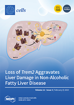
Genetic ablation of the triggering receptors expressed in myeloid cells 2 (Trem2) in mice leads to an impaired response to lipopolysaccharide (LPS)-laden triglyceride-rich lipoprotein particles (TLR) in vivo and reduced clearance of apoptotic hepatocytes by liver macrophages in vitro. Blocking the engagement of TREM2, and therefore its synergy with the LPS co-receptor CD14, contributes to the generation of a pathological environment characterized by the secretion of pro-inflammatory cytokines such as IL-6, TNFα and IL-1β. Hence, in a murine model of non-alcoholic fatty liver disease (NAFLD), Trem2 deficiency increases alanine transaminase (ALT) levels and fibrotic markers, and thus aggravates liver damage. View this paper
  • Issues are regarded as officially published after their release is announced to the table of contents alert mailing list.
  • You may sign up for e-mail alerts to receive table of contents of newly released issues.
  • PDF is the official format for papers published in both, html and pdf forms. To view the papers in pdf format, click on the "PDF Full-text" link, and use the free Adobe Reader to open them.
Order results
Result details
Section
Select all
Export citation of selected articles as:
20 pages, 3079 KiB  
Review
Purification of Myosin from Bovine Tracheal Smooth Muscle, Filament Formation and Endogenous Association of Its Regulatory Complex
by Lu Wang, Isabel J. Sobieszek, Chun Y. Seow and Apolinary Sobieszek
Cells 2023, 12(3), 514; https://doi.org/10.3390/cells12030514 - 03 Feb 2023
Viewed by 1515
Abstract
Dynamic regulation of myosin filaments is a crucial factor in the ability of airway smooth muscle (ASM) to adapt to a wide length range. Increased stability or robustness of myosin filaments may play a role in the pathophysiology of asthmatic airways. Biochemical techniques [...] Read more.
Dynamic regulation of myosin filaments is a crucial factor in the ability of airway smooth muscle (ASM) to adapt to a wide length range. Increased stability or robustness of myosin filaments may play a role in the pathophysiology of asthmatic airways. Biochemical techniques for the purification of myosin and associated regulatory proteins could help elucidate potential alterations in myosin filament properties of asthmatic ASM. An effective myosin purification approach was originally developed for chicken gizzard smooth muscle myosin. More recently, we successfully adapted the procedure to bovine tracheal smooth muscle. This method yields purified myosin with or without the endogenous regulatory complex of myosin light chain kinase and myosin light chain phosphatase. The tight association of the regulatory complex with the assembled myosin filaments can be valuable in functional experiments. The purification protocol discussed here allows for enzymatic comparisons of myosin regulatory proteins. Furthermore, we detail the methodology for quantification and removal of the co-purified regulatory enzymes as a tool for exploring potentially altered phenotypes of the contractile apparatus in diseases such as asthma. Full article
(This article belongs to the Special Issue Airway Smooth Muscle and Asthma)
Show Figures

Figure 1

19 pages, 3114 KiB  
Article
Pharmacological Targeting of the RAGE-NFκB Signalling Axis Impedes Monocyte Activation under Diabetic Conditions through the Repression of SHP-2 Tyrosine Phosphatase Function
by Marc Dorenkamp, Madina Nasiry, Dilvin Semo, Sybille Koch, Ivonne Löffler, Gunter Wolf, Holger Reinecke and Rinesh Godfrey
Cells 2023, 12(3), 513; https://doi.org/10.3390/cells12030513 - 03 Feb 2023
Cited by 3 | Viewed by 1896
Abstract
Monocytes play a vital role in the development of cardiovascular diseases. Type 2 diabetes mellitus (T2DM) is a major CVD risk factor, and T2DM-induced aberrant activation and enhanced migration of monocytes is a vital pathomechanism that leads to atherogenesis. We recently reported the [...] Read more.
Monocytes play a vital role in the development of cardiovascular diseases. Type 2 diabetes mellitus (T2DM) is a major CVD risk factor, and T2DM-induced aberrant activation and enhanced migration of monocytes is a vital pathomechanism that leads to atherogenesis. We recently reported the upregulation of SHP-2 phosphatase expression in mediating the VEGF resistance of T2DM patient-derived monocytes or methylglyoxal- (MG, a glucose metabolite and advanced glycation end product (AGE) precursor) treated monocytes. However, the exact mechanisms leading to SHP-2 upregulation in hyperglycemic monocytes are unknown. Since inflammation and accumulation of AGEs is a hallmark of T2DM, we hypothesise that inflammation and AGE-RAGE (Receptor-for-AGEs) signalling drive SHP-2 expression in monocytes and blockade of these pathways will repress SHP-2 function. Indeed, monocytes from T2DM patients revealed an elevated SHP-2 expression. Under normoglycemic conditions, the serum from T2DM patients strongly induced SHP-2 expression, indicating that the T2DM serum contains critical factors that directly regulate SHP-2 expression. Activation of pro-inflammatory TNFα signalling cascade drove SHP-2 expression in monocytes. In line with this, linear regression analysis revealed a significant positive correlation between TNFα expression and SHP-2 transcript levels in T2DM monocytes. Monocytes exposed to MG or AGE mimetic AGE-BSA, revealed an elevated SHP-2 expression and co-treatment with an NFκB inhibitor or genetic inhibition of p65 reversed it. The pharmacological inhibition of RAGE was sufficient to block MG- or AGE-BSA-induced SHP-2 expression and activity. Confirming the importance of RAGE-NFκB signalling in regulating SHP-2 expression, the elevated binding of NFκB to the SHP-2 promoter—induced by MG or AGE-BSA—was reversed by RAGE and NFκB inhibition. Besides, we detected elevated RAGE levels in human and murine T2DM monocytes and monocytes exposed to MG or AGE-BSA. Importantly, MG and AGE-BSA treatment of non-T2DM monocytes phenocopied the aberrant pro-migratory phenotype of T2DM monocytes, which was reversed entirely by either SHP-2- or RAGE inhibition. In conclusion, these findings suggest a new therapeutic approach to prevent accelerated atherosclerosis in T2DM patients since inhibiting the RAGE-NFκB-SHP-2 axis impeded the T2DM-driven, SHP-2-dependent monocyte activation. Full article
Show Figures

Figure 1

11 pages, 5067 KiB  
Review
The Role of TP53 in Adaptation and Evolution
by Konstantinos Voskarides and Nefeli Giannopoulou
Cells 2023, 12(3), 512; https://doi.org/10.3390/cells12030512 - 03 Feb 2023
Cited by 12 | Viewed by 3646
Abstract
The TP53 gene is a major player in cancer formation, and it is considered the most important tumor suppressor gene. The p53 protein acts as a transcription factor, and it is involved in DNA repair, senescence, cell-cycle control, autophagy, and apoptosis. Beyond cancer, [...] Read more.
The TP53 gene is a major player in cancer formation, and it is considered the most important tumor suppressor gene. The p53 protein acts as a transcription factor, and it is involved in DNA repair, senescence, cell-cycle control, autophagy, and apoptosis. Beyond cancer, there is evidence that TP53 is associated with fertility, aging, and longevity. Additionally, more evidence exists that genetic variants in TP53 are associated with environmental adaptation. Special TP53 amino-acid residues or pathogenic TP53 mutations seem to be adaptive for animals living in hypoxic and cold environments or having been exposed to starvation, respectively. At the somatic level, it has recently been proven that multiple cancer genes, including TP53, are under positive selection in healthy human tissues. It is not clear why these driver mutations do not transform these tissues into cancerous ones. Other studies have shown that elephants have multiple TP53 copies, probably this being the reason for the very low cancer incidence in these large animals. This may explain the famous Peto’s paradox. This review discusses in detail the multilevel role of TP53 in adaptation, according to the published evidence. This role is complicated, and it extends from cells to individuals and to populations. Full article
(This article belongs to the Special Issue Role of TP53 Gene in Preventing Cancer and Promoting Adaptation)
Show Figures

Figure 1

15 pages, 3632 KiB  
Article
Endothelial Specific Deletion of Autotaxin Improves Stroke Outcomes
by Susmita Bhattarai, Utsab Subedi, Shrivats Manikandan, Sudha Sharma, Papori Sharma, Chloe Miller, Md Shenuarin Bhuiyan, Srivatsan Kidambi, Vassilis Aidinis, Hong Sun, Sumitra Miriyala and Manikandan Panchatcharam
Cells 2023, 12(3), 511; https://doi.org/10.3390/cells12030511 - 03 Feb 2023
Cited by 6 | Viewed by 1943
Abstract
Autotaxin (ATX) is an extracellular secretory enzyme (lysophospholipase D) that catalyzes the hydrolysis of lysophosphatidyl choline to lysophosphatidic acid (LPA). The ATX–LPA axis is a well-known pathological mediator of liver fibrosis, metastasis in cancer, pulmonary fibrosis, atherosclerosis, and neurodegenerative diseases. Additionally, it is [...] Read more.
Autotaxin (ATX) is an extracellular secretory enzyme (lysophospholipase D) that catalyzes the hydrolysis of lysophosphatidyl choline to lysophosphatidic acid (LPA). The ATX–LPA axis is a well-known pathological mediator of liver fibrosis, metastasis in cancer, pulmonary fibrosis, atherosclerosis, and neurodegenerative diseases. Additionally, it is believed that LPA may cause vascular permeability. In ischemic stroke, vascular permeability leading to hemorrhagic transformation is a major limitation for therapies and an obstacle to stroke management. Therefore, in this study, we generated an endothelial-specific ATX deletion in mice (ERT2 ATX−/−) to observe stroke outcomes in a mouse stroke model to analyze the role of endothelial ATX. The AR2 probe and Evans Blue staining were used to perform the ATX activity and vascular permeability assays, respectively. Laser speckle imaging was used to observe the cerebral blood flow following stroke. In this study, we observed that stroke outcomes were alleviated with the endothelial deletion of ATX. Permeability and infarct volume were reduced in ERT2 ATX−/− mice compared to ischemia–reperfusion (I/R)-only mice. In addition, the cerebral blood flow was retained in ERT2 ATX−/− compared to I/R mice. The outcomes in the stroke model are alleviated due to the limited LPA concentration, reduced ATX concentration, and ATX activity in ERT2 ATX−/− mice. This study suggests that endothelial-specific ATX leads to increased LPA in the brain vasculature following ischemic–reperfusion and ultimately disrupts vascular permeability, resulting in adverse stroke outcomes. Full article
Show Figures

Figure 1

18 pages, 12615 KiB  
Article
Increased Nuclear FOXP2 Is Related to Reduced Neural Stem Cell Number and Increased Neurogenesis in the Dorsal Telencephalon of Embryos of Diabetic Rats through Histamine H1 Receptors
by Diana Sarahi De la Merced-García, Ángel Sánchez-Barrera, Juan Hernández-Yonca, Ismael Mancilla, Guadalupe García-López, Néstor Fabián Díaz, Luis Ignacio Terrazas and Anayansi Molina-Hernández
Cells 2023, 12(3), 510; https://doi.org/10.3390/cells12030510 - 03 Feb 2023
Viewed by 1676
Abstract
Diabetic rat embryos have increased cortical neurogenesis and neuron maturation, and their offspring presented altered neuron polarity, lamination, and diminished neuron excitability. The FOXP2 overexpression results in higher cortical neurogenesis by increasing the transition of radial glia to the intermediate progenitor. Similarly, histamine [...] Read more.
Diabetic rat embryos have increased cortical neurogenesis and neuron maturation, and their offspring presented altered neuron polarity, lamination, and diminished neuron excitability. The FOXP2 overexpression results in higher cortical neurogenesis by increasing the transition of radial glia to the intermediate progenitor. Similarly, histamine through H1-receptor activation increases cortical neuron differentiation. Indeed, blocking the H1-receptor by the systemic administration of chlorpheniramine to diabetic pregnant rats prevents increased neurogenesis. Here, we explore the relationship between the H1-receptor and FOXP2 on embryo neurogenesis from diabetic dams. Through qRT-PCR, Western blot, immunohistofluorescence, and flow cytometry, we showed an increased FOXP2 expression and nuclear localization, a reduced Nestin expression and -positive cells number, and a higher PKCα expression in the cortical neuroepithelium of fourteen-day-old embryos from diabetic rats. Interestingly, this scenario was prevented by the chlorpheniramine systemic administration to diabetic pregnant rats at embryo day twelve. These data, together with the bioinformatic analysis, suggest that higher H1-receptor activity in embryos under high glucose increases FOXP2 nuclear translocation, presumably through PKCα phosphorylation, impairing the transition of radial glia to intermediate progenitor and increasing neuron differentiation in embryos of diabetic rats. Full article
(This article belongs to the Special Issue Neural Differentiation and Development)
Show Figures

Figure 1

2 pages, 176 KiB  
Editorial
Mesenchymal Stromal Cells “Think” Globally, but Act Locally
by Günther Eissner
Cells 2023, 12(3), 509; https://doi.org/10.3390/cells12030509 - 03 Feb 2023
Cited by 1 | Viewed by 985
Abstract
In this Special Issue of Cells, entitled “Immunomodulation by Mesenchymal Stem Cells 2020”, you can find five excellent papers on the role of mesenchymal stem/stromal cells (MSCs) in immunomodulation, which also includes regenerative processes, such as wound healing [...] Full article
(This article belongs to the Special Issue Immunomodulation by Mesenchymal Stem Cells 2020)
16 pages, 3011 KiB  
Article
Novel Zebrafish Patient-Derived Tumor Xenograft Methodology for Evaluating Efficacy of Immune-Stimulating BCG Therapy in Urinary Bladder Cancer
by Saskia Kowald, Ylva Huge, Decky Tandiono, Zaheer Ali, Gabriela Vazquez-Rodriguez, Anna Erkstam, Anna Fahlgren, Amir Sherif, Yihai Cao and Lasse D. Jensen
Cells 2023, 12(3), 508; https://doi.org/10.3390/cells12030508 - 03 Feb 2023
Cited by 3 | Viewed by 2375
Abstract
Background: Bacillus Calmette-Guérin (BCG) immunotherapy is the standard-of-care adjuvant therapy for non-muscle-invasive bladder cancer in patients at considerable risk of disease recurrence. Although its exact mechanism of action is unknown, BCG significantly reduces this risk in responding patients but is mainly associated with [...] Read more.
Background: Bacillus Calmette-Guérin (BCG) immunotherapy is the standard-of-care adjuvant therapy for non-muscle-invasive bladder cancer in patients at considerable risk of disease recurrence. Although its exact mechanism of action is unknown, BCG significantly reduces this risk in responding patients but is mainly associated with toxic side-effects in those facing treatment resistance. Methods that allow the identification of BCG responders are, therefore, urgently needed. Methods: Fluorescently labelled UM-UC-3 cells and dissociated patient tumor samples were used to establish zebrafish tumor xenograft (ZTX) models. Changes in the relative primary tumor size and cell dissemination to the tail were evaluated via fluorescence microscopy at three days post-implantation. The data were compared to the treatment outcomes of the corresponding patients. Toxicity was evaluated based on gross morphological evaluation of the treated zebrafish larvae. Results: BCG-induced toxicity was avoided by removing the water-soluble fraction of the BCG formulation prior to use. BCG treatment via co-injection with the tumor cells resulted in significant and dose-dependent primary tumor size regression. Heat-inactivation of BCG decreased this effect, while intravenous BCG injections were ineffective. ZTX models were successfully established for six of six patients based on TUR-B biopsies. In two of these models, significant tumor regression was observed, which, in both cases, corresponded to the treatment response in the patients. Conclusions: The observed BCG-related anti-tumor effect indicates that ZTX models might predict the BCG response and thereby improve treatment planning. More experiments and clinical studies are needed, however, to elucidate the BCG mechanism and estimate the predictive value. Full article
(This article belongs to the Collection Advances in 3D Cell Culture)
Show Figures

Figure 1

22 pages, 825 KiB  
Article
Exploring the Interactions of Oncolytic Viral Therapy and Immunotherapy of Anti-CTLA-4 for Malignant Melanoma Mice Model
by Jui-Ling Yu, Sophia R.-J. Jang and Kwei-Yan Liu
Cells 2023, 12(3), 507; https://doi.org/10.3390/cells12030507 - 03 Feb 2023
Cited by 2 | Viewed by 2055
Abstract
Oncolytic ability to direct target and lyse tumor cells makes oncolytic virus therapy (OVT) a promising approach to treating cancer. Despite its therapeutic potential to stimulate anti-tumor immune responses, it also has immunosuppressive effects. The efficacy of OVTs as monotherapies can be enhanced [...] Read more.
Oncolytic ability to direct target and lyse tumor cells makes oncolytic virus therapy (OVT) a promising approach to treating cancer. Despite its therapeutic potential to stimulate anti-tumor immune responses, it also has immunosuppressive effects. The efficacy of OVTs as monotherapies can be enhanced by appropriate adjuvant therapy such as anti-CTLA-4. In this paper, we propose a mathematical model to explore the interactions of combined therapy of oncolytic viruses and a checkpoint inhibitor, anti-CTLA-4. The model incorporates both the susceptible and infected tumor populations, natural killer cell population, virus population, tumor-specific immune populations, virus-specific immune populations, tumor suppressive cytokine IFN-g, and the effect of immune checkpoint inhibitor CTLA-4. In particular, we distinguish the tumor-specific immune abilities of CD8+ T, NK cells, and CD4+ T cells and describe the destructive ability of cytokine on tumor cells as well as the inhibitory capacity of CTLA-4 on various components. Our model is validated through the experimental results. We also investigate various dosing strategies to improve treatment outcomes. Our study reveals that tumor killing rate by cytokines, cytokine decay rate, and tumor growth rate play important roles on both the OVT monotherapy and the combination therapy. Moreover, parameters related to CD8+ T cell killing have a large impact on treatment outcomes with OVT alone, whereas parameters associated with IFN-g strongly influence treatment responses for the combined therapy. We also found that virus killing by NK cells may halt the desired spread of OVs and enhance the probability of tumor escape during the treatment. Our study reveals that it is the activation of host anti-tumor immune system responses rather than its direct destruction of the tumor cells plays a major biological function of the combined therapy. Full article
(This article belongs to the Special Issue Immunogenic Cell Death in Cancer and Infectious Disease)
Show Figures

Figure 1

19 pages, 2754 KiB  
Article
Cancer Stem Cell and Aggressiveness Traits Are Promoted by Stable Endothelin-Converting Enzyme-1c in Glioblastoma Cells
by Ignacio Niechi, José I. Erices, Diego Carrillo-Beltrán, Atenea Uribe-Ojeda, Ángelo Torres, José Dellis Rocha, Daniel Uribe, María A. Toro, Karla Villalobos-Nova, Belén Gaete-Ramírez, Gabriel Mingo, Gareth I. Owen, Manuel Varas-Godoy, Lilian Jara, Francisco Aguayo, Verónica A. Burzio, Claudia Quezada-Monrás and Julio C. Tapia
Cells 2023, 12(3), 506; https://doi.org/10.3390/cells12030506 - 03 Feb 2023
Cited by 1 | Viewed by 2332
Abstract
Glioblastoma (GBM) is the most common and aggressive type of brain tumor due to its elevated recurrence following treatments. This is mainly mediated by a subpopulation of cells with stemness traits termed glioblastoma stem-like cells (GSCs), which are extremely resistant to anti-neoplastic drugs. [...] Read more.
Glioblastoma (GBM) is the most common and aggressive type of brain tumor due to its elevated recurrence following treatments. This is mainly mediated by a subpopulation of cells with stemness traits termed glioblastoma stem-like cells (GSCs), which are extremely resistant to anti-neoplastic drugs. Thus, an advancement in the understanding of the molecular processes underlying GSC occurrence should contribute significantly towards progress in reducing aggressiveness. High levels of endothelin-converting enzyme-1 (ECE1), key for endothelin-1 (ET-1) peptide activation, have been linked to the malignant progression of GBM. There are four known isoforms of ECE1 that activate ET-1, which only differ in their cytoplasmic N-terminal sequences. Isoform ECE1c is phosphorylated at Ser-18 and Ser-20 by protein kinase CK2, which increases its stability and hence promotes aggressiveness traits in colon cancer cells. In order to study whether ECE1c exerts a malignant effect in GBM, we designed an ECE1c mutant by switching a putative ubiquitination lysine proximal to the phospho-serines Lys-6-to-Arg (i.e., K6R). This ECE1cK6R mutant was stably expressed in U87MG, T98G, and U251 GBM cells, and their behavior was compared to either mock or wild-type ECE1c-expressing clone cells. ECE1cK6R behaved as a highly stable protein in all cell lines, and its expression promoted self-renewal and the enrichment of a stem-like population characterized by enhanced neurospheroid formation, as well as increased expression of stem-like surface markers. These ECE1cK6R-derived GSC-like cells also displayed enhanced resistance to the GBM-related chemotherapy drugs temozolomide and gemcitabine and increased expression of the ABCG2 efflux pump. In addition, ECE1cK6R cells displayed enhanced metastasis-associated traits, such as the modulation of adhesion and the enhancement of cell migration and invasion. In conclusion, the acquisition of a GSC-like phenotype, together with heightened chemoresistance and invasiveness traits, allows us to suggest phospho-ECE1c as a novel marker for poor prognosis as well as a potential therapeutic target for GBM. Full article
(This article belongs to the Section Cell Signaling)
Show Figures

Figure 1

15 pages, 3148 KiB  
Article
Injection of Ultra-Purified Stem Cells with Sodium Alginate Reduces Discogenic Pain in a Rat Model
by Hisataka Suzuki, Katsuro Ura, Daisuke Ukeba, Takashi Suyama, Norimasa Iwasaki, Masatoki Watanabe, Yumi Matsuzaki, Katsuhisa Yamada and Hideki Sudo
Cells 2023, 12(3), 505; https://doi.org/10.3390/cells12030505 - 03 Feb 2023
Cited by 4 | Viewed by 1838
Abstract
Intervertebral disc (IVD) degeneration is a major cause of low back pain. However, treatments directly approaching the etiology of IVD degeneration and discogenic pain are not yet established. We previously demonstrated that intradiscal implantation of cell-free bioresorbable ultra-purified alginate (UPAL) gel promotes tissue [...] Read more.
Intervertebral disc (IVD) degeneration is a major cause of low back pain. However, treatments directly approaching the etiology of IVD degeneration and discogenic pain are not yet established. We previously demonstrated that intradiscal implantation of cell-free bioresorbable ultra-purified alginate (UPAL) gel promotes tissue repair and reduces discogenic pain, and a combination of ultra-purified, Good Manufacturing Practice (GMP)-compliant, human bone marrow mesenchymal stem cells (rapidly expanding clones; RECs), and the UPAL gel increasingly enhanced IVD regeneration in animal models. This study investigated the therapeutic efficacy of injecting a mixture of REC and UPAL non-gelling solution for discogenic pain and IVD regeneration in a rat caudal nucleus pulposus punch model. REC and UPAL mixture and UPAL alone suppressed not only the expression of TNF-α, IL-6, and TrkA (p < 0.01, respectively), but also IVD degeneration and nociceptive behavior compared to punching alone (p < 0.01, respectively). Furthermore, REC and UPAL mixture suppressed these expression levels and nociceptive behavior compared to UPAL alone (p < 0.01, respectively). These results suggest that this minimally invasive treatment strategy with a single injection may be applied to treat discogenic pain and as a regenerative therapy. Full article
Show Figures

Figure 1

14 pages, 46944 KiB  
Article
NFATc1 Regulation of Dexamethasone-Induced TGFB2 Expression Is Cell Cycle Dependent in Trabecular Meshwork Cells
by Mark S. Filla, Jennifer A. Faralli, Caleigh R. Dunn, Haania Khan and Donna M. Peters
Cells 2023, 12(3), 504; https://doi.org/10.3390/cells12030504 - 03 Feb 2023
Cited by 1 | Viewed by 1483
Abstract
Although elevated TGFβ2 levels appear to be a causative factor in glaucoma pathogenesis, little is known about how TGFβ2 expression is regulated in the trabecular meshwork (TM). Here, we investigated if activation of the cytokine regulator NFATc1 controlled transcription of TGFβ2 in human [...] Read more.
Although elevated TGFβ2 levels appear to be a causative factor in glaucoma pathogenesis, little is known about how TGFβ2 expression is regulated in the trabecular meshwork (TM). Here, we investigated if activation of the cytokine regulator NFATc1 controlled transcription of TGFβ2 in human TM cells by using dexamethasone (DEX) to induce NFATc1 activity. The study used both proliferating and cell cycle arrested quiescent cells. Cell cycle arrest was achieved by either cell–cell contact inhibition or serum starvation. β-catenin staining and p21 and Ki-67 nuclear labeling were used to verify the formation of cell–cell contacts and activity of the cell cycle. NFATc1 inhibitors cyclosporine A (CsA) or 11R-VIVIT were used to determine the role of NFATc1. mRNA levels were determined by RT-qPCR. DEX increased TGFβ2 mRNA expression by 3.5-fold in proliferating cells but not in quiescent cells or serum-starved cells, and both CsA and 11R-VIVIT inhibited this increase. In contrast, the expression of other DEX/NFATc1-induced mRNAs (myocilin and β3 integrin) occurred regardless of the proliferative state of the cells. These studies show that NAFTc1 regulates TGFβ2 transcription in TM cells and reveals a previously unknown connection between the TM cell cycle and modulation of gene expression by NFATc1 and/or DEX in TM cells. Full article
(This article belongs to the Special Issue Mechanisms of TGF-β Signaling in Disease Progression)
Show Figures

Figure 1

20 pages, 6322 KiB  
Article
Targeting Human Endothelial Cells with Glutathione and Alanine Increases the Crossing of a Polypeptide Nanocarrier through a Blood–Brain Barrier Model and Entry to Human Brain Organoids
by Mária Mészáros, Thi Ha My Phan, Judit P. Vigh, Gergő Porkoláb, Anna Kocsis, Emese K. Páli, Tamás F. Polgár, Fruzsina R. Walter, Silvia Bolognin, Jens C. Schwamborn, Jeng-Shiung Jan, Mária A. Deli and Szilvia Veszelka
Cells 2023, 12(3), 503; https://doi.org/10.3390/cells12030503 - 03 Feb 2023
Cited by 6 | Viewed by 2256
Abstract
Nanoparticles (NPs) are the focus of research efforts that aim to develop successful drug delivery systems for the brain. Polypeptide nanocarriers are versatile platforms and combine high functionality with good biocompatibility and biodegradability. The key to the efficient brain delivery of NPs is [...] Read more.
Nanoparticles (NPs) are the focus of research efforts that aim to develop successful drug delivery systems for the brain. Polypeptide nanocarriers are versatile platforms and combine high functionality with good biocompatibility and biodegradability. The key to the efficient brain delivery of NPs is the specific targeting of cerebral endothelial cells that form the blood–brain barrier (BBB). We have previously discovered that the combination of two different ligands of BBB nutrient transporters, alanine and glutathione, increases the permeability of vesicular NPs across the BBB. Our aim here was to investigate whether the combination of these molecules can also promote the efficient transfer of 3-armed poly(l-glutamic acid) NPs across a human endothelial cell and brain pericyte BBB co-culture model. Alanine and glutathione dual-targeted polypeptide NPs showed good cytocompatibility and elevated cellular uptake in a time-dependent and active manner. Targeted NPs had a higher permeability across the BBB model and could subsequently enter midbrain-like organoids derived from healthy and Parkinson’s disease patient-specific stem cells. These results indicate that poly(l-glutamic acid) NPs can be used as nanocarriers for nervous system application and that the right combination of molecules that target cerebral endothelial cells, in this case alanine and glutathione, can facilitate drug delivery to the brain. Full article
(This article belongs to the Collection Emerging Topics in Vascular Endothelial Cell Biology)
Show Figures

Graphical abstract

9 pages, 1417 KiB  
Editorial
Introduction to the Special Issue “Skeletal Muscle Atrophy: Mechanisms at a Cellular Level”
by Emanuela Zuccaro, Caterina Marchioretti, Marco Pirazzini and Maria Pennuto
Cells 2023, 12(3), 502; https://doi.org/10.3390/cells12030502 - 03 Feb 2023
Viewed by 3850
Abstract
Skeletal muscle is the most abundant tissue in the body and requires high levels of energy to function properly. Skeletal muscle allows voluntary movement and body posture, which require different types of fiber, innervation, energy, and metabolism. Here, we summarize the contribution received [...] Read more.
Skeletal muscle is the most abundant tissue in the body and requires high levels of energy to function properly. Skeletal muscle allows voluntary movement and body posture, which require different types of fiber, innervation, energy, and metabolism. Here, we summarize the contribution received at the time of publication of this Introductory Issue for the Special Issue dedicated to “Skeletal Muscle Atrophy: Mechanisms at a Cellular Level”. The Special Issue is divided into three sections. The first is dedicated to skeletal muscle pathophysiology, the second to disease mechanisms, and the third to therapeutic development. Full article
(This article belongs to the Special Issue Skeletal Muscle Atrophy: Mechanisms at a Cellular Level)
Show Figures

Figure 1

28 pages, 4532 KiB  
Article
Lens Epithelial Explants Treated with Vitreous Humor Undergo Alterations in Chromatin Landscape with Concurrent Activation of Genes Associated with Fiber Cell Differentiation and Innate Immune Response
by Anil Upreti, Stephanie L. Padula, Jared A. Tangeman, Brad D. Wagner, Michael J. O’Connell, Tycho J. Jaquish, Raye K. Palko, Courtney J. Mantz, Deepti Anand, Frank J. Lovicu, Salil A. Lachke and Michael L. Robinson
Cells 2023, 12(3), 501; https://doi.org/10.3390/cells12030501 - 03 Feb 2023
Cited by 4 | Viewed by 2114
Abstract
Lens epithelial explants are comprised of lens epithelial cells cultured in vitro on their native basement membrane, the lens capsule. Biologists have used lens epithelial explants to study many different cellular processes including lens fiber cell differentiation. In these studies, fiber differentiation is [...] Read more.
Lens epithelial explants are comprised of lens epithelial cells cultured in vitro on their native basement membrane, the lens capsule. Biologists have used lens epithelial explants to study many different cellular processes including lens fiber cell differentiation. In these studies, fiber differentiation is typically measured by cellular elongation and the expression of a few proteins characteristically expressed by lens fiber cells in situ. Chromatin and RNA was collected from lens epithelial explants cultured in either un-supplemented media or media containing 50% bovine vitreous humor for one or five days. Chromatin for ATAC-sequencing and RNA for RNA-sequencing was prepared from explants to assess regions of accessible chromatin and to quantitatively measure gene expression, respectively. Vitreous humor increased chromatin accessibility in promoter regions of genes associated with fiber differentiation and, surprisingly, an immune response, and this was associated with increased transcript levels for these genes. In contrast, vitreous had little effect on the accessibility of the genes highly expressed in the lens epithelium despite dramatic reductions in their mRNA transcripts. An unbiased analysis of differentially accessible regions revealed an enrichment of cis-regulatory motifs for RUNX, SOX and TEAD transcription factors that may drive differential gene expression in response to vitreous. Full article
(This article belongs to the Special Issue New Advances in Lens Biology and Pathology)
Show Figures

Figure 1

23 pages, 1254 KiB  
Review
Beyond Pellagra—Research Models and Strategies Addressing the Enduring Clinical Relevance of NAD Deficiency in Aging and Disease
by Morgan B. Feuz, Mirella L. Meyer-Ficca and Ralph G. Meyer
Cells 2023, 12(3), 500; https://doi.org/10.3390/cells12030500 - 03 Feb 2023
Cited by 5 | Viewed by 3471
Abstract
Research into the functions of nicotinamide adenine dinucleotide (NAD) has intensified in recent years due to the insight that abnormally low levels of NAD are involved in many human pathologies including metabolic disorders, neurodegeneration, reproductive dysfunction, cancer, and aging. Consequently, the development and [...] Read more.
Research into the functions of nicotinamide adenine dinucleotide (NAD) has intensified in recent years due to the insight that abnormally low levels of NAD are involved in many human pathologies including metabolic disorders, neurodegeneration, reproductive dysfunction, cancer, and aging. Consequently, the development and validation of novel NAD-boosting strategies has been of central interest, along with the development of models that accurately represent the complexity of human NAD dynamics and deficiency levels. In this review, we discuss pioneering research and show how modern researchers have long since moved past believing that pellagra is the overt and most dramatic clinical presentation of NAD deficiency. The current research is centered on common human health conditions associated with moderate, but clinically relevant, NAD deficiency. In vitro and in vivo research models that have been developed specifically to study NAD deficiency are reviewed here, along with emerging strategies to increase the intracellular NAD concentrations. Full article
Show Figures

Figure 1

3 pages, 184 KiB  
Editorial
B Cell Signaling and Activation in Autoimmunity
by Rudi W. Hendriks and Odilia B. J. Corneth
Cells 2023, 12(3), 499; https://doi.org/10.3390/cells12030499 - 03 Feb 2023
Cited by 1 | Viewed by 1179
Abstract
Autoreactive B cells play a key role in the initiation or aggravation of many systemic and tissue-specific autoimmune disorders [...] Full article
(This article belongs to the Special Issue B Cell Signaling and Activation in Autoimmunity)
5 pages, 210 KiB  
Editorial
Exploring the Complex Link between Autophagy, Regulated Cell Death, and Cell Fate Pathways in Cancer Pathogenesis and Therapy
by Mohammad Amin Moosavi and Mojgan Djavaheri-Mergny
Cells 2023, 12(3), 498; https://doi.org/10.3390/cells12030498 - 03 Feb 2023
Viewed by 1385
Abstract
Autophagy is a catabolic lysosomal-dependent pathway involved in the degradation of cellular materials, supplying precursor compounds and energy for macromolecule synthesis and metabolic needs [...] Full article
21 pages, 7455 KiB  
Article
C/EBPβ Coupled with E2F2 Promoted the Proliferation of hESC-Derived Hepatocytes through Direct Binding to the Promoter Regions of Cell-Cycle-Related Genes
by Shoupei Liu, Jue Wang, Sen Chen, Zonglin Han, Haibin Wu, Honglin Chen and Yuyou Duan
Cells 2023, 12(3), 497; https://doi.org/10.3390/cells12030497 - 02 Feb 2023
Viewed by 1655
Abstract
Human embryonic stem cells (hESCs) hold the potential to solve the problem of the shortage of functional hepatocytes in clinical applications and drug development. However, a large number of usable hepatocytes derived from hESCs cannot be effectively obtained due to the limited proliferation [...] Read more.
Human embryonic stem cells (hESCs) hold the potential to solve the problem of the shortage of functional hepatocytes in clinical applications and drug development. However, a large number of usable hepatocytes derived from hESCs cannot be effectively obtained due to the limited proliferation capacity. In this study, we found that enhancement of liver transcription factor C/EBPβ during hepatic differentiation could not only significantly promote the expression of hepatic genes, such as albumin, alpha fetoprotein, and alpha-1 antitrypsin, but also dramatically reinforce proliferation-related phenotypes, including increasing the expression of proliferative genes, such as CDC25C, CDC45L, and PCNA, and the activation of cell cycle and DNA replication pathways. In addition, the analysis of CUT&Tag sequencing further revealed that C/EBPβ is directly bound to the promoter region of proliferating genes to promote cell proliferation; this interaction between C/EBPβ and DNA sequences of the promoters was verified by luciferase assay. On the contrary, the knockdown of C/EBPβ could significantly inhibit the expression of the aforementioned proliferative genes. RNA transcriptome analysis and GSEA enrichment indicated that the E2F family was enriched, and the expression of E2F2 was changed with the overexpression or knockdown of C/EBPβ. Moreover, the results of CUT&Tag sequencing showed that C/EBPβ also directly bound the promoter of E2F2, regulating E2F2 expression. Interestingly, Co-IP analysis exhibited a direct binding between C/EBPβ and E2F2 proteins, and this interaction between these two proteins was also verified in the LO2 cell line, a hepatic progenitor cell line. Thus, our results demonstrated that C/EBPβ first initiated E2F2 expression and then coupled with E2F2 to regulate the expression of proliferative genes in hepatocytes during the differentiation of hESCs. Therefore, our findings open a new avenue to provide an in vitro efficient approach to generate proliferative hepatocytes to potentially meet the demands for use in cell-based therapeutics as well as for pharmaceutical and toxicological studies. Full article
Show Figures

Figure 1

16 pages, 1795 KiB  
Review
Prothymosin α Plays Role as a Brain Guardian through Ecto-F1 ATPase-P2Y12 Complex and TLR4/MD2
by Hiroshi Ueda
Cells 2023, 12(3), 496; https://doi.org/10.3390/cells12030496 - 02 Feb 2023
Cited by 4 | Viewed by 1816
Abstract
Prothymosin alpha (ProTα) was discovered to be a necrosis inhibitor from the conditioned medium of a primary culture of rat cortical neurons under starved conditions. This protein carries out a neuronal cell-death-mode switch from necrosis to apoptosis, which is, in turn, suppressed by [...] Read more.
Prothymosin alpha (ProTα) was discovered to be a necrosis inhibitor from the conditioned medium of a primary culture of rat cortical neurons under starved conditions. This protein carries out a neuronal cell-death-mode switch from necrosis to apoptosis, which is, in turn, suppressed by a variety of neurotrophic factors (NTFs). This type of NTF-assisted survival action of ProTα is reproduced in cerebral and retinal ischemia–reperfusion models. Further studies that used a retinal ischemia–reperfusion model revealed that ProTα protects retinal cells via ecto-F1 ATPase coupled with the Gi-coupled P2Y12 receptor and Toll-like receptor 4 (TLR4)/MD2 coupled with a Toll–IL-1 receptor domain-containing adaptor inducing IFN-β (TRIF). In cerebral ischemia–reperfusion models, ProTα has additional survival mechanisms via an inhibition of matrix metalloproteases in microglia and vascular endothelial cells. Heterozygous or conditional ProTα knockout mice show phenotypes of anxiety, memory learning impairment, and a loss of neurogenesis. There are many reports that ProTα has multiple intracellular functions for cell survival and proliferation through a variety of protein–protein interactions. Overall, it is suggested that ProTα plays a key role as a brain guardian against ischemia stress through a cell-death-mode switch assisted by NTFs and a role of neurogenesis. Full article
(This article belongs to the Special Issue Immune- and Neurobiology of Prothymosin Alpha)
Show Figures

Figure 1

17 pages, 2356 KiB  
Review
Innovative Therapeutic Approaches for the Treatment of the Ocular Morbidities in Patients with EEC Syndrome
by Vanessa Barbaro, Filippo Bonelli, Stefano Ferrari, Giulia La Vella and Enzo Di Iorio
Cells 2023, 12(3), 495; https://doi.org/10.3390/cells12030495 - 02 Feb 2023
Cited by 2 | Viewed by 2139
Abstract
Ectrodactyly-Ectodermal dysplasia-Clefting (EEC) syndrome is caused by heterozygous missense point mutations in the p63 gene, an important transcription factor during embryogenesis and for stem cell differentiation in stratified epithelia. Most of the cases are sporadic, related to de novo mutations arising during early-stage [...] Read more.
Ectrodactyly-Ectodermal dysplasia-Clefting (EEC) syndrome is caused by heterozygous missense point mutations in the p63 gene, an important transcription factor during embryogenesis and for stem cell differentiation in stratified epithelia. Most of the cases are sporadic, related to de novo mutations arising during early-stage development. Familial cases show an autosomic dominant inheritance. The major cause of visual morbidity is limbal stem cell failure, which develops in the second to third decade of life. Patients often show ocular surface alterations, such as recurrent blepharitis and conjunctivitis, superficial microlesions of the cornea, and spontaneous corneal perforation and ulceration, leading to progressive corneal clouding and eventually visual loss. No definitive cures are currently available, and treatments to alleviate symptoms are only palliative. In this review, we will discuss the proposed therapeutic strategies that have been tested or are under development for the management of the ocular defects in patients affected by EEC syndrome: (i) gene therapy-based approaches by means of Allele-Specific (AS) siRNAs to correct the p63 mutations; (ii) cell therapy-based approaches to replenish the pool of limbal stem cells; and (iii) drug therapy to correct/bypass the genetic defect. However, as the number of patients with EEC syndrome is too limited, further studies are still necessary to prove the effectiveness (and safety) of these innovative therapeutic approaches to counteract the premature differentiation of limbal stem cells. Full article
Show Figures

Figure 1

15 pages, 2080 KiB  
Article
Protective Effect of NO2-OA on Oxidative Stress, Gliosis, and Pro-Angiogenic Response in Müller Glial Cells
by María V. Vaglienti, Paula V. Subirada, Mariana B. Joray, Gustavo Bonacci and María C. Sánchez
Cells 2023, 12(3), 494; https://doi.org/10.3390/cells12030494 - 02 Feb 2023
Cited by 5 | Viewed by 1484
Abstract
Inflammation and oxidative and nitrosative stress are involved in the pathogenesis of proliferative retinopathies (PR). In PR, a loss of balance between pro-angiogenic and anti-angiogenic factors favors the secretion of vascular endothelial growth factor (VEGF). This vascular change results in alterations in the [...] Read more.
Inflammation and oxidative and nitrosative stress are involved in the pathogenesis of proliferative retinopathies (PR). In PR, a loss of balance between pro-angiogenic and anti-angiogenic factors favors the secretion of vascular endothelial growth factor (VEGF). This vascular change results in alterations in the blood–retinal barrier, with extravasation of plasma proteins such as α2-macroglobulin (α2M) and gliosis in Müller glial cells (MGCs, such as MIO-M1). It is well known that MGCs play important roles in healthy and sick retinas, including in PR. Nitro-fatty acids are electrophilic lipid mediators with anti-inflammatory and cytoprotective properties. Our aim was to investigate whether nitro-oleic acid (NO2-OA) is beneficial against oxidative stress, gliosis, and the pro-angiogenic response in MGCs. Pure synthetic NO2-OA increased HO-1 expression in a time- and concentration-dependent manner, which was abrogated by the Nrf2 inhibitor trigonelline. In response to phorbol 12-myristate 13-acetate (PMA) and lipopolysaccharide (LPS), NO2-OA prevented the ROS increase and reduced the gliosis induced by α2M. Finally, when hypoxic MGCs were incubated with NO2-OA, the increase in VEGF mRNA expression was not affected, but under hypoxia and inflammation (IL-1β), NO2-OA significantly reduced VEGF mRNA levels. Furthermore, NO2-OA inhibited endothelial cell (BAEC) tubulogenesis. Our results highlight NO2-OA’s protective effect on oxidative damage, gliosis; and the exacerbated pro-angiogenic response in MGCs. Full article
(This article belongs to the Special Issue Angiogenesis and Inflammation in the Eye)
Show Figures

Figure 1

25 pages, 1890 KiB  
Review
Targeted Sequencing Approach and Its Clinical Applications for the Molecular Diagnosis of Human Diseases
by Xiao Meng Pei, Martin Ho Yin Yeung, Alex Ngai Nick Wong, Hin Fung Tsang, Allen Chi Shing Yu, Aldrin Kay Yuen Yim and Sze Chuen Cesar Wong
Cells 2023, 12(3), 493; https://doi.org/10.3390/cells12030493 - 02 Feb 2023
Cited by 9 | Viewed by 5661
Abstract
The outbreak of COVID-19 has positively impacted the NGS market recently. Targeted sequencing (TS) has become an important routine technique in both clinical and research settings, with advantages including high confidence and accuracy, a reasonable turnaround time, relatively low cost, and fewer data [...] Read more.
The outbreak of COVID-19 has positively impacted the NGS market recently. Targeted sequencing (TS) has become an important routine technique in both clinical and research settings, with advantages including high confidence and accuracy, a reasonable turnaround time, relatively low cost, and fewer data burdens with the level of bioinformatics or computational demand. Since there are no clear consensus guidelines on the wide range of next-generation sequencing (NGS) platforms and techniques, there is a vital need for researchers and clinicians to develop efficient approaches, especially for the molecular diagnosis of diseases in the emergency of the disease and the global pandemic outbreak of COVID-19. In this review, we aim to summarize different methods of TS, demonstrate parameters for TS assay designs, illustrate different TS panels, discuss their limitations, and present the challenges of TS concerning their clinical application for the molecular diagnosis of human diseases. Full article
(This article belongs to the Special Issue Autophagy in COVID-19 and/or Autoimmune Diseases)
Show Figures

Figure 1

17 pages, 50462 KiB  
Article
Actin-Cytoskeleton Drives Caveolae Signaling to Mitochondria during Postconditioning
by Francisco Correa, Cristina Enríquez-Cortina, Alejandro Silva-Palacios, Nadia Román-Anguiano, Aurora Gil-Hernández, Marcos Ostolga-Chavarría, Elizabeth Soria-Castro, Sharik Hernández-Rizo, Paola de los Heros, María Chávez-Canales and Cecilia Zazueta
Cells 2023, 12(3), 492; https://doi.org/10.3390/cells12030492 - 02 Feb 2023
Viewed by 2134
Abstract
Caveolae-associated signaling toward mitochondria contributes to the cardioprotective mechanisms against ischemia-reperfusion (I/R) injury induced by ischemic postconditioning. In this work, we evaluated the role that the actin-cytoskeleton network exerts on caveolae-mitochondria communication during postconditioning. Isolated rat hearts subjected to I/R and to postconditioning [...] Read more.
Caveolae-associated signaling toward mitochondria contributes to the cardioprotective mechanisms against ischemia-reperfusion (I/R) injury induced by ischemic postconditioning. In this work, we evaluated the role that the actin-cytoskeleton network exerts on caveolae-mitochondria communication during postconditioning. Isolated rat hearts subjected to I/R and to postconditioning were treated with latrunculin A, a cytoskeleton disruptor. Cardiac function was compared between these hearts and those exposed only to I/R and to the cardioprotective maneuver. Caveolae and mitochondria structures were determined by electron microscopy and maintenance of the actin-cytoskeleton was evaluated by phalloidin staining. Caveolin-3 and other putative caveolae-conforming proteins were detected by immunoblot analysis. Co-expression of caveolin-3 and actin was evaluated both in lipid raft fractions and in heart tissue from the different groups. Mitochondrial function was assessed by respirometry and correlated with cholesterol levels. Treatment with latrunculin A abolishes the cardioprotective postconditioning effect, inducing morphological and structural changes in cardiac tissue, reducing F-actin staining and diminishing caveolae formation. Latrunculin A administration to post-conditioned hearts decreases the interaction between caveolae-forming proteins, the co-localization of caveolin with actin and inhibits oxygen consumption rates in both subsarcolemmal and interfibrillar mitochondria. We conclude that actin-cytoskeleton drives caveolae signaling to mitochondria during postconditioning, supporting their functional integrity and contributing to cardiac adaption against reperfusion injury. Full article
Show Figures

Figure 1

13 pages, 1271 KiB  
Viewpoint
Immunogenomic Biomarkers and Validation in Lynch Syndrome
by Ramadhani Chambuso, Mbali Mthembu, Eveline Kaambo, Barbara Robertson and Raj Ramesar
Cells 2023, 12(3), 491; https://doi.org/10.3390/cells12030491 - 02 Feb 2023
Viewed by 1796
Abstract
Lynch syndrome (LS) is an inherited disorder in which affected individuals have a significantly higher-than-average risk of developing colorectal and non-colorectal cancers, often before the age of 50 years. In LS, mutations in DNA repair genes lead to a dysfunctional post-replication repair system. [...] Read more.
Lynch syndrome (LS) is an inherited disorder in which affected individuals have a significantly higher-than-average risk of developing colorectal and non-colorectal cancers, often before the age of 50 years. In LS, mutations in DNA repair genes lead to a dysfunctional post-replication repair system. As a result, the unrepaired errors in coding regions of the genome produce novel proteins, called neoantigens. Neoantigens are recognised by the immune system as foreign and trigger an immune response. Due to the invasive nature of cancer screening tests, universal cancer screening guidelines unique for LS (primarily colonoscopy) are poorly adhered to by LS variant heterozygotes (LSVH). Currently, it is unclear whether immunogenomic components produced as a result of neoantigen formation can be used as novel biomarkers in LS. We hypothesise that: (i) LSVH produce measurable and dynamic immunogenomic components in blood, and (ii) these quantifiable immunogenomic components correlate with cancer onset and stage. Here, we discuss the feasibility to: (a) identify personalised novel immunogenomic biomarkers and (b) validate these biomarkers in various clinical scenarios in LSVH. Full article
(This article belongs to the Special Issue Cancers: Genetics and Cellular Perspective)
Show Figures

Figure 1

19 pages, 1101 KiB  
Review
Encoding and Decoding of p53 Dynamics in Cellular Response to Stresses
by Ping Wang, Hang-Yu Wang, Xing-Jie Gao, Hua-Xia Zhu, Xiao-Peng Zhang, Feng Liu and Wei Wang
Cells 2023, 12(3), 490; https://doi.org/10.3390/cells12030490 - 02 Feb 2023
Cited by 4 | Viewed by 2055
Abstract
In the cellular response to stresses, the tumor suppressor p53 is activated to maintain genomic integrity and fidelity. As a transcription factor, p53 exhibits rich dynamics to allow for discrimination of the type and intensity of stresses and to direct the selective activation [...] Read more.
In the cellular response to stresses, the tumor suppressor p53 is activated to maintain genomic integrity and fidelity. As a transcription factor, p53 exhibits rich dynamics to allow for discrimination of the type and intensity of stresses and to direct the selective activation of target genes involved in different processes including cell cycle arrest and apoptosis. In this review, we focused on how stresses are encoded into p53 dynamics and how the dynamics are decoded into cellular outcomes. Theoretical modeling may provide a global view of signaling in the p53 network by coupling the encoding and decoding processes. We discussed the significance of modeling in revealing the mechanisms of the transition between p53 dynamic modes. Moreover, we shed light on the crosstalk between the p53 network and other signaling networks. This review may advance the understanding of operating principles of the p53 signaling network comprehensively and provide insights into p53 dynamics-based cancer therapy. Full article
(This article belongs to the Special Issue p53 Signaling and Cancer)
Show Figures

Figure 1

3 pages, 220 KiB  
Editorial
Autophagy Meets Aging: An Overview
by Anna Picca, Emanuele Marzetti and Christiaan Leeuwenburgh
Cells 2023, 12(3), 489; https://doi.org/10.3390/cells12030489 - 02 Feb 2023
Cited by 1 | Viewed by 1486
Abstract
Aging is characterized by biological disarrangements that increase vulnerability to stressors, the development of chronic diseases (e [...] Full article
(This article belongs to the Special Issue Autophagy Meets Aging)
29 pages, 10632 KiB  
Article
Understanding Intra- and Inter-Species Variability in Neural Stem Cells’ Biology Is Key to Their Successful Cryopreservation, Culture, and Propagation
by Klaudia Radoszkiewicz, Katarzyna Jezierska-Woźniak, Tomasz Waśniewski and Anna Sarnowska
Cells 2023, 12(3), 488; https://doi.org/10.3390/cells12030488 - 02 Feb 2023
Cited by 1 | Viewed by 1557
Abstract
Although clinical trials on human neural stem cells (hNSCs) have already been implemented in the treatment of neurological diseases and they have demonstrated their therapeutic effects, many questions remain in the field of preclinical research regarding the biology of these cells, their therapeutic [...] Read more.
Although clinical trials on human neural stem cells (hNSCs) have already been implemented in the treatment of neurological diseases and they have demonstrated their therapeutic effects, many questions remain in the field of preclinical research regarding the biology of these cells, their therapeutic properties, and their neurorestorative potential. Unfortunately, scientific reports are inconsistent and much of the NSCs research has been conducted on rodents rather than human cells for ethical reasons or due to insufficient cell material. Therefore, a question arises as to whether or which conclusions drawn on the isolation, cell survival, proliferation, or cell fate observed in vitro in rodent NSCs can be introduced into clinical applications. This paper presents the effects of different spatial, nutritional, and dissociation conditions on NSCs’ functional properties, which are highly species-dependent. Our study confirmed that the discrepancies in the available literature on NSCs survival, proliferation, and fate did not only depend on intra-species factors and applied environmental conditions, but they were also affected by significant inter-species variability. Human and rodent NSCs share one feature, i.e., the necessity to be cultured immediately after isolation, which significantly maintains their survival. Additionally, in the absence of experiments on human cells, rat NSCs biology (neurosphere formation potential and neural differentiation stage) seems closer to that of humans rather than mice in response to environmental factors. Full article
(This article belongs to the Special Issue Neural Stem Cells: Developmental Mechanisms and Disease Modelling)
Show Figures

Figure 1

28 pages, 5781 KiB  
Article
The Absence of FAIM Leads to a Delay in Dark Adaptation and Hampers Arrestin-1 Translocation upon Light Reception in the Retina
by Anna Sirés, Mateo Pazo-González, Joaquín López-Soriano, Ana Méndez, Enrique J. de la Rosa, Pedro de la Villa, Joan X. Comella, Catalina Hernández-Sánchez and Montse Solé
Cells 2023, 12(3), 487; https://doi.org/10.3390/cells12030487 - 02 Feb 2023
Viewed by 1879
Abstract
The short and long isoforms of FAIM (FAIM-S and FAIM-L) hold important functions in the central nervous system, and their expression levels are specifically enriched in the retina. We previously described that Faim knockout (KO) mice present structural and molecular alterations in the [...] Read more.
The short and long isoforms of FAIM (FAIM-S and FAIM-L) hold important functions in the central nervous system, and their expression levels are specifically enriched in the retina. We previously described that Faim knockout (KO) mice present structural and molecular alterations in the retina compatible with a neurodegenerative phenotype. Here, we aimed to study Faim KO retinal functions and molecular mechanisms leading to its alterations. Electroretinographic recordings showed that aged Faim KO mice present functional loss of rod photoreceptor and ganglion cells. Additionally, we found a significant delay in dark adaptation from early adult ages. This functional deficit is exacerbated by luminic stress, which also caused histopathological alterations. Interestingly, Faim KO mice present abnormal Arrestin-1 redistribution upon light reception, and we show that Arrestin-1 is ubiquitinated, a process that is abrogated by either FAIM-S or FAIM-L in vitro. Our results suggest that FAIM assists Arrestin-1 light-dependent translocation by a process that likely involves ubiquitination. In the absence of FAIM, this impairment could be the cause of dark adaptation delay and increased light sensitivity. Multiple retinal diseases are linked to deficits in photoresponse termination, and hence, investigating the role of FAIM could shed light onto the underlying mechanisms of their pathophysiology. Full article
Show Figures

Figure 1

28 pages, 26339 KiB  
Article
Effect of the Ketone Body, D-β-Hydroxybutyrate, on Sirtuin2-Mediated Regulation of Mitochondrial Quality Control and the Autophagy–Lysosomal Pathway
by Juan Carlos Gómora-García, Teresa Montiel, Melanie Hüttenrauch, Ashley Salcido-Gómez, Lizbeth García-Velázquez, Yazmin Ramiro-Cortés, Juan Carlos Gomora, Susana Castro-Obregón and Lourdes Massieu
Cells 2023, 12(3), 486; https://doi.org/10.3390/cells12030486 - 02 Feb 2023
Cited by 13 | Viewed by 4181
Abstract
Mitochondrial activity and quality control are essential for neuronal homeostasis as neurons rely on glucose oxidative metabolism. The ketone body, D-β-hydroxybutyrate (D-BHB), is metabolized to acetyl-CoA in brain mitochondria and used as an energy fuel alternative to glucose. We have previously reported that [...] Read more.
Mitochondrial activity and quality control are essential for neuronal homeostasis as neurons rely on glucose oxidative metabolism. The ketone body, D-β-hydroxybutyrate (D-BHB), is metabolized to acetyl-CoA in brain mitochondria and used as an energy fuel alternative to glucose. We have previously reported that D-BHB sustains ATP production and stimulates the autophagic flux under glucose deprivation in neurons; however, the effects of D-BHB on mitochondrial turnover under physiological conditions are still unknown. Sirtuins (SIRTs) are NAD+-activated protein deacetylases involved in the regulation of mitochondrial biogenesis and mitophagy through the activation of transcription factors FOXO1, FOXO3a, TFEB and PGC1α coactivator. Here, we aimed to investigate the effect of D-BHB on mitochondrial turnover in cultured neurons and the mechanisms involved. Results show that D-BHB increased mitochondrial membrane potential and regulated the NAD+/NADH ratio. D-BHB enhanced FOXO1, FOXO3a and PGC1α nuclear levels in an SIRT2-dependent manner and stimulated autophagy, mitophagy and mitochondrial biogenesis. These effects increased neuronal resistance to energy stress. D-BHB also stimulated the autophagic–lysosomal pathway through AMPK activation and TFEB-mediated lysosomal biogenesis. Upregulation of SIRT2, FOXOs, PGC1α and TFEB was confirmed in the brain of ketogenic diet (KD)-treated mice. Altogether, the results identify SIRT2, for the first time, as a target of D-BHB in neurons, which is involved in the regulation of autophagy/mitophagy and mitochondrial quality control. Full article
Show Figures

Figure 1

22 pages, 3830 KiB  
Article
Metabolic Hijacking of Hexose Metabolism to Ascorbate Synthesis Is the Unifying Biochemical Basis of Murine Liver Fibrosis
by Diren Beyoğlu, Pinzhu Huang, Disha Skelton-Badlani, Christine Zong, Yury V. Popov and Jeffrey R. Idle
Cells 2023, 12(3), 485; https://doi.org/10.3390/cells12030485 - 02 Feb 2023
Cited by 2 | Viewed by 2336
Abstract
We wished to understand the metabolic reprogramming underlying liver fibrosis progression in mice. Administration to male C57BL/6J mice of the hepatotoxins carbon tetrachloride (CCl4), thioacetamide (TAA), or a 60% high-fat diet, choline-deficient, amino-acid-defined diet (HF-CDAA) was conducted using standard protocols. Livers collected at [...] Read more.
We wished to understand the metabolic reprogramming underlying liver fibrosis progression in mice. Administration to male C57BL/6J mice of the hepatotoxins carbon tetrachloride (CCl4), thioacetamide (TAA), or a 60% high-fat diet, choline-deficient, amino-acid-defined diet (HF-CDAA) was conducted using standard protocols. Livers collected at different times were analyzed by gas chromatography–mass spectrometry-based metabolomics. RNA was extracted from liver and assayed by qRT-PCR for mRNA expression of 11 genes potentially involved in the synthesis of ascorbic acid from hexoses, Gck, Adpgk, Hk1, Hk2, Ugp2, Ugdh, Ugt1a1, Akr1a4, Akr1b3, Rgn and Gulo. All hepatotoxins resulted in similar metabolic changes during active fibrogenesis, despite different etiology and resultant scarring pattern. Diminished hepatic glucose, galactose, fructose, pentose phosphate pathway intermediates, glucuronic acid and long-chain fatty acids were compensated by elevated ascorbate and the product of collagen prolyl 4-hydroxylase, succinate and its downstream metabolites fumarate and malate. Recovery from the HF-CDAA diet challenge (F2 stage fibrosis) after switching to normal chow was accompanied by increased glucose, galactose, fructose, ribulose 5-phosphate, glucuronic acid, the ascorbate metabolite threonate and diminished ascorbate. During the administration of CCl4, TAA and HF-CDAA, aldose reductase Akr1b3 transcription was induced six- to eightfold, indicating increased conversion of glucuronic acid to gulonic acid, a precursor of ascorbate synthesis. Triggering hepatic fibrosis by three independent mechanisms led to the hijacking of glucose and galactose metabolism towards ascorbate synthesis, to satisfy the increased demand for ascorbate as a cofactor for prolyl 4-hydroxylase for mature collagen production. This metabolic reprogramming and causal gene expression changes were reversible. The increased flux in this pathway was mediated predominantly by increased transcription of aldose reductase Akr1b3. Full article
(This article belongs to the Section Cellular Metabolism)
Show Figures

Figure 1

Previous Issue
Back to TopTop