Next Issue
Volume 12, January-2
Previous Issue
Volume 11, December-2


Cells, Volume 12, Issue 1 (January-1 2023) – 203 articles

Cover Story (view full-size image): 
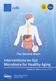
The gut microbiota plays an important role in maintaining the host’s health by delicately balancing commensal and pathogenic bacteria, thus influencing their metabolic, oxidative and cognitive status. Despite there being interindividual differences, it is known that the composition of the microbiota in the elderly differs significantly from that of young people. Even with an accumulation of new evidence regarding the efficacy of microbiota therapies, communicating this to the elderly is a difficult challenge. A detailed understanding of the actions of gut microorganisms is useful to delay the onset of age-related pathologies and to develop strategies which target individual needs. In this study, we discuss the effects of physical activity, dietary interventions and supplements as part of an integrated strategy to implement microbiota health with the goal of healthy aging. View this paper
  • Issues are regarded as officially published after their release is announced to the table of contents alert mailing list.
  • You may sign up for e-mail alerts to receive table of contents of newly released issues.
  • PDF is the official format for papers published in both, html and pdf forms. To view the papers in pdf format, click on the "PDF Full-text" link, and use the free Adobe Reader to open them.
Order results
Result details
Section
Select all
Export citation of selected articles as:
15 pages, 2995 KiB  
Article
Dynamic 3D Modeling for Human Sperm Motility through the Female Cervical Canal and Uterine Cavity to Predict Sperm Chance of Reaching the Oocyte
by Mayssam Nassir, Mattan Levi and Natan T. Shaked
Cells 2023, 12(1), 203; https://doi.org/10.3390/cells12010203 - 03 Jan 2023
Cited by 2 | Viewed by 3609
Abstract
Sperm motility in the female genital tract is a key factor in the natural selection of competent cells that will produce a healthy offspring. We created a dynamic three-dimensional (3D) mechanical model of human sperm cells swimming inside cervical canal and uterine cavity [...] Read more.
Sperm motility in the female genital tract is a key factor in the natural selection of competent cells that will produce a healthy offspring. We created a dynamic three-dimensional (3D) mechanical model of human sperm cells swimming inside cervical canal and uterine cavity dynamic 3D models, all generated based on experimental studies. Using these simulations, we described the sperm cells’ behaviors during swimming inside the 3D tract model as a function of 3D displacement and time. We evaluated normal- and abnormal-morphology sperm cells according to their chances of reaching the oocyte site. As expected, we verified that the number of normal sperm cells that succeeded in reaching the fallopian tube sites is greater than the number of abnormal sperm cells. However, interestingly, after inspecting various abnormal sperm cells, we found out that their scores changed compared to swimming in an infinite medium, as is the case with in vitro fertilization. Thus, the interactions of abnormal sperm cells and the complicated geometry and dynamics of the uterus are significant factors in the filtering of abnormal sperm cells until they reach the oocyte site. Our study provides an advanced tool for sperm analysis and selection criteria for fertility treatments. Full article
(This article belongs to the Section Reproductive Cells and Development)
Show Figures

Figure 1

22 pages, 14976 KiB  
Article
Cell Pair Algorithm-Based Immune Infiltrating Cell Signature for Improving Outcomes and Treatment Responses in Patients with Hepatocellular Carcinoma
by Xiao Zhang, Jun Xie, Dan He, Xin Yan and Jian Chen
Cells 2023, 12(1), 202; https://doi.org/10.3390/cells12010202 - 03 Jan 2023
Cited by 1 | Viewed by 2059
Abstract
Background: Immune interactions play important roles in the regulation of T cells’ cytotoxic function, further impacting the anti-tumor efficacy of immunotherapy. A comprehensive analysis of immune cell types in HCC and immune-cell-related signatures predicting prognosis and monitoring immunotherapy efficacy is still absent. Methods: [...] Read more.
Background: Immune interactions play important roles in the regulation of T cells’ cytotoxic function, further impacting the anti-tumor efficacy of immunotherapy. A comprehensive analysis of immune cell types in HCC and immune-cell-related signatures predicting prognosis and monitoring immunotherapy efficacy is still absent. Methods: More than 1,300 hepatocellular carcinomas (HCC) patients were collected from public databases and included in the present study. The ssGSEA algorithm was applied to calculate the infiltration level of 28 immunocyte subpopulations. A cell pair algorithm was applied to construct an immune-cell-related prognostic index (ICRPI). Survival analyses were performed to measure the survival difference across ICRPI risk groups. Spearman’s correlation analyses were used for the relevance assessment. A Wilcoxon test was used to measure the expression level’s differences. Results: In this study, 28 immune subpopulations were retrieved, and 374 immune cell pairs (ICPs) were established, 38 of which were picked out by the least absolute shrinkage and selection operator (LASSO) algorithm. By using the selected ICPs, the ICRPI was constructed and validated to play crucial roles in survival stratification and dynamic monitoring of immunotherapy effect. We also explored several candidate drugs targeting ICRPI. A composite ICRPI and clinical prognostic index (ICPI) was then constructed, which achieved a more accurate estimation of HCC’s survival and is a better choice for prognosis predictions in HCC. Conclusions: In conclusion, we constructed and validated ICRPI based on the cell pair algorithm in this study, which might provide some novel insights for increasing the survival estimation and clinical response to immune therapy for individual HCC patients and contribute to the personalized precision immunotherapy strategy of HCC. Full article
(This article belongs to the Special Issue Early Biomarkers of Cancer: Diagnosis and Progression)
Show Figures

Figure 1

25 pages, 7901 KiB  
Article
The E3 Ligase TRIM25 Impairs Apoptotic Cell Death in Colon Carcinoma Cells via Destabilization of Caspase-7 mRNA: A Possible Role of hnRNPH1
by Usman Nasrullah, Kristina Stanke, Victoria Recknagel, Süleyman Bozkurt, Patrick Wurzel, Stefan Gauer, Gergely Imre, Christian Münch, Josef Pfeilschifter and Wolfgang Eberhardt
Cells 2023, 12(1), 201; https://doi.org/10.3390/cells12010201 - 03 Jan 2023
Cited by 1 | Viewed by 4865
Abstract
Therapy resistance is still a major reason for treatment failure in colorectal cancer (CRC). Previously, we identified the E3 ubiquitin ligase TRIM25 as a novel suppressor of caspase-2 translation which contributes to the apoptosis resistance of CRC cells towards chemotherapeutic drugs. Here, we [...] Read more.
Therapy resistance is still a major reason for treatment failure in colorectal cancer (CRC). Previously, we identified the E3 ubiquitin ligase TRIM25 as a novel suppressor of caspase-2 translation which contributes to the apoptosis resistance of CRC cells towards chemotherapeutic drugs. Here, we report the executioner caspase-7 as being a further target of TRIM25. The results from the gain- and loss-of-function approaches and the actinomycin D experiments indicate that TRIM25 attenuates caspase-7 expression mainly through a decrease in mRNA stability. The data from the RNA pulldown assays with immunoprecipitated TRIM25 truncations indicate a direct TRIM25 binding to caspase-7 mRNA, which is mediated by the PRY/SPRY domain, which is also known to be highly relevant for protein–protein interactions. By employing TRIM25 immunoprecipitation, we identified the heterogeneous nuclear ribonucleoprotein H1 (hnRNPH1) as a novel TRIM25 binding protein with a functional impact on caspase-7 mRNA stability. Notably, the interaction of both proteins was highly sensitive to RNase A treatment and again depended on the PRY/SPRY domain, thus indicating an indirect interaction of both proteins which is achieved through a common RNA binding. Ubiquitin affinity chromatography showed that both proteins are targets of ubiquitin modification. Functionally, the ectopic expression of caspase-7 in CRC cells caused an increase in poly ADP-ribose polymerase (PARP) cleavage concomitant with a significant increase in apoptosis. Collectively, the negative regulation of caspase-7 by TRIM25, which is possibly executed by hnRNPH1, implies a novel survival mechanism underlying the chemotherapeutic drug resistance of CRC cells. The targeting of TRIM25 could therefore offer a promising strategy for the reduction in therapy resistance in CRC patients. Full article
(This article belongs to the Special Issue Factsheets of Cell Death)
Show Figures

Figure 1

23 pages, 4769 KiB  
Article
A Live Cell Protein Complementation Assay for ORFeome-Wide Probing of Human HOX Interactomes
by Yunlong Jia, Jonathan Reboulet, Benjamin Gillet, Sandrine Hughes, Christelle Forcet, Violaine Tribollet, Nawal Hajj Sleiman, Cindy Kundlacz, Jean-Marc Vanacker, Françoise Bleicher and Samir Merabet
Cells 2023, 12(1), 200; https://doi.org/10.3390/cells12010200 - 03 Jan 2023
Cited by 1 | Viewed by 2668
Abstract
Biological pathways rely on the formation of intricate protein interaction networks called interactomes. Getting a comprehensive map of interactomes implies the development of tools that allow one to capture transient and low-affinity protein–protein interactions (PPIs) in live conditions. Here we presented an experimental [...] Read more.
Biological pathways rely on the formation of intricate protein interaction networks called interactomes. Getting a comprehensive map of interactomes implies the development of tools that allow one to capture transient and low-affinity protein–protein interactions (PPIs) in live conditions. Here we presented an experimental strategy: the Cell-PCA (cell-based protein complementation assay), which was based on bimolecular fluorescence complementation (BiFC) for ORFeome-wide screening of proteins that interact with different bait proteins in the same live cell context, by combining high-throughput sequencing method. The specificity and sensitivity of the Cell-PCA was established by using a wild-type and a single-amino-acid-mutated HOXA9 protein, and the approach was subsequently applied to seven additional human HOX proteins. These proof-of-concept experiments revealed novel molecular properties of HOX interactomes and led to the identification of a novel cofactor of HOXB13 that promoted its proliferative activity in a cancer cell context. Taken together, our work demonstrated that the Cell-PCA was pertinent for revealing and, importantly, comparing the interactomes of different or highly related bait proteins in the same cell context. Full article
(This article belongs to the Section Cell Methods)
Show Figures

Figure 1

37 pages, 696 KiB  
Review
Pseudomonas aeruginosa: Infections, Animal Modeling, and Therapeutics
by Stephen J. Wood, Timothy M. Kuzel and Sasha H. Shafikhani
Cells 2023, 12(1), 199; https://doi.org/10.3390/cells12010199 - 03 Jan 2023
Cited by 35 | Viewed by 7754
Abstract
Pseudomonas aeruginosa is an important Gram-negative opportunistic pathogen which causes many severe acute and chronic infections with high morbidity, and mortality rates as high as 40%. What makes P. aeruginosa a particularly challenging pathogen is its high intrinsic and acquired resistance to many [...] Read more.
Pseudomonas aeruginosa is an important Gram-negative opportunistic pathogen which causes many severe acute and chronic infections with high morbidity, and mortality rates as high as 40%. What makes P. aeruginosa a particularly challenging pathogen is its high intrinsic and acquired resistance to many of the available antibiotics. In this review, we review the important acute and chronic infections caused by this pathogen. We next discuss various animal models which have been developed to evaluate P. aeruginosa pathogenesis and assess therapeutics against this pathogen. Next, we review current treatments (antibiotics and vaccines) and provide an overview of their efficacies and their limitations. Finally, we highlight exciting literature on novel antibiotic-free strategies to control P. aeruginosa infections. Full article
(This article belongs to the Special Issue Immunopathogenesis of Bacterial Infection)
13 pages, 672 KiB  
Article
Investigating the Association between Diabetic Neuropathy and Vitamin D in Emirati Patients with Type 2 Diabetes Mellitus
by Tahra Al Ali, Alizeh Ashfaq, Narjes Saheb Sharif-Askari, Salah Abusnana and Bashair M. Mussa
Cells 2023, 12(1), 198; https://doi.org/10.3390/cells12010198 - 03 Jan 2023
Cited by 3 | Viewed by 2623
Abstract
(1) Background: Vitamin D deficiency is a common public health problem in the United Arab Emirates (UAE) and globally, and interestingly, improvements in diabetic neuropathy after taking Vitamin D supplementation for a short time have been reported. Despite living in a country that [...] Read more.
(1) Background: Vitamin D deficiency is a common public health problem in the United Arab Emirates (UAE) and globally, and interestingly, improvements in diabetic neuropathy after taking Vitamin D supplementation for a short time have been reported. Despite living in a country that is sunny all year round, hypovitaminosis D, indicated by an obvious low serum vitamin D level, has been recurrently noted in the UAE, as well as in the surrounding Arabian Gulf countries. This problem is receiving much attention and attracting clinical and academic interest. Therefore, the main objective of the present study is to identify the association, if any, between vitamin D deficiency and the development of diabetic neuropathy in the UAE population with T2DM. (2) Methods: a total of 600 Emirati patients (male and female) with T2DM, aged between 20 and 80, were recruited from University Hospital Sharjah (UHS). The medical records of the patients were reviewed and analyzed. (3) Results: The results of the present study showed that among the 600 patients, 50% were affected with diabetic neuropathy. Vitamin D level in patients with neuropathy were estimated to be around 20 ng/mL (IQR 14–25), and vitamin D levels were significantly higher (33 ng/mL (IQR 20–42)) among patients without neuropathy, with p < 0.001. Another important finding was that patients without neuropathy had a better vitamin D status, with only 19% being deficient and 18% having insufficient vitamin D levels, compared to patients with neuropathy, where 39% were deficient (vitamin D < 20 ng/mL) and 44% had insufficient vitamin D levels (20–30 ng/mL). (4) Conclusion: The findings of the present study show that the prevalence of vitamin D deficiency (low serum 25-hydroxy vitamin D 25-OHD level) is significantly high in diabetic neuropathy in Emirati patients with T2DM. Full article
Show Figures

Figure 1

19 pages, 4507 KiB  
Article
Sigma-1 Receptor as a Protective Factor for Diabetes-Associated Cognitive Dysfunction via Regulating Astrocytic Endoplasmic Reticulum-Mitochondrion Contact and Endoplasmic Reticulum Stress
by Mengyu Du, Tao Jiang, Shuxuan He, Bo Cheng, Xin Zhang, Liya Li, Lan Yang, Wei Gao, Yansong Li and Qiang Wang
Cells 2023, 12(1), 197; https://doi.org/10.3390/cells12010197 - 03 Jan 2023
Cited by 1 | Viewed by 2339
Abstract
The prevalence of diabetes-associated cognitive dysfunction (DACD) has increased to 13.5%. Dementia, as the most severe DACD, is the second leading cause of death in patients with diabetes mellitus. Hence, the potential mechanisms of DACD for slowing or halting its progression need to [...] Read more.
The prevalence of diabetes-associated cognitive dysfunction (DACD) has increased to 13.5%. Dementia, as the most severe DACD, is the second leading cause of death in patients with diabetes mellitus. Hence, the potential mechanisms of DACD for slowing or halting its progression need to be urgently explored. Given that the sigma-1 receptor (Sig-1R), a chaperone protein located in the endoplasmic reticulum (ER)-mitochondrion contact membranes to regulate ER stress (ERS), is associated with cognitive outcomes in neurodegenerative diseases, this study aimed to investigate the role of astrocytic Sig-1R in DACD and its underlying mechanism. Here, we examined the levels of ERS and complement component 3/3a (C3/C3a) from primary astrocytes with different concentrations of glucose and treatment. Subsequently, HT22 neurons were cultured in different astrocyte-conditioned medium, and the expression of synaptic proteins was detected. We constructed type 1 diabetes mellitus (T1DM) model to evaluate the astrocytic Sig-1R mechanism on synapse and cognitive function changes. In vitro, high glucose concentration downregulated Sig-1R and aggravated ERS in astrocytes, resulting in synapse deficits. PRE-084, a high-affinity and selective Sig-1R agonist, inhibited astrocytic ERS and complement cascades and restored synaptic damage, while the Sig-1R antagonist displayed the opposite results. Moreover, C3a receptor antagonist (C3aRA) could mimic the effect of PRE-084 and exerted neuroprotective effects. In vivo, PRE-084 substantially reduced ER-mitochondrion contact, activation of ERS, and C3/C3a secretion in mice with T1DM. Additionally, the synaptic loss and neurobehavioral dysfunction of mice with T1DM were less pronounced in both the PRE-084 and C3aRA treatment groups. These findings demonstrated that Sig-1R activation reduced the astrocytic ER-mitochondrion contact, ERS activation, and complement-mediated synaptic damage in T1DM. This study suggested the mechanisms and potential therapeutic approaches for treating DACD. Full article
Show Figures

Figure 1

22 pages, 2554 KiB  
Systematic Review
Meta-Analysis of MS-Based Proteomics Studies Indicates Interferon Regulatory Factor 4 and Nucleobindin1 as Potential Prognostic and Drug Resistance Biomarkers in Diffuse Large B Cell Lymphoma
by Mostafa Ejtehadifar, Sara Zahedi, Paula Gameiro, José Cabeçadas, Maria Gomes da Silva, Hans C. Beck, Ana Sofia Carvalho and Rune Matthiesen
Cells 2023, 12(1), 196; https://doi.org/10.3390/cells12010196 - 03 Jan 2023
Cited by 1 | Viewed by 2851
Abstract
The prognosis of diffuse large B cell lymphoma (DLBCL) is inaccurately predicted using clinical features and immunohistochemistry (IHC) algorithms. Nomination of a panel of molecules as the target for therapy and predicting prognosis in DLBCL is challenging because of the divergences in the [...] Read more.
The prognosis of diffuse large B cell lymphoma (DLBCL) is inaccurately predicted using clinical features and immunohistochemistry (IHC) algorithms. Nomination of a panel of molecules as the target for therapy and predicting prognosis in DLBCL is challenging because of the divergences in the results of molecular studies. Mass spectrometry (MS)-based proteomics in the clinic represents an analytical tool with the potential to improve DLBCL diagnosis and prognosis. Previous proteomics studies using MS-based proteomics identified a wide range of proteins. To achieve a consensus, we reviewed MS-based proteomics studies and extracted the most consistently significantly dysregulated proteins. These proteins were then further explored by analyzing data from other omics fields. Among all significantly regulated proteins, interferon regulatory factor 4 (IRF4) was identified as a potential target by proteomics, genomics, and IHC. Moreover, annexinA5 (ANXA5) and nucleobindin1 (NUCB1) were two of the most up-regulated proteins identified in MS studies. Functional enrichment analysis identified the light zone reactions of the germinal center (LZ-GC) together with cytoskeleton locomotion functions as enriched based on consistent, significantly dysregulated proteins. In this study, we suggest IRF4 and NUCB1 proteins as potential biomarkers that deserve further investigation in the field of DLBCL sub-classification and prognosis. Full article
(This article belongs to the Special Issue Molecular Mechanism of Lymphoma)
Show Figures

Figure 1

33 pages, 2735 KiB  
Review
Pseudomonas aeruginosa Cytotoxins: Mechanisms of Cytotoxicity and Impact on Inflammatory Responses
by Stephen J. Wood, Josef W. Goldufsky, Michelle Y. Seu, Amir H. Dorafshar and Sasha H. Shafikhani
Cells 2023, 12(1), 195; https://doi.org/10.3390/cells12010195 - 03 Jan 2023
Cited by 12 | Viewed by 4572
Abstract
Pseudomonas aeruginosa is one of the most virulent opportunistic Gram-negative bacterial pathogens in humans. It causes many acute and chronic infections with morbidity and mortality rates as high as 40%. P. aeruginosa owes its pathogenic versatility to a large arsenal of cell-associated and [...] Read more.
Pseudomonas aeruginosa is one of the most virulent opportunistic Gram-negative bacterial pathogens in humans. It causes many acute and chronic infections with morbidity and mortality rates as high as 40%. P. aeruginosa owes its pathogenic versatility to a large arsenal of cell-associated and secreted virulence factors which enable this pathogen to colonize various niches within hosts and protect it from host innate immune defenses. Induction of cytotoxicity in target host cells is a major virulence strategy for P. aeruginosa during the course of infection. P. aeruginosa has invested heavily in this strategy, as manifested by a plethora of cytotoxins that can induce various forms of cell death in target host cells. In this review, we provide an in-depth review of P. aeruginosa cytotoxins based on their mechanisms of cytotoxicity and the possible consequences of their cytotoxicity on host immune responses. Full article
(This article belongs to the Special Issue Immunopathogenesis of Bacterial Infection)
Show Figures

Figure 1

18 pages, 3315 KiB  
Article
Synergism of Feeding and Digestion Regulated by the Neuropeptide F System in Ostrinia furnacalis Larvae
by Jiajia Zhao, Yu Song, Xuemin Jiang, Lei He, Liya Wei and Zhangwu Zhao
Cells 2023, 12(1), 194; https://doi.org/10.3390/cells12010194 - 03 Jan 2023
Cited by 4 | Viewed by 1811
Abstract
Feeding is crucial for the growth and survival of animals, including humans, but relatively little is known about how it is regulated. Here, we show that larval feeding in Ostrinia furnacalis is regulated by neuropeptide F (NPF, the homologous peptide of mammalian NPY) [...] Read more.
Feeding is crucial for the growth and survival of animals, including humans, but relatively little is known about how it is regulated. Here, we show that larval feeding in Ostrinia furnacalis is regulated by neuropeptide F (NPF, the homologous peptide of mammalian NPY) via the insulin signalling pathway in the midgut. Furthermore, the genes pi3k and mtor in the insulin pathway positively regulate α-amylase and lipase of the midgut by recruiting the transcription factors c-Myc and PPARγ for binding to the promotors of these two enzymes. Importantly, we find that the feeding behaviour and the digestive system of midgut in O. furnacalis larvae are closely related and interactive in that knocking down α-amylase or lipase induces a reduction in larval feeding, while food-deprived larvae lead to fewer expressions of α-amylase and lipase. Importantly, it is the gut NPF that regulates the α-amylase and lipase, while variations of α-amylase and lipase may feed back to the brain NPF. This current study reveals a molecular feedback mechanism between feeding behaviour and the digestive system that is regulated by the conserved NPF via insulin signalling systems in the midgut of O. furnacalis larvae. Full article
(This article belongs to the Section Cellular Metabolism)
Show Figures

Figure 1

10 pages, 1514 KiB  
Article
Pervasive Platelet Secretion Defects in Patients with Severe Acute Respiratory Syndrome Coronavirus 2 (SARS-CoV-2)
by Johannes Kalbhenn, Jan-Steffen Pooth, Georg Trummer, David Kranzhöfer, Axel Schlagenhauf and Barbara Zieger
Cells 2023, 12(1), 193; https://doi.org/10.3390/cells12010193 - 03 Jan 2023
Cited by 3 | Viewed by 1957
Abstract
Critically ill COVID-19 patients suffer from thromboembolic as well as bleeding events. Endothelial dysfunction, spiking of von Willebrand factor (vWF), and excessive cytokine signaling result in coagulopathy associated with substantial activation of plasmatic clotting factors. Thrombocytopenia secondary to extensive platelet activation is a [...] Read more.
Critically ill COVID-19 patients suffer from thromboembolic as well as bleeding events. Endothelial dysfunction, spiking of von Willebrand factor (vWF), and excessive cytokine signaling result in coagulopathy associated with substantial activation of plasmatic clotting factors. Thrombocytopenia secondary to extensive platelet activation is a frequent finding, but abnormal platelet dysfunction may also exist in patients with normal platelet counts. In this study, we performed analyses of platelet function and of von Willebrand factor in critically ill COVID-19 patients (n = 13). Platelet aggregometry was performed using ADP, collagen, epinephrin, and ristocetin. VWF and fibrinogen binding of platelets and CD62 and CD63 expression after thrombin stimulation were analyzed via flow cytometry. In addition, VWF antigen (VWF:Ag), collagen binding capacity (VWF:CB), and multimer analysis were performed next to routine coagulation parameters. All patients exhibited reduced platelet aggregation and decreased CD62 and CD63 expression. VWF binding of platelets was reduced in 12/13 patients. VWF:CB/VWF:Ag ratios were pathologically decreased in 2/13 patients and elevated in 2/13 patients. Critically ill COVID-19 patients exhibit platelet secretion defects independent of thrombocytopenia. Platelet exhaustion and VWF dysfunction may result in impaired primary hemostasis and should be considered when treating coagulopathy in these patients. Full article
(This article belongs to the Special Issue Impact of Platelet Defects on Pathophysiological Processes)
Show Figures

Figure 1

14 pages, 3335 KiB  
Article
Morphological and Functional Effects of Ultrasound on Blood–Brain Barrier Transitory Opening: An In Vitro Study on Rat Brain Endothelial Cells
by Jacopo Junio Valerio Branca, Matteo Boninsegna, Gabriele Morucci, Donatello Carrino, Claudio Nicoletti, Ferdinando Paternostro, Massimo Gulisano, Leonardo Bocchi and Alessandra Pacini
Cells 2023, 12(1), 192; https://doi.org/10.3390/cells12010192 - 03 Jan 2023
Viewed by 1864
Abstract
With the recent advances in medicine, human life expectancy is increasing; however, the extra years of life are not necessarily spent in good health or free from disability, resulting in a significantly higher incidence of age-associated pathologies. Among these disorders, neurodegenerative diseases have [...] Read more.
With the recent advances in medicine, human life expectancy is increasing; however, the extra years of life are not necessarily spent in good health or free from disability, resulting in a significantly higher incidence of age-associated pathologies. Among these disorders, neurodegenerative diseases have a significant impact. To this end, the presence of the protective blood–brain barrier (BBB) represents a formidable obstacle to the delivery of therapeutics. Thus, this makes it imperative to define strategies to bypass the BBB in order to successfully target the brain with the appropriate drugs. It has been demonstrated that targeting the BBB by ultrasound (US) can transiently make this anatomical barrier permeable and in so doing, allow the delivery of therapeutics. Thus, our aim was to carry out an in depth in vitro molecular and morphological study on the effects of US treatment on the BBB. The rat brain endothelial (RBE4) cell line was challenged with exposure to 12 MHz diagnostic US treatment for 10, 20, and 30 min. Cell viability assays, Western blotting analysis on the endoplasmic reticulum (ER), and oxidative stress marker evaluation were then performed, along with cytological and immunofluorescence staining, in order to evaluate the effects of US on the intercellular spaces and tight junction distribution of the brain endothelial cells. We observed that the US treatment exerted no toxic effects on either RBE4 cell viability or the upregulation/dislocation of the ER and oxidative stress marker (GRP78 and cytochrome C, respectively). Further, we observed that the application of US induced an increase in the intercellular spaces, as shown by Papanicolaou staining, mainly due to the altered distribution of the tight junction protein zonula occludens-1 (ZO-1). This latter US-dependent effect was transient and disappeared 20 min after the removal of the stimulus. In conclusion, our results show that US induces a transient alteration of the BBB, without altering the intracellular signaling pathways such as the ER and oxidative stress that could potentially be toxic for endothelial cells. These results suggested that US treatment could represent a potential strategy for improving drug delivery to the brain. Full article
(This article belongs to the Special Issue Blood–Brain Barrier: From Physiology to Disease and Back)
Show Figures

Figure 1

22 pages, 985 KiB  
Review
The Consequences of GBA Deficiency in the Autophagy–Lysosome System in Parkinson’s Disease Associated with GBA
by Eddie Pradas and Marta Martinez-Vicente
Cells 2023, 12(1), 191; https://doi.org/10.3390/cells12010191 - 03 Jan 2023
Cited by 9 | Viewed by 3812
Abstract
GBA gene variants were the first genetic risk factor for Parkinson’s disease. GBA encodes the lysosomal enzyme glucocerebrosidase (GBA), which is involved in sphingolipid metabolism. GBA exhibits a complex physiological function that includes not only the degradation of its substrate glucosylceramide but also [...] Read more.
GBA gene variants were the first genetic risk factor for Parkinson’s disease. GBA encodes the lysosomal enzyme glucocerebrosidase (GBA), which is involved in sphingolipid metabolism. GBA exhibits a complex physiological function that includes not only the degradation of its substrate glucosylceramide but also the metabolism of other sphingolipids and additional lipids such as cholesterol, particularly when glucocerebrosidase activity is deficient. In the context of Parkinson’s disease associated with GBA, the loss of GBA activity has been associated with the accumulation of α-synuclein species. In recent years, several hypotheses have proposed alternative and complementary pathological mechanisms to explain why lysosomal enzyme mutations lead to α-synuclein accumulation and become important risk factors in Parkinson’s disease etiology. Classically, loss of GBA activity has been linked to a dysfunctional autophagy–lysosome system and to a subsequent decrease in autophagy-dependent α-synuclein turnover; however, several other pathological mechanisms underlying GBA-associated parkinsonism have been proposed. This review summarizes and discusses the different hypotheses with a special focus on autophagy-dependent mechanisms, as well as autophagy-independent mechanisms, where the role of other players such as sphingolipids, cholesterol and other GBA-related proteins make important contributions to Parkinson’s disease pathogenesis. Full article
(This article belongs to the Collection Feature Papers in Autophagy)
Show Figures

Figure 1

22 pages, 6306 KiB  
Systematic Review
Effect of Different Application Modalities on the Bonding Performance of Adhesive Systems to Dentin: A Systematic Review and Meta-Analysis
by Louis Hardan, Rim Bourgi, Carlos Enrique Cuevas-Suárez, Walter Devoto, Maciej Zarow, Paulo Monteiro, Natalia Jakubowicz, Amine El Zoghbi, Dariusz Skaba, Davide Mancino, Naji Kharouf, Youssef Haïkel and Monika Lukomska-Szymanska
Cells 2023, 12(1), 190; https://doi.org/10.3390/cells12010190 - 03 Jan 2023
Cited by 8 | Viewed by 3780
Abstract
Diverse types of dental adhesives exhibit different cytotoxic outcomes on cells in vitro. Currently, no standard adhesive application technique has so far been decisive for clinicians for better durability of resin–dentin bonds of adhesive systems. The purpose of this study was to systematically [...] Read more.
Diverse types of dental adhesives exhibit different cytotoxic outcomes on cells in vitro. Currently, no standard adhesive application technique has so far been decisive for clinicians for better durability of resin–dentin bonds of adhesive systems. The purpose of this study was to systematically review the literature to evaluate the bonding performance of adhesive systems to dentin by using different application modalities. The systematic research strategy was conducted by two reviewers among multiple databases: PubMed, Scopus, Web of Science, Embase, and Scielo. In vitro studies reporting the effects of additional steps for the application of adhesive systems on the bond strength to dentin were selected. Meta-analysis was performed using Review Manager Software version 5.3.5 using the random effects model. The methodological quality of each in vitro study was assessed according to the parameters of a previous systematic review. The electronic research through different databases generated a total of 8318 references. After the examination of titles and abstracts, a total of 106 potentially relevant studies accessed the full-text evaluation phase. After full-text examination, 78 publications were included for the qualitative analysis, and 68 studies were included in the meta-analysis. Regarding the etch-and-rinse adhesive systems, the application modalities that improved the overall bond strength were the application of a hydrophobic resin layer (p = 0.005), an extended application time (p < 0.001), an application assisted by an electric current (p < 0.001), a double-layer application (p = 0.05), the agitation technique (p = 0.02), and the active application of the adhesive (p < 0.001). For self-etch adhesive systems, the techniques that improved the overall bond strength were the application of a hydrophobic resin layer (p < 0.001), an extended application time (p = 0.001), an application assisted by an electric current (p < 0.001), a double-layer application (p < 0.001), the agitation technique (p = 0.01), and the active application of the adhesive (p < 0.001). The in vitro evidence suggests that the application of adhesive systems using alternative techniques or additional strategies may be beneficial for improving their bond strength to dentin. The application modalities that favored the overall bond strength to dentin were an extended application time, a double-layer application, an application assisted by an electric current, the active application of the adhesive, and the application of a hydrophobic resin layer. Worth mentioning is that some techniques are intended to increase the degree of the conversion of the materials, and therefore, improvements in the biocompatibility of the materials can be expected. Full article
(This article belongs to the Special Issue Mineralized Tissues Repair and Regeneration)
Show Figures

Figure 1

28 pages, 4672 KiB  
Article
Maternal Hyperhomocysteinemia Disturbs the Mechanisms of Embryonic Brain Development and Its Maturation in Early Postnatal Ontogenesis
by Dmitrii S. Vasilev, Anastasiia D. Shcherbitskaia, Natalia L. Tumanova, Anastasiia V. Mikhel, Yulia P. Milyutina, Anna A. Kovalenko, Nadezhda M. Dubrovskaya, Daria B. Inozemtseva, Irina V. Zalozniaia and Alexander V. Arutjunyan
Cells 2023, 12(1), 189; https://doi.org/10.3390/cells12010189 - 03 Jan 2023
Cited by 5 | Viewed by 1791
Abstract
Maternal hyperhomocysteinemia causes the disruption of placental blood flow and can lead to serious disturbances in the formation of the offspring’s brain. In the present study, the effects of prenatal hyperhomocysteinemia (PHHC) on the neuronal migration, neural tissue maturation, and the expression of [...] Read more.
Maternal hyperhomocysteinemia causes the disruption of placental blood flow and can lead to serious disturbances in the formation of the offspring’s brain. In the present study, the effects of prenatal hyperhomocysteinemia (PHHC) on the neuronal migration, neural tissue maturation, and the expression of signaling molecules in the rat fetal brain were described. Maternal hyperhomocysteinemia was induced in female rats by per os administration of 0.15% aqueous methionine solution in the period of days 4–21 of pregnancy. Behavioral tests revealed a delay in PHHC male pups maturing. Ultrastructure of both cortical and hippocampus tissue demonstrated the features of the developmental delay. PHHC was shown to disturb both generation and radial migration of neuroblasts into the cortical plate. Elevated Bdnf expression, together with changes in proBDNF/mBDNF balance, might affect neuronal cell viability, positioning, and maturation in PHHC pups. Reduced Kdr gene expression and the content of SEMA3E might lead to impaired brain development. In the brain tissue of E20 PHHC fetuses, the content of the procaspase-8 was decreased, and the activity level of the caspase-3 was increased; this may indicate the development of apoptosis. PHHC disturbs the mechanisms of early brain development leading to a delay in brain tissue maturation and formation of the motor reaction of pups. Full article
(This article belongs to the Collection How Perinatal Stress Affects Brain Plasticity in Ontogenesis)
Show Figures

Figure 1

3 pages, 195 KiB  
Editorial
Editorial: Highlights in Autophagy—From Basic Mechanisms to Human Disorder Treatments
by Pei-Hui Lin and Lydie Combaret
Cells 2023, 12(1), 188; https://doi.org/10.3390/cells12010188 - 03 Jan 2023
Cited by 2 | Viewed by 1435
Abstract
Autophagy is an evolutionarily conserved catabolic process and represents a field of research that is constantly growing [...] Full article
15 pages, 1305 KiB  
Review
Multiple Roles of PLK1 in Mitosis and Meiosis
by Jaroslav Kalous and Daria Aleshkina
Cells 2023, 12(1), 187; https://doi.org/10.3390/cells12010187 - 02 Jan 2023
Cited by 7 | Viewed by 3242
Abstract
Cells are equipped with a diverse network of signaling and regulatory proteins that function as cell cycle regulators and checkpoint proteins to ensure the proper progression of cell division. A key regulator of cell division is polo-like kinase 1 (PLK1), a member of [...] Read more.
Cells are equipped with a diverse network of signaling and regulatory proteins that function as cell cycle regulators and checkpoint proteins to ensure the proper progression of cell division. A key regulator of cell division is polo-like kinase 1 (PLK1), a member of the serine/threonine kinase family that plays an important role in regulating the mitotic and meiotic cell cycle. The phosphorylation of specific substrates mediated by PLK1 controls nuclear envelope breakdown (NEBD), centrosome maturation, proper spindle assembly, chromosome segregation, and cytokinesis. In mammalian oogenesis, PLK1 is essential for resuming meiosis before ovulation and for establishing the meiotic spindle. Among other potential roles, PLK1 regulates the localized translation of spindle-enriched mRNAs by phosphorylating and thereby inhibiting the translational repressor 4E-BP1, a downstream target of the mTOR (mammalian target of rapamycin) pathway. In this review, we summarize the functions of PLK1 in mitosis, meiosis, and cytokinesis and focus on the role of PLK1 in regulating mRNA translation. However, knowledge of the role of PLK1 in the regulation of meiosis remains limited. Full article
(This article belongs to the Section Cell Proliferation and Division)
Show Figures

Figure 1

17 pages, 3986 KiB  
Article
Transcriptomic Analysis Reveals JAK2/MPL-Independent Effects of Calreticulin Mutations in a C. elegans Model
by Ana Guijarro-Hernández, Laura Eder-Azanza, Cristina Hurtado, David Navarro-Herrera, Begoña Ezcurra, Francisco Javier Novo, Juan Cabello and José Luis Vizmanos
Cells 2023, 12(1), 186; https://doi.org/10.3390/cells12010186 - 02 Jan 2023
Cited by 1 | Viewed by 1879
Abstract
There is growing evidence that Ph-negative myeloproliferative neoplasms (MPNs) are disorders in which multiple molecular mechanisms are significantly disturbed. Since their discovery, CALR driver mutations have been demonstrated to trigger pathogenic mechanisms apart from the well-documented activation of JAK2/MPL-related pathways, but the [...] Read more.
There is growing evidence that Ph-negative myeloproliferative neoplasms (MPNs) are disorders in which multiple molecular mechanisms are significantly disturbed. Since their discovery, CALR driver mutations have been demonstrated to trigger pathogenic mechanisms apart from the well-documented activation of JAK2/MPL-related pathways, but the lack of experimental models harboring CALR mutations in a JAK2/MPL knockout background has hindered the research on these non-canonical mechanisms. In this study, CRISPR/Cas9 was performed to introduce homozygous patient-like calreticulin mutations in a C. elegans model that naturally lacks JAK2 and MPL orthologs. Whole-genome transcriptomic analysis of these worms was conducted, and some of the genes identified to be associated with processes involved in the pathogenesis of MPNs were further validated by qPCR. Some of the transcriptomic alterations corresponded to typically altered genes and processes in cancer and Ph-negative MPN patients that are known to be triggered by mutant calreticulin without the intervention of JAK2/MPL. However, interestingly, we have also found altered other processes described in these diseases that had not been directly attributed to calreticulin mutations without the intervention of JAK2 or MPL. Thus, these results point to a new experimental model for the study of the JAK2/MPL-independent mechanisms of mutant calreticulin that induce these biological alterations, which could be useful to study unknown non-canonical effects of the mutant protein. The comparison with a calreticulin null strain revealed that the alteration of all of these processes seems to be a consequence of a loss of function of mutant calreticulin in the worm, except for the dysregulation of Hedgehog signaling and flh-3. Further analysis of this model could help to delineate these mechanisms, and the verification of these results in mammalian models may unravel new potential therapeutic targets in MPNs. As far as we know, this is the first time that a C. elegans strain with patient-like mutations is proposed as a potential model for leukemia research. Full article
Show Figures

Figure 1

15 pages, 1987 KiB  
Article
Azilsartan Modulates HMGB1/NF-κB/p38/ERK1/2/JNK and Apoptosis Pathways during Renal Ischemia Reperfusion Injury
by Rania Alaaeldin, Sally M. Bakkar, Reham H. Mohyeldin, Fares E. M. Ali, Nehad M. Reda Abdel-Maqsoud and Moustafa Fathy
Cells 2023, 12(1), 185; https://doi.org/10.3390/cells12010185 - 02 Jan 2023
Cited by 7 | Viewed by 2153
Abstract
Renal ischemia/reperfusion (IR) injury is characterized by an unexpected impairment of blood flow to the kidney. Azilsartan is an angiotensin receptor blocker that is approved for the management of hypertension. The present study aimed to investigate, on molecular basics, the nephroprotective activity of [...] Read more.
Renal ischemia/reperfusion (IR) injury is characterized by an unexpected impairment of blood flow to the kidney. Azilsartan is an angiotensin receptor blocker that is approved for the management of hypertension. The present study aimed to investigate, on molecular basics, the nephroprotective activity of azilsartan on renal IR injury in rats. Rats were assigned into four groups: (1) Sham group, (2) Azilsartan group, (3) IR group, and (4) IR/Azilsartan-treated group. Histological examination and renal function were evaluated. Levels of KIM-1, HMGB1, caspase 3, GPX, SOD, NF-κB, and p53 proteins were investigated using ELISA. mRNA levels of IL-1β, IL6, IL10, TNF-α, NF-κB, p53, and bax were assessed by qRT-PCR. Expression of p38, JNK, and ERK1/2 proteins was investigated by Western blotting. IR injury resulted in tissue damage, elevation of creatinine, BUN, KIM-1, HMGB1, caspase 3, NF-κB, and p53 levels, decreasing GPX and SOD activities, and up-regulation of NF-κB, IL-1β, IL6, TNF-α, p53, and bax genes. Furthermore, it up-regulated the expression of phosphorylated/total ratio of p38, ERK1/2, and JNK proteins. Interestingly, treatment of the injured rats with azilsartan significantly alleviated IR injury-induced histopathological and biochemical changes. It reduced the creatinine, BUN, KIM-1, HMGB1, caspase-3, NF-κB, and p53 levels, elevated GPX and SOD activities, down-regulated the expression of NF-κB, IL-1β, IL6, TNF-α, p53, and bax genes, and up-regulated IL10 gene expression. Furthermore, it decreased the phosphorylated/total ratio of p38, ERK1/2, and JNK proteins. Azilsartan exhibited nephroprotective activity in IR-injured rats via its antioxidant effect, suppression of inflammation, attenuation of apoptosis, and inhibition of HMGB1/NF-κB/p38/ERK1/2/JNK signaling pathway. Full article
Show Figures

Figure 1

33 pages, 1345 KiB  
Review
Probiotics Mechanism of Action on Immune Cells and Beneficial Effects on Human Health
by Chiara Mazziotta, Mauro Tognon, Fernanda Martini, Elena Torreggiani and John Charles Rotondo
Cells 2023, 12(1), 184; https://doi.org/10.3390/cells12010184 - 02 Jan 2023
Cited by 74 | Viewed by 13520
Abstract
Immune cells and commensal microbes in the human intestine constantly communicate with and react to each other in a stable environment in order to maintain healthy immune activities. Immune system-microbiota cross-talk relies on a complex network of pathways that sustain the balance between [...] Read more.
Immune cells and commensal microbes in the human intestine constantly communicate with and react to each other in a stable environment in order to maintain healthy immune activities. Immune system-microbiota cross-talk relies on a complex network of pathways that sustain the balance between immune tolerance and immunogenicity. Probiotic bacteria can interact and stimulate intestinal immune cells and commensal microflora to modulate specific immune functions and immune homeostasis. Growing evidence shows that probiotic bacteria present important health-promoting and immunomodulatory properties. Thus, the use of probiotics might represent a promising approach for improving immune system activities. So far, few studies have been reported on the beneficial immune modulatory effect of probiotics. However, many others, which are mainly focused on their metabolic/nutritional properties, have been published. Therefore, the mechanisms behind the interaction between host immune cells and probiotics have only been partially described. The present review aims to collect and summarize the most recent scientific results and the resulting implications of how probiotic bacteria and immune cells interact to improve immune functions. Hence, a description of the currently known immunomodulatory mechanisms of probiotic bacteria in improving the host immune system is provided. Full article
Show Figures

Figure 1

12 pages, 2496 KiB  
Article
Relationship between Mitochondrial Quality Control Markers, Lower Extremity Tissue Composition, and Physical Performance in Physically Inactive Older Adults
by Anna Picca, Matthew Triolo, Stephanie E. Wohlgemuth, Matthew S. Martenson, Robert T. Mankowski, Stephen D. Anton, Emanuele Marzetti, Christiaan Leeuwenburgh and David A. Hood
Cells 2023, 12(1), 183; https://doi.org/10.3390/cells12010183 - 02 Jan 2023
Cited by 7 | Viewed by 2297
Abstract
Altered mitochondrial quality and function in muscle may be involved in age-related physical function decline. The role played by the autophagy–lysosome system, a major component of mitochondrial quality control (MQC), is incompletely understood. This study was undertaken to obtain initial indications on the [...] Read more.
Altered mitochondrial quality and function in muscle may be involved in age-related physical function decline. The role played by the autophagy–lysosome system, a major component of mitochondrial quality control (MQC), is incompletely understood. This study was undertaken to obtain initial indications on the relationship between autophagy, mitophagy, and lysosomal markers in muscle and measures of physical performance and lower extremity tissue composition in young and older adults. Twenty-three participants were enrolled, nine young (mean age: 24.3 ± 4.3 years) and 14 older adults (mean age: 77.9 ± 6.3 years). Lower extremity tissue composition was quantified volumetrically by magnetic resonance imaging and a tissue composition index was calculated as the ratio between muscle and intermuscular adipose tissue volume. Physical performance in older participants was assessed via the Short Physical Performance Battery (SPPB). Protein levels of the autophagy marker p62, the mitophagy mediator BCL2/adenovirus E1B 19 kDa protein-interacting protein 3 (BNIP3), the lysosomal markers transcription factor EB, vacuolar-type ATPase, and lysosomal-associated membrane protein 1 were measured by Western immunoblotting in vastus lateralis muscle biopsies. Older adults had smaller muscle volume and lower tissue composition index than young participants. The protein content of p62 and BNIP3 was higher in older adults. A negative correlation was detected between p62 and BNIP3 and the tissue composition index. p62 and BNIP3 were also related to the performance on the 5-time sit-to-stand test of the SPPB. Our results suggest that an altered expression of markers of the autophagy/mitophagy–lysosomal system is related to deterioration of lower extremity tissue composition and muscle dysfunction. Additional studies are needed to clarify the role of defective MQC in human muscle aging and identify novel biological targets for drug development. Full article
(This article belongs to the Special Issue Autophagy Meets Aging Ⅱ)
Show Figures

Figure 1

18 pages, 20921 KiB  
Article
CXCR3 Inhibition Blocks the NF-κB Signaling Pathway by Elevating Autophagy to Ameliorate Lipopolysaccharide-Induced Intestinal Dysfunction in Mice
by Cheng Zhang, Yian Deng, Yingsi Zhang, Tongtong Ba, Sai Niu, Yiqin Chen, Yuan Gao and Hanchuan Dai
Cells 2023, 12(1), 182; https://doi.org/10.3390/cells12010182 - 01 Jan 2023
Cited by 9 | Viewed by 2649
Abstract
Autophagy is a cellular catabolic process in the evolutionarily conservative turnover of intracellular substances in eukaryotes, which is involved in both immune homeostasis and injury repairment. CXCR3 is an interferon-induced chemokine receptor that participates in immune regulation and inflammatory responses. However, CXCR3 regulating [...] Read more.
Autophagy is a cellular catabolic process in the evolutionarily conservative turnover of intracellular substances in eukaryotes, which is involved in both immune homeostasis and injury repairment. CXCR3 is an interferon-induced chemokine receptor that participates in immune regulation and inflammatory responses. However, CXCR3 regulating intestine injury via autophagy along with the precise underlying mechanism have yet to be elucidated. In the current study, we employed an LPS-induced inflammatory mouse model and confirmed that CXCR3 knockout significantly attenuates intestinal mucosal structural damage and increases tight junction protein expression. CXCR3 knockout alleviated the LPS-induced increase in the expression of inflammatory factors including TNF-α, IL-6, p-65, and JNK-1 and enhanced autophagy by elevating LC3II, ATG12, and PINK1/Parkin expression. Mechanistically, the function of CXCR3 regarding autophagy and immunity was investigated in IPEC-J2 cells. CXCR3 inhibition by AMG487 enhanced autophagy and reduced the inflammatory response, as well as blocked the NF-κB signaling pathway and elevated the expression of the tight junction protein marker Claudin-1. Correspondingly, these effects were abolished by autophagy inhibition with the selective blocker, 3-MA. Moreover, the immunofluorescence assay results further demonstrated that CXCR3 inhibition-mediated autophagy blocked p65 nuclear translocation, and the majority of Claudin-1 was located at the tight junctions. In conclusion, CXCR3 inhibition reversed LPS-induced intestinal barrier damage and alleviated the NF-κB signaling pathway via enhancing autophagy. These data provided a theoretical basis for elucidating the immunoregulatory mechanism by targeting CXCR3 to prevent intestinal dysfunction. Full article
(This article belongs to the Topic Animal Models of Human Disease)
Show Figures

Figure 1

14 pages, 1420 KiB  
Review
Anti-Oxidant and Pro-Oxidant Effects of Peroxiredoxin 6: A Potential Target in Respiratory Diseases
by Wenhui Jia, Chunling Dong and Bo Li
Cells 2023, 12(1), 181; https://doi.org/10.3390/cells12010181 - 01 Jan 2023
Cited by 6 | Viewed by 2049
Abstract
Peroxiredoxin 6 (PRDX6) is widely distributed in several organs, especially the lungs. The role of PRDX6 in oxidative stress is controversial and even contradictory, as indicated by research conducted over the past 20 years. PRDX6 has anti-oxidant or pro-oxidant effects on oxidative stress [...] Read more.
Peroxiredoxin 6 (PRDX6) is widely distributed in several organs, especially the lungs. The role of PRDX6 in oxidative stress is controversial and even contradictory, as indicated by research conducted over the past 20 years. PRDX6 has anti-oxidant or pro-oxidant effects on oxidative stress in different diseases. It can even exhibit both anti-oxidant and pro-oxidant effects in the same disease. These findings are attributed to the fact that PRDX6 is a multifunctional enzyme. The peroxidase and phospholipase A2 activity of PRDX6 is closely related to its anti-oxidant and pro-oxidant effects, which leads to the conflicting regulatory effects of PRDX6 on oxidative stress in respiratory diseases. Moreover, PRDX6 interacts with multiple redox signaling pathways to interfere with cell proliferation and apoptosis. PRDX6 has become a new target in respiratory disease research due to its important regulatory role in oxidative stress. In this paper, the role of PRDX6 in oxidative stress in respiratory diseases and the research progress in targeting PRDX6 are reviewed. Full article
Show Figures

Figure 1

30 pages, 99652 KiB  
Article
Identification and Validation of the Prognostic Panel in Clear Cell Renal Cell Carcinoma Based on Resting Mast Cells for Prediction of Distant Metastasis and Immunotherapy Response
by Yang Su, Tianxiang Zhang, Jinsen Lu, Lei Qian, Yang Fei, Li Zhang, Song Fan, Jun Zhou, Jieqiong Tang, Haige Chen and Chaozhao Liang
Cells 2023, 12(1), 180; https://doi.org/10.3390/cells12010180 - 01 Jan 2023
Cited by 3 | Viewed by 2368
Abstract
Clear cell renal cell carcinoma (ccRCC) has a high metastatic rate, and its incidence and mortality are still rising. The aim of this study was to identify the key tumor-infiltrating immune cells (TIICs) affecting the distant metastasis and prognosis of patients with ccRCC [...] Read more.
Clear cell renal cell carcinoma (ccRCC) has a high metastatic rate, and its incidence and mortality are still rising. The aim of this study was to identify the key tumor-infiltrating immune cells (TIICs) affecting the distant metastasis and prognosis of patients with ccRCC and to construct a relevant prognostic panel to predict immunotherapy response. Based on ccRCC bulk RNA sequencing data, resting mast cells (RMCs) were screened and verified using the CIBERSORT algorithm, survival analysis, and expression analysis. Distant metastasis-associated genes were identified using single-cell RNA sequencing data. Subsequently, a three-gene (CFB, PPP1R18, and TOM1L1) panel with superior distant metastatic and prognostic performance was established and validated, which stratified patients into high- and low-risk groups. The high-risk group exhibited lower infiltration of RMCs, higher tumor mutation burden (TMB), and worse prognosis. Therapeutically, the high-risk group was more sensitive to anti-PD-1 and anti-CTLA-4 immunotherapy, whereas the low-risk group displayed a better response to anti-PD-L1 immunotherapy. Furthermore, two immune clusters revealing distinct immune, clinical, and prognosis heterogeneity were distinguished. Immunohistochemistry of ccRCC samples verified the expression patterns of the three key genes. Collectively, the prognostic panel based on RMCs is able to predict distant metastasis and immunotherapy response in patients with ccRCC, providing new insight for the treatment of advanced ccRCC. Full article
Show Figures

Figure 1

17 pages, 4683 KiB  
Article
Oral Squamous Cell Carcinoma Cells with Acquired Resistance to Erlotinib Are Sensitive to Anti-Cancer Effect of Quercetin via Pyruvate Kinase M2 (PKM2)
by Chien-Yi Chan, Shih-Cing Hong, Chin-Ming Chang, Yuan-Hong Chen, Pin-Chen Liao and Chun-Yin Huang
Cells 2023, 12(1), 179; https://doi.org/10.3390/cells12010179 - 01 Jan 2023
Cited by 10 | Viewed by 2339
Abstract
Oral squamous cell carcinoma (OSCC) frequently carries high epidermal growth factor receptor (EGFR) expression. Erlotinib, a small molecule tyrosine kinase inhibitor (TKI), is an effective inhibitor of EGFR activity; however, resistance to this drug can occur, limiting therapeutic outcomes. Therefore, in the current [...] Read more.
Oral squamous cell carcinoma (OSCC) frequently carries high epidermal growth factor receptor (EGFR) expression. Erlotinib, a small molecule tyrosine kinase inhibitor (TKI), is an effective inhibitor of EGFR activity; however, resistance to this drug can occur, limiting therapeutic outcomes. Therefore, in the current study, we aimed to unveil key intracellular molecules and adjuvant reagents to overcome erlotinib resistance. First, two HSC-3-derived erlotinib-resistant cell lines, ERL-R5 and ERL-R10, were established; both exhibited relatively higher growth rates, glucose utilization, epithelial-mesenchymal transition (EMT), and invasiveness compared with parental cells. Cancer aggressiveness-related proteins, such as N-cadherin, Vimentin, Twist, MMP-2, MMP-9, and MMP-13, and the glycolytic enzymes PKM2 and GLUT1 were upregulated in ERL-R cells. Notably, ERL-R cells were sensitive to quercetin, a naturally-existing flavonol phytochemical with anti-cancer properties against various cancer cells. At a concentration of 5 μM, quercetin effectively arrested cell growth, reduced glucose utilization, and inhibited cellular invasiveness. An ERL-R5-derived xenograft mouse model confirmed the growth-inhibitory efficacy of quercetin. Additionally, knock-down of PKM2 by siRNA mimicked the effect of quercetin and re-sensitized ERL-R cells to erlotinib. Furthermore, adding quercetin blocked the development of erlotinib-mediated resistance by enhancing apoptosis. In conclusion, our data support the application of quercetin in anti-erlotinib-resistant OSCC and indicate that PKM2 is a determinant factor in erlotinib resistance and quercetin sensitivity. Full article
Show Figures

Figure 1

26 pages, 5303 KiB  
Article
The Metabolic Changes between Monolayer (2D) and Three-Dimensional (3D) Culture Conditions in Human Mesenchymal Stem/Stromal Cells Derived from Adipose Tissue
by Paulina Rybkowska, Klaudia Radoszkiewicz, Maria Kawalec, Dorota Dymkowska, Barbara Zabłocka, Krzysztof Zabłocki and Anna Sarnowska
Cells 2023, 12(1), 178; https://doi.org/10.3390/cells12010178 - 01 Jan 2023
Cited by 10 | Viewed by 3026
Abstract
Introduction: One of the key factors that may influence the therapeutic potential of mesenchymal stem/stromal cells (MSCs) is their metabolism. The switch between mitochondrial respiration and glycolysis can be affected by many factors, including the oxygen concentration and the spatial form of culture. [...] Read more.
Introduction: One of the key factors that may influence the therapeutic potential of mesenchymal stem/stromal cells (MSCs) is their metabolism. The switch between mitochondrial respiration and glycolysis can be affected by many factors, including the oxygen concentration and the spatial form of culture. This study compared the metabolic features of adipose-derived mesenchymal stem/stromal cells (ASCs) and dedifferentiated fat cells (DFATs) cultivated as monolayer or spheroid culture under 5% O2 concentration (physiological normoxia) and their impact on MSCs therapeutic abilities. Results: We observed that the cells cultured as spheroids had a slightly lower viability and a reduced proliferation rate but a higher expression of the stemness-related transcriptional factors compared to the cells cultured in monolayer. The three-dimensional culture form increased mtDNA content, oxygen consumption rate (OCR) and extracellular acidification rate (ECAR), especially in DFATs-3D population. The DFATs spheroids also demonstrated increased levels of Complex V proteins and higher rates of ATP production. Moreover, increased reactive oxygen species and lower intracellular lactic acid levels were also found in 3D culture. Conclusion: Our results may suggest that metabolic reconfiguration accompanies the transition from 2D to 3D culture and the processes of both mitochondrial respiration and glycolysis become more active. Intensified metabolism might be associated with the increased demand for energy, which is needed to maintain the expression of pluripotency genes and stemness state. Full article
(This article belongs to the Special Issue Stem Cells, Metabolism and Neurodegenerative Diseases)
Show Figures

Figure 1

20 pages, 4240 KiB  
Article
SESN2 Knockdown Increases Betulinic Acid-Induced Radiosensitivity of Hypoxic Breast Cancer Cells
by Antje Güttler, Claus Weinholdt, Elisabeth Ruff, Judith Reidt, Elisa Darnstaedt, Alicia Wildemann, Marina Petrenko, Jacqueline Keßler, Matthias Kappler, Ivo Grosse, Dirk Vordermark and Matthias Bache
Cells 2023, 12(1), 177; https://doi.org/10.3390/cells12010177 - 31 Dec 2022
Viewed by 1995
Abstract
Betulinic acid (BA) is a natural compound well known for its anti-inflammatory, anti-viral, anti-bacterial, anti-malarial effects and anti-tumor properties. Its enhanced cytotoxicity in tumor cells and induction of cell death in various cancer entities qualifies BA as an interesting candidate for novel treatment [...] Read more.
Betulinic acid (BA) is a natural compound well known for its anti-inflammatory, anti-viral, anti-bacterial, anti-malarial effects and anti-tumor properties. Its enhanced cytotoxicity in tumor cells and induction of cell death in various cancer entities qualifies BA as an interesting candidate for novel treatment concepts. Our analyses showed enhanced cytotoxicity and radiosensitization under hypoxic conditions in human breast cancer cells. So far, the underlying mechanisms are unknown. Therefore, we investigated the BA-treated human breast cancer cell lines MDA-MB-231 and MCF-7 under normoxic and hypoxic conditions based on microarray technology. Hypoxia and BA regulated a variety of genes in both breast cancer cell lines. KEGG pathway analysis identified an enrichment of the p53 pathway in MCF-7 cells (wtp53) under hypoxia. In MDA-MB-231 cells (mtp53) an additional BA incubation was required to activate the p53 signaling pathway. Fourteen down-regulated and up-regulated genes of the p53 pathway were selected for further validation via qRT-PCR in a panel of five breast cancer cell lines. The stress-induced gene Sestrin-2 (SESN2) was identified as one of the most strongly up-regulated genes after BA treatment. Knockdown of SESN2 enhanced BA-induced ROS production, DNA damage, radiosensitivity and reduced autophagy in breast cancer cells. Our results identified SESN2 as an important target to enhance the radiobiological and anti-tumor effects of BA on breast cancer cells. Full article
(This article belongs to the Special Issue Cancer and Radiation Therapy)
Show Figures

Figure 1

15 pages, 3093 KiB  
Article
Effects of Microbeam Irradiation on Rodent Esophageal Smooth Muscle Contraction
by Bernd Frerker, Stefan Fiedler, Timo Kirschstein, Falko Lange, Katrin Porath, Tina Sellmann, Leonie Kutzner, Fabian Wilde, Julian Moosmann, Rüdiger Köhling, Guido Hildebrandt and Elisabeth Schültke
Cells 2023, 12(1), 176; https://doi.org/10.3390/cells12010176 - 31 Dec 2022
Cited by 1 | Viewed by 1670
Abstract
Background: High-dose-rate radiotherapy has shown promising results with respect to normal tissue preservation. We developed an ex vivo model to study the physiological effects of experimental radiotherapy in the rodent esophageal smooth muscle. Methods: We assessed the physiological parameters of the esophageal function [...] Read more.
Background: High-dose-rate radiotherapy has shown promising results with respect to normal tissue preservation. We developed an ex vivo model to study the physiological effects of experimental radiotherapy in the rodent esophageal smooth muscle. Methods: We assessed the physiological parameters of the esophageal function in ex vivo preparations of the proximal, middle, and distal segments in the organ bath. High-dose-rate synchrotron irradiation was conducted using both the microbeam irradiation (MBI) technique with peak doses greater than 200 Gy and broadbeam irradiation (BBI) with doses ranging between 3.5–4 Gy. Results: Neither MBI nor BBI affected the function of the contractile apparatus. While peak latency and maximal force change were not affected in the BBI group, and no changes were seen in the proximal esophagus segments after MBI, a significant increase in peak latency and a decrease in maximal force change was observed in the middle and distal esophageal segments. Conclusion: No severe changes in physiological parameters of esophageal contraction were determined after high-dose-rate radiotherapy in our model, but our results indicate a delayed esophageal function. From the clinical perspective, the observed increase in peak latency and decreased maximal force change may indicate delayed esophageal transit. Full article
(This article belongs to the Topic Innovative Radiation Therapies)
Show Figures

Figure 1

17 pages, 4176 KiB  
Article
Modeling the Differentiation of Embryonic Limb Chondroprogenitors by Cell Death and Cell Senescence in High Density Micromass Cultures and Their Regulation by FGF Signaling
by Cristina Duarte-Olivenza, Juan M. Hurle, Juan A. Montero and Carlos I. Lorda-Diez
Cells 2023, 12(1), 175; https://doi.org/10.3390/cells12010175 - 31 Dec 2022
Cited by 1 | Viewed by 1581
Abstract
Considering the importance of programmed cell death in the formation of the skeleton during embryonic development, the aim of the present study was to analyze whether regulated cell degeneration also accompanies the differentiation of embryonic limb skeletal progenitors in high-density tridimensional cultures (micromass [...] Read more.
Considering the importance of programmed cell death in the formation of the skeleton during embryonic development, the aim of the present study was to analyze whether regulated cell degeneration also accompanies the differentiation of embryonic limb skeletal progenitors in high-density tridimensional cultures (micromass cultures). Our results show that the formation of primary cartilage nodules in the micromass culture assay involves a patterned process of cell death and cell senescence, complementary to the pattern of chondrogenesis. As occurs in vivo, the degenerative events were preceded by DNA damage detectable by γH2AX immunolabeling and proceeded via apoptosis and cell senescence. Combined treatments of the cultures with growth factors active during limb skeletogenesis, including FGF, BMP, and WNT revealed that FGF signaling modulates the response of progenitors to signaling pathways implicated in cell death. Transcriptional changes induced by FGF treatments suggested that this function is mediated by the positive regulation of the genetic machinery responsible for apoptosis and cell senescence together with hypomethylation of the Sox9 gene promoter. We propose that FGF signaling exerts a primordial function in the embryonic limb conferring chondroprogenitors with their biological properties. Full article
Show Figures

Figure 1

36 pages, 1771 KiB  
Review
Endocrine Disrupting Chemicals in Polycystic Ovary Syndrome: The Relevant Role of the Theca and Granulosa Cells in the Pathogenesis of the Ovarian Dysfunction
by Malgorzata Jozkowiak, Hanna Piotrowska-Kempisty, Dominik Kobylarek, Natalia Gorska, Paul Mozdziak, Bartosz Kempisty, Dominik Rachon and Robert Z. Spaczynski
Cells 2023, 12(1), 174; https://doi.org/10.3390/cells12010174 - 31 Dec 2022
Cited by 11 | Viewed by 5257
Abstract
Polycystic ovary syndrome (PCOS) is the most common heterogeneous endocrine disorder among women of reproductive age. The pathogenesis of PCOS remains elusive; however, there is evidence suggesting the potential contribution of genetic interactions or predispositions combined with environmental factors. Among these, endocrine disrupting [...] Read more.
Polycystic ovary syndrome (PCOS) is the most common heterogeneous endocrine disorder among women of reproductive age. The pathogenesis of PCOS remains elusive; however, there is evidence suggesting the potential contribution of genetic interactions or predispositions combined with environmental factors. Among these, endocrine disrupting chemicals (EDCs) have been proposed to potentially contribute to the etiology of PCOS. Granulosa and theca cells are known to cooperate to maintain ovarian function, and any disturbance can lead to endocrine disorders, such as PCOS. This article provides a review of the recent knowledge on PCOS pathophysiology, the role of granulosa and theca cells in PCOS pathogenesis, and the evidence linking exposure to EDCs with reproductive disorders such as PCOS. Full article
Show Figures

Figure 1

Previous Issue
Back to TopTop