Next Issue
Volume 12, January-1
Previous Issue
Volume 11, December-1


Cells, Volume 11, Issue 24 (December-2 2022) – 193 articles

Cover Story (view full-size image): 
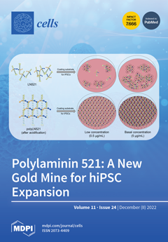
Laminins (LNs) play a central role in the self-assembly and maintenance of basement membranes and are involved in critical interactions between cells and other extracellular matrix proteins. Polylaminin (polyLN) is an acidification-induced LN mimic that recapitulates the native-like polymeric array in cell-free systems. Here, we characterize the native-like hexagonal structure of polyLN-521 and demonstrate that polyLN521 improves the adhesion and proliferation of human induced pluripotent stem cells (hiPSCs) compared with LN521 or Matrigel. Furthermore, hiPSCs cultivated on a low concentration of polyLN521 maintain the pluripotent state. Thus, polyLN521 is a feasible and cost-effective candidate for a chemically defined, xeno-free coating in the large-scale expansion of hiPSCs. These findings further illustrate that the biological activity of LNs is enhanced when assembled in acidic pH. View this paper
  • Issues are regarded as officially published after their release is announced to the table of contents alert mailing list.
  • You may sign up for e-mail alerts to receive table of contents of newly released issues.
  • PDF is the official format for papers published in both, html and pdf forms. To view the papers in pdf format, click on the "PDF Full-text" link, and use the free Adobe Reader to open them.
Order results
Result details
Section
Select all
Export citation of selected articles as:
11 pages, 4295 KiB  
Article
Sensing Cells-Peptide Hydrogel Interaction In Situ via Scanning Ion Conductance Microscopy
by Tatiana N. Tikhonova, Vasilii S. Kolmogorov, Roman V. Timoshenko, Alexander N. Vaneev, Dana Cohen-Gerassi, Liubov A. Osminkina, Petr V. Gorelkin, Alexander S. Erofeev, Nikolay N. Sysoev, Lihi Adler-Abramovich and Evgeny A. Shirshin
Cells 2022, 11(24), 4137; https://doi.org/10.3390/cells11244137 - 19 Dec 2022
Cited by 2 | Viewed by 1585
Abstract
Peptide-based hydrogels were shown to serve as good matrices for 3D cell culture and to be applied in the field of regenerative medicine. The study of the cell-matrix interaction is important for the understanding of cell attachment, proliferation, and migration, as well as [...] Read more.
Peptide-based hydrogels were shown to serve as good matrices for 3D cell culture and to be applied in the field of regenerative medicine. The study of the cell-matrix interaction is important for the understanding of cell attachment, proliferation, and migration, as well as for the improvement of the matrix. Here, we used scanning ion conductance microscopy (SICM) to study the growth of cells on self-assembled peptide-based hydrogels. The hydrogel surface topography, which changes during its formation in an aqueous solution, were studied at nanoscale resolution and compared with fluorescence lifetime imaging microscopy (FLIM). Moreover, SICM demonstrated the ability to map living cells inside the hydrogel. A zwitterionic label-free pH nanoprobe with a sensitivity > 0.01 units was applied for the investigation of pH mapping in the hydrogel to estimate the hydrogel applicability for cell growth. The SICM technique that was applied here to evaluate the cell growth on the peptide-based hydrogel can be used as a tool to study functional living cells. Full article
(This article belongs to the Special Issue Nanobiophotonics: From Cell Imaging to Clinical Applications)
Show Figures

Figure 1

3 pages, 197 KiB  
Editorial
Acute Coronary Syndrome: Unravelling the Biology to Identify New Therapies
by Bradley Tucker and Sanjay Patel
Cells 2022, 11(24), 4136; https://doi.org/10.3390/cells11244136 - 19 Dec 2022
Cited by 1 | Viewed by 1270
Abstract
Acute coronary syndrome (ACS) encompasses a spectrum of presentations including unstable angina, non-ST elevation myocardial infarction (NSTEMI) and ST-elevation myocardial infarction (STEMI) [...] Full article
14 pages, 5529 KiB  
Article
Single-Cell RNAseq Resolve the Potential Effects of LanCL1 Gene in the Mouse Testis
by Jiangting Lu, Jinling Liao, Min Qin, Hui Li, Qingyuan Zhang, Yang Chen and Jiwen Cheng
Cells 2022, 11(24), 4135; https://doi.org/10.3390/cells11244135 - 19 Dec 2022
Cited by 1 | Viewed by 1486
Abstract
Infertility affects lots of couples, half of which are caused by male factors. The LanCL1 gene is highly expressed in testis specifically, which might affect the development of sperms. In order to understand the potential functions of the LanCL1 gene in the testis, [...] Read more.
Infertility affects lots of couples, half of which are caused by male factors. The LanCL1 gene is highly expressed in testis specifically, which might affect the development of sperms. In order to understand the potential functions of the LanCL1 gene in the testis, this study was conducted with constructed transgenic LanCL1 knockout mice. The mouse breeding experiment, semen analysis and single-cell RNAseq of testicular tissue were performed. Results suggested that the LanCL1 gene would significantly influence the reproduction ability and sperm motility of male mice. Single-cell RNAseq also confirmed the high expression of the LanCL1 gene in the spermatocytes and spermatids. Downregulating the LanCL1 gene expression could promote M2 macrophage polarity to maintain testicular homeostasis. Moreover, the LanCL1 gene could affect both the germ cells and stromal cells through various pathways such as the P53 signaling and the PPAR signaling pathway to disturb the normal process of spermatogenesis. However, no effects of the LanCL1 gene in testosterone synthesis and serum testosterone level were shown. Further studies are needed to discuss the mechanisms of the LanCL1 gene in the various cells of the testis independently. Full article
(This article belongs to the Section Reproductive Cells and Development)
Show Figures

Figure 1

13 pages, 1484 KiB  
Review
Therapeutic Potential of VEGF-B in Coronary Heart Disease and Heart Failure: Dream or Vision?
by Rahul Mallick and Seppo Ylä-Herttuala
Cells 2022, 11(24), 4134; https://doi.org/10.3390/cells11244134 - 19 Dec 2022
Cited by 5 | Viewed by 2267
Abstract
Coronary heart disease (CHD) is the leading cause of death around the world. Based on the roles of vascular endothelial growth factor (VEGF) family members to regulate blood and lymphatic vessels and metabolic functions, several therapeutic approaches have been attempted during the last [...] Read more.
Coronary heart disease (CHD) is the leading cause of death around the world. Based on the roles of vascular endothelial growth factor (VEGF) family members to regulate blood and lymphatic vessels and metabolic functions, several therapeutic approaches have been attempted during the last decade. However proangiogenic therapies based on classical VEGF-A have been disappointing. Therefore, it has become important to focus on other VEGFs such as VEGF-B, which is a novel member of the VEGF family. Recent studies have shown the very promising potential of the VEGF-B to treat CHD and heart failure. The aim of this review article is to present the role of VEGF-B in endothelial biology and as a potential therapeutic agent for CHD and heart failure. In addition, key differences between the VEGF-A and VEGF-B effects on endothelial functions are demonstrated. Full article
(This article belongs to the Collection Emerging Topics in Vascular Endothelial Cell Biology)
Show Figures

Figure 1

15 pages, 4625 KiB  
Article
Lactobacillus salivarius SNK-6 Regulates Liver Lipid Metabolism Partly via the miR-130a-5p/MBOAT2 Pathway in a NAFLD Model of Laying Hens
by Lihui Zhu, Rongrong Liao, Jiwen Huang, Changfeng Xiao, Yunzhou Yang, Huiying Wang, Daqian He, Huaxiang Yan and Changsuo Yang
Cells 2022, 11(24), 4133; https://doi.org/10.3390/cells11244133 - 19 Dec 2022
Cited by 9 | Viewed by 1998
Abstract
Lactobacillus spp., as probiotics, have shown efficacy in alleviating nonalcoholic fatty liver disease (NAFLD). Here, we screened a new probiotic strain, Lactobacillus salivarius SNK-6 (L. salivarius SNK-6), which was isolated from the ileum of healthy Xinyang black-feather laying hens in China. We [...] Read more.
Lactobacillus spp., as probiotics, have shown efficacy in alleviating nonalcoholic fatty liver disease (NAFLD). Here, we screened a new probiotic strain, Lactobacillus salivarius SNK-6 (L. salivarius SNK-6), which was isolated from the ileum of healthy Xinyang black-feather laying hens in China. We investigated the beneficial activity of L. salivarius SNK-6 in a NAFLD model in laying hens and found that L. salivarius SNK-6 inhibited liver fat deposition and decreased serum triglyceride levels and activity of aspartate transaminase and alanine transaminase. MBOAT2 (membrane-bound O-acyltransferase domain containing 2) was directly targeted by miR-130a-5p, which was downregulated in the liver of NAFLD laying hens but reversed after L. salivarius SNK-6 treatment. Downregulation of MBOAT2, L. salivarius SNK-6 supplementation in vivo, and L. salivarius SNK-6 cell culture treatment in vitro suppressed the mRNA expression of genes involved in the PPAR/SREBP pathway. In addition, 250 metabolites were identified in the supernatants of L. salivarius SNK-6 culture media, and most of them participated in metabolic pathways, including amino acid, carbohydrate, and lipid metabolism. Targeted metabolomic analysis revealed that acetate, butyrate, and propionate were the most abundant short-chain fatty acids, while cholic acid, ursodeoxycholic acid, chenodeoxycholic acid, and tauroursodeoxycholic acid were the four most-enriched bile acids among L. salivarius SNK-6 metabolites. This may have contributed to the reparative effect of L. salivarius SNK-6 in the NAFLD chicken model. Our study suggested that L. salivarius SNK-6 alleviated liver damage partly via the miR-130a-5p/MBOAT2 signaling pathway. Full article
(This article belongs to the Section Cell Signaling)
Show Figures

Figure 1

28 pages, 1550 KiB  
Review
PCSK9 in Liver Cancers at the Crossroads between Lipid Metabolism and Immunity
by Malak Alannan, Nabil G. Seidah and Aksam J. Merched
Cells 2022, 11(24), 4132; https://doi.org/10.3390/cells11244132 - 19 Dec 2022
Cited by 5 | Viewed by 2902
Abstract
Metabolic rewiring and defective immune responses are considered to be the main driving forces sustaining cell growth and oncogenesis in many cancers. The atypical enzyme, proprotein convertase subtilisin/kexin type 9 (PCSK9), is produced by the liver in large amounts and plays a major [...] Read more.
Metabolic rewiring and defective immune responses are considered to be the main driving forces sustaining cell growth and oncogenesis in many cancers. The atypical enzyme, proprotein convertase subtilisin/kexin type 9 (PCSK9), is produced by the liver in large amounts and plays a major role in lipid metabolism via the control of the low density lipoprotein receptor (LDLR) and other cell surface receptors. In this context, many clinical studies have clearly demonstrated the high efficacy of PCSK9 inhibitors in treating hyperlipidemia and cardiovascular diseases. Recent data implicated PCSK9 in the degradation of major histocompatibility complex I (MHC-I) receptors and the immune system as well as in other physiological activities. This review highlights the complex crosstalk between PCSK9, lipid metabolism and immunosuppression and underlines the latest advances in understanding the involvement of this convertase in other critical functions. We present a comprehensive assessment of the different strategies targeting PCSK9 and show how these approaches could be extended to future therapeutic options to treat cancers with a main focus on the liver. Full article
(This article belongs to the Collection Immuno-Metabolic Crosstalk in Oncogenesis)
Show Figures

Graphical abstract

11 pages, 532 KiB  
Article
Redox Genetic Risk Score and the Incidence of End-Stage Kidney Disease in People with Type 1 Diabetes
by Kamel Mohammedi, Michel Marre, Samy Hadjadj, Louis Potier and Gilberto Velho
Cells 2022, 11(24), 4131; https://doi.org/10.3390/cells11244131 - 19 Dec 2022
Viewed by 1120
Abstract
End-stage kidney disease (ESKD) is a multifactorial condition influenced by genetic background, but the extent to which a genetic risk score (GRS) improves ESKD prediction is unknown. We built a redox GRS on the base of previous association studies (six polymorphisms from six [...] Read more.
End-stage kidney disease (ESKD) is a multifactorial condition influenced by genetic background, but the extent to which a genetic risk score (GRS) improves ESKD prediction is unknown. We built a redox GRS on the base of previous association studies (six polymorphisms from six redox genes) and tested its relationship with ESKD in three cohorts of people with type 1 diabetes. Among 1012 participants, ESKD (hemodialysis requirement, kidney transplantation, eGFR < 15 mL/min/1.73 m2) occurred in 105 (10.4%) during a 14-year follow-up. High redox GRS was associated with increased ESKD risk (adjusted HR for the upper versus the lowest GRS tertile: 2.60 (95% CI, 1.51–4.48), p = 0.001). Each additional risk-allele was associated with a 20% increased risk of ESKD (95% CI, 8–33, p < 0.0001). High GRS yielded a relevant population attributable fraction (30%), but only a marginal enhancement in c-statistics index (0.928 [0.903–0.954]) over clinical factors 0.921 (0.892–0.950), p = 0.04). This is the first report of an independent association between redox GRS and increased risk of ESKD in type 1 diabetes. Our results do not support the use of this GRS in clinical practice but provide new insights into the involvement of oxidative stress genetic factors in ESKD risk in type 1 diabetes. Full article
(This article belongs to the Special Issue Recent Advances of Oxidative Stress and Inflammation in Diabetes)
Show Figures

Figure 1

11 pages, 1424 KiB  
Article
Expression and Kinetics of Endogenous Cannabinoids in the Brain and Spinal Cord of a Spare Nerve Injury (SNI) Model of Neuropathic Pain
by Kenta Kurosu, Ariful Islam, Tomohito Sato, Tomoaki Kahyo, Tomohiro Banno, Noriko Sato, Yukihiro Matsuyama and Mitsutoshi Setou
Cells 2022, 11(24), 4130; https://doi.org/10.3390/cells11244130 - 19 Dec 2022
Cited by 2 | Viewed by 1610
Abstract
The role of endogenous cannabinoids in neuropathic pain has been actively studied, among which 2-arachidonoyl glycerol (2-AG) has received the most attention. However, owing to its chemical properties, direct detection of 2-AG distribution in tissues is difficult. Moreover, although desorption electrospray ionization mass [...] Read more.
The role of endogenous cannabinoids in neuropathic pain has been actively studied, among which 2-arachidonoyl glycerol (2-AG) has received the most attention. However, owing to its chemical properties, direct detection of 2-AG distribution in tissues is difficult. Moreover, although desorption electrospray ionization mass spectrometry imaging (DESI-MSI) has enabled the detection of 2-AG, its distribution in the brain and spinal cord of neuropathic pain models has not been reported. In this study, the expression and distribution of 2-AG in the brain and spinal cord of a spare nerve injury (SNI) mice model of neuropathic pain was examined using DESI-MSI. The brain and lumbar spinal cord were collected and analyzed on days 3, 7, and 21 after treatment. On days 3 and 7 after treatment, 2-AG expression in the SNI model was decreased in the hypothalamus, midbrain, and especially in the periaqueductal gray (PAG) region but increased in the lumbar spinal cord. On day 21, the SNI model showed decreased 2-AG expression in the hypothalamus, but the difference from the control was not significant. Furthermore, there were no differences in 2-AG expression between the lumbar spinal cord, midbrain, or PAG. These data suggest that 2-AG might be involved in pain control. Full article
Show Figures

Figure 1

29 pages, 846 KiB  
Review
Therapeutic Potential of Mesenchymal Stem Cells in the Treatment of Epilepsy and Their Interaction with Antiseizure Medications
by Maryam Rahimi Tesiye, Mohammad Gol, Mohammad Rajabi Fadardi, Seyede Nasim Mousavi Kani, Anna-Maria Costa, Maryam Ghasemi-Kasman and Giuseppe Biagini
Cells 2022, 11(24), 4129; https://doi.org/10.3390/cells11244129 - 19 Dec 2022
Cited by 5 | Viewed by 2304
Abstract
Epilepsy is a life-threatening neurological disease that affects approximately 70 million people worldwide. Although the vast majority of patients may be successfully managed with currently used antiseizure medication (ASM), the search for alternative therapies is still necessary due to pharmacoresistance in about 30% [...] Read more.
Epilepsy is a life-threatening neurological disease that affects approximately 70 million people worldwide. Although the vast majority of patients may be successfully managed with currently used antiseizure medication (ASM), the search for alternative therapies is still necessary due to pharmacoresistance in about 30% of patients with epilepsy. Here, we review the effects of ASMs on stem cell treatment when they could be, as expected, co-administered. Indeed, it has been reported that ASMs produce significant effects on the differentiation and determination of stem cell fate. In addition, we discuss more recent findings on mesenchymal stem cells (MSCs) in pre-clinical and clinical investigations. In this regard, their ability to differentiate into various cell types, reach damaged tissues and produce and release biologically active molecules with immunomodulatory/anti-inflammatory and regenerative properties make them a high-potential therapeutic tool to address neuroinflammation in different neurological disorders, including epilepsy. Overall, the characteristics of MSCs to be genetically engineered, in order to replace dysfunctional elements with the aim of restoring normal tissue functioning, suggested that these cells could be good candidates for the treatment of epilepsy refractory to ASMs. Further research is required to understand the potential of stem cell treatment in epileptic patients and its interaction with ASMs. Full article
(This article belongs to the Special Issue Mesenchymal Stem Cells and Their Role in Neurodegenerative Diseases)
Show Figures

Figure 1

19 pages, 6558 KiB  
Article
Relative Importance of Different Elements of Mitochondrial Oxidative Phosphorylation in Maintaining the Barrier Integrity of Retinal Endothelial Cells: Implications for Vascular-Associated Retinal Diseases
by Shaimaa Eltanani, Thangal Yumnamcha, Andrew Gregory, Mahmoud Elshal, Mohamed Shawky and Ahmed S. Ibrahim
Cells 2022, 11(24), 4128; https://doi.org/10.3390/cells11244128 - 19 Dec 2022
Cited by 4 | Viewed by 1595
Abstract
Purpose: Mitochondrial dysfunction is central to breaking the barrier integrity of retinal endothelial cells (RECs) in various blinding eye diseases such as diabetic retinopathy and retinopathy of prematurity. Therefore, we aimed to investigate the role of different mitochondrial constituents, specifically those of oxidative [...] Read more.
Purpose: Mitochondrial dysfunction is central to breaking the barrier integrity of retinal endothelial cells (RECs) in various blinding eye diseases such as diabetic retinopathy and retinopathy of prematurity. Therefore, we aimed to investigate the role of different mitochondrial constituents, specifically those of oxidative phosphorylation (OxPhos), in maintaining the barrier function of RECs. Methods: Electric cell-substrate impedance sensing (ECIS) technology was used to assess in real time the role of different mitochondrial components in the total impedance (Z) of human RECs (HRECs) and its components: capacitance (C) and the total resistance (R). HRECs were treated with specific mitochondrial inhibitors that target different steps in OxPhos: rotenone for complex I, oligomycin for complex V (ATP synthase), and FCCP for uncoupling OxPhos. Furthermore, data were modeled to investigate the effects of these inhibitors on the three parameters that govern the total resistance of cells: Cell–cell interactions (Rb), cell–matrix interactions (α), and cell membrane permeability (Cm). Results: Rotenone (1 µM) produced the greatest reduction in Z, followed by FCCP (1 µM), whereas no reduction in Z was observed after oligomycin (1 µM) treatment. We then further deconvoluted the effects of these inhibitors on the Rb, α, and Cm parameters. Rotenone (1 µM) completely abolished the resistance contribution of Rb, as the Rb became zero immediately after the treatment. Secondly, FCCP (1 µM) eliminated the resistance contribution of Rb only after 2.5 h and increased Cm without a significant effect on α. Lastly, of all the inhibitors used, oligomycin had the lowest impact on Rb, as evidenced by the fact that this value became similar to that of the control group at the end of the experiment without noticeable effects on Cm or α. Conclusion: Our study demonstrates the differential roles of complex I, complex V, and OxPhos coupling in maintaining the barrier functionality of HRECs. We specifically showed that complex I is the most important component in regulating HREC barrier integrity. These observed differences are significant since they could serve as the basis for future pharmacological and gene expression studies aiming to improve the activity of complex I and thereby provide avenues for therapeutic modalities in endothelial-associated retinal diseases. Full article
Show Figures

Figure 1

15 pages, 4460 KiB  
Article
C-MYC Inhibited Ferroptosis and Promoted Immune Evasion in Ovarian Cancer Cells through NCOA4 Mediated Ferritin Autophagy
by Yanping Jin, Jianping Qiu, Xiufang Lu and Guowei Li
Cells 2022, 11(24), 4127; https://doi.org/10.3390/cells11244127 - 19 Dec 2022
Cited by 23 | Viewed by 2814
Abstract
Objective: We aimed to construct the ferritin autophagy regulatory network and illustrate its mechanism in ferroptosis, TME immunity and malignant phenotypes of ovarian cancer. Methods: First, we used Western blot assays and immunohistochemistry to detect the pathway expression in ovarian cancer samples (C-MYC, [...] Read more.
Objective: We aimed to construct the ferritin autophagy regulatory network and illustrate its mechanism in ferroptosis, TME immunity and malignant phenotypes of ovarian cancer. Methods: First, we used Western blot assays and immunohistochemistry to detect the pathway expression in ovarian cancer samples (C-MYC, NCOA4). Then, we performed RIP and FISH analysis to verify the targeted binding of these factors after which we constructed ovarian cancer cell models and detected pathway regulator expression (NCOA4). Co-localization and Western blot assays were used to detect ferritin autophagy in different experimental groups. We selected corresponding kits to assess ROS contents in ovarian cancer cells. MMP was measured using flow cytometry and mitochondrial morphology was observed through TEM. Then, we chose Clone, EdU and Transwell to evaluate the proliferation and invasion abilities of ovarian cancer cells. We used Western blot assays to measure the DAMP content in ovarian cancer cell supernatants. Finally, we constructed tumor bearing models to study the effect of the C-MYC pathway on ovarian cancer tumorigenesis and TME immune infiltration in in vivo conditions. Results: Through pathway expression detection, we confirmed that C-MYC was obviously up-regulated and NCOA4 was obviously down-regulated in ovarian cancer samples, while their expression levels were closely related to the malignancy degree of ovarian cancer. RIP, FISH and cell model detection revealed that C-MYC could down-regulate NCOA4 expression through directly targeted binding with its mRNA. Ferritin autophagy and ferroptosis detection showed that C-MYC could inhibit ferroptosis through NCOA4-mediated ferritin autophagy, thus reducing ROS and inhibiting mitophagy in ovarian cancer cells. Cell function tests showed that C-MYC could promote the proliferation and invasion of ovarian cancer cells through the NCOA4 axis. The Western blot assay revealed that C-MYC could reduce HMGB1 release in ovarian cancer cells through the NCOA4 axis. In vivo experiments showed that C-MYC could promote tumorigenesis and immune evasion in ovarian cancer cells through inhibiting HMGB1 release induced by NCOA4-mediated ferroptosis. Conclusion: According to these results, we concluded that C-MYC could down-regulate NCOA4 expression through directly targeted binding, thus inhibiting ferroptosis and promoting malignant phenotype/immune evasion in ovarian cancer cells through inhibiting ferritin autophagy. Full article
Show Figures

Figure 1

1 pages, 167 KiB  
Correction
Correction: Gill, H. Lysine-Specific Demethylase 1 (LSD1/KDM1A) Inhibition as a Target for Disease Modification in Myelofibrosis. Cells 2022, 11, 2107
by Harinder Gill
Cells 2022, 11(24), 4126; https://doi.org/10.3390/cells11244126 - 19 Dec 2022
Cited by 2 | Viewed by 717
Abstract
The authors would like to make the following corrections to the published paper [...] Full article
15 pages, 2402 KiB  
Article
QTLs Related to Rice Callus Regeneration Ability: Localization and Effect Verification of qPRR3
by Jiemin Wu, Xinlei Chang, Chuanhong Li, Zhaoyang Zhang, Jianguo Zhang, Changxi Yin, Weihua Ma, Hao Chen, Fei Zhou and Yongjun Lin
Cells 2022, 11(24), 4125; https://doi.org/10.3390/cells11244125 - 19 Dec 2022
Cited by 4 | Viewed by 1270
Abstract
Mature and efficient tissue culture systems are already available for most japonica rice varieties (Oryza sativa ssp. geng). However, it remains challenging to regenerate the majority of indica rice varieties (Oryza sativa ssp. xian). In this study, quantitative trait loci [...] Read more.
Mature and efficient tissue culture systems are already available for most japonica rice varieties (Oryza sativa ssp. geng). However, it remains challenging to regenerate the majority of indica rice varieties (Oryza sativa ssp. xian). In this study, quantitative trait loci (QTLs) associated with rice callus regeneration ability were identified based on the plant regeneration rate (PRR) and total green plant rate (TGPR) of the 93-11 × Nip recombinant inbred line population. Significant positive correlations were found between PRR and TGPR. A total of three QTLs (one for PRR and two for TGPR) were identified. qPRR3 (located on chromosome 3) was detected for both traits, which could explain 13.40% and 17.07% of the phenotypic variations of PRR and TGPR, respectively. Subsequently, the effect of qPRR3 on callus regeneration ability was validated by cryptographically tagged near-isogenic lines (NILs), and the QTL was narrowed to an interval of approximately 160 kb. The anatomical structure observation of the regenerated callus of the NILs revealed that qPRR3 can improve the callus regeneration ability by promoting the regeneration of shoots. Full article
(This article belongs to the Section Plant, Algae and Fungi Cell Biology)
Show Figures

Figure 1

25 pages, 19569 KiB  
Article
AP-2δ Is the Most Relevant Target of AP-2 Family-Focused Cancer Therapy and Affects Genome Organization
by Damian Kołat, Lin-Yong Zhao, Mateusz Kciuk, Elżbieta Płuciennik and Żaneta Kałuzińska-Kołat
Cells 2022, 11(24), 4124; https://doi.org/10.3390/cells11244124 - 19 Dec 2022
Cited by 3 | Viewed by 2257
Abstract
Formerly hailed as “undruggable” proteins, transcription factors (TFs) are now under investigation for targeted therapy. In cancer, this may alter, inter alia, immune evasion or replicative immortality, which are implicated in genome organization, a process that accompanies multi-step tumorigenesis and which frequently develops [...] Read more.
Formerly hailed as “undruggable” proteins, transcription factors (TFs) are now under investigation for targeted therapy. In cancer, this may alter, inter alia, immune evasion or replicative immortality, which are implicated in genome organization, a process that accompanies multi-step tumorigenesis and which frequently develops in a non-random manner. Still, targeting-related research on some TFs is scarce, e.g., among AP-2 proteins, which are known for their altered functionality in cancer and prognostic importance. Using public repositories, bioinformatics tools, and RNA-seq data, the present study examined the ligandability of all AP-2 members, selecting the best one, which was investigated in terms of mutations, targets, co-activators, correlated genes, and impact on genome organization. AP-2 proteins were found to have the conserved “TF_AP-2” domain, but manifested different binding characteristics and evolution. Among them, AP-2δ has not only the highest number of post-translational modifications and extended strands but also contains a specific histidine-rich region and cleft that can receive a ligand. Uterine, colon, lung, and stomach tumors are most susceptible to AP-2δ mutations, which also co-depend with cancer hallmark genes and drug targets. Considering AP-2δ targets, some of them were located proximally in the spatial genome or served as co-factors of the genes regulated by AP-2δ. Correlation and functional analyses suggested that AP-2δ affects various processes, including genome organization, via its targets; this has been eventually verified in lung adenocarcinoma using expression and immunohistochemistry data of chromosomal conformation-related genes. In conclusion, AP-2δ affects chromosomal conformation and is the most appropriate target for cancer therapy focused on the AP-2 family. Full article
(This article belongs to the Special Issue Determination of AP-2 Transcription Factors Role in Carcinogenesis)
Show Figures

Graphical abstract

10 pages, 2528 KiB  
Article
UC-BSCs Exosomes Regulate Th17/Treg Balance in Patients with Systemic Lupus Erythematosus via miR-19b/KLF13
by Jianxin Tu, Nan Zheng, Chentong Mao, Shan Liu, Hongxing Zhang and Li Sun
Cells 2022, 11(24), 4123; https://doi.org/10.3390/cells11244123 - 19 Dec 2022
Cited by 9 | Viewed by 1895
Abstract
Umbilical cord blood mesenchymal stem cells (UC-BSCs) are cells with low immunogenicity and differentiation potential, and the transfer of exosomes carried by UC-BSCs can regulate innate and adaptive immunity and affect immune homeostasis. This is an area of focus for autoimmune illnesses such [...] Read more.
Umbilical cord blood mesenchymal stem cells (UC-BSCs) are cells with low immunogenicity and differentiation potential, and the transfer of exosomes carried by UC-BSCs can regulate innate and adaptive immunity and affect immune homeostasis. This is an area of focus for autoimmune illnesses such as systemic lupus erythematosus (SLE). The target of this research was to investigate the immunomodulatory effect of exosomes produced from mesenchymal stem cells on SLE and its mechanism. After isolation of peripheral blood mononuclear cells (PBMC) from the SLE group and healthy group and treatment of SLE-derived PBMCs with UC-BSC-derived exosomes, the mRNA levels of corresponding factors in cells under different treatments were determined by RT-PCR, Th17/Treg content was analyzed by FCM (flow cytometry), and the targeted binding of microRNA-19b (miR-19b) to KLF13 was identified by in vitro experiments and bioinformatics analysis. The findings demonstrated that PBMC cells from SLE patients had higher proportions of Th17 subsets than the control group, whereas Treg subgroups with lower percentages were discovered. miR-19b’s expression level was markedly reduced, which was inversely associated to the concentration of KLF13. In vitro experiments show that UC-BSC-derived exosome treatment can target KLF13 expression by increasing the miR-19b level, thereby regulating Th17/Treg balance and inhibiting the expression of inflammatory factors. According to the study’s findings, SLE patients have dysregulated expression of the genes miR-19b and KLF13, and UC-BSC exosomes could regulate Th17/Treg cell balance and inflammatory factor expression in SLE patients through miR-19b/KLF13. Full article
Show Figures

Figure 1

3 pages, 202 KiB  
Editorial
10th Anniversary of Cells: Advances in Cellular Immunology—Regulation of Autoimmune Response and Antitumor Reactivity: Are They Two Side of the Same Coin?
by Alessandro Poggi
Cells 2022, 11(24), 4122; https://doi.org/10.3390/cells11244122 - 19 Dec 2022
Viewed by 855
Abstract
The innate and adaptive arms of the immune system are involved in maintaining organism homeostasis [...] Full article
(This article belongs to the Special Issue 10th Anniversary of Cells—Advances in Cellular Immunology)
18 pages, 4188 KiB  
Article
An Approach for Systems-Level Understanding of Prostate Cancer from High-Throughput Data Integration to Pathway Modeling and Simulation
by Mohammad Mobashir, S. Pauliina Turunen, Mohammad Asrar Izhari, Ibraheem Mohammed Ashankyty, Thomas Helleday and Kaisa Lehti
Cells 2022, 11(24), 4121; https://doi.org/10.3390/cells11244121 - 19 Dec 2022
Cited by 7 | Viewed by 2368
Abstract
To understand complex diseases, high-throughput data are generated at large and multiple levels. However, extracting meaningful information from large datasets for comprehensive understanding of cell phenotypes and disease pathophysiology remains a major challenge. Despite tremendous advances in understanding molecular mechanisms of cancer and [...] Read more.
To understand complex diseases, high-throughput data are generated at large and multiple levels. However, extracting meaningful information from large datasets for comprehensive understanding of cell phenotypes and disease pathophysiology remains a major challenge. Despite tremendous advances in understanding molecular mechanisms of cancer and its progression, current knowledge appears discrete and fragmented. In order to render this wealth of data more integrated and thus informative, we have developed a GECIP toolbox to investigate the crosstalk and the responsible genes’/proteins’ connectivity of enriched pathways from gene expression data. To implement this toolbox, we used mainly gene expression datasets of prostate cancer, and the three datasets were GSE17951, GSE8218, and GSE1431. The raw samples were processed for normalization, prediction of differentially expressed genes, and the prediction of enriched pathways for the differentially expressed genes. The enriched pathways have been processed for crosstalk degree calculations for which number connections per gene, the frequency of genes in the pathways, sharing frequency, and the connectivity have been used. For network prediction, protein–protein interaction network database FunCoup2.0 was used, and cytoscape software was used for the network visualization. In our results, we found that there were enriched pathways 27, 45, and 22 for GSE17951, GSE8218, and GSE1431, respectively, and 11 pathways in common between all of them. From the crosstalk results, we observe that focal adhesion and PI3K pathways, both experimentally proven central for cellular output upon perturbation of numerous individual/distinct signaling pathways, displayed highest crosstalk degree. Moreover, we also observe that there were more critical pathways which appear to be highly significant, and these pathways are HIF1a, hippo, AMPK, and Ras. In terms of the pathways’ components, GSK3B, YWHAE, HIF1A, ATP1A3, and PRKCA are shared between the aforementioned pathways and have higher connectivity with the pathways and the other pathway components. Finally, we conclude that the focal adhesion and PI3K pathways are the most critical pathways, and since for many other pathways, high-rank enrichment did not translate to high crosstalk degree, the global impact of one pathway on others appears distinct from enrichment. Full article
(This article belongs to the Collection Emerging Cancer Target Genes)
Show Figures

Figure 1

2 pages, 196 KiB  
Editorial
Nuclear Shape-Shifters: Lipid and Protein Dynamics at the Nuclear Envelope
by Wolfram Antonin and Symeon Siniossoglou
Cells 2022, 11(24), 4120; https://doi.org/10.3390/cells11244120 - 19 Dec 2022
Viewed by 1267
Abstract
The nuclear envelope constitutes a selective barrier that segregates chromatin into the nucleus of eukaryotic cells [...] Full article
(This article belongs to the Special Issue Lipid and Protein Dynamics at the Nuclear Envelope)
15 pages, 5923 KiB  
Article
Easy and Affordable: A New Method for the Studying of Bacterial Biofilm Formation
by Dan Alexandru Toc, Alexandra Csapai, Florin Popa, Catalin Popa, Violeta Pascalau, Nicoleta Tosa, Alexandru Botan, Razvan Marian Mihaila, Carmen Anca Costache, Ioana Alina Colosi and Lia Monica Junie
Cells 2022, 11(24), 4119; https://doi.org/10.3390/cells11244119 - 19 Dec 2022
Cited by 4 | Viewed by 2123
Abstract
Background: Bacterial biofilm formation (BBF) proves itself to be in the spotlight of microbiology research due to the wide variety of infections that it can be associated with, the involvement in food spoilage, industrial biofouling and perhaps sewage treatment. However, BBF remains difficult [...] Read more.
Background: Bacterial biofilm formation (BBF) proves itself to be in the spotlight of microbiology research due to the wide variety of infections that it can be associated with, the involvement in food spoilage, industrial biofouling and perhaps sewage treatment. However, BBF remains difficult to study due to the lack of standardization of the existing methods and the expensive equipment needed. We aim to describe a new inexpensive and easy to reproduce protocol for a 3D-printed microfluidic device that can be used to study BBF in a dynamic manner. Methods: We used the SolidWorks 3D CAD Software (EducationEdition 2019–2020, Dassault Systèmes, Vélizy-Villacoublay, France) to design the device and the Creality3D Ender 5 printer (Shenzhen Creality 3D Technology Co., Ltd., Shenzhen, China) for its manufacture. We cultivated strains of Enterococcus faecalis, Staphylococcus aureus, Klebsiella pneumoniae and Pseudomonas aeruginosa. For the biofilm evaluation we used optical coherence tomography (OCT), scanning electron microscopy (SEM), Fourier Transform Infrared (FTIR) spectroscopy and crystal violet staining technique. Results: Based on the analysis, Enterococcus faecalis seems to produce more biofilm in the first hours while Pseudomonas aeruginosa started to take the lead on biofilm production after 24 h. Conclusions: With an estimated cost around €0.1285 for one microfluidic device, a relatively inexpensive and easy alternative for the study of BBF was developed. Full article
(This article belongs to the Special Issue Advances in Cell Culture Technology)
Show Figures

Figure 1

18 pages, 5116 KiB  
Article
CBP and p300 Jointly Maintain Neural Progenitor Viability but Play Unique Roles in the Differentiation of Neural Lineages
by Rocío González-Martínez, Angel Márquez-Galera, Beatriz Del Blanco, Jose P. López-Atalaya, Angel Barco and Eloísa Herrera
Cells 2022, 11(24), 4118; https://doi.org/10.3390/cells11244118 - 18 Dec 2022
Cited by 2 | Viewed by 2303
Abstract
The paralogous lysine acetyltransferases 3 (KAT3), CBP and P300, play critical roles during neurodevelopment, but their specific roles in neural precursors maintenance and differentiation remain obscure. In fact, it is still unclear whether these proteins are individually or jointly essential in processes such [...] Read more.
The paralogous lysine acetyltransferases 3 (KAT3), CBP and P300, play critical roles during neurodevelopment, but their specific roles in neural precursors maintenance and differentiation remain obscure. In fact, it is still unclear whether these proteins are individually or jointly essential in processes such as proliferation of neural precursors, differentiation to specific neural cell types, or both. Here, we use subventricular zone-derived neurospheres as a potential ex vivo developmental model to analyze the proliferation and differentiation of neural stem cells (NSCs) lacking CBP, p300, or both proteins. The results showed that CBP and p300 are not individually essential for maintenance and proliferation of NSCs, although their combined ablation seriously compromised cell division. In turn, the absence of either of the two proteins compromised the differentiation of NSC into the neuronal and astrocytic lineages. Single-nucleus RNA sequencing analysis of neural cell cultures derived from CBP or p300 mutant neurospheres revealed divergent trajectories of neural differentiation upon CBP or p300 ablation, confirming unique functions and nonredundant roles in neural development. These findings contribute to a better understanding of the shared and individual roles of KAT3 proteins in neural differentiation and the etiology of neurodevelopmental disorders caused by their deficiency. Full article
(This article belongs to the Special Issue Neural Stem Cells: Developmental Mechanisms and Disease Modelling)
Show Figures

Figure 1

20 pages, 3315 KiB  
Article
Hepatic Polarized Differentiation Promoted the Maturity and Liver Function of Human Embryonic Stem Cell-Derived Hepatocytes via Activating Hippo and AMPK Signaling Pathways
by Jue Wang, Ping Situ, Sen Chen, Haibin Wu, Xueyan Zhang, Shoupei Liu, Yiyu Wang, Jinghe Xie, Honglin Chen and Yuyou Duan
Cells 2022, 11(24), 4117; https://doi.org/10.3390/cells11244117 - 18 Dec 2022
Cited by 2 | Viewed by 1453
Abstract
Hepatocytes exhibit a multi-polarized state under the in vivo physiological environment, however, human embryonic stem cell-derived hepatocytes (hEHs) rarely exhibit polarity features in a two-dimensional (2D) condition. Thus, we hypothesized whether the polarized differentiation might enhance the maturity and liver function of hEHs. [...] Read more.
Hepatocytes exhibit a multi-polarized state under the in vivo physiological environment, however, human embryonic stem cell-derived hepatocytes (hEHs) rarely exhibit polarity features in a two-dimensional (2D) condition. Thus, we hypothesized whether the polarized differentiation might enhance the maturity and liver function of hEHs. In this study, we obtained the polarized hEHs (phEHs) by using 2D differentiation in conjunct with employing transwell-based polarized culture. Our results showed that phEHs directionally secreted albumin, urea and bile acids, and afterward, the apical membrane and blood–bile barrier (BBIB) were identified to form in phEHs. Moreover, phEHs exhibited a higher maturity and capacitity of cellular secretory and drug metabolism than those of non-phEHs. Through transcriptome analysis, it was found that the polarized differentiation induced obvious changes in gene expression profiles of cellular adhesion and membrane transport in hEHs. Our further investigation revealed that the activation of Hippo and AMPK signaling pathways made contributions to the regulation of function and cellular polarity in phEHs, further verifying that the liver function of hEHs was closely related with their polarization state. These results not only demonstrated that the polarized differentiation enhanced the maturity and liver function of hEHs, but also identified the molecular targets that regulated the polarization state of hEHs. Full article
(This article belongs to the Section Stem Cells)
Show Figures

Figure 1

28 pages, 5418 KiB  
Article
Human PSEN1 Mutant Glia Improve Spatial Learning and Memory in Aged Mice
by Henna Jäntti, Minna Oksanen, Pinja Kettunen, Stella Manta, Lionel Mouledous, Hennariikka Koivisto, Johanna Ruuth, Kalevi Trontti, Hiramani Dhungana, Meike Keuters, Isabelle Weert, Marja Koskuvi, Iiris Hovatta, Anni-Maija Linden, Claire Rampon, Tarja Malm, Heikki Tanila, Jari Koistinaho and Taisia Rolova
Cells 2022, 11(24), 4116; https://doi.org/10.3390/cells11244116 - 18 Dec 2022
Cited by 1 | Viewed by 2632
Abstract
The PSEN1 ΔE9 mutation causes a familial form of Alzheimer’s disease (AD) by shifting the processing of amyloid precursor protein (APP) towards the generation of highly amyloidogenic Aβ42 peptide. We have previously shown that the PSEN1 ΔE9 mutation in human-induced pluripotent stem cell [...] Read more.
The PSEN1 ΔE9 mutation causes a familial form of Alzheimer’s disease (AD) by shifting the processing of amyloid precursor protein (APP) towards the generation of highly amyloidogenic Aβ42 peptide. We have previously shown that the PSEN1 ΔE9 mutation in human-induced pluripotent stem cell (iPSC)-derived astrocytes increases Aβ42 production and impairs cellular responses. Here, we injected PSEN1 ΔE9 mutant astrosphere-derived glial progenitors into newborn mice and investigated mouse behavior at the ages of 8, 12, and 16 months. While we did not find significant behavioral changes in younger mice, spatial learning and memory were paradoxically improved in 16-month-old PSEN1 ΔE9 glia-transplanted male mice as compared to age-matched isogenic control-transplanted animals. Memory improvement was associated with lower levels of soluble, but not insoluble, human Aβ42 in the mouse brain. We also found a decreased engraftment of PSEN1 ΔE9 mutant cells in the cingulate cortex and significant transcriptional changes in both human and mouse genes in the hippocampus, including the extracellular matrix-related genes. Overall, the presence of PSEN1 ΔE9 mutant glia exerted a more beneficial effect on aged mouse brain than the isogenic control human cells likely as a combination of several factors. Full article
(This article belongs to the Special Issue Glial Cells in Synaptic Plasticity)
Show Figures

Figure 1

19 pages, 2359 KiB  
Article
Synovial Fluid in Knee Osteoarthritis Extends Proinflammatory Niche for Macrophage Polarization
by Priya Kulkarni, Vanshika Srivastava, Kaspar Tootsi, Ali Electricwala, Avinash Kharat, Ramesh Bhonde, Sulev Koks, Aare Martson and Abhay Harsulkar
Cells 2022, 11(24), 4115; https://doi.org/10.3390/cells11244115 - 18 Dec 2022
Cited by 6 | Viewed by 2739
Abstract
Macrophage polarization is a steering factor of osteoarthritis (OA) progression. Synovial fluid (SF) obtained from OA patients with different Kellgren–Lawrence grades (KL grades) holds several proinflammatory factors and was hypothesized to induce macrophage differentiation and polarization by providing the needed microenvironment. U937 cells [...] Read more.
Macrophage polarization is a steering factor of osteoarthritis (OA) progression. Synovial fluid (SF) obtained from OA patients with different Kellgren–Lawrence grades (KL grades) holds several proinflammatory factors and was hypothesized to induce macrophage differentiation and polarization by providing the needed microenvironment. U937 cells and peripheral-blood-mononuclear-cell-derived monocytes (PBMC-derived CD14+ cells) were induced with SFs of progressive KL grades for 48 h, and the status of the differentiated cells was evaluated by cell surface markers representing M1 and M2 macrophage phenotypes. Functional viability assessment of the differentiated cells was performed by cytokine estimation. The fraction of macrophages and their phenotypes were estimated by immunophenotyping of SF-isolated cells of different KL grades. A grade-wise proteome analysis of SFs was performed in search of the factors which are influential in macrophage differentiation and polarization. In the assay on U937 cells, induction with SF of KL grade III and IV showed a significant increase in M1 type (CD86+). The percentage of M2 phenotype (CD163+) was significantly higher after the induction with SF of KL grade II. A Significantly higher M1/M2 ratio was estimated in the cells induced with KL grade III and IV. The cell differentiation pattern in the assay on PBMC-derived CD14+ cells showed a grade-wise decline in both M1 (CD11C+, CD86+) and M2 phenotype (CD163+). Cytokine estimation specific to M1 (TNF-α, IL-6, IL-1β, IFN-γ) and M2 (IL-4 and IL-10) macrophages corelated with the differentiation pattern in the U937 cell assay, while it did not reveal any significant changes in the PBMC-derived CD14+ cells assay. SF cells’ immunophenotyping showed the highest percentage of CD14+ macrophages in KL grade II; CD86+ and CD163+ cells were minimal in all KL grades’ SFs. The proteome analysis revealed significantly expressed MIF, CAPG/MCP, osteopontin, and RAS-related RAB proteins in KL grade III and IV samples, which are linked with macrophages’ movement, polarization, and migration-behavior. In conclusion, this study demonstrated that SF in OA joints acts as a niche and facilitates M1 phenotype polarization by providing a proinflammatory microenvironment. Full article
(This article belongs to the Section Cellular Immunology)
Show Figures

Figure 1

14 pages, 2866 KiB  
Article
D4Z4 Methylation Levels Combined with a Machine Learning Pipeline Highlight Single CpG Sites as Discriminating Biomarkers for FSHD Patients
by Valerio Caputo, Domenica Megalizzi, Carlo Fabrizio, Andrea Termine, Luca Colantoni, Cristina Bax, Juliette Gimenez, Mauro Monforte, Giorgio Tasca, Enzo Ricci, Carlo Caltagirone, Emiliano Giardina, Raffaella Cascella and Claudia Strafella
Cells 2022, 11(24), 4114; https://doi.org/10.3390/cells11244114 - 18 Dec 2022
Cited by 4 | Viewed by 2067
Abstract
The study describes a protocol for methylation analysis integrated with Machine Learning (ML) algorithms developed to classify Facio-Scapulo-Humeral Dystrophy (FSHD) subjects. The DNA methylation levels of two D4Z4 regions (DR1 and DUX4-PAS) were assessed by an in-house protocol based on bisulfite sequencing [...] Read more.
The study describes a protocol for methylation analysis integrated with Machine Learning (ML) algorithms developed to classify Facio-Scapulo-Humeral Dystrophy (FSHD) subjects. The DNA methylation levels of two D4Z4 regions (DR1 and DUX4-PAS) were assessed by an in-house protocol based on bisulfite sequencing and capillary electrophoresis, followed by statistical and ML analyses. The study involved two independent cohorts, namely a training group of 133 patients with clinical signs of FSHD and 150 healthy controls (CTRL) and a testing set of 27 FSHD patients and 25 CTRL. As expected, FSHD patients showed significantly reduced methylation levels compared to CTRL. We utilized single CpG sites to develop a ML pipeline able to discriminate FSHD subjects. The model identified four CpGs sites as the most relevant for the discrimination of FSHD subjects and showed high metrics values (accuracy: 0.94, sensitivity: 0.93, specificity: 0.96). Two additional models were developed to differentiate patients with lower D4Z4 size and patients who might carry pathogenic variants in FSHD genes, respectively. Overall, the present model enables an accurate classification of FSHD patients, providing additional evidence for DNA methylation as a powerful disease biomarker that could be employed for prioritizing subjects to be tested for FSHD. Full article
Show Figures

Figure 1

11 pages, 565 KiB  
Review
Mitochondria Drive Immune Responses in Critical Disease
by Shilpa Tiwari-Heckler, Simon C. Robson and Maria Serena Longhi
Cells 2022, 11(24), 4113; https://doi.org/10.3390/cells11244113 - 18 Dec 2022
Cited by 5 | Viewed by 1954
Abstract
Mitochondria engage in multiple cellular and extracellular signaling pathways ranging from metabolic control, antiviral and antibacterial host defense to the modulation of inflammatory responses following cellular damage and stress. The remarkable contributions of these organelles to innate and adaptive immunity, shape cell phenotype [...] Read more.
Mitochondria engage in multiple cellular and extracellular signaling pathways ranging from metabolic control, antiviral and antibacterial host defense to the modulation of inflammatory responses following cellular damage and stress. The remarkable contributions of these organelles to innate and adaptive immunity, shape cell phenotype and modulate their functions during infection, after trauma and in the setting of inflammatory disease. We review the latest knowledge of mitochondrial biology and then discuss how these organelles may impact immune cells to drive aberrant immune responses in critical disease. Full article
(This article belongs to the Section Mitochondria)
Show Figures

Figure 1

6 pages, 751 KiB  
Editorial
An Important Step in Neuroscience: Camillo Golgi and His Discoveries
by Vicentiu Mircea Saceleanu, Razvan-Adrian Covache-Busuioc, Horia-Petre Costin, Luca-Andrei Glavan and Alexandru Vlad Ciurea
Cells 2022, 11(24), 4112; https://doi.org/10.3390/cells11244112 - 18 Dec 2022
Cited by 9 | Viewed by 2137
Abstract
Camillo Golgi (Figure 1) is one of the most prestigious personalities of modern medicine [...] Full article
Show Figures

Figure 1

25 pages, 2304 KiB  
Review
Astrocytic MicroRNAs and Transcription Factors in Alzheimer’s Disease and Therapeutic Interventions
by Ajmal Nassar, Triveni Kodi, Sairaj Satarker, Prasada Chowdari Gurram, Dinesh Upadhya, Fayaz SM, Jayesh Mudgal and Madhavan Nampoothiri
Cells 2022, 11(24), 4111; https://doi.org/10.3390/cells11244111 - 17 Dec 2022
Cited by 10 | Viewed by 2797
Abstract
Astrocytes are important for maintaining cholesterol metabolism, glutamate uptake, and neurotransmission. Indeed, inflammatory processes and neurodegeneration contribute to the altered morphology, gene expression, and function of astrocytes. Astrocytes, in collaboration with numerous microRNAs, regulate brain cholesterol levels as well as glutamatergic and inflammatory [...] Read more.
Astrocytes are important for maintaining cholesterol metabolism, glutamate uptake, and neurotransmission. Indeed, inflammatory processes and neurodegeneration contribute to the altered morphology, gene expression, and function of astrocytes. Astrocytes, in collaboration with numerous microRNAs, regulate brain cholesterol levels as well as glutamatergic and inflammatory signaling, all of which contribute to general brain homeostasis. Neural electrical activity, synaptic plasticity processes, learning, and memory are dependent on the astrocyte–neuron crosstalk. Here, we review the involvement of astrocytic microRNAs that potentially regulate cholesterol metabolism, glutamate uptake, and inflammation in Alzheimer’s disease (AD). The interaction between astrocytic microRNAs and long non-coding RNA and transcription factors specific to astrocytes also contributes to the pathogenesis of AD. Thus, astrocytic microRNAs arise as a promising target, as AD conditions are a worldwide public health problem. This review examines novel therapeutic strategies to target astrocyte dysfunction in AD, such as lipid nanodiscs, engineered G protein-coupled receptors, extracellular vesicles, and nanoparticles. Full article
(This article belongs to the Special Issue The Function of Astrocytes in Neurodegenerative Diseases)
Show Figures

Figure 1

22 pages, 1714 KiB  
Article
The Association between Maternal Oral Inflammation and Neutrophil Phenotypes and Poly-Unsaturated Fatty Acids Composition in Human Milk: A Prospective Cohort Study
by Rana Badewy, Amir Azarpazhooh, Howard Tenenbaum, Kristin L. Connor, Jim Yuan Lai, Michael Sgro, Richard P. Bazinet, Noah Fine, Erin Watson, Chunxiang Sun, Sourav Saha and Michael Glogauer
Cells 2022, 11(24), 4110; https://doi.org/10.3390/cells11244110 - 17 Dec 2022
Cited by 1 | Viewed by 1831
Abstract
This prospective cohort study aimed to investigate the impact of maternal oral inflammation on human milk composition including neutrophil counts, activation state (based on cluster of differentiation (CD) markers expression), and fatty acid levels. Fifty mothers were recruited from St. Michael’s hospital, Toronto, [...] Read more.
This prospective cohort study aimed to investigate the impact of maternal oral inflammation on human milk composition including neutrophil counts, activation state (based on cluster of differentiation (CD) markers expression), and fatty acid levels. Fifty mothers were recruited from St. Michael’s hospital, Toronto, and followed up from 2–4 weeks until 4 months postpartum. Oral rinse and human milk samples were collected at both timepoints. Oral polymorphonuclear neutrophils (oPMNs) within the rinses were quantified using flow cytometry and the participants’ oral health state was categorized into three groups (i.e., healthy, moderate, and severe) based on the oPMNs counts. Fatty acids were identified and quantified using a gas chromatography-flame ionization detector (GC-FID). Compared to mothers with a healthy oral health state, mothers with moderate to severe oral inflammation had a statistically significant decrease in the expression of CD64 biomarker, an increase in the expression of CD14 biomarker on human milk neutrophils and a decrease in the levels of eicosapentaenoic acid (C20:5n-3) in their human milk at follow-up compared to baseline. This study demonstrates for the first time that maternal oral inflammation can affect human milk composition. The mechanism by which these alterations can affect infant health outcomes in the long term critically needs to be considered. Full article
(This article belongs to the Collection Emerging Roles of Neutrophil in Inflammation and Immunity)
Show Figures

Figure 1

21 pages, 8337 KiB  
Article
Ubiquitination Occurs in the Mitochondrial Matrix by Eclipsed Targeted Components of the Ubiquitination Machinery
by Yu Zhang, Ofri Karmon, Koyeli Das, Reuven Wiener, Norbert Lehming and Ophry Pines
Cells 2022, 11(24), 4109; https://doi.org/10.3390/cells11244109 - 17 Dec 2022
Cited by 6 | Viewed by 3314
Abstract
Ubiquitination is a critical type of post-translational modification in eukaryotic cells. It is involved in regulating nearly all cellular processes in the cytosol and nucleus. Mitochondria, known as the metabolism heart of the cell, are organelles that evolved from bacteria. Using the subcellular [...] Read more.
Ubiquitination is a critical type of post-translational modification in eukaryotic cells. It is involved in regulating nearly all cellular processes in the cytosol and nucleus. Mitochondria, known as the metabolism heart of the cell, are organelles that evolved from bacteria. Using the subcellular compartment-dependent α-complementation, we detect multiple components of ubiquitination machinery as being eclipsed distributed to yeast mitochondria. Ubiquitin conjugates and mono-ubiquitin can be detected in lysates of isolated mitochondria from cells expressing HA-Ub and treated with trypsin. By expressing MTS (mitochondrial targeting sequence) targeted HA-tagged ubiquitin, we demonstrate that certain ubiquitination events specifically occur in yeast mitochondria and are independent of proteasome activity. Importantly, we show that the E2 Rad6 affects the pattern of protein ubiquitination in mitochondria and provides an in vivo assay for its activity in the matrix of the organelle. This study shows that ubiquitination occurs in the mitochondrial matrix by eclipsed targeted components of the ubiquitin machinery, providing a new perspective on mitochondrial and ubiquitination research. Full article
(This article belongs to the Special Issue Multitasking Proteins and Their Involvement in Pathogenesis)
Show Figures

Graphical abstract

15 pages, 3010 KiB  
Article
Cathepsins Trigger Cell Death and Regulate Radioresistance in Glioblastoma
by Xiaofeng Ding, Chen Zhang, Huajian Chen, Minghua Ren and Xiaodong Liu
Cells 2022, 11(24), 4108; https://doi.org/10.3390/cells11244108 - 17 Dec 2022
Cited by 4 | Viewed by 1510
Abstract
Treatment of glioblastoma (GBM) remains very challenging, and it is particularly important to find sensitive and specific molecular targets. In this work, we reveal the relationship between the expression of cathepsins and radioresistance in GBM. We analyzed cathepsins (cathepsin B, cathepsin D, cathepsin [...] Read more.
Treatment of glioblastoma (GBM) remains very challenging, and it is particularly important to find sensitive and specific molecular targets. In this work, we reveal the relationship between the expression of cathepsins and radioresistance in GBM. We analyzed cathepsins (cathepsin B, cathepsin D, cathepsin L, and cathepsin Z/X), which are highly associated with the radioresistance of GBM by regulating different types of cell death. Cathepsins could be potential targets for GBM treatment. Full article
Show Figures

Figure 1

Previous Issue
Back to TopTop