Next Issue
Volume 11, December-2
Previous Issue
Volume 11, November-2


Cells, Volume 11, Issue 23 (December-1 2022) – 238 articles

Cover Story (view full-size image): 
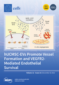
Extracellular vesicles (EVs) have emerged in recent years as key cellular communicators. Similarly, mesenchymal stromal cell (MSC)-derived EVs are novel tools in regenerative medicine. Angiogenesis modulation is of wide interest for tissue regeneration. In our study, hUCMSC-derived EVs affected VEGFR2 signalling by enhancing the phosphorylation of AKT, which reduced apoptosis in human microvascular endothelial cells. Interestingly, hypoxia-derived EVs revealed slightly enhanced effects. Upon longer-term study of an angiogenesis model, both EV populations positively affected vessel formation, evidenced by an increase in vessel networks with larger tubes. Therefore, hUCMSC-derived EVs demonstrate selective targeting of components in the angiogenic signalling pathway and may be of therapeutic interest to support endothelial cell survival. View this paper
  • Issues are regarded as officially published after their release is announced to the table of contents alert mailing list.
  • You may sign up for e-mail alerts to receive table of contents of newly released issues.
  • PDF is the official format for papers published in both, html and pdf forms. To view the papers in pdf format, click on the "PDF Full-text" link, and use the free Adobe Reader to open them.
Order results
Result details
Section
Select all
Export citation of selected articles as:
15 pages, 1698 KiB  
Article
Role of S100A8/A9 in Platelet–Neutrophil Complex Formation during Acute Inflammation
by Julian Revenstorff, Nadine Ludwig, Annika Hilger, Sina Mersmann, Martin Lehmann, Julia Chiara Grenzheuser, Marina Kardell, Julia Bone, Niklas Martin Kötting, Nina Christine Marx, Johannes Roth, Thomas Vogl and Jan Rossaint
Cells 2022, 11(23), 3944; https://doi.org/10.3390/cells11233944 - 06 Dec 2022
Cited by 5 | Viewed by 2870
Abstract
Acute respiratory distress syndrome (ARDS) due to pulmonary infections is associated with high morbidity and mortality. Upon inflammation, the alarmin S100A8/A9 is released and stimulates neutrophil recruitment mainly via binding to Toll-like receptor 4 (TLR4). TLR4 is also expressed on platelets, which modulate [...] Read more.
Acute respiratory distress syndrome (ARDS) due to pulmonary infections is associated with high morbidity and mortality. Upon inflammation, the alarmin S100A8/A9 is released and stimulates neutrophil recruitment mainly via binding to Toll-like receptor 4 (TLR4). TLR4 is also expressed on platelets, which modulate the immune response through direct interaction with leukocytes. In a murine model of Klebsiella pneumoniae-induced pulmonary inflammation, global S100A9 deficiency resulted in diminished neutrophil recruitment into the lung alveoli and neutrophil accumulation in the intravascular space, indicating an impaired neutrophil migration. A lack of TLR4 on platelets resulted in reduced neutrophil counts in the whole lung, emphasising the impact of TLR4-mediated platelet activity on neutrophil behaviour. Flow cytometry-based analysis indicated elevated numbers of platelet–neutrophil complexes in the blood of S100A9−/− mice. Intravital microscopy of the murine cremaster muscle confirmed these findings and further indicated a significant increase in neutrophil–platelet complex formation in S100A9−/− mice, which was reversed by administration of the S100A8/A9 tetramer. An in vitro bilayer assay simulated the murine alveolar capillary barrier during inflammation and validated significant differences in transmigration behaviour between wild-type and S100A9−/− neutrophils. This study demonstrates the role of S100A8/A9 during platelet–neutrophil interactions and neutrophil recruitment during pulmonary inflammation. Full article
Show Figures

Graphical abstract

29 pages, 3484 KiB  
Review
Osteometabolism: Metabolic Alterations in Bone Pathologies
by Rupesh K. Srivastava, Leena Sapra and Pradyumna K. Mishra
Cells 2022, 11(23), 3943; https://doi.org/10.3390/cells11233943 - 06 Dec 2022
Cited by 16 | Viewed by 4492
Abstract
Renewing interest in the study of intermediate metabolism and cellular bioenergetics is brought on by the global increase in the prevalence of metabolic illnesses. Understanding of the mechanisms that integrate energy metabolism in the entire organism has significantly improved with the application of [...] Read more.
Renewing interest in the study of intermediate metabolism and cellular bioenergetics is brought on by the global increase in the prevalence of metabolic illnesses. Understanding of the mechanisms that integrate energy metabolism in the entire organism has significantly improved with the application of contemporary biochemical tools for quantifying the fuel substrate metabolism with cutting-edge mouse genetic procedures. Several unexpected findings in genetically altered mice have prompted research into the direction of intermediate metabolism of skeletal cells. These findings point to the possibility of novel endocrine connections through which bone cells can convey their energy status to other metabolic control centers. Understanding the expanded function of skeleton system has in turn inspired new lines of research aimed at characterizing the energy needs and bioenergetic characteristics of these bone cells. Bone-forming osteoblast and bone-resorbing osteoclast cells require a constant and large supply of energy substrates such as glucose, fatty acids, glutamine, etc., for their differentiation and functional activity. According to latest research, important developmental signaling pathways in bone cells are connected to bioenergetic programs, which may accommodate variations in energy requirements during their life cycle. The present review article provides a unique perspective of the past and present research in the metabolic characteristics of bone cells along with mechanisms governing energy substrate utilization and bioenergetics. In addition, we discussed the therapeutic inventions which are currently being utilized for the treatment and management of bone-related diseases such as osteoporosis, rheumatoid arthritis (RA), osteogenesis imperfecta (OIM), etc., by modulating the energetics of bone cells. We further emphasized on the role of GUT-associated metabolites (GAMs) such as short-chain fatty acids (SCFAs), medium-chain fatty acids (MCFAs), indole derivates, bile acids, etc., in regulating the energetics of bone cells and their plausible role in maintaining bone health. Emphasis is importantly placed on highlighting knowledge gaps in this novel field of skeletal biology, i.e., “Osteometabolism” (proposed by our group) that need to be further explored to characterize the physiological importance of skeletal cell bioenergetics in the context of human health and bone related metabolic diseases. Full article
Show Figures

Figure 1

21 pages, 4836 KiB  
Article
RIPOR2 Expression Decreased by HPV-16 E6 and E7 Oncoproteins: An Opportunity in the Search for Prognostic Biomarkers in Cervical Cancer
by Leslie Olmedo-Nieva, J. Omar Muñoz-Bello, Imelda Martínez-Ramírez, Antonio Daniel Martínez-Gutiérrez, Yunuen Ortiz-Pedraza, Claudia González-Espinosa, Vicente Madrid-Marina, Kirvis Torres-Poveda, Margarita Bahena-Roman and Marcela Lizano
Cells 2022, 11(23), 3942; https://doi.org/10.3390/cells11233942 - 06 Dec 2022
Cited by 4 | Viewed by 1765
Abstract
High-risk human papillomavirus (HPV) infection is the main risk factor for cervical cancer (CC) development, where the continuous expression of E6 and E7 oncoproteins maintain the malignant phenotype. In Mexico, around 70% of CC cases are diagnosed in advanced stages, impacting the survival [...] Read more.
High-risk human papillomavirus (HPV) infection is the main risk factor for cervical cancer (CC) development, where the continuous expression of E6 and E7 oncoproteins maintain the malignant phenotype. In Mexico, around 70% of CC cases are diagnosed in advanced stages, impacting the survival of patients. The aim of this work was to identify biomarkers affected by HPV-16 E6 and E7 oncoproteins that impact the prognosis of CC patients. Expression profiles dependent on E6 and E7 oncoproteins, as well as their relationship with biological processes and cellular signaling pathways, were analyzed in CC cells. A comparison among expression profiles of E6- and E7-expressing cells and that from a CC cohort obtained from The Cancer Genome Atlas (TCGA) demonstrated that the expression of 13 genes impacts the overall survival (OS). A multivariate analysis revealed that the downregulated expression of RIPOR2 was strongly associated with a worse OS. RIPOR2, including its transcriptional variants, were overwhelmingly depleted in E6- and E7-expressing cells. Finally, in a Mexican cohort, it was found that in premalignant cervical lesions, RIPOR2 expression decreases as the lesions progress; meanwhile, decreased RIPOR2 expression was also associated with a worse OS in CC patients. Full article
(This article belongs to the Special Issue Molecular and Immunological Mechanisms of HPV-Induced Carcinogenesis)
Show Figures

Figure 1

12 pages, 595 KiB  
Review
Insulitis in Human Type 1 Diabetic Pancreas: From Stem Cell Grafting to Islet Organoids for a Successful Cell-Based Therapy
by Marcella La Noce, Giovanni Francesco Nicoletti, Gianpaolo Papaccio, Vitale Del Vecchio and Federica Papaccio
Cells 2022, 11(23), 3941; https://doi.org/10.3390/cells11233941 - 06 Dec 2022
Cited by 4 | Viewed by 3216
Abstract
Type 1 diabetes (T1D) is an autoimmune disease with immune cells’ islet infiltration (called “insulitis”), which leads to beta cell loss. Despite being the critical element of T1D occurrence and pathogenesis, insulitis is often present in a limited percentage of islets, also at [...] Read more.
Type 1 diabetes (T1D) is an autoimmune disease with immune cells’ islet infiltration (called “insulitis”), which leads to beta cell loss. Despite being the critical element of T1D occurrence and pathogenesis, insulitis is often present in a limited percentage of islets, also at diagnosis. Therefore, it is needed to define reproducible methods to detect insulitis and beta-cell decline, to allow accurate and early diagnosis and to monitor therapy. However, this goal is still far due to the morphological aspect of islet microvasculature, which is rather dense and rich, and is considerably rearranged during insulitis. More studies on microvasculature are required to understand if contrast-enhanced ultrasound sonography measurements of pancreatic blood-flow dynamics may provide a clinically deployable predictive marker to predict disease progression and therapeutic reversal in pre-symptomatic T1D patients. Therefore, it is needed to clarify the relation between insulitis and the dynamics of β cell loss and with coexisting mechanisms of dysfunction, according to clinical stage, as well as the micro vessels’ dynamics and microvasculature reorganization. Moreover, the ideal cell-based therapy of T1D should start from an early diagnosis allowing a sufficient isolation of specific Procr+ progenitors, followed by the generation and expansion of islet organoids, which could be transplanted coupled to an immune-regulatory therapy which will permit the maintenance of pancreatic islets and an effective and long-lasting insulitis reversal. Full article
Show Figures

Figure 1

15 pages, 2023 KiB  
Article
Differential Expression of Lonp1 Isoforms in Cancer Cells
by Giada Zanini, Valentina Selleri, Anna De Gaetano, Lara Gibellini, Mara Malerba, Anna Vittoria Mattioli, Milena Nasi, Nadezda Apostolova and Marcello Pinti
Cells 2022, 11(23), 3940; https://doi.org/10.3390/cells11233940 - 06 Dec 2022
Cited by 3 | Viewed by 1617
Abstract
Lonp1 is a mitochondrial protease that degrades oxidized and damaged proteins, assists protein folding, and contributes to the maintenance of mitochondrial DNA. A higher expression of LonP1 has been associated with higher tumour aggressiveness. Besides the full-length isoform (ISO1), we identified two other [...] Read more.
Lonp1 is a mitochondrial protease that degrades oxidized and damaged proteins, assists protein folding, and contributes to the maintenance of mitochondrial DNA. A higher expression of LonP1 has been associated with higher tumour aggressiveness. Besides the full-length isoform (ISO1), we identified two other isoforms of Lonp1 in humans, resulting from alternative splicing: Isoform-2 (ISO2) lacking aa 42-105 and isoform-3 (ISO3) lacking aa 1-196. An inspection of the public database TSVdb showed that ISO1 was upregulated in lung, bladder, prostate, and breast cancer, ISO2 in all the cancers analysed (including rectum, colon, cervical, bladder, prostate, breast, head, and neck), ISO3 did not show significant changes between cancer and normal tissue. We overexpressed ISO1, ISO2, and ISO3 in SW620 cells and found that the ISO1 isoform was exclusively mitochondrial, ISO2 was present in the organelle and in the cytoplasm, and ISO3 was exclusively cytoplasmatic. The overexpression of ISO1 and, at a letter extent, of ISO2 enhanced basal, ATP-linked, and maximal respiration without altering the mitochondria number or network, mtDNA amount. or mitochondrial dynamics. A higher extracellular acidification rate was observed in ISO1 and ISO2, overexpressing cells, suggesting an increase in glycolysis. Cells overexpressing the different isoforms did not show a difference in the proliferation rate but showed a great increase in anchorage-independent growth. ISO1 and ISO2, but not ISO3, determined an upregulation of EMT-related proteins, which appeared unrelated to higher mitochondrial ROS production, nor due to the activation of the MEK ERK pathway, but rather to global metabolic reprogramming of cells. Full article
(This article belongs to the Section Mitochondria)
Show Figures

Figure 1

26 pages, 6307 KiB  
Article
Phytocannabinoids Stimulate Rejuvenation and Prevent Cellular Senescence in Human Dermal Fibroblasts
by Marta Gerasymchuk, Gregory Ian Robinson, Alyssa Groves, Lucie Haselhorst, Sanjana Nandakumar, Cora Stahl, Olga Kovalchuk and Igor Kovalchuk
Cells 2022, 11(23), 3939; https://doi.org/10.3390/cells11233939 - 06 Dec 2022
Cited by 10 | Viewed by 21972
Abstract
In light of the increased popularity of phytocannabinoids (pCBs) and their appearance in beauty products without rigorous research on their rejuvenation efficacy, we decided to investigate the potential role of pCBs in skin rejuvenation. Utilizing healthy and stress-induced premature senescent (SIPS) CCD-1064Sk skin [...] Read more.
In light of the increased popularity of phytocannabinoids (pCBs) and their appearance in beauty products without rigorous research on their rejuvenation efficacy, we decided to investigate the potential role of pCBs in skin rejuvenation. Utilizing healthy and stress-induced premature senescent (SIPS) CCD-1064Sk skin fibroblasts, the effects of pCBs on cellular viability, functional activity, metabolic function, and nuclear architecture were tested. Both delta-9-tetrahydrocannabinol (THC) and cannabidiol (CBD) within the range of 0.5 µM to 2.0 µM increased cell growth in a dose-dependent manner while significantly decreasing senescence as measured by beta-galactosidase activity. Utilizing a scratch assay, both THC and CBD (2.0 µM) significantly improved wound healing in both healthy and SIPS fibroblasts. THC and CBD altered nuclear architecture and mRNA levels of cell cycle regulators and genes involved in ECM production. Subsequently, we found ELN, Cyclin D1, PCNA, and BID protein levels altered by SIPS but ameliorated after pCBs exposure in human dermal fibroblasts. Lastly, we compared the efficacy of THC and CBD with common anti-aging nutrient signaling regulators in replicative senescent adult human dermal fibroblasts, CCD-1135Sk. Both THC and CBD were found to improve wound healing better than metformin, rapamycin, and triacetylresveratrol in replicative senescent CCD-1135Sk fibroblasts. Therefore, pCBs can be a valuable source of biologically active substances used in cosmetics, and more studies using clinical trials should be performed to confirm the efficacy of phytocannabinoids. Full article
(This article belongs to the Special Issue Molecular and Cell Basis of Skin Diseases and Aging)
Show Figures

Figure 1

20 pages, 1436 KiB  
Review
Mushroom Natural Products in Neurodegenerative Disease Drug Discovery
by Arjuna Abitbol, Brody Mallard, Evelin Tiralongo and Joe Tiralongo
Cells 2022, 11(23), 3938; https://doi.org/10.3390/cells11233938 - 06 Dec 2022
Cited by 5 | Viewed by 3229
Abstract
The variety of drugs available to treat neurodegenerative diseases is limited. Most of these drug’s efficacy is restricted by individual genetics and disease stages and usually do not prevent neurodegeneration acting long after irreversible damage has already occurred. Thus, drugs targeting the molecular [...] Read more.
The variety of drugs available to treat neurodegenerative diseases is limited. Most of these drug’s efficacy is restricted by individual genetics and disease stages and usually do not prevent neurodegeneration acting long after irreversible damage has already occurred. Thus, drugs targeting the molecular mechanisms underlying subsequent neurodegeneration have the potential to negate symptom manifestation and subsequent neurodegeneration. Neuroinflammation is a common feature of neurodegenerative diseases such as Alzheimer’s disease, Parkinson’s disease, Huntington’s disease, and multiple sclerosis, and is associated with the activation of the NLRP3 inflammasome, which in turn leads to neurodegeneration. Inflammasome activation and oligomerisation is suggested to be a major driver of disease progression occurring in microglia. With several natural products and natural product derivatives currently in clinical trials, mushrooms have been highlighted as a rich and largely untapped source of biologically active compounds in both in vitro and in vivo neurodegenerative disease models, partially supported by successful clinical trial evaluations. Additionally, novel high-throughput methods for the screening of natural product compound libraries are being developed to help accelerate the neurodegenerative disease drug discovery process, targeting neuroinflammation. However, the breadth of research relating to mushroom natural product high-throughput screening is limited, providing an exciting opportunity for further detailed investigations. Full article
Show Figures

Figure 1

13 pages, 1923 KiB  
Article
Caveolin-1 rs1997623 Single Nucleotide Polymorphism Creates a New Binding Site for the Early B-Cell Factor 1 That Instigates Adipose Tissue CAV1 Protein Overexpression
by Ashraf Al Madhoun, Dania Haddad, Rasheeba Nizam, Lavina Miranda, Shihab Kochumon, Reeby Thomas, Thangavel Alphonse Thanaraj, Rasheed Ahmad, Milad S. Bitar and Fahd Al-Mulla
Cells 2022, 11(23), 3937; https://doi.org/10.3390/cells11233937 - 06 Dec 2022
Cited by 1 | Viewed by 1496
Abstract
Caveolin-1 (CAV1) is implicated in the pathophysiology of diabetes and obesity. Previously, we demonstrated an association between the CAV1 rs1997623 C > A variant and metabolic syndrome (MetS). Here, we decipher the functional role of rs1997623 in CAV1 gene regulation. A cohort of [...] Read more.
Caveolin-1 (CAV1) is implicated in the pathophysiology of diabetes and obesity. Previously, we demonstrated an association between the CAV1 rs1997623 C > A variant and metabolic syndrome (MetS). Here, we decipher the functional role of rs1997623 in CAV1 gene regulation. A cohort of 38 patients participated in this study. The quantitative MetS scores (siMS) of the participants were computed. CAV1 transcript and protein expression were tested in subcutaneous adipose tissue using RT-PCR and immunohistochemistry. Chromatin immunoprecipitation assays were performed using primary preadipocytes isolated from individuals with different CAV1 rs1997623 genotypes (AA, AC, and CC). The regulatory region flanking the variant was cloned into a luciferase reporter plasmid and expressed in human preadipocytes. Additional knockdown and overexpression assays were carried out. We show a significant correlation between siMS and CAV1 transcript levels and protein levels in human adipose tissue collected from an Arab cohort. We found that the CAV1 rs1997623 A allele generates a transcriptionally active locus and a new transcription factor binding site for early B-cell factor 1 (EBF1), which enhanced CAV1 expression. Our in vivo and in vitro combined study implicates, for the first time, EBF1 in regulating CAV1 expression in individuals harboring the rs1997623 C > A variant. Full article
Show Figures

Figure 1

21 pages, 4122 KiB  
Article
HMGB1 Promotes In Vitro and In Vivo Skeletal Muscle Atrophy through an IL-18-Dependent Mechanism
by Trung-Loc Ho, Chih-Hsin Tang, Sunny Li-Yun Chang, Chun-Hao Tsai, Hsien-Te Chen and Chen-Ming Su
Cells 2022, 11(23), 3936; https://doi.org/10.3390/cells11233936 - 06 Dec 2022
Cited by 4 | Viewed by 2762
Abstract
Skeletal muscle atrophy occurs due to muscle wasting or reductions in protein associated with aging, injury, and inflammatory processes. High-mobility group box-1 (HMGB1) protein is passively released from necrotic cells and actively secreted by inflammatory cells, and is implicated in the pathogenesis of [...] Read more.
Skeletal muscle atrophy occurs due to muscle wasting or reductions in protein associated with aging, injury, and inflammatory processes. High-mobility group box-1 (HMGB1) protein is passively released from necrotic cells and actively secreted by inflammatory cells, and is implicated in the pathogenesis of various inflammatory and immune diseases. HMGB1 is upregulated in muscle inflammation, and circulating levels of the proinflammatory cytokine interleukin-18 (IL-18) are upregulated in patients with sarcopenia, a muscle-wasting disease. We examined whether an association exists between HMGB1 and IL-18 signaling in skeletal muscle atrophy. HMGB1-induced increases of IL-18 levels enhanced the expression of muscle atrophy markers and inhibited myogenic marker expression in C2C12 and G7 myoblast cell lines. HMGB1-induced increases of IL-18 production in C2C12 cells involved the RAGE/p85/Akt/mTOR/c-Jun signaling pathway. HMGB1 short hairpin RNA (shRNA) treatment rescued the expression of muscle-specific differentiation markers in murine C2C12 myotubes and in mice with glycerol-induced muscle atrophy. HMGB1 and IL-18 signaling was suppressed in the mice after HMGB1 shRNA treatment. These findings suggest that the HMGB1/IL-18 axis is worth targeting for the treatment of skeletal muscle atrophy. Full article
Show Figures

Figure 1

16 pages, 1177 KiB  
Review
Cross-Generational Impact of Innate Immune Memory Following Pregnancy Complications
by Nakeisha A. Lodge-Tulloch, Alexa J. Toews, Aline Atallah, Tiziana Cotechini, Sylvie Girard and Charles H. Graham
Cells 2022, 11(23), 3935; https://doi.org/10.3390/cells11233935 - 06 Dec 2022
Cited by 5 | Viewed by 2217
Abstract
Pregnancy complications can have long-term negative effects on the health of the affected mothers and their children. In this review, we highlight the underlying inflammatory etiologies of common pregnancy complications and discuss how aberrant inflammation may lead to the acquisition of innate immune [...] Read more.
Pregnancy complications can have long-term negative effects on the health of the affected mothers and their children. In this review, we highlight the underlying inflammatory etiologies of common pregnancy complications and discuss how aberrant inflammation may lead to the acquisition of innate immune memory. The latter can be described as a functional epigenetic reprogramming of innate immune cells following an initial exposure to an inflammatory stimulus, ultimately resulting in an altered response following re-exposure to a similar inflammatory stimulus. We propose that aberrant maternal inflammation associated with complications of pregnancy increases the cross-generational risk of developing noncommunicable diseases (i.e., pregnancy complications, cardiovascular disease, and metabolic disease) through a process mediated by innate immune memory. Elucidating a role for innate immune memory in the cross-generational health consequences of pregnancy complications may lead to the development of novel strategies aimed at reducing the long-term risk of disease. Full article
(This article belongs to the Special Issue Signaling Pathways in Pregnancy)
Show Figures

Figure 1

19 pages, 1844 KiB  
Review
Wnt Signaling in the Development of Bone Metastasis
by Noa Ben-Ghedalia-Peled and Razi Vago
Cells 2022, 11(23), 3934; https://doi.org/10.3390/cells11233934 - 05 Dec 2022
Cited by 9 | Viewed by 2769
Abstract
Wnt signaling occurs through evolutionarily conserved pathways that affect cellular proliferation and fate decisions during development and tissue maintenance. Alterations in these highly regulated pathways, however, play pivotal roles in various malignancies, promoting cancer initiation, growth and metastasis and the development of drug [...] Read more.
Wnt signaling occurs through evolutionarily conserved pathways that affect cellular proliferation and fate decisions during development and tissue maintenance. Alterations in these highly regulated pathways, however, play pivotal roles in various malignancies, promoting cancer initiation, growth and metastasis and the development of drug resistance. The ability of cancer cells to metastasize is the primary cause of cancer mortality. Bone is one of the most frequent sites of metastases that generally arise from breast, prostate, lung, melanoma or kidney cancer. Upon their arrival to the bone, cancer cells can enter a long-term dormancy period, from which they can be reactivated, but can rarely be cured. The activation of Wnt signaling during the bone metastasis process was found to enhance proliferation, induce the epithelial-to-mesenchymal transition, promote the modulation of the extracellular matrix, enhance angiogenesis and immune tolerance and metastasize and thrive in the bone. Due to the complexity of Wnt pathways and of the landscape of this mineralized tissue, Wnt function during metastatic progression within bone is not yet fully understood. Therefore, we believe that a better understanding of these pathways and their roles in the development of bone metastasis could improve our understanding of the disease and may constitute fertile ground for potential therapeutics. Full article
(This article belongs to the Special Issue From Mechanisms to Therapeutics: Wnt Signaling in Cancer)
Show Figures

Figure 1

21 pages, 3622 KiB  
Article
Targeting of the ELR+CXCL/CXCR1/2 Pathway Is a Relevant Strategy for the Treatment of Paediatric Medulloblastomas
by Manon Penco-Campillo, Clément Molina, Patricia Piris, Nouha Soufi, Manon Carré, Marina Pagnuzzi-Boncompagni, Vincent Picco, Maeva Dufies, Cyril Ronco, Rachid Benhida, Sonia Martial and Gilles Pagès
Cells 2022, 11(23), 3933; https://doi.org/10.3390/cells11233933 - 05 Dec 2022
Cited by 1 | Viewed by 2012
Abstract
Medulloblastoma (MB) is the most common and aggressive paediatric brain tumour. Although the cure rate can be as high as 70%, current treatments (surgery, radio- and chemotherapy) excessively affect the patients’ quality of life. Relapses cannot be controlled by conventional or targeted treatments [...] Read more.
Medulloblastoma (MB) is the most common and aggressive paediatric brain tumour. Although the cure rate can be as high as 70%, current treatments (surgery, radio- and chemotherapy) excessively affect the patients’ quality of life. Relapses cannot be controlled by conventional or targeted treatments and are usually fatal. The strong heterogeneity of the disease (four subgroups and several subtypes) is related to innate or acquired resistance to reference treatments. Therefore, more efficient and less-toxic therapies are needed. Here, we demonstrated the efficacy of a novel inhibitor (C29) of CXCR1/2 receptors for ELR+CXCL cytokines for the treatment of childhood MB. The correlation between ELR+CXCL/CXCR1/2 expression and patient survival was determined using the R2: Genomics Analysis and Visualization platform. In vitro efficacy of C29 was evaluated by its ability to inhibit proliferation, migration, invasion, and pseudo-vessel formation of MB cell lines sensitive or resistant to radiotherapy. The growth of experimental MB obtained by MB spheroids on organotypic mouse cerebellar slices was also assayed. ELR+CXCL/CXCR1/2 levels correlated with shorter survival. C29 inhibited proliferation, clone formation, CXCL8/CXCR1/2-dependent migration, invasion, and pseudo-vessel formation by sensitive and radioresistant MB cells. C29 reduced experimental growth of MB in the ex vivo organotypic mouse model and crossed the blood–brain barrier. Targeting CXCR1/2 represents a promising therapeutic strategy for the treatment of paediatric MB in first-line treatment or after relapse following conventional therapy. Full article
(This article belongs to the Special Issue Mechanisms of Cancer Cell Invasion)
Show Figures

Figure 1

21 pages, 3409 KiB  
Article
PARP1 Activation Controls Stress Granule Assembly after Oxidative Stress and DNA Damage
by Anastasia S. Singatulina, Maria V. Sukhanova, Bénédicte Desforges, Vandana Joshi, David Pastré and Olga I. Lavrik
Cells 2022, 11(23), 3932; https://doi.org/10.3390/cells11233932 - 05 Dec 2022
Cited by 1 | Viewed by 2298
Abstract
DNA damage causes PARP1 activation in the nucleus to set up the machinery responsible for the DNA damage response. Here, we report that, in contrast to cytoplasmic PARPs, the synthesis of poly(ADP-ribose) by PARP1 opposes the formation of cytoplasmic mRNA-rich granules after arsenite [...] Read more.
DNA damage causes PARP1 activation in the nucleus to set up the machinery responsible for the DNA damage response. Here, we report that, in contrast to cytoplasmic PARPs, the synthesis of poly(ADP-ribose) by PARP1 opposes the formation of cytoplasmic mRNA-rich granules after arsenite exposure by reducing polysome dissociation. However, when mRNA-rich granules are pre-formed, whether in the cytoplasm or nucleus, PARP1 activation positively regulates their assembly, though without additional recruitment of poly(ADP-ribose) in stress granules. In addition, PARP1 promotes the formation of TDP-43- and FUS-rich granules in the cytoplasm, two RNA-binding proteins which form neuronal cytoplasmic inclusions observed in certain neurodegenerative diseases such as amyotrophic lateral sclerosis and frontotemporal lobar degeneration. Together, the results therefore reveal a dual role of PARP1 activation which, on the one hand, prevents the early stage of stress granule assembly and, on the other hand, enables the persistence of cytoplasmic mRNA-rich granules in cells which may be detrimental in aging neurons. Full article
(This article belongs to the Special Issue Systems Biology of Apoptotic and Non-apoptotic Signaling)
Show Figures

Figure 1

6 pages, 253 KiB  
Editorial
The Centrosome: Conclusions and Perspectives
by Rustem E. Uzbekov and Tomer Avidor-Reiss
Cells 2022, 11(23), 3931; https://doi.org/10.3390/cells11233931 - 05 Dec 2022
Cited by 1 | Viewed by 1190
Abstract
The centrosome consists of two centrioles surrounded by pericentriolar material [...] Full article
(This article belongs to the Section Intracellular and Plasma Membranes)
16 pages, 1070 KiB  
Review
An Altered Skin and Gut Microbiota Are Involved in the Modulation of Itch in Atopic Dermatitis
by Catharina Sagita Moniaga, Mitsutoshi Tominaga and Kenji Takamori
Cells 2022, 11(23), 3930; https://doi.org/10.3390/cells11233930 - 05 Dec 2022
Cited by 16 | Viewed by 5934
Abstract
Skin and gut microbiota play an important role in the pathogenesis of atopic dermatitis (AD). An alteration of the microbiota diversity modulates the development and course of AD, e.g., decreased microbiome diversity correlates with disease severity, particularly in lesional skin of AD. Itch [...] Read more.
Skin and gut microbiota play an important role in the pathogenesis of atopic dermatitis (AD). An alteration of the microbiota diversity modulates the development and course of AD, e.g., decreased microbiome diversity correlates with disease severity, particularly in lesional skin of AD. Itch is a hallmark of AD with unsatisfying treatment until now. Recent evidence suggests a possible role of microbiota in altering itch in AD through gut–skin–brain interactions. The microbial metabolites, proinflammatory cytokines, and impaired immune response lead to a modulation of histamine-independent itch, disruption of epidermal barrier, and central sensitization of itch mechanisms. The positive impact of probiotics in alleviating itch in AD supports this hypothesis, which may lead to novel strategies for managing itchy skin in AD patients. This review summarizes the emerging findings on the correlation between an altered microbiota and gut–skin–brain axis in AD, especially in modulating itchy skin. Full article
(This article belongs to the Special Issue Atopic Dermatitis: Pathogenesis and Emerging Therapies)
Show Figures

Figure 1

18 pages, 1700 KiB  
Review
FGFR Inhibitors in Cholangiocarcinoma—A Novel Yet Primary Approach: Where Do We Stand Now and Where to Head Next in Targeting This Axis?
by Paulina Chmiel, Katarzyna Gęca, Karol Rawicz-Pruszyński, Wojciech P. Polkowski and Magdalena Skórzewska
Cells 2022, 11(23), 3929; https://doi.org/10.3390/cells11233929 - 05 Dec 2022
Cited by 5 | Viewed by 2301
Abstract
Cholangiocarcinomas (CCAs) are rare but aggressive tumours with poor diagnosis and limited treatment options. Molecular targeted therapies became a promising proposal for patients after progression under first-line chemical treatment. In light of an escalating prevalence of CCA, it is crucial to fully comprehend [...] Read more.
Cholangiocarcinomas (CCAs) are rare but aggressive tumours with poor diagnosis and limited treatment options. Molecular targeted therapies became a promising proposal for patients after progression under first-line chemical treatment. In light of an escalating prevalence of CCA, it is crucial to fully comprehend its pathophysiology, aetiology, and possible targets in therapy. Such knowledge would play a pivotal role in searching for new therapeutic approaches concerning diseases’ symptoms and their underlying causes. Growing evidence showed that fibroblast growth factor/fibroblast growth factor receptor (FGF/FGFR) pathway dysregulation is involved in a variety of processes during embryonic development and homeostasis as well as tumorigenesis. CCA is known for its close correlation with the FGF/FGFR pathway and targeting this axis has been proposed in treatment guidelines. Bearing in mind the significance of molecular targeted therapies in different neoplasms, it seems most reasonable to move towards intensive research and testing on these in the case of CCA. However, there is still a need for more data covering this topic. Although positive results of many pre-clinical and clinical studies are discussed in this review, many difficulties lie ahead. Furthermore, this review presents up-to-date literature regarding the outcomes of the latest clinical data and discussion over future directions of FGFR-directed therapies in patients with CCA. Full article
Show Figures

Figure 1

28 pages, 2453 KiB  
Review
CRISPR/Cas Genome Editing Technologies for Plant Improvement against Biotic and Abiotic Stresses: Advances, Limitations, and Future Perspectives
by Yaxin Wang, Naeem Zafar, Qurban Ali, Hakim Manghwar, Guanying Wang, Lu Yu, Xiao Ding, Fang Ding, Ni Hong, Guoping Wang and Shuangxia Jin
Cells 2022, 11(23), 3928; https://doi.org/10.3390/cells11233928 - 05 Dec 2022
Cited by 14 | Viewed by 5965
Abstract
Crossbreeding, mutation breeding, and traditional transgenic breeding take much time to improve desirable characters/traits. CRISPR/Cas-mediated genome editing (GE) is a game-changing tool that can create variation in desired traits, such as biotic and abiotic resistance, increase quality and yield in less time with [...] Read more.
Crossbreeding, mutation breeding, and traditional transgenic breeding take much time to improve desirable characters/traits. CRISPR/Cas-mediated genome editing (GE) is a game-changing tool that can create variation in desired traits, such as biotic and abiotic resistance, increase quality and yield in less time with easy applications, high efficiency, and low cost in producing the targeted edits for rapid improvement of crop plants. Plant pathogens and the severe environment cause considerable crop losses worldwide. GE approaches have emerged and opened new doors for breeding multiple-resistance crop varieties. Here, we have summarized recent advances in CRISPR/Cas-mediated GE for resistance against biotic and abiotic stresses in a crop molecular breeding program that includes the modification and improvement of genes response to biotic stresses induced by fungus, virus, and bacterial pathogens. We also discussed in depth the application of CRISPR/Cas for abiotic stresses (herbicide, drought, heat, and cold) in plants. In addition, we discussed the limitations and future challenges faced by breeders using GE tools for crop improvement and suggested directions for future improvements in GE for agricultural applications, providing novel ideas to create super cultivars with broad resistance to biotic and abiotic stress. Full article
(This article belongs to the Special Issue Plant Genome Editing: State-of-the-Art and Perspectives in China)
Show Figures

Figure 1

21 pages, 6670 KiB  
Article
Nrf2 Deficiency Exacerbated CLP-Induced Pulmonary Injury and Inflammation through Autophagy- and NF-κB/PPARγ-Mediated Macrophage Polarization
by Jing Luo, Jin Wang, Jing Zhang, Aming Sang, Xujun Ye, Zhenshun Cheng and Xinyi Li
Cells 2022, 11(23), 3927; https://doi.org/10.3390/cells11233927 - 04 Dec 2022
Cited by 10 | Viewed by 3114
Abstract
The balance between M1 and M2 macrophage polarization is involved in the regulation of pulmonary inflammation. Nuclear factor erythroid-derived 2-like 2 (Nfe2l2, also known as Nrf2), a nuclear transcription factor, is reported to play protective roles in acute lung injury (ALI) and inflammation, [...] Read more.
The balance between M1 and M2 macrophage polarization is involved in the regulation of pulmonary inflammation. Nuclear factor erythroid-derived 2-like 2 (Nfe2l2, also known as Nrf2), a nuclear transcription factor, is reported to play protective roles in acute lung injury (ALI) and inflammation, and increasing evidence indicates that the protective effects of Nrf2 are closely related to autophagy. This study aimed to explore whether Nrf2 is involved in sepsis-induced acute pulmonary injury and inflammation and in the role of macrophage polarization in the process. In the present study, sepsis patients, an Nrf2 knockout mouse that underwent cecal ligation and puncture (CLP), and lipopolysaccharide (LPS)-treated macrophage cell lines were employed to investigate the potential functions of Nrf2 in sepsis-induced lung injury and the underlying mechanisms. Clinical studies showed that the NRF2 mRNA level was inversely correlated with pulmonary inflammation and disease severity in patients with sepsis. Analyses in a CLP-treated Nrf2 knockout mouse model indicated that an Nrf2 deficiency promoted a CLP-induced increase in M1 macrophage polarization and apoptosis and inhibited CLP-induced upregulation of the autophagy level in lung tissues. Experiments in RAW264.7 cells revealed that Nrf2 overexpression inhibited M1 macrophage polarization but promoted M2 macrophage polarization by improving the autophagy, and Nrf2 overexpression promoted PPARγ but inhibited NF-κB nuclear translocation. In conclusion, these results indicate that Nrf2 plays a protective role in sepsis-induced pulmonary injury and inflammation through the regulation of autophagy- and NF-κB/PPARγ-mediated macrophage polarization. Full article
(This article belongs to the Special Issue The Autophagic Process in Human Physiology and Pathogenesis)
Show Figures

Figure 1

23 pages, 3790 KiB  
Article
Oxidative Stress and Extracellular Matrix Remodeling Are Signature Pathways of Extracellular Vesicles Released upon Morphine Exposure on Human Brain Microvascular Endothelial Cells
by Tatjana Vujić, Domitille Schvartz, Izadora Liranço Furlani, Isabel Meister, Víctor González-Ruiz, Serge Rudaz and Jean-Charles Sanchez
Cells 2022, 11(23), 3926; https://doi.org/10.3390/cells11233926 - 04 Dec 2022
Cited by 8 | Viewed by 2034
Abstract
Morphine, a commonly used antinociceptive drug in hospitals, is known to cross the blood-brain barrier (BBB) by first passing through brain endothelial cells. Despite its pain-relieving effect, morphine also has detrimental effects, such as the potential induction of redox imbalance in the brain. [...] Read more.
Morphine, a commonly used antinociceptive drug in hospitals, is known to cross the blood-brain barrier (BBB) by first passing through brain endothelial cells. Despite its pain-relieving effect, morphine also has detrimental effects, such as the potential induction of redox imbalance in the brain. However, there is still insufficient evidence of these effects on the brain, particularly on the brain endothelial cells and the extracellular vesicles that they naturally release. Indeed, extracellular vesicles (EVs) are nanosized bioparticles produced by almost all cell types and are currently thought to reflect the physiological state of their parent cells. These vesicles have emerged as a promising source of biomarkers by indicating the functional or dysfunctional state of their parent cells and, thus, allowing a better understanding of the biological processes involved in an adverse state. However, there is very little information on the morphine effect on human brain microvascular endothelial cells (HBMECs), and even less on their released EVs. Therefore, the current study aimed at unraveling the detrimental mechanisms of morphine exposure (at 1, 10, 25, 50 and 100 µM) for 24 h on human brain microvascular endothelial cells as well as on their associated EVs. Isolation of EVs was carried out using an affinity-based method. Several orthogonal techniques (NTA, western blotting and proteomics analysis) were used to validate the EVs enrichment, quality and concentration. Data-independent mass spectrometry (DIA-MS)-based proteomics was applied in order to analyze the proteome modulations induced by morphine on HBMECs and EVs. We were able to quantify almost 5500 proteins in HBMECs and 1500 proteins in EVs, of which 256 and 148, respectively, were found to be differentially expressed in at least one condition. Pathway enrichment analysis revealed that the “cell adhesion and extracellular matrix remodeling” process and the “HIF1 pathway”, a pathway related to oxidative stress responses, were significantly modulated upon morphine exposure in HBMECs and EVs. Altogether, the combination of proteomics and bioinformatics findings highlighted shared pathways between HBMECs exposed to morphine and their released EVs. These results put forward molecular signatures of morphine-induced toxicity in HBMECs that were also carried by EVs. Therefore, EVs could potentially be regarded as a useful tool to investigate brain endothelial cells dysfunction, and to a different extent, the BBB dysfunction in patient circulation using these “signature pathways”. Full article
(This article belongs to the Collection Emerging Topics in Vascular Endothelial Cell Biology)
Show Figures

Figure 1

18 pages, 4528 KiB  
Article
Modeling the Endothelial Glycocalyx Layer in the Human Conventional Aqueous Outflow Pathway
by Alireza Karimi, Mahdi Halabian, Reza Razaghi, J. Crawford Downs, Mary J. Kelley and Ted S. Acott
Cells 2022, 11(23), 3925; https://doi.org/10.3390/cells11233925 - 04 Dec 2022
Cited by 2 | Viewed by 1685
Abstract
A layer of proteoglycans and glycoproteins known as glycocalyx covers the surface of the trabecular meshwork (TM), juxtacanalicular tissue (JCT), and Schlemm’s canal (SC) inner wall of the conventional aqueous outflow pathway in the eye. This has been shown to play a role [...] Read more.
A layer of proteoglycans and glycoproteins known as glycocalyx covers the surface of the trabecular meshwork (TM), juxtacanalicular tissue (JCT), and Schlemm’s canal (SC) inner wall of the conventional aqueous outflow pathway in the eye. This has been shown to play a role in the mechanotransduction of fluid shear stress and in the regulation of the outflow resistance. The outflow resistance in the conventional outflow pathway is the main determinant of the intraocular pressure (IOP) through an active, two-way, fluid–structure interaction coupling between the outflow tissues and aqueous humor. A 3D microstructural finite element (FE) model of a healthy human eye TM/JCT/SC complex with interspersed aqueous humor was constructed. A very thin charged double layer that represents the endothelial glycocalyx layer covered the surface of the elastic outflow tissues. The aqueous humor was modeled as electroosmotic flow that is charged when it is in contact with the outflow tissues. The electrical–fluid–structure interaction (EFSI) method was used to couple the charged double layer (glycocalyx), fluid (aqueous humor), and solid (outflow tissues). When the IOP was elevated to 15 mmHg, the maximum aqueous humor velocity in the EFSI model was decreased by 2.35 mm/s (9%) compared to the fluid–structure interaction (FSI) model. The charge or electricity in the living human conventional outflow pathway generated by the charged endothelial glycocalyx layer plays a minor biomechanical role in the resultant stresses and strains as well as the hydrodynamics of the aqueous humor. Full article
Show Figures

Figure 1

19 pages, 10547 KiB  
Article
Unbalanced Glutamine Partitioning between CD8T Cells and Cancer Cells Accompanied by Immune Cell Dysfunction in Hepatocellular Carcinoma
by Jianfei Chen, Rui Wang, Zhongliang Liu, Jun Fan, Shenglu Liu, Shunde Tan, Xinkai Li, Bo Li and Xiaoli Yang
Cells 2022, 11(23), 3924; https://doi.org/10.3390/cells11233924 - 04 Dec 2022
Cited by 13 | Viewed by 2042
Abstract
Glutamine metabolism is critical both for the proliferation of cancer cells and the activation of CD8T cells to kill cancer cells. We aim to explore the relationship between the glutamine metabolism of CD8T cells and cancer cells and tumor immunity in the tumor [...] Read more.
Glutamine metabolism is critical both for the proliferation of cancer cells and the activation of CD8T cells to kill cancer cells. We aim to explore the relationship between the glutamine metabolism of CD8T cells and cancer cells and tumor immunity in the tumor microenvironment. In a TCGA cohort, we found that patients with high scores of glutamine-metabolism-related genes showed poor prognoses, and that a high score of glutamine-metabolism-related genes was an independent risk factor for HCC patients. In single-cell RNA-seq data, we found that, in some patients, the glutamine metabolism gene scores of tumor cells were significantly higher than those of CD8T cells, while decreased ratios of CD8-Tef-GZMA and suppressed tumor-killing activity of CD8-Tef-APOC2 were observed. A further genetic dynamics pseudotime analysis suggested that immune remodeling of these two subpopulations was accompanied by metabolic reprogramming. CD8-Tef-APOC2 in the dominant group tended to metabolize exogenous lipids, while the metabolic program of CD8-Tef-GZMA in the nondominant group was characterized by amino acid and endogenous lipid synthesis. In addition, we found that the glutamine metabolism inhibitor JHU083 promoted the proliferation of CD8T cells and improved the efficacy of PD-1 blockers. We proposed a new tool to quantify the glutamine partitioning between tumor cells and CD8T cells, through which the unique immune microenvironment could be identified at the transcriptome level. Furthermore, the simultaneous destruction of the glutamine metabolism in tumor cells and CD8T cells facilitated the enrichment of tumor-infiltrating CD8T cells and enhanced the efficacy of immunotherapy. Full article
(This article belongs to the Topic Cancer Cell Metabolism)
Show Figures

Figure 1

29 pages, 5103 KiB  
Article
Micro-RNAs Shuttled by Extracellular Vesicles Secreted from Mesenchymal Stem Cells Dampen Astrocyte Pathological Activation and Support Neuroprotection in In-Vitro Models of ALS
by Francesca Provenzano, Sophie Nyberg, Debora Giunti, Carola Torazza, Benedetta Parodi, Tiziana Bonifacino, Cesare Usai, Nicole Kerlero de Rosbo, Marco Milanese, Antonio Uccelli, Pamela J. Shaw, Laura Ferraiuolo and Giambattista Bonanno
Cells 2022, 11(23), 3923; https://doi.org/10.3390/cells11233923 - 04 Dec 2022
Cited by 14 | Viewed by 2881
Abstract
Amyotrophic lateral sclerosis (ALS) is a neurodegenerative disease with no effective cure. Astrocytes display a toxic phenotype in ALS and contribute to motoneuron (MN) degeneration. Modulating astrocytes’ neurotoxicity can reduce MN death. Our previous studies showed the beneficial effect of mesenchymal stem cell [...] Read more.
Amyotrophic lateral sclerosis (ALS) is a neurodegenerative disease with no effective cure. Astrocytes display a toxic phenotype in ALS and contribute to motoneuron (MN) degeneration. Modulating astrocytes’ neurotoxicity can reduce MN death. Our previous studies showed the beneficial effect of mesenchymal stem cell (MSC) administration in SOD1G93A ALS mice, but the mechanisms are still unclear. We postulated that the effects could be mediated by extracellular vesicles (EVs) secreted by MSCs. We investigated, by immunohistochemical, molecular, and in vitro functional analyses, the activity of MSC-derived EVs on the pathological phenotype and neurotoxicity of astrocytes isolated from the spinal cord of symptomatic SOD1G93A mice and human astrocytes (iAstrocytes) differentiated from inducible neural progenitor cells (iNPCs) of ALS patients. In vitro EV exposure rescued mouse and human ALS astrocytes’ neurotoxicity towards MNs. EVs significantly dampened the pathological phenotype and neuroinflammation in SOD1G93A astrocytes. In iAstrocytes, exposure to EVs increased the antioxidant factor Nrf2 and reduced reactive oxygen species. We previously found nine miRNAs upregulated in MSC-derived EVs. Here, the transfection of SOD1G93A astrocytes with single miRNA mimics reduced astrocytes’ activation and the expression of neuroinflammatory factors. Moreover, miR-466q and miR-467f mimics downregulate Mapk11, while miR-466m-5p and miR-466i-3p mimics promote the nuclear translocation of Nrf2. In iAstrocytes, transfection with miR-29b-3p mimic upregulated NQO1 antioxidant activity and reduced neurotoxicity towards MNs. MSC-derived EVs modulate astrocytes’ reactive phenotype and neurotoxicity through anti-inflammatory and antioxidant-shuttled miRNAs, thus representing a therapeutic strategy in ALS. Full article
Show Figures

Figure 1

11 pages, 785 KiB  
Review
Ectopic Tumor VCAM-1 Expression in Cancer Metastasis and Therapy Resistance
by Kristen A. VanHeyst, Sung Hee Choi, Daniel T. Kingsley and Alex Y. Huang
Cells 2022, 11(23), 3922; https://doi.org/10.3390/cells11233922 - 04 Dec 2022
Cited by 8 | Viewed by 2723
Abstract
Vascular Cell Adhesion Molecule-1 (VCAM-1; CD106) is a membrane protein that contributes critical physiologic functional roles in cellular immune response, including leukocyte extravasation in inflamed and infected tissues. Expressed as a cell membrane protein, VCAM-1 can also be cleaved from the cell surface [...] Read more.
Vascular Cell Adhesion Molecule-1 (VCAM-1; CD106) is a membrane protein that contributes critical physiologic functional roles in cellular immune response, including leukocyte extravasation in inflamed and infected tissues. Expressed as a cell membrane protein, VCAM-1 can also be cleaved from the cell surface into a soluble form (sVCAM-1). The integrin α4β1 (VLA-4) was identified as the first major ligand for VCAM-1. Ongoing studies suggest that, in addition to mediating physiologic immune functions, VCAM-1/VLA-4 signaling plays an increasingly vital role in the metastatic progression of various tumors. Additionally, elevated concentrations of sVCAM-1 have been found in the peripheral blood of patients with cancer, suggesting the tumor microenvironment (TME) as the source of sVCAM-1. Furthermore, over-expression of VLA-4 was linked to tumor progression in various malignancies when VCAM-1 was also up-regulated. This review explores the functional role of VCAM-1 expression in cancer metastasis and therapy resistance, and the potential for the disruption of VCAM-1/VLA-4 signaling as a novel immunotherapeutic approach in cancer, including osteosarcoma, which disproportionately affects the pediatric, adolescent and young adult population, as an unmet medical need. Full article
(This article belongs to the Special Issue Harnessing the Immune System to Fight Pediatric Cancer)
Show Figures

Figure 1

14 pages, 906 KiB  
Article
Enterotoxin Gene Cluster and selX Are Associated with Atopic Dermatitis Severity—A Cross-Sectional Molecular Study of Staphylococcus aureus Superantigens
by Leszek Blicharz, Maciej Żochowski, Ksenia Szymanek-Majchrzak, Joanna Czuwara, Mohamad Goldust, Krzysztof Skowroński, Grażyna Młynarczyk, Małgorzata Olszewska, Zbigniew Samochocki and Lidia Rudnicka
Cells 2022, 11(23), 3921; https://doi.org/10.3390/cells11233921 - 03 Dec 2022
Cited by 5 | Viewed by 1604
Abstract
Staphylococcus aureus superantigens (SAgs) have been reported to aggravate atopic dermatitis. However, comprehensive analyses of these molecules in multiple microniches are lacking. The present study involved 50 adult patients with active atopic dermatitis. S. aureus was isolated from the lesional skin, nonlesional skin, [...] Read more.
Staphylococcus aureus superantigens (SAgs) have been reported to aggravate atopic dermatitis. However, comprehensive analyses of these molecules in multiple microniches are lacking. The present study involved 50 adult patients with active atopic dermatitis. S. aureus was isolated from the lesional skin, nonlesional skin, and anterior nares. Multiplex-PCR was performed to identify genes encoding (1) selX (core genome); (2) seg, selI, selM, selN, selO, selU (enterotoxin gene cluster, EGC); and (3) sea, seb, sec, sed, see, tstH (classic SAgs encoded on other mobile genetic elements). The results were correlated to clinical parameters of the study group. selx and EGC were the most prevalent in all microniches. The number of SAg-encoding genes correlated between the anterior nares and nonlesional skin, and between the nonlesional and lesional skin. On lesional skin, the total number of SAg genes correlated with disease severity (total and objective SCORAD, intensity, erythema, edema/papulation, lichenification and dryness). Linear regression revealed that AD severity was predicted only by selx and EGC. This study revealed that selX and EGC are associated with atopic dermatitis severity. Anterior nares and nonlesional skin could be reservoirs of SAg-positive S. aureus. Restoring the physiological microbiome could reduce the SAg burden and alleviate syndromes of atopic dermatitis. Full article
(This article belongs to the Special Issue Cellular Mechanisms of Skin Diseases)
Show Figures

Figure 1

16 pages, 4514 KiB  
Article
Low WIP1 Expression Accelerates Ovarian Aging by Promoting Follicular Atresia and Primordial Follicle Activation
by Su Zhou, Yueyue Xi, Yingying Chen, Fangfang Fu, Wei Yan, Milu Li, Yaling Wu, Aiyue Luo, Ya Li and Shixuan Wang
Cells 2022, 11(23), 3920; https://doi.org/10.3390/cells11233920 - 03 Dec 2022
Cited by 1 | Viewed by 1681
Abstract
Our previous study demonstrated that ovarian wild-type P53-induced phosphatase 1 (WIP1) expression decreased with age. We hypothesized that WIP1 activity was related to ovarian aging. The role of WIP1 in regulating ovarian aging and its mechanisms remain to be elucidated. Adult female mice [...] Read more.
Our previous study demonstrated that ovarian wild-type P53-induced phosphatase 1 (WIP1) expression decreased with age. We hypothesized that WIP1 activity was related to ovarian aging. The role of WIP1 in regulating ovarian aging and its mechanisms remain to be elucidated. Adult female mice with or without WIP1 inhibitor (GSK2830371) treatment were divided into three groups (Veh, GSK-7.5, GSK-15) to evaluate the effect of WIP1 on ovarian endocrine and reproductive function and the ovarian reserve. In vitro follicle culture and primary granulosa cell culture were applied to explore the mechanisms of WIP1 in regulating follicular development. This study revealed that WIP1 expression in atretic follicle granulosa cells is significantly lower than that in healthy follicles. Inhibiting WIP1 phosphatase activity in mice induced irregular estrous cycles, caused fertility declines, and decreased the ovarian reserve through triggering excessive follicular atresia and primordial follicle activation. Primordial follicle depletion was accelerated via PI3K-AKT-rpS6 signaling pathway activation. In vitro follicle culture experiments revealed that inhibiting WIP1 activity impaired follicular development and oocyte quality. In vitro granulosa cell experiments further indicated that downregulating WIP1 expression promoted granulosa cell death via WIP1-p53-BAX signaling pathway-mediated apoptosis. These findings suggest that appropriate WIP1 expression is essential for healthy follicular development, and decreased WIP1 expression accelerates ovarian aging by promoting follicular atresia and primordial follicle activation. Full article
(This article belongs to the Section Reproductive Cells and Development)
Show Figures

Graphical abstract

15 pages, 5572 KiB  
Article
Systematic Methods for Isolating High Purity Nuclei from Ten Important Plants for Omics Interrogation
by Ming-Chao Yang, Zi-Chen Wu, Liang-Liang Huang, Farhat Abbas and Hui-Cong Wang
Cells 2022, 11(23), 3919; https://doi.org/10.3390/cells11233919 - 03 Dec 2022
Cited by 4 | Viewed by 3478
Abstract
Recent advances in developmental biology have been made possible by using multi-omic studies at single cell resolution. However, progress in plants has been slowed, owing to the tremendous difficulty in protoplast isolation from most plant tissues and/or oversize protoplasts during flow cytometry purification. [...] Read more.
Recent advances in developmental biology have been made possible by using multi-omic studies at single cell resolution. However, progress in plants has been slowed, owing to the tremendous difficulty in protoplast isolation from most plant tissues and/or oversize protoplasts during flow cytometry purification. Surprisingly, rapid innovations in nucleus research have shed light on plant studies in single cell resolution, which necessitates high quality and efficient nucleus isolation. Herein, we present efficient nuclei isolation protocols from the leaves of ten important plants including Arabidopsis, rice, maize, tomato, soybean, banana, grape, citrus, apple, and litchi. We provide a detailed procedure for nucleus isolation, flow cytometry purification, and absolute nucleus number quantification. The nucleus isolation buffer formula of the ten plants tested was optimized, and the results indicated a high nuclei yield. Microscope observations revealed high purity after flow cytometry sorting, and the DNA and RNA quality extract from isolated nuclei were monitored by using the nuclei in cell division cycle and single nucleus RNA sequencing (snRNA-seq) studies, with detailed procedures provided. The findings indicated that nucleus yield and quality meet the requirements of snRNA-seq, cell division cycle, and likely other omic studies. The protocol outlined here makes it feasible to perform plant omic studies at single cell resolution. Full article
(This article belongs to the Special Issue Advances in Cell Techniques)
Show Figures

Figure 1

23 pages, 7726 KiB  
Article
Nucleophagic Degradation of Progerin Ameliorates Defenestration in Liver Sinusoidal Endothelium Due to SIRT1-Mediated Deacetylation of Nuclear LC3
by Yangqiu Bai, Jinying Liu, Xiaoke Jiang, Xiuling Li, Bingyong Zhang and Xiaoying Luo
Cells 2022, 11(23), 3918; https://doi.org/10.3390/cells11233918 - 03 Dec 2022
Cited by 2 | Viewed by 1571
Abstract
Progerin, a permanently farnesylated prelamin A protein in cell nuclei, is potentially implicated in the defenestration of liver sinusoidal endothelial cells (LSECs) and liver fibrogenesis. Autophagy regulates the degradation of nuclear components, called nucleophagy, in response to damage. However, little is known about [...] Read more.
Progerin, a permanently farnesylated prelamin A protein in cell nuclei, is potentially implicated in the defenestration of liver sinusoidal endothelial cells (LSECs) and liver fibrogenesis. Autophagy regulates the degradation of nuclear components, called nucleophagy, in response to damage. However, little is known about the role of nucleophagy in LSEC defenestration. Herein, we aim to dissect the underlying mechanism of progerin and nucleophagy in LSEC phenotype. We found an abnormal accumulation of progerin and a loss of SIRT1 in the nucleus of intrahepatic cells in human fibrotic liver tissue. In vivo, nuclear progerin abnormally accumulated in defenestrated LSECs, along with a depletion of SIRT1 and Cav-1 during liver fibrogenesis, whereas these effects were reversed by the overexpression of SIRT1 with the adenovirus vector. In vitro, H2O2 induced the excessive accumulation of progeirn, with the depletion of Lamin B1 and Cav-1 to aggravate LSEC defenestration. NAC and mito-TEMPO, classical antioxidants, inhibited NOX2- and NOX4-dependent oxidative stress to improve the depletion of Lamin B1 and Cav-1 and promoted progerin-related nucleophagy, leading to a reverse in H2O2-induced LSEC defenestration. However, rapamycin aggravated the H2O2-induced depletion of Lamin B1 and Cav-1 due to excessive autophagy, despite promoting progerin nucleophagic degradation. In addition, overexpressing SIRT1 with the adenovirus vector inhibited oxidative stress to rescue the production of Lamin B1 and Cav-1. Moreover, the SIRT1-mediated deacetylation of nuclear LC3 promoted progerin nucleophagic degradation and subsequently inhibited the degradation of Lamin B1 and Cav-1, as well as improved F-actin remodeling, contributing to maintaining LSEC fenestrae. Hence, our findings indicate a new strategy for reversing LSEC defenestration by promoting progerin clearance via the SIRT1-mediated deacetylation of nuclear LC3. Full article
Show Figures

Figure 1

17 pages, 6341 KiB  
Article
CHRDL1 Regulates Stemness in Glioma Stem-like Cells
by Inka Berglar, Stephanie Hehlgans, Andrej Wehle, Caterina Roth, Christel Herold-Mende, Franz Rödel, Donat Kögel and Benedikt Linder
Cells 2022, 11(23), 3917; https://doi.org/10.3390/cells11233917 - 03 Dec 2022
Cited by 1 | Viewed by 1841
Abstract
Glioblastoma (GBM) still presents as one of the most aggressive tumours in the brain, which despite enormous research efforts, remains incurable today. As many theories evolve around the persistent recurrence of this malignancy, the assumption of a small population of cells with a [...] Read more.
Glioblastoma (GBM) still presents as one of the most aggressive tumours in the brain, which despite enormous research efforts, remains incurable today. As many theories evolve around the persistent recurrence of this malignancy, the assumption of a small population of cells with a stem-like phenotype remains a key driver of its infiltrative nature. In this article, we research Chordin-like 1 (CHRDL1), a secreted protein, as a potential key regulator of the glioma stem-like cell (GSC) phenotype. It has been shown that CHRDL1 antagonizes the function of bone morphogenic protein 4 (BMP4), which induces GSC differentiation and, hence, reduces tumorigenicity. We, therefore, employed two previously described GSCs spheroid cultures and depleted them of CHRDL1 using the stable transduction of a CHRDL1-targeting shRNA. We show with in vitro cell-based assays (MTT, limiting dilution, and sphere formation assays), Western blots, irradiation procedures, and quantitative real-time PCR that the depletion of the secreted BMP4 antagonist CHRDL1 prominently decreases functional and molecular stemness traits resulting in enhanced radiation sensitivity. As a result, we postulate CHRDL1 as an enforcer of stemness in GSCs and find additional evidence that high CHRDL1 expression might also serve as a marker protein to determine BMP4 susceptibility. Full article
(This article belongs to the Special Issue Novel Therapeutic Strategies for the Treatment of Brain Tumors)
Show Figures

Graphical abstract

17 pages, 3324 KiB  
Article
Targeting mGlu1 Receptors in the Treatment of Motor and Cognitive Dysfunctions in Mice Modeling Type 1 Spinocerebellar Ataxia
by Francesca Liberatore, Nico Antenucci, Daniel Tortolani, Giada Mascio, Federico Fanti, Manuel Sergi, Giuseppe Battaglia, Valeria Bruno, Ferdinando Nicoletti, Mauro Maccarrone and Serena Notartomaso
Cells 2022, 11(23), 3916; https://doi.org/10.3390/cells11233916 - 03 Dec 2022
Cited by 2 | Viewed by 1752
Abstract
Type 1 spinocerebellar ataxia (SCA1) is a progressive neurodegenerative disorder with no effective treatment to date. Using mice modeling SCA1, it has been demonstrated that a drug that amplifies mGlu1 receptor activation (mGlu1 receptor PAM, Ro0711401) improves motor coordination without the development of [...] Read more.
Type 1 spinocerebellar ataxia (SCA1) is a progressive neurodegenerative disorder with no effective treatment to date. Using mice modeling SCA1, it has been demonstrated that a drug that amplifies mGlu1 receptor activation (mGlu1 receptor PAM, Ro0711401) improves motor coordination without the development of tolerance when cerebellar dysfunction manifests (i.e., in 30-week-old heterozygous ataxin-1 [154Q/2Q] transgenic mice). SCA1 is also associated with cognitive dysfunction, which may precede cerebellar motor signs. Here, we report that otherwise healthy, 8-week-old SCA1 mice showed a defect in spatial learning and memory associated with reduced protein levels of mGlu1α receptors, the GluN2B subunit of NMDA receptors, and cannabinoid CB1 receptors in the hippocampus. Systemic treatment with Ro0711401 (10 mg/kg, s.c.) partially corrected the learning deficit in the Morris water maze and restored memory retention in the SCA1 mice model. This treatment also enhanced hippocampal levels of the endocannabinoid, anandamide, without changing the levels of 2-arachidonylglycerol. These findings suggest that mGlu1 receptor PAMs may be beneficial in the treatment of motor and nonmotor signs associated with SCA1 and encourage further studies in animal models of SCA1 and other types of SCAs. Full article
(This article belongs to the Special Issue The Role of Metabotropic Glutamate Receptors in Health and Disease)
Show Figures

Figure 1

20 pages, 5158 KiB  
Article
Deciphering Transcriptional Networks during Human Cardiac Development
by Robin Canac, Bastien Cimarosti, Aurore Girardeau, Virginie Forest, Pierre Olchesqui, Jeremie Poschmann, Richard Redon, Patricia Lemarchand, Nathalie Gaborit and Guillaume Lamirault
Cells 2022, 11(23), 3915; https://doi.org/10.3390/cells11233915 - 03 Dec 2022
Cited by 2 | Viewed by 1471
Abstract
Human heart development is governed by transcription factor (TF) networks controlling dynamic and temporal gene expression alterations. Therefore, to comprehensively characterize these transcriptional regulations, day-to-day transcriptomic profiles were generated throughout the directed cardiac differentiation, starting from three distinct human- induced pluripotent stem cell [...] Read more.
Human heart development is governed by transcription factor (TF) networks controlling dynamic and temporal gene expression alterations. Therefore, to comprehensively characterize these transcriptional regulations, day-to-day transcriptomic profiles were generated throughout the directed cardiac differentiation, starting from three distinct human- induced pluripotent stem cell lines from healthy donors (32 days). We applied an expression-based correlation score to the chronological expression profiles of the TF genes, and clustered them into 12 sequential gene expression waves. We then identified a regulatory network of more than 23,000 activation and inhibition links between 216 TFs. Within this network, we observed previously unknown inferred transcriptional activations linking IRX3 and IRX5 TFs to three master cardiac TFs: GATA4, NKX2-5 and TBX5. Luciferase and co-immunoprecipitation assays demonstrated that these five TFs could (1) activate each other’s expression; (2) interact physically as multiprotein complexes; and (3) together, finely regulate the expression of SCN5A, encoding the major cardiac sodium channel. Altogether, these results unveiled thousands of interactions between TFs, generating multiple robust hypotheses governing human cardiac development. Full article
(This article belongs to the Special Issue iPS Cells (iPSCs) for Modelling and Treatment of Human Diseases 2022)
Show Figures

Figure 1

Previous Issue
Back to TopTop