Next Issue
Volume 11, October-1
Previous Issue
Volume 11, September-1


J. Clin. Med., Volume 11, Issue 18 (September-2 2022) – 240 articles

Cover Story (view full-size image): 
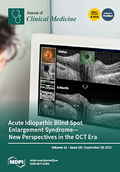
Acute idiopathic blind spot enlargement syndrome (AIBSES) predominantly affects young adults and is often misdiagnosed as optic neuritis because of low awareness. Only around 100 cases have been published. A careful taking of history and unprejudiced ophthalmological workup help in diagnosing AIBSES in patients with unilateral visual field loss in the blind spot area, acute onset photopsia, and funduscopically few or no optic disc changes. As it is a disease of the outer retina, optical coherence tomography (OCT) has become the gold standard in diagnosing AIBSES. In our case series, we present three consecutive patients with AIBSES that are followed prospectively with and without steroid therapy. All of the patients reported full recovery of their symptoms and partial restoration of the outer retinal layer anatomy. View this paper
  • Issues are regarded as officially published after their release is announced to the table of contents alert mailing list.
  • You may sign up for e-mail alerts to receive table of contents of newly released issues.
  • PDF is the official format for papers published in both, html and pdf forms. To view the papers in pdf format, click on the "PDF Full-text" link, and use the free Adobe Reader to open them.
Order results
Result details
Section
Select all
Export citation of selected articles as:
16 pages, 3222 KiB  
Systematic Review
Effectiveness of Inferior Mesenteric Artery Embolization on Type II Endoleak-Related Complications after Endovascular Aortic Repair (EVAR): Systematic Review and Meta-Analysis
by Natalia Niklas, Michalina Malec, Piotr Gutowski, Arkadiusz Kazimierczak and Paweł Rynio
J. Clin. Med. 2022, 11(18), 5491; https://doi.org/10.3390/jcm11185491 - 19 Sep 2022
Viewed by 1705
Abstract
Type II endoleak is one of the most common and problematic complications after endovascular aneurysm repair. It has been suggested that the inferior mesenteric artery (IMA) embolization could prevent further adverse events and postoperative complications. This article is a systematic review and meta-analysis [...] Read more.
Type II endoleak is one of the most common and problematic complications after endovascular aneurysm repair. It has been suggested that the inferior mesenteric artery (IMA) embolization could prevent further adverse events and postoperative complications. This article is a systematic review and meta-analysis following PRISMA guidelines. The Medline, PubMed, Embase, and Cochrane databases were used to identify studies that investigated the effect of IMA embolization on the occurrence of type II endoleaks and secondary interventions in a group of patients with abdominal aortic aneurysm who underwent EVAR compared with results after EVAR procedure without embolization. A random effects meta-analysis was performed. Of 3510 studies, 6 studies involving 659 patients were included. Meta-analysis of all studies showed that the rate of secondary interventions was smaller in patients with IMA embolization (OR, 0.17; SE, 0.45; 95% CI, 0.07 to 0.41; p < 0.01; I2 = 0%). The occurrence of type II endoleaks was also smaller in the embolization group (OR, 0.37; SE, 0.21; 95% CI, 0.25 to 0.57; p < 0.01; I2 = 16.20%). This meta-analysis suggests that IMA embolization correlates with lower rates of type II endoleaks and secondary interventions. Full article
(This article belongs to the Special Issue Abdominal Aortic Aneurysm: Clinical Updates and Perspectives)
Show Figures

Figure 1

14 pages, 321 KiB  
Review
The Unhappy Shoulder: A Conceptual Review of the Psychosomatics of Shoulder Pain
by Matthias Vogel, Marius Binneböse, Hannah Wallis, Christoph H. Lohmann, Florian Junne, Alexander Berth and Christian Riediger
J. Clin. Med. 2022, 11(18), 5490; https://doi.org/10.3390/jcm11185490 - 19 Sep 2022
Cited by 2 | Viewed by 2146
Abstract
Introduction: Chronic pain is a multifaceted disorder genuinely entangled with psychic and psychosomatic symptoms, which are typically involved in the processes of chronification. The impingement syndrome of the shoulder is no exception to this rule, but several studies have shown respective peculiarities among [...] Read more.
Introduction: Chronic pain is a multifaceted disorder genuinely entangled with psychic and psychosomatic symptoms, which are typically involved in the processes of chronification. The impingement syndrome of the shoulder is no exception to this rule, but several studies have shown respective peculiarities among those with pain and impingement of the shoulder. Notably, chronic pain is a lateralized experience, and, similarly, its psychosomatic correlates may be attached to the hemispheres functionally. Aim: The present review therefore gives an overview of the respective findings, with regard not only to psychopathology, but also to personality factors and psychologic trauma, since the latter are reportedly associated with chronic pain. Moreover, we acknowledge symmetry as a possible pathogenic factor. Methods: This narrative review followed the current standards for conducting narrative studies. Based on prior findings, our research strategy included the relevance of psychotraumatologic and symmetrical aspects, as well as comorbidity. We retrieved the relevant literature reporting on the impact of psychopathology as well as personality features on shoulder pain, as published up to January 2022 from the Medline database (1966–2022). Study selecton: We included numerous studies, and considered the contextual relevance of studies referring to the neuropsychosomatics of chronic pain. Results: Pain-specific fears, depression, and anxiety are important predictors of shoulder pain, and the latter is generally overrepresented in those with trauma and PTSD. Moreover, associations of shoulder pain with psychological variables are stronger as regards surgical therapies as compared to conservative ones. This may point to a specific and possibly trauma-related vulnerability for perioperative maladaptation. Additionally, functional hemispheric lateralization may explain some of those results given that limb pain is a naturally lateralized experience. Not least, psychosocial risk factors are shared between shoulder pain and its physical comorbidities (e.g., hypertension), and the incapacitated state of the shoulder is a massive threat to the function of the human body as a whole. Conclusions: This review suggests the involvement of psychosomatic and psychotraumatologic factors in shoulder impingement-related chronic pain, but the inconclusiveness and heterogeneity of the literature in the field is possibly suggestive of other determinants such as laterality. Full article
(This article belongs to the Special Issue Chronic Pain: Clinical Updates and Perspectives)
7 pages, 6664 KiB  
Article
Three Different Types of Fat Grafting for Facial Systemic Sclerosis: A Case Series
by Antonio Arena, Umberto Committeri, Fabio Maglitto, Giovanni Salzano, Giovanni Dell’Aversana Orabona, Luigi Angelo Vaira, Pasquale Piombino, Michela Apolito, Gianluca Renato De Fazio and Luigi Califano
J. Clin. Med. 2022, 11(18), 5489; https://doi.org/10.3390/jcm11185489 - 19 Sep 2022
Cited by 2 | Viewed by 1897
Abstract
Systemic sclerosis (SSc) is a heterogeneous, chronic connective tissue disease, characterized by skin fibrosis as well as vascular and visceral lesions. It can involve the lungs, heart, kidneys, gastrointestinal tract, and bones. The orofacial manifestations of SSc can cause functional, aesthetic, and social [...] Read more.
Systemic sclerosis (SSc) is a heterogeneous, chronic connective tissue disease, characterized by skin fibrosis as well as vascular and visceral lesions. It can involve the lungs, heart, kidneys, gastrointestinal tract, and bones. The orofacial manifestations of SSc can cause functional, aesthetic, and social distress, resulting in significant psychological implications for the patients. In recent decades, fat grafting improved the aesthetic outcomes in terms of volume deficiency, contour asymmetry, and skin elasticity of the face thanks to the regenerative action of the stem cells contained within it. We describe five cases of a patient with SSc treated with fat grafting used to correct volume loss and facial elasticity of the lips and perioral region on the middle and lower third of the face. All the patients received regular postoperative checks at weeks 1 and 2. A multiple choice questionnaire was administered to assess the degree of tolerability of the procedure. The reliability of the questionnaire was evaluated by calculating the Cronbach alpha using the MedCalc Statistical Software version 20.113. The aim of our study is to describe three different types of fat grafting used to correct volume loss and restore facial elasticity of the lips and perioral region on the middle and lower third of the face. Full article
(This article belongs to the Special Issue Current Challenges and Advances in Skin Repair and Regeneration)
Show Figures

Figure 1

10 pages, 4031 KiB  
Review
Robotic Retrograde Intrarenal Surgery: A Journey from “Back to the Future”
by Vineet Gauhar, Olivier Traxer, Sung-Yong Cho, Jeremy Yuen-Chun Teoh, Alba Sierra, Vishesh Gauhar, Kemal Sarica, Bhaskar Somani and Daniele Castellani
J. Clin. Med. 2022, 11(18), 5488; https://doi.org/10.3390/jcm11185488 - 19 Sep 2022
Cited by 12 | Viewed by 3158
Abstract
The introduction of robotics has revolutionized surgery. Robotic platforms have also recently been introduced in clinical practice specifically for flexible ureteroscopy. In this paper, we look at the robotic platforms currently available for flexible ureteroscopy, describing their advantages and limitations. The following robotic [...] Read more.
The introduction of robotics has revolutionized surgery. Robotic platforms have also recently been introduced in clinical practice specifically for flexible ureteroscopy. In this paper, we look at the robotic platforms currently available for flexible ureteroscopy, describing their advantages and limitations. The following robotic platforms are discussed: Roboflex Avicenna®, EasyUretero®, and ILY® robot. Finally, potential future advancements in this field are presented. Full article
(This article belongs to the Section Nephrology & Urology)
Show Figures

Figure 1

15 pages, 619 KiB  
Systematic Review
Correlation between Oral Lichen Planus and Viral Infections Other Than HCV: A Systematic Review
by Alberta Lucchese, Dario Di Stasio, Antonio Romano, Fausto Fiori, Guido Paolo De Felice, Carlo Lajolo, Rosario Serpico, Francesco Cecchetti and Massimo Petruzzi
J. Clin. Med. 2022, 11(18), 5487; https://doi.org/10.3390/jcm11185487 - 19 Sep 2022
Cited by 13 | Viewed by 2272
Abstract
Objectives: This review aimed to evaluate the correlation between viral infections (HPV, EBV, HSV-1, CMV) other than HCV and oral lichen planus to assess if there is sufficient evidence to establish if these viruses can play a role in the etiopathogenesis of the [...] Read more.
Objectives: This review aimed to evaluate the correlation between viral infections (HPV, EBV, HSV-1, CMV) other than HCV and oral lichen planus to assess if there is sufficient evidence to establish if these viruses can play a role in the etiopathogenesis of the disease. Materials and methods: We reviewed the literature using different search engines (PubMed, ISI Web of Science, and the Cochrane Library), employing MeSH terms such as “oral lichen planus” and “OLP” in conjunction with other terms. We utilized the Population, Intervention, Comparison, Outcomes, and Study design (PICOS) method to define our study eligibility criteria. Results: A total of 43 articles of the 1219 results initially screened were included in the study. We allocated the 43 selected items into four groups, according to each related virus: HPV, EBV, HSV-1, and CMV. Conclusions: Heterogeneous results neither confirm nor exclude a direct correlation between the investigated viral infections and oral lichen planus etiopathogenesis and its feasible malignant transformation. Many viral agents can cause oral lesions and act as cancerizing agents. Future studies could be desirable to produce comparable statistical analyses and enhance the quantity and quality of the outcomes to promote the translation of research into clinical practice. Full article
(This article belongs to the Topic Bridging Oral Medicine and Systemic Disease)
Show Figures

Figure 1

12 pages, 636 KiB  
Review
Weight Gain and Asthenia Following Thyroidectomy: Current Knowledge from Literature Review
by Gregorio Scerrino, Giuseppe Salamone, Alessandro Corigliano, Pierina Richiusa, Maria Pia Proclamà, Stefano Radellini, Gianfranco Cocorullo, Giuseppina Orlando, Giuseppina Melfa and Nunzia Cinzia Paladino
J. Clin. Med. 2022, 11(18), 5486; https://doi.org/10.3390/jcm11185486 - 19 Sep 2022
Cited by 2 | Viewed by 2584
Abstract
Background: Thyroidectomy is a common procedure all over the world. Its complication rate is low, but some patients complain of weight gain and/or asthenia. The aim of this review is to investigate the correlation between thyroidectomy and weight change and asthenia. Materials and [...] Read more.
Background: Thyroidectomy is a common procedure all over the world. Its complication rate is low, but some patients complain of weight gain and/or asthenia. The aim of this review is to investigate the correlation between thyroidectomy and weight change and asthenia. Materials and Methods: Seven papers concerning weight gain and four concerning asthenia were found. Results: Weight gain would seem to be more related to the change in habits after surgery. Asthenia seems to be more linked to endocrine mechanisms not yet clarified although a deficiency of triiodothyronine and its metabolites could explain some of its aspects. Conclusion: Patients who are candidates for thyroidectomy should be adequately informed of the onset of both possible implications of the surgical act in terms of weight gain and chronic asthenia. Full article
(This article belongs to the Section General Surgery)
Show Figures

Figure 1

14 pages, 1520 KiB  
Article
High Inferior Vena Cava Diameter with High Left Ventricular End Systolic Diameter as a Risk Factor for Major Adverse Cardiovascular Events, Cardiovascular and Overall Mortality among Chronic Hemodialysis Patients
by Chung-Kuan Wu, Noi Yar, Zih-Kai Kao, Ming-Tsang Chuang and Tzu-Hao Chang
J. Clin. Med. 2022, 11(18), 5485; https://doi.org/10.3390/jcm11185485 - 19 Sep 2022
Cited by 2 | Viewed by 1381
Abstract
Background: Little is known about the association of inferior vena cava diameter (IVCD) and left ventricular end-systolic diameter (LVESD) with mortality in patients undergoing hemodialysis (HD). Methods: The single medical center observational cohort study enrolled 241 adult chronic HD patients from 1 October [...] Read more.
Background: Little is known about the association of inferior vena cava diameter (IVCD) and left ventricular end-systolic diameter (LVESD) with mortality in patients undergoing hemodialysis (HD). Methods: The single medical center observational cohort study enrolled 241 adult chronic HD patients from 1 October 2018 to 31 December 2018. Echocardiography results of IVCD and LVESD prior to dialysis were retrieved and patients were divided into high IVCD and low IVCD groups. Patients who received HD via a tunneled cuffed catheter were excluded. Study outcomes included all-cause mortality, cardiovascular mortality, and major adverse cardiovascular events (MACE). Subgroup analyses of HD patients with high and low LVESD were also performed. Results: The incidence of all-cause mortality, cardiovascular mortality, and MACE were higher in chronic HD patients with high IVCD (p < 0.01). High IVCD patients had significantly greater all-cause mortality, cardiovascular mortality, and MACE (log-rank test; p < 0.05). High IVCD patients are also associated with an increased risk of all-cause mortality and MACE relative to low IVCD patients (aHRs, 2.88 and 3.42; 95% CIs, 1.06–7.86 and 1.73–6.77, respectively; all p < 0.05). In the subgroup analysis of patients with high or low LVESD, the high IVCD remained a significant risk factor for all-cause mortality and MACE, and the HR is especially high in the high LVESD group. Conclusions: Dilated IVCD is a risk factor for all-cause mortality and MACE in chronic HD patients. In addition, these patients with high LVESD also have a significantly higher HR of all-cause mortality and MACE. Full article
(This article belongs to the Special Issue Hot Cardiovascular Topics in Chronic and End-Stage Renal Disease)
Show Figures

Figure 1

15 pages, 2508 KiB  
Article
Different Machine Learning Approaches for Implementing Telehealth-Based Cancer Pain Management Strategies
by Marco Cascella, Sergio Coluccia, Federica Monaco, Daniela Schiavo, Davide Nocerino, Mariacinzia Grizzuti, Maria Cristina Romano and Arturo Cuomo
J. Clin. Med. 2022, 11(18), 5484; https://doi.org/10.3390/jcm11185484 - 19 Sep 2022
Cited by 13 | Viewed by 2084
Abstract
Background: The most effective strategy for managing cancer pain remotely should be better defined. There is a need to identify those patients who require increased attention and calibrated follow-up programs. Methods: Machine learning (ML) models were developed using the data prospectively obtained from [...] Read more.
Background: The most effective strategy for managing cancer pain remotely should be better defined. There is a need to identify those patients who require increased attention and calibrated follow-up programs. Methods: Machine learning (ML) models were developed using the data prospectively obtained from a single-center program of telemedicine-based cancer pain management. These models included random forest (RF), gradient boosting machine (GBM), artificial neural network (ANN), and the LASSO–RIDGE algorithm. Thirteen demographic, social, clinical, and therapeutic variables were adopted to define the conditions that can affect the number of teleconsultations. After ML validation, the risk analysis for more than one remote consultation was assessed in target individuals. Results: The data from 158 patients were collected. In the training set, the accuracy was about 95% and 98% for ANN and RF, respectively. Nevertheless, the best accuracy on the test set was obtained with RF (70%). The ML-based simulations showed that young age (<55 years), lung cancer, and occurrence of breakthrough cancer pain help to predict the number of remote consultations. Elderly patients (>75 years) with bone metastases may require more telemedicine-based clinical evaluations. Conclusion: ML-based analyses may enable clinicians to identify the best model for predicting the need for more remote consultations. It could be useful for calibrating care interventions and resource allocation. Full article
Show Figures

Figure 1

11 pages, 2735 KiB  
Article
The Impact of Non-Invasive Ventilation on Sleep Quality in COPD Patients
by Maximilian Wollsching-Strobel, Iris Anna Bauer, Johannes Julian Baur, Daniel Sebastian Majorski, Friederike Sophie Magnet, Jan Hendrik Storre, Wolfram Windisch and Sarah Bettina Schwarz
J. Clin. Med. 2022, 11(18), 5483; https://doi.org/10.3390/jcm11185483 - 19 Sep 2022
Cited by 2 | Viewed by 1802
Abstract
Background: Non-invasive ventilation (NIV) has been shown to be the most appropriate therapy for COPD patients with chronic respiratory failure. While physiological parameters and long-term outcome frequently serve as primary outcomes, very few studies have primarily addressed the impact of NIV initiation on [...] Read more.
Background: Non-invasive ventilation (NIV) has been shown to be the most appropriate therapy for COPD patients with chronic respiratory failure. While physiological parameters and long-term outcome frequently serve as primary outcomes, very few studies have primarily addressed the impact of NIV initiation on sleep quality in COPD. Methods: This single-center prospective cohort study comprised NIV-naïve patients with COPD. All patients underwent polysomnographic evaluation both at baseline and at 3 months follow-up, accompanied by the assessment of health-related quality of life (HRQL) using the Severe Respiratory Insufficiency Questionnaire (SRI) and the Epworth Sleepiness Scale (ESS). A subgroup evaluation was performed to address the impact of comorbid obstructive sleep apnea syndrome (OSAS). Results: Forty-six patients were enrolled and twenty-five patients completed the follow-up period (66.7 ± 7.4 years). NIV resulted in an increase in slow-wave sleep (+2% (−3.5/7.5), p = 0.465) and rapid eye movement sleep (+2.2% (−1.0/5.4), p = 0.174), although no statistical significance could be detected. ESS (−1.7(−3.6/0.1), p = 0.066) also showed a positive trend. Significant improvements in the Respiratory Disturbance Index (RDI) (−12.6(−23.7/−1.5), p = 0.027), lung function parameters, transcutaneous PCO2 and the SRI summary scale (4.5(0.9/8), p = 0.016) were observed. Conclusion: NIV therapy does not decrease sleep quality and is even capable of improving HRQL, transcutaneous PaCO2, daytime sleepiness and RDI, and the latter especially holds true for patients with comorbid OSAS. Full article
(This article belongs to the Section Pulmonology)
Show Figures

Figure 1

9 pages, 608 KiB  
Article
Vitamin D Status and Steatohepatitis in Obese Diabetic and Non-Diabetic Patients
by Laura Taban, Dana Stoian, Bogdan Timar, Daniela Amzar, Calin Adela, Alexandru Motofelea, Andreea Borlea, Romain Frisoni and Nadege Laguerre
J. Clin. Med. 2022, 11(18), 5482; https://doi.org/10.3390/jcm11185482 - 18 Sep 2022
Viewed by 1576
Abstract
Background and Aims: The presence of steatohepatitis in obese patients can be multifactorial. The current study tries to determine the differences between diabetic and non-diabetic patients regarding the presence of steatohepatitis. We evaluated sequential liver samples and collected the times of bariatric surgery [...] Read more.
Background and Aims: The presence of steatohepatitis in obese patients can be multifactorial. The current study tries to determine the differences between diabetic and non-diabetic patients regarding the presence of steatohepatitis. We evaluated sequential liver samples and collected the times of bariatric surgery to assess the presence of NASH in patients with obesity, in the circuit of bariatric surgery. Methods: We performed a retrospective study of 49 patients presenting high-grade obesity in the circuit of bariatric surgery, with liver biopsy. The patients underwent bariatric surgery at a single center in France and were followed for 2 years. The liver biopsies were performed intraoperatively on all 49 patients before the bariatric surgery. The primary endpoint of the study was to evaluate the relationships between steatohepatitis/liver fibrosis and the presence of diabetes and to evaluate the current relationships between the biochemical work-ups. Special importance was accorded to the correlations between vitamin D levels and the presence of hepatic steatosis, due to the antifibrogenic pattern in the liver, as shown in many important papers in the field. Results: Significant correlations were found between the presence of liver fibrosis and the presence of diabetes (p = 0.022), but not regarding the antidiabetic treatment. An important correlation was found between the vitamin D levels and the presence of liver fibrosis, as well as with the levels of A1C hemoglobin and LDL cholesterol levels. Conclusions: Vitamin D deficiency presents a strong correlation with hepatic steatosis in individuals with morbid obesity. Correcting vitamin D deficiency may present a beneficial role in treating hepatic steatosis, diabetes, and cardiovascular risk in patients with morbid obesity. Full article
(This article belongs to the Special Issue Clinical Management and Challenges in Metabolic Syndrome)
Show Figures

Figure 1

14 pages, 1287 KiB  
Article
MicroShunt versus Trabeculectomy for Surgical Management of Glaucoma: A Retrospective Analysis
by Michael X. Fu, Eduardo M. Normando, Sheila M. H. Luk, Mira Deshmukh, Faisal Ahmed, Laura Crawley, Sally Ameen, Niten Vig, Maria Francesca Cordeiro and Philip A. Bloom
J. Clin. Med. 2022, 11(18), 5481; https://doi.org/10.3390/jcm11185481 - 18 Sep 2022
Cited by 2 | Viewed by 1608
Abstract
This case-control study aims to compare the efficacy, safety, and postoperative burden of MicroShunt versus trabeculectomy. The first consecutive cohort of MicroShunt procedures (n = 101) was matched to recent historical trabeculectomy procedures (n = 101) at two London hospital trusts. Primary endpoints [...] Read more.
This case-control study aims to compare the efficacy, safety, and postoperative burden of MicroShunt versus trabeculectomy. The first consecutive cohort of MicroShunt procedures (n = 101) was matched to recent historical trabeculectomy procedures (n = 101) at two London hospital trusts. Primary endpoints included changes in intraocular pressure (IOP) and glaucoma medications. Secondary outcome measures included changes in retinal nerve fibre layer (RNFL) thickness, rates of complications, further theatre interventions, and the number of postoperative visits. From the baseline to Month-18, the median [interquartile range] IOP decreased from 22 [17–29] mmHg (on 4 [3–4] medications) to 15 [10–17] mmHg (on 0 [0–2] medications) and from 20 [16–28] mmHg (on 4 [3–4] medications) to 11 [10–13] mmHg (on 0 [0–0] medications) in the MicroShunt and trabeculectomy groups, respectively. IOP from Month-3 was significantly higher in the MicroShunt group (p = 0.006), with an increased number of medications from Month-12 (p = 0.024). There were greater RNFL thicknesses from Month-6 in the MicroShunt group (p = 0.005). The rates of complications were similar (p = 0.060) but with fewer interventions (p = 0.031) and postoperative visits (p = 0.001) in the MicroShunt group. Therefore, MicroShunt has inferior efficacy to trabeculectomy in lowering IOP and medications but provides a better safety profile and postoperative burden and may delay RNFL loss. Full article
(This article belongs to the Special Issue Advances in Glaucoma Surgery)
Show Figures

Figure 1

33 pages, 1407 KiB  
Systematic Review
Can Swallowing Cerebral Neurophysiology Be Evaluated during Ecological Food Intake Conditions? A Systematic Literature Review
by Yohan Gallois, Fabrice Neveu, Muriel Gabas, Xavier Cormary, Pascal Gaillard, Eric Verin, Renée Speyer and Virginie Woisard
J. Clin. Med. 2022, 11(18), 5480; https://doi.org/10.3390/jcm11185480 - 18 Sep 2022
Cited by 3 | Viewed by 1829
Abstract
Swallowing is a complex function that relies on both brainstem and cerebral control. Cerebral neurofunctional evaluations are mostly based on functional magnetic resonance imaging (fMRI) and positron emission tomography (PET), performed with the individual laying down; which is a non-ecological/non-natural position for swallowing. [...] Read more.
Swallowing is a complex function that relies on both brainstem and cerebral control. Cerebral neurofunctional evaluations are mostly based on functional magnetic resonance imaging (fMRI) and positron emission tomography (PET), performed with the individual laying down; which is a non-ecological/non-natural position for swallowing. According to the PRISMA guidelines, a review of the non-invasive non-radiating neurofunctional tools, other than fMRI and PET, was conducted to explore the cerebral activity in swallowing during natural food intake, in accordance with the PRISMA guidelines. Using Embase and PubMed, we included human studies focusing on neurofunctional imaging during an ecologic swallowing task. From 5948 unique records, we retained 43 original articles, reporting on three different techniques: electroencephalography (EEG), magnetoencephalography (MEG) and functional near infra-red spectroscopy (fNIRS). During swallowing, all three techniques showed activity of the pericentral cortex. Variations were associated with the modality of the swallowing process (volitional or non-volitional) and the substance used (mostly water and saliva). All techniques have been used in both healthy and pathological conditions to explore the precise time course, localization or network structure of the swallowing cerebral activity, sometimes even more precisely than fMRI. EEG and MEG are the most advanced and mastered techniques but fNIRS is the most ready-to-use and the most therapeutically promising. Ongoing development of these techniques will support and improve our future understanding of the cerebral control of swallowing. Full article
(This article belongs to the Section Otolaryngology)
Show Figures

Figure 1

14 pages, 1488 KiB  
Article
Composition of the Gut Microbiota Associated with the Response to Immunotherapy in Advanced Cancer Patients: A Chinese Real-World Pilot Study
by Xi Cheng, Jiawei Wang, Liu Gong, Yong Dong, Jiawei Shou, Hongming Pan, Zhaonan Yu and Yong Fang
J. Clin. Med. 2022, 11(18), 5479; https://doi.org/10.3390/jcm11185479 - 18 Sep 2022
Cited by 5 | Viewed by 2079
Abstract
Background: The composition of the gut microbiota is associated with the response to immunotherapy for different cancers. However, the majority of previous studies have focused on a single cancer and a single immune checkpoint inhibitor. Here, we investigated the relationship between the gut [...] Read more.
Background: The composition of the gut microbiota is associated with the response to immunotherapy for different cancers. However, the majority of previous studies have focused on a single cancer and a single immune checkpoint inhibitor. Here, we investigated the relationship between the gut microbiota and the clinical response to anti-programmed cell death protein 1 (PD-1) immunotherapy in patients with advanced cancers. Method: In this comprehensive study, 16S rRNA sequencing was performed on the gut microbiota of pre-immunotherapy and post-immunotherapy, of 72 advanced cancer patients in China. Results: At the phylum level, Firmicutes, Bacteroidetes, Proteobacteria, and Actinobacteria were the main components of the microbiota in the 72 advanced cancer patients. At the genus level, Bacteroides and Prevotella were the dominant microbiota among these 72 patients. The PD_whole_tree, Chao1, Observed_species and Shannon indices of R.0 and R.T were higher than those of NR.0 and NR.T. The results of LEfSe showed that Archaea, Lentisphaerae, Victivallaceae, Victivallales, Lentisphaeria, Methanobacteriaceae, Methanobacteria, Euryarchaeota, Methanobrevibacter, and Methanobacteriales were significantly enriched in the response group before immunotherapy (R.0), and the Clostridiaceae was significantly enriched in the non-response group before immunotherapy (NR.0) (p < 0.05). Lachnospiraceae and Thermus were significantly enriched in the response group after immunotherapy (R.T), and Leuconostoc was significantly enriched in R.0 (p < 0.05). ROC analysis showed that the microbiota of R.T (AUC = 0.70) had obvious diagnostic value in differentiating Chinese cancer patients based on their response to immunotherapy. Conclusions: We demonstrated that the gut microbiota was associated with the clinical response to anti-PD-1 immunotherapy in cancer patients. Taxonomic signatures enriched in responders were effective biomarkers to predict the clinical response. Our findings provide a new strategy to improve the efficiency of responses to immunotherapy among cancer patients. Full article
(This article belongs to the Special Issue Target and Im-Oncology Therapeutic Approaches for Lung Cancer)
Show Figures

Figure 1

9 pages, 1516 KiB  
Article
Tibial Lengthening along Submuscular Plate with Simultaneous Acute Tibial Deformity Correction by High-Energy Osteotomy: A Comparative Study
by Kuei-Yu Liu, Kuan-Wen Wu, Chia-Che Lee, Sheng-Chieh Lin, Ken N. Kuo and Ting-Ming Wang
J. Clin. Med. 2022, 11(18), 5478; https://doi.org/10.3390/jcm11185478 - 18 Sep 2022
Cited by 1 | Viewed by 1811
Abstract
Submuscular plating and osteotomy using power saw have shown the benefits in certain situations of limb lengthening. However, no previous studies combining both procedures have been conducted for acute tibial deformity correction and limb lengthening. Nineteen cases were enrolled in this study. Ten [...] Read more.
Submuscular plating and osteotomy using power saw have shown the benefits in certain situations of limb lengthening. However, no previous studies combining both procedures have been conducted for acute tibial deformity correction and limb lengthening. Nineteen cases were enrolled in this study. Ten patients received tibial lengthening with acute knee angular deformity correction using high-energy osteotomy (Group 1), and nine patients received tibial lengthening only with osteotomy using multiple drills and osteotome (Group 2). Radiographic parameters retrieved before and after the operation included leg-length discrepancy, tibial length, length gained, mechanical lateral distal femoral angle (mLDFA), medial proximal tibial angle (MPTA), and mechanical axis deviation (MAD). There were significant differences between groups in terms of external fixator index (EFI) (p = 0.013) and healing index (HI) (p = 0.014), but no significance in the length gained (p = 0.356). The latest postoperative mLDFA (p = 0.315), MPTA (p = 0.497), and MAD (p = 0.211) of Group 1 were not distinguishable from Group 2. The functional outcomes were excellent, and there were no permanent complications. Despite showing a longer healing time, this alternative lengthening procedure which combines fixator-assisted plate lengthening in the tibia with simultaneous surgical intervention of acute tibial deformity correction using an oscillating saw is appropriate for patients with leg-length discrepancy and angular deformity of the tibia. Full article
Show Figures

Figure 1

20 pages, 1709 KiB  
Article
Energy Homeostasis Gene Nucleotide Variants and Survival of Hemodialysis Patients—A Genetic Cohort Study
by Monika Katarzyna Świderska, Adrianna Mostowska, Damian Skrypnik, Paweł Piotr Jagodziński, Paweł Bogdański and Alicja Ewa Grzegorzewska
J. Clin. Med. 2022, 11(18), 5477; https://doi.org/10.3390/jcm11185477 - 18 Sep 2022
Cited by 1 | Viewed by 1834
Abstract
Background: Patients undergoing hemodialysis (HD) therapy have an increased risk of death compared to the general population. We investigated whether selected single nucleotide variants (SNVs) involved in glucose and lipid metabolism are associated with mortality risk in HD patients. Methods: The study included [...] Read more.
Background: Patients undergoing hemodialysis (HD) therapy have an increased risk of death compared to the general population. We investigated whether selected single nucleotide variants (SNVs) involved in glucose and lipid metabolism are associated with mortality risk in HD patients. Methods: The study included 805 HD patients tested for 11 SNVs in FOXO3, IGFBP3, FABP1, PCSK9, ANGPTL6, and DOCK6 using HRM analysis and TaqMan assays. FOXO3, IGFBP3, L-FABP, PCSK9, ANGPTL6, and ANGPTL8 plasma concentrations were measured by ELISA in 86 individuals. The Kaplan–Meier method and Cox proportional hazards models were used for survival analyses. Results: We found out that the carriers of a C allele in ANGPTL6 rs8112063 had an increased risk of all-cause, cardiovascular, and cardiac mortality. In addition, the C allele of DOCK6 rs737337 was associated with all-cause and cardiac mortality. The G allele of DOCK6 rs17699089 was correlated with the mortality risk of patients initiating HD therapy. The T allele of FOXO3 rs4946936 was negatively associated with cardiac and cardiovascular mortality in HD patients. We observed no association between the tested proteins’ circulating levels and the survival of HD patients. Conclusions: The ANGPTL6 rs8112063, FOXO3 rs4946936, DOCK6 rs737337, and rs17699089 nucleotide variants are predictors of survival in patients undergoing HD. Full article
Show Figures

Graphical abstract

8 pages, 941 KiB  
Article
Effects of Compression of the Ulnar Artery on the Radial Artery Catheterization
by Cho-Long Kim, Seung-Wan Hong and Seong-Hyop Kim
J. Clin. Med. 2022, 11(18), 5476; https://doi.org/10.3390/jcm11185476 - 18 Sep 2022
Viewed by 1249
Abstract
Background: The study was designed to evaluate the effects of compression of the ulnar artery on blood flow (BF) and internal cross-sectional area (CSAi) of the radial artery. We also evaluated the success rate and time of successful ultrasound-guided radial artery catheterization at [...] Read more.
Background: The study was designed to evaluate the effects of compression of the ulnar artery on blood flow (BF) and internal cross-sectional area (CSAi) of the radial artery. We also evaluated the success rate and time of successful ultrasound-guided radial artery catheterization at the first attempt with or without compression of the ulnar artery. Methods: Patients were randomly allocated to the Compression group or Standard group to be treated with or without the application of ulnar artery compression, respectively. Hemodynamic stability was confirmed, and ultrasound-guided radial artery catheterization was performed. In the Compression group, an assistant compressed the ulnar artery at 5 cm above the wrist crease and the catheterization was performed after the loss of the distal ulnar artery BF. In the Standard group, the catheterization was performed without compression of the ulnar artery. Before and after the catheterization, BF and CSAi of the radial artery were evaluated. Success rate and time to successful catheterization at the first attempt were recorded. Results: BF and CSAi of the radial artery were similar in the two groups (37.5 [19.3–66] vs. 37.0 [20.6–53.7] mL/min, respectively, p = 0.63; 4.0 [4.0–6.0] vs. 4.0 [3.0–5.0] mm2, respectively, p = 0.095). In the Compression group, BF and CSAi were changed to 80.9 [35.9–128.5] mL/min (p < 0.001) and 5.0 [4.0–7.0] mm2 (p < 0.001), respectively, after compression of the ulnar artery. There was a trend that the success rate of ultrasound-guided radial artery catheterization at the first attempt was higher in the Compression group than in the Standard group (58/59 vs. 53/59, respectively, p = 0.05), although the difference was not statistically significant. However, the time to successful ultrasound-guided radial artery catheterization at the first attempt was significantly shorter in the Compression group than in the Standard group (34 [27–41] s vs. 46 [36–60] s, p < 0.001). Conclusion: Compression of the ulnar artery augmented BF and CSAi of the radial artery. It resulted in a significantly shorter success time for ultrasound-guided radial artery catheterization at the first attempt. Full article
(This article belongs to the Section Anesthesiology)
Show Figures

Figure 1

12 pages, 683 KiB  
Article
Head and Neck Cutaneous Soft-Tissue Sarcoma Demonstrate Sex and Racial/Ethnic Disparities in Incidence and Socioeconomic Disparities in Survival
by Muhammad Umar Jawad, Lauren N. Zeitlinger, Arnaud F. Bewley, Edmond F. O’Donnell III, Sophia A. Traven, Janai R. Carr-Ascher, Arta M. Monjazeb, Robert J. Canter, Steven W. Thorpe and R. Lor Randall
J. Clin. Med. 2022, 11(18), 5475; https://doi.org/10.3390/jcm11185475 - 17 Sep 2022
Cited by 3 | Viewed by 1589
Abstract
Background: Cutaneous soft-tissue sarcoma (CSTS) of the head and neck are rare and are known to have aggressive clinical course. The current study utilizes a population-based registry in the U.S. to characterize these malignancies and explore disparities. Methods: National Cancer Institute’s (NCI) Surveillance, [...] Read more.
Background: Cutaneous soft-tissue sarcoma (CSTS) of the head and neck are rare and are known to have aggressive clinical course. The current study utilizes a population-based registry in the U.S. to characterize these malignancies and explore disparities. Methods: National Cancer Institute’s (NCI) Surveillance, Epidemiology and End Result (SEER) database from 2000 to 2018 was queried to report incidence and survival data in 4253 cases in the U.S. Results: Males were 5.37 times more likely and Non-Hispanic-White people (NHW) were 4.62 times more likely than females and Non-Hispanic-Black people (NHB) to develop CSTS of the head and neck. The overall incidence was 0.27 per 100,000 persons in 2018, with a significant increase since 2000. Advanced age and stage, histologic group other than ‘fibromatous sarcoma’ and lower SES groups were independent factors for worse overall survival. Conclusions: CSTS of the head and neck demonstrate sex and racial/ethnic disparities in incidence and socioeconomic disparities in overall survival. Level of evidence: II. Full article
Show Figures

Figure 1

11 pages, 3041 KiB  
Review
Inner Ear Diagnostics and Drug Delivery via Microneedles
by Stephen Leong, Aykut Aksit, Sharon J. Feng, Jeffrey W. Kysar and Anil K. Lalwani
J. Clin. Med. 2022, 11(18), 5474; https://doi.org/10.3390/jcm11185474 - 17 Sep 2022
Cited by 4 | Viewed by 1747
Abstract
Objectives: Precision medicine for inner ear disorders has seen significant advances in recent years. However, unreliable access to the inner ear has impeded diagnostics and therapeutic delivery. The purpose of this review is to describe the development, production, and utility of novel microneedles [...] Read more.
Objectives: Precision medicine for inner ear disorders has seen significant advances in recent years. However, unreliable access to the inner ear has impeded diagnostics and therapeutic delivery. The purpose of this review is to describe the development, production, and utility of novel microneedles for intracochlear access. Methods: We summarize the current work on microneedles developed using two-photon polymerization (2PP) lithography for perforation of the round window membrane (RWM). We contextualize our findings with the existing literature in intracochlear diagnostics and delivery. Results: Two-photon polymerization lithography produces microneedles capable of perforating human and guinea pig RWMs without structural or functional damage. Solid microneedles may be used to perforate guinea pig RWMs in vivo with full reconstitution of the membrane in 48–72 h, and hollow microneedles may be used to aspirate perilymph or inject therapeutics into the inner ear. Microneedles produced with two-photon templated electrodeposition (2PTE) have greater strength and biocompatibility and may be used to perforate human RWMs. Conclusions: Microneedles produced with 2PP lithography and 2PTE can safely and reliably perforate the RWM for intracochlear access. This technology is groundbreaking and enabling in the field of inner ear precision medicine. Full article
(This article belongs to the Special Issue Innovative Technologies and Translational Therapies for Deafness)
Show Figures

Figure 1

7 pages, 519 KiB  
Article
Exploring the Risk Factors for Poor Survival in Lupus Pericarditis Patients: A Retrospective Cohort Study
by Yen-Fu Chen, Meng-Ru Hsieh, Che-Tzu Chang, Ping-Han Tsai and Yao-Fan Fang
J. Clin. Med. 2022, 11(18), 5473; https://doi.org/10.3390/jcm11185473 - 17 Sep 2022
Cited by 1 | Viewed by 1446
Abstract
Patients with systemic lupus erythematosus (SLE) have a higher risk of pericarditis, which could be fatal. The goal of this study was to identify the prognostic factors for mortality in patients with lupus pericarditis. Patients with lupus pericarditis treated at Chang Gung Memorial [...] Read more.
Patients with systemic lupus erythematosus (SLE) have a higher risk of pericarditis, which could be fatal. The goal of this study was to identify the prognostic factors for mortality in patients with lupus pericarditis. Patients with lupus pericarditis treated at Chang Gung Memorial Hospital were included in this observational cohort study. This study conducted univariate and multivariate COX regression, as well as Kaplan–Meier survival curve analysis, to investigate mortality risk in SLE patients. The average age at admission was 40.78 ± 15.92 years. A total of 113 (16.4%) of the 689 patients had lupus pericarditis. Patients with lupus pericarditis exhibited older age, shorter follow-up, higher disease activities, and higher incidence rates of comorbidities than patients without pericarditis. Cox regression adjusted analysis indicated that lupus pericarditis (hazard ratio = 1.963, 95% CI = 1.315, 2.963, p = 0.001), old age at admission (HR = 1.053, 95% CI = 1.040, 1.065, p < 0.001), high SLEDAI score (HR = 1.079, 95% CI = 1.043, 1.116, p < 0.001), and end-stage kidney disease (ESKD) (HR = 2.533, 95% CI = 1.620, 3.961, p < 0.001) were all linked to increased mortality. Moreover, the Kaplan–Meier survival curve analysis revealed that patients with pericarditis compared to those without pericarditis had a higher mortality rate (log-rank test, p < 0.001). A high proportion of SLE patients have manifestations of lupus pericarditis. Moreover, patients with lupus pericarditis have a greater risk of mortality even if they have no pericardial tamponade. Therefore, these patients need prompt diagnosis and treatment. Full article
(This article belongs to the Special Issue Systemic Immune Inflammatory Disease: New Updates)
Show Figures

Figure 1

13 pages, 4163 KiB  
Review
The Small Posterior Cranial Fossa Syndrome and Chiari Malformation Type 0
by Enver I. Bogdanov, Aisylu T. Faizutdinova and John D. Heiss
J. Clin. Med. 2022, 11(18), 5472; https://doi.org/10.3390/jcm11185472 - 17 Sep 2022
Cited by 4 | Viewed by 2826
Abstract
Patients showing typical Chiari malformation type 1 (CM1) signs and symptoms frequently undergo cranial and cervical MRI. In some patients, MRI documents >5 mm of cerebellar tonsillar herniation (TH) and the diagnosis of CM1. Patients with 3–5 mm TH have “borderline” CM1. Patients [...] Read more.
Patients showing typical Chiari malformation type 1 (CM1) signs and symptoms frequently undergo cranial and cervical MRI. In some patients, MRI documents >5 mm of cerebellar tonsillar herniation (TH) and the diagnosis of CM1. Patients with 3–5 mm TH have “borderline” CM1. Patients with less than 3 mm of TH and an associated cervical syrinx are diagnosed with Chiari “zero” malformation (CM0). However, patients reporting CM1 symptoms are usually not diagnosed with CM if MRI shows less than 3–5 mm of TH and no syrinx. Recent MRI morphometric analysis of the posterior fossa and upper cervical spine detected anatomical abnormalities in and around the foramen magnum (FM) that explain these patients’ symptoms. The abnormalities include a reduced size of the posterior fossa, FM, and upper cervical spinal canal and extension of the cerebellar tonsils around the medulla rather than inferior to the foramen magnum, as in CM1. These morphometric findings lead some neurologists and neurosurgeons to diagnose CM0 in patients with typical CM1 signs and symptoms, with or without cervical syringes. This article reviews recent findings and controversies about CM0 diagnosis and updates current thinking about the clinical and radiological relationship between CM0, borderline CM1, and CM1. Full article
Show Figures

Figure 1

11 pages, 411 KiB  
Article
Neuropathic-like Pain Symptoms and Their Association with Muscle Strength in Patients with Chronic Musculoskeletal Pain
by Hee Jung Kim, Min Gi Ban, Kyung Bong Yoon, Woohyuk Jeon and Shin Hyung Kim
J. Clin. Med. 2022, 11(18), 5471; https://doi.org/10.3390/jcm11185471 - 17 Sep 2022
Cited by 4 | Viewed by 1488
Abstract
The relationship between sarcopenia and pain remains unclear; thus, this study evaluated whether muscle strength is independently associated with neuropathic-like pain symptoms in patients with chronic musculoskeletal pain. A cut-off score of painDETECT ≥13 was used to indicate a possible neuropathic component. Handgrip [...] Read more.
The relationship between sarcopenia and pain remains unclear; thus, this study evaluated whether muscle strength is independently associated with neuropathic-like pain symptoms in patients with chronic musculoskeletal pain. A cut-off score of painDETECT ≥13 was used to indicate a possible neuropathic component. Handgrip strength was measured, and muscle mass was estimated. A total of 2599 patients, including 439 patients who reported neuropathic-like pain symptoms (16.9%), were included for analysis. Handgrip strength was significantly lower in patients experiencing neuropathic-like pain symptoms (23.23 ± 10.57 vs. 24.82 ± 10.43 kg, p < 0.001), and this result was chiefly found in female patients. However, there was no difference in estimated muscle mass. Shorter duration of pain, opioid usage, pain in lower limbs, sleep disturbance, and lower handgrip strength were significantly associated with neuropathic-like pain symptoms. In patients with handgrip strength below the reference values by sex, experiencing radiating pain and at least moderate sensory symptoms by light touch and thermal stimulation were more frequently reported. In conclusion, lower handgrip strength appeared to be an independent factor associated with symptoms suggestive of neuropathic pain in this population. Interventional studies are required to determine whether improvement in muscle strength can reduce the neuropathic pain component in chronic musculoskeletal pain. Full article
(This article belongs to the Special Issue Rehabilitation and Management of Frailty, Sarcopenia and Malnutrition)
Show Figures

Figure 1

4 pages, 182 KiB  
Editorial
New Concepts in Diagnosis, Risk Factors and Work Ability in Patients with Hematogenous Spinal Infection
by Panagiotis Korovessis
J. Clin. Med. 2022, 11(18), 5470; https://doi.org/10.3390/jcm11185470 - 17 Sep 2022
Cited by 1 | Viewed by 910
Abstract
In the last decades, there is an increasing incidence of hematogenous septic spinal infection (HIS) [...] Full article
(This article belongs to the Section Orthopedics)
10 pages, 1333 KiB  
Article
Transesophageal Endoscopic Ultrasound Fine Needle Biopsy for the Diagnosis of Mediastinal Masses: A Retrospective Real-World Analysis
by Daniela Assisi, Filippo Tommaso Gallina, Daniele Forcella, Riccardo Tajè, Enrico Melis, Paolo Visca, Federico Pierconti, Emanuela Venti and Francesco Facciolo
J. Clin. Med. 2022, 11(18), 5469; https://doi.org/10.3390/jcm11185469 - 17 Sep 2022
Cited by 3 | Viewed by 2025
Abstract
Background: Endoscopic ultrasound (EUS) plays an important role in the diagnosis and staging of thoracic disease. Our report studies the diagnostic performance and clinical impact of EUS fine needle aspiration (FNA) in a homogenous cohort of patients according to the distribution of the [...] Read more.
Background: Endoscopic ultrasound (EUS) plays an important role in the diagnosis and staging of thoracic disease. Our report studies the diagnostic performance and clinical impact of EUS fine needle aspiration (FNA) in a homogenous cohort of patients according to the distribution of the enlarged MLNs or pulmonary masses. Methods: We retrospectively reviewed the diagnostic performance of 211 EUS-FNA in 200 consecutive patients with enlarged or PET-positive MLNs and para-mediastinal masses who were referred to our oncological center between January 2019 and May 2020. Results: The overall sensitivity of EUS-FNA was 85% with a corresponding negative predictive value (NPV) of 56% and an accuracy of 87.5%. The sensitivity and accuracy in patients with abnormal MLNs were 81.1% and 84.4%, respectively. In those with para-mediastinal masses, sensitivity and accuracy were 96.4% and 96.8%. The accuracy for both masses and lymph nodes was 100%, and in the LAG (left adrenal gland), it was 66.6%. Conclusions: Our results show that, in patients with suspected mediastinal masses, EUS-FNA is an accurate technique to evaluate all reachable mediastinal nodal stations, including station 5. Full article
Show Figures

Figure 1

6 pages, 223 KiB  
Editorial
Interval Training in Sports Medicine: Current Thoughts on an Old Idea
by Sascha Ketelhut, Reinhard G. Ketelhut, Burkhard Weisser and Claudio R. Nigg
J. Clin. Med. 2022, 11(18), 5468; https://doi.org/10.3390/jcm11185468 - 17 Sep 2022
Cited by 2 | Viewed by 1981
Abstract
In light of the global physical inactivity pandemic, the increasing prevalence of non-committable diseases, and mounting healthcare costs, effective and feasible prevention and treatment approaches are urgently needed [...] Full article
(This article belongs to the Special Issue Interval-Training in Sports Medicine)
8 pages, 948 KiB  
Review
Optic Nerve Changes Detected with Ocular Ultrasonography during Different Surgical Procedures: A Narrative Review
by Maddalena De Bernardo, Livio Vitiello, Martina De Luca, Aniello La Marca and Nicola Rosa
J. Clin. Med. 2022, 11(18), 5467; https://doi.org/10.3390/jcm11185467 - 16 Sep 2022
Cited by 5 | Viewed by 1991
Abstract
Ultrasonographic appraisal of the optic nerve sheath diameter has become popular in recent years as a useful diagnostic tool to detect intracranial pressure variations. Intracranial hypertension is a life-threatening disease with possible poor clinical outcomes and can be caused by a variety of [...] Read more.
Ultrasonographic appraisal of the optic nerve sheath diameter has become popular in recent years as a useful diagnostic tool to detect intracranial pressure variations. Intracranial hypertension is a life-threatening disease with possible poor clinical outcomes and can be caused by a variety of neurological and non-neurological conditions. Considering the latter, increases in intracranial pressure have also been described during several surgical procedures. Ocular ultrasonography might be utilized to identify intracranial pressure increases by evaluating optic nerve sheath diameter variations. The aim of this review is to provide a wide overview on the use of the optic nerve ultrasound evaluation to detect intracranial pressure changes during surgical procedures, also discussing the pitfalls of the B-scan technique, the most widely used for such a purpose. PubMed medical database, Web of Science and Scopus were used to carry out this review. The present review showed that ocular ultrasonography could be considered a valuable diagnostic tool in the surgical setting to indirectly assess intracranial pressure. However, the use of the B-scan ultrasound should always be coupled with the standardized A-scan technique for a more accurate, precise and trustworthy ultrasound assessment. Full article
(This article belongs to the Section Ophthalmology)
Show Figures

Figure 1

10 pages, 1200 KiB  
Article
Alanine-Serine-Cysteine Transporter 2 Inhibition Suppresses Prostate Cancer Cell Growth In Vitro
by Masanobu Saruta, Kiyoshi Takahara, Atsuhiko Yoshizawa, Atsuko Niimi, Toshiyuki Takeuchi, Takuhisa Nukaya, Masashi Takenaka, Kenji Zennami, Manabu Ichino, Hitomi Sasaki, Mamoru Kusaka, Motoshi Suzuki, Makoto Sumitomo and Ryoichi Shiroki
J. Clin. Med. 2022, 11(18), 5466; https://doi.org/10.3390/jcm11185466 - 16 Sep 2022
Cited by 1 | Viewed by 1487
Abstract
Alanine-serine-cysteine transporter 2 (ASCT2) has been associated with increased levels of metabolism in various malignant tumors. However, its biological significance in the proliferation of prostate cancer (PCa) cells remains under investigation. We used the cBioPortal database to assess the effect of ASCT2 expression [...] Read more.
Alanine-serine-cysteine transporter 2 (ASCT2) has been associated with increased levels of metabolism in various malignant tumors. However, its biological significance in the proliferation of prostate cancer (PCa) cells remains under investigation. We used the cBioPortal database to assess the effect of ASCT2 expression on the oncological outcomes of 108 PCa patients. To evaluate the function of ASCT2 in castration-sensitive PCa (CSPC) and castration-resistant PCa (CRPC), LNCaP cells and the ARV7-positive PCa cell line, 22Rv1, were assessed using cell proliferation assays and Western blot analyses. The ASCT2 expression level was associated with biochemical recurrence-free survival after prostatectomy in patients with a Gleason score ≥ 7. In vitro experiments indicated that the growth of LNCaP cells after combination therapy of ASCT2 siRNA and enzalutamide treatment was significantly reduced, compared to that following treatment with enzalutamide alone or ASCT2 siRNA transfection alone (p < 0.01, 0.01, respectively). After ASCT2 inhibition by siRNA transfection, the growth of 22Rv1 cells was significantly suppressed as compared with negative control siRNA via downregulation of ARV7 both in fetal bovine serum and androgen-deprivation conditions (p < 0.01, 0.01, respectively). We demonstrated that ASCT2 inhibition significantly reduced the proliferation rates of both CSPC and CRPC cells in vitro. Full article
(This article belongs to the Section Oncology)
Show Figures

Figure 1

19 pages, 7616 KiB  
Review
Imaging in Hip Arthroplasty Management—Part 1: Templating: Past, Present and Future
by Edouard Germain, Charles Lombard, Fatma Boubaker, Mathias Louis, Alain Blum, Pedro Augusto Gondim-Teixeira and Romain Gillet
J. Clin. Med. 2022, 11(18), 5465; https://doi.org/10.3390/jcm11185465 - 16 Sep 2022
Cited by 5 | Viewed by 2547
Abstract
Hip arthroplasty is a frequently used procedure with high success rates. Its main indications are primary or secondary advanced osteoarthritis, due to acute fracture, osteonecrosis of the femoral head, and hip dysplasia. The goals of HA are to reduce pain and restore normal [...] Read more.
Hip arthroplasty is a frequently used procedure with high success rates. Its main indications are primary or secondary advanced osteoarthritis, due to acute fracture, osteonecrosis of the femoral head, and hip dysplasia. The goals of HA are to reduce pain and restore normal hip biomechanics, allowing a return to the patient’s normal activities. To reach those goals, the size of implants must suit, and their positioning must meet, quality criteria, which can be determined by preoperative imaging. Moreover, mechanical complications can be influenced by implant size and position, and could be avoided by precise preoperative templating. Templating used to rely on standard radiographs, but recently the use of EOS® imaging and CT has been growing, given the 3D approach provided by these methods. However, there is no consensus on the optimal imaging work-up, which may have an impact on the outcomes of the procedure. This article reviews the current principles of templating, the various imaging techniques used for it, as well as their advantages and drawbacks, and their expected results. Full article
(This article belongs to the Special Issue Diagnostic Imaging of Arthritis)
Show Figures

Figure 1

19 pages, 1277 KiB  
Article
Early Determinants of Adverse Motor Outcomes in Preschool Children with a Critical Congenital Heart Defect
by Maaike C. A. Sprong, Barbara C. H. Huijgen, Linda S. de Vries, Hanna Talacua, Kim van Loon, Rian M. J. C. Eijsermans, Joppe Nijman, Johannes M. P. J. Breur, Marco van Brussel and Martijn G. Slieker
J. Clin. Med. 2022, 11(18), 5464; https://doi.org/10.3390/jcm11185464 - 16 Sep 2022
Cited by 1 | Viewed by 1729
Abstract
Neurodevelopmental disabilities are common in infants with critical congenital heart disease (CCHD). A prospective, longitudinal cohort study was conducted to establish the prevalence and early determinants of adverse motor outcomes in infants who underwent cardiac surgery with cardiopulmonary bypass before six months of [...] Read more.
Neurodevelopmental disabilities are common in infants with critical congenital heart disease (CCHD). A prospective, longitudinal cohort study was conducted to establish the prevalence and early determinants of adverse motor outcomes in infants who underwent cardiac surgery with cardiopulmonary bypass before six months of age. Motor development was assessed in 147 preschoolers using the Movement Assessment Battery for children-II. Although the majority displayed an average motor development, 22% of preschool children with CCHD deteriorated in their motor developmental score compared to their previous assessment at 18 months, especially in those with an aortic arch anomaly (AAA) (35%). Individual stability over time appeared to be moderate and the number of children with a motor delay increased, up to 20% in children with AAA. Motor development up to 42 months was best predicted by gestational age, cardio pulmonary bypass time, aortic cross clamp time, number of heart catheterizations up to 18 months and early motor outcomes. The increase in number of preschool children with a motor delay underlines the importance of longitudinal screening of motor skills in children with CCHD at risk for adverse motor outcomes. Offering early interventions may protect their current and future cardiovascular health as motor development is an independent predictor of exercise capacity, physical activity and participation in daily living. Full article
(This article belongs to the Section Clinical Rehabilitation)
Show Figures

Figure 1

15 pages, 3918 KiB  
Article
Measures of Corticalization
by Marcin Kozakiewicz
J. Clin. Med. 2022, 11(18), 5463; https://doi.org/10.3390/jcm11185463 - 16 Sep 2022
Cited by 7 | Viewed by 1566
Abstract
After the insertion of dental implants into living bone, the condition of the peri-implant bone changes with time. Implant-loading phenomena can induce bone remodeling in the form of the corticalization of the trabecular bone. The aim of this study was to see how [...] Read more.
After the insertion of dental implants into living bone, the condition of the peri-implant bone changes with time. Implant-loading phenomena can induce bone remodeling in the form of the corticalization of the trabecular bone. The aim of this study was to see how bone index (BI) values behave in areas of bone loss (radiographically translucent non-trabecular areas) and to propose other indices specifically dedicated to detecting corticalization in living bone. Eight measures of corticalization in clinical standardized intraoral radiographs were studied: mean optical density, entropy, differential entropy, long-run emphasis moment, BI, corticalization index ver. 1 and ver. 2 (CI v.1, CI v.2) and corticalization factor (CF). The analysis was conducted on 40 cortical bone image samples, 40 cancellous bone samples and 40 soft tissue samples. It was found that each measure distinguishes corticalization significantly (p < 0.001), but only CI v.1 and CI v.2 do so selectively. CF or the inverse of BI can serve as a measure of peri-implant bone corticalization. However, better measures are CIs as they are dedicated to detecting this phenomenon and allowing clear clinical deduction. Full article
Show Figures

Figure 1

10 pages, 521 KiB  
Article
Muscular Strength and Carotid Intima–Media Thickness in Physically Fit Young Adults: The CHIEF Atherosclerosis Study
by Gen-Min Lin, Kun-Zhe Tsai, Yun-Chen Chang, Wei-Chun Huang, Xuemei Sui and Carl J. Lavie
J. Clin. Med. 2022, 11(18), 5462; https://doi.org/10.3390/jcm11185462 - 16 Sep 2022
Cited by 6 | Viewed by 1465
Abstract
Background: Greater muscular strength (MusS) has been found to have an inverse association with subclinical atherosclerosis in children, as well as with mortality from cardiovascular diseases (CVDs) in middle-aged and elderly individuals. However, the association of the degree of MusS with atherosclerosis may [...] Read more.
Background: Greater muscular strength (MusS) has been found to have an inverse association with subclinical atherosclerosis in children, as well as with mortality from cardiovascular diseases (CVDs) in middle-aged and elderly individuals. However, the association of the degree of MusS with atherosclerosis may differ by sex and has not been clarified in young adults. Methods and Results: A total of 1021 Taiwanese military personnel, aged 18–40 years, participated in annual health examinations in 2018–2020. MusS was separately assessed by 2-min push-up and 2-min sit-up numbers. Subclinical atherosclerosis was measured by the left carotid bulb intima–media thickness (cIMT) using high-resolution ultrasonography. Multiple linear regression with adjustments for age, sex, alcohol intake, cigarette smoking, anthropometric indices, blood pressure, and lipid profiles was utilized to determine the correlation between MusS and cIMT. Both 2-min push-up and 2-min sit-up numbers were inversely correlated with cIMT (standardized β: −0.089 and −0.072, respectively; both p-values < 0.05). In men, both 2-min push-up and 2-min sit-up numbers were inversely correlated with cIMT (standardized β: −0.076 and −0.086, respectively; both p-values < 0.05), while in women, 2-min push-up numbers but not 2-min sit-up numbers were inversely correlated with cIMT (standardized β: −0.204 and −0.01; p = 0.03 and 0.99, respectively). Conclusions: Among young adults, there was an inverse association between MusS and cIMT, emphasizing the beneficial impact of MusS on the regression of atherosclerosis. The study also revealed a sex difference and suggested that training of the upper arm muscles may be an effective preventive measure for young women to reduce the risk of early cardiovascular diseases. Full article
(This article belongs to the Section Sports Medicine)
Show Figures

Figure 1

Previous Issue
Back to TopTop