Article

Energy Homeostasis Gene Nucleotide Variants and Survival of Hemodialysis Patients—A Genetic Cohort Study

by
Monika Katarzyna Świderska
1,2,*,
Adrianna Mostowska
3,
Damian Skrypnik
4,
Paweł Piotr Jagodziński
3,
Paweł Bogdański
4 and
Alicja Ewa Grzegorzewska
3,†
1
Department of Nephrology, Transplantology and Internal Diseases, Poznan University of Medical Sciences, Przybyszewskiego 49, 60-355 Poznań, Poland
2
Department of Diagnostic and Interventional Radiology and Nuclear Medicine, University Medical Center Hamburg-Eppendorf, Martinistraße 52, 20246 Hamburg, Germany
3
Department of Biochemistry and Molecular Biology, Poznan University of Medical Sciences, Święcickiego 6, 60-781 Poznań, Poland
4
Department of Treatment of Obesity, Metabolic Disorders, and Clinical Dietetics, Poznań University of Medical Sciences, Szamarzewskiego 82/84, 60-569 Poznań, Poland
*
Author to whom correspondence should be addressed.
The author died prior to the submission of this paper. This is one of her last works.
J. Clin. Med. 2022, 11(18), 5477; https://doi.org/10.3390/jcm11185477
Submission received: 28 June 2022 / Revised: 11 September 2022 / Accepted: 13 September 2022 / Published: 18 September 2022

Abstract

:
Background: Patients undergoing hemodialysis (HD) therapy have an increased risk of death compared to the general population. We investigated whether selected single nucleotide variants (SNVs) involved in glucose and lipid metabolism are associated with mortality risk in HD patients. Methods: The study included 805 HD patients tested for 11 SNVs in FOXO3, IGFBP3, FABP1, PCSK9, ANGPTL6, and DOCK6 using HRM analysis and TaqMan assays. FOXO3, IGFBP3, L-FABP, PCSK9, ANGPTL6, and ANGPTL8 plasma concentrations were measured by ELISA in 86 individuals. The Kaplan–Meier method and Cox proportional hazards models were used for survival analyses. Results: We found out that the carriers of a C allele in ANGPTL6 rs8112063 had an increased risk of all-cause, cardiovascular, and cardiac mortality. In addition, the C allele of DOCK6 rs737337 was associated with all-cause and cardiac mortality. The G allele of DOCK6 rs17699089 was correlated with the mortality risk of patients initiating HD therapy. The T allele of FOXO3 rs4946936 was negatively associated with cardiac and cardiovascular mortality in HD patients. We observed no association between the tested proteins’ circulating levels and the survival of HD patients. Conclusions: The ANGPTL6 rs8112063, FOXO3 rs4946936, DOCK6 rs737337, and rs17699089 nucleotide variants are predictors of survival in patients undergoing HD.

Graphical Abstract

1. Introduction

Chronic kidney disease (CKD) is a major global health burden, affecting up to 15% of adults [1], with an increase in stage 1 CKD of 15% being observed in the last decade [1]. The most common comorbidities in CKD patients include diabetes mellitus, arterial hypertension, coronary artery disease, peripheral artery disease, anemia, and obesity, which are known mortality risk factors in the general population [1,2,3,4,5,6]. The risk of death in the end-stage renal disease (ESRD) population is four times higher compared to the age and sex-adjusted general population. For example, among individuals aged 40–44 years old, there is more than a 25-year difference in the lifespan between men receiving dialysis and men in the general population and a more than a 30-year difference for women [1].
ESRD is associated with increased levels of oxidative stress and significant abnormalities in circulating lipoproteins and in renal lipid and glucose metabolism [7,8,9]. However, a paradoxical relationship between the lipid profile and obesity and survival has been observed in hemodialysis patients; contrary to the general population, hypercholesterolemia and obesity appear to provide a survival benefit in ESRD patients [10,11]. One possible explanation for both the “cholesterol paradox” and “obesity paradox” is that systemic inflammation and malnutrition are significant confounders of the association between the lipid profile and body mass index (BMI) and mortality in this group of patients [12]. Multiple modifiable and non-modifiable factors are involved in regulating lipid and carbohydrate metabolism, including diet, body composition, levels of physical activity, and genetic factors [13,14]. Therefore, a question arises as to whether the factors associated with lipid and carbohydrate metabolism in general populations would have similar effects on clinical outcomes in hemodialysis patients.
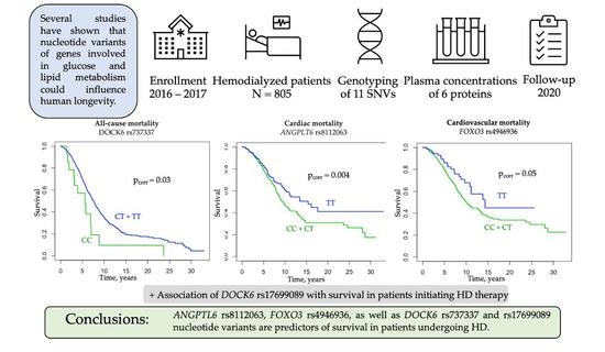
Some studies have shown that nucleotide variants in the insulin growth factor-1 (IGF-1) signaling pathway genes involved in glucose metabolism could influence human longevity [15]. For example, the forkhead box protein O3 gene (FOXO3, OMIM*602681) encoding the IGF-1 pathway downstream transcription factor (TF), forkhead box protein O3 (FOXO3), has been found to be strongly associated with human longevity and the prevalence of diabetes and arterial hypertension [16]. Previous in vitro studies found that FOXO3 plays a crucial role in regulating the insulin/insulin-like growth factor 1 (IGF-1)/phosphatidylinositol-3 kinase (PI3K)/AKT (Protein Kinase B) metabolic pathway [17]. Moreover, the transcriptional targets of FOXOs include genes involved in cell cycle arrest, oxidative resistance, apoptosis, autophagy, DNA damage repair, and energy metabolism [18]. Another IGF-1 signaling pathway protein, insulin-like growth factor binding protein 3 (IGFBP-3), the most abundant of the six known IGFBPs, circulates in the bloodstream and transports IGFs that have been sequestered in the form of a ternary complex to the IGF receptors (IGFRs) to initiate a cascade of downstream signaling events [19]. The insulin-like growth factor binding protein-3 gene (IGFBP3, OMIM*146732) influences lipid parameters in adolescents and cancer susceptibility in the general population [20,21].
Angiopoietin-like proteins (ANGPTL) are members of a protein family named according to their structural similarity to angiopoietins [22,23,24,25]. Angiopoietin-like protein 6, a liver-derived circulating factor, is considered to be a regulator of metabolic homeostasis [25]. It is encoded by the angiopoietin-like protein 6 gene (ANGPTL6, OMIM*609336) and is capable of counteracting both obesity and obesity-related insulin resistance [24]. The production of angiopoietin-like protein 8 (ANGPLT8) in the liver and adipose tissue is induced by insulin via PI3K/AKT signaling [26]. Angiopoietin-like protein 8 gene (ANGPTL8) is located in the corresponding intron of dedicator of cytokinesis 6 (DOCK6, OMIM*614194) [25].
Moreover, numerous genes associated with lipid metabolism have been found to influence the risk of cardiovascular disease and dyslipidemia in the general population, including the fatty acid binding protein 1 gene (FABP1, OMIM*134650). Fatty acid-binding proteins are a family of small and highly conserved cytoplasmic proteins with the ability to bind to long-chain fatty acids and other hydrophobic ligands and participate in the intracellular transportation of lipids [22,27].
Proprotein convertase subtilisin/kexin type 9 (PCSK9) is a serine protease that promotes the catabolism of low-density lipoprotein (LDL) receptors (LDLR) and consecutively controls lipid metabolism [28]. Variants of the proprotein convertase subtilisin/kexin type 9 gene (PCSK9, OMIM*607786) have recently been associated with cardiovascular risk in the general population [29].
Our study aimed to investigate whether the FOXO3, IGFBP3, FABP1, PCSK9, and ANGPTL6, DOCK6 single nucleotide variants and FOXO3, IGFBP3, L-FABP, PCSK9, ANGPTL6, and ANGPTL8 plasma concentrations are associated with the survival probability of Polish hemodialyzed patients

2. Materials and Methods

2.1. Study Design

The study was designed as a genetic cohort study. STROBE (STrengthening the Reporting of OBservational studies in Epidemiology) guidelines were employed. The study protocol was approved by the Ethics Committee of the Poznań University of Medical Sciences and fulfilled the requirements of the Declaration of Helsinki. The patients were enrolled in the study between June 2016 and December 2017. Mortality was the primary outcome of the study. In deceased individuals, causes of death were registered based on medical documentation and were categorized as cardiac (reported as myocardial infarction, sudden cardiac death, severe arrhythmias, cardiomyopathies, or cardiac failure), vascular (reported as cerebrovascular events, cerebral stroke, or generalized atherosclerosis), infection-related (reported as sepsis, pneumonia, limb necrosis, pyonephrosis, or acute abdomen with peritonitis), cancer-related, and other or unknown. Cardiovascular deaths were defined as deaths caused by cardiac and/or vascular events. Additionally, a subgroup analysis of 83 patients initiating HD therapy was performed. Patients initiating therapy were defined as individuals who had been dialyzed for less than 6 months prior to enrollment in the study. Secondary outcomes were cardiovascular events, diagnosis of coronary artery disease (CAD), BMI, serum total cholesterol, serum high-density lipoprotein cholesterol (HDL-cholesterol), serum low-density cholesterol (LDL-cholesterol), and serum triglycerides (TG). The patient outcomes (death, renal transplantation, moving to a non-collaborating center) were evaluated in January 2020.

2.2. Study Participants

Prevalent HD patients (n = 805) undergoing dialysis at 27 dialysis centers in the Greater Poland region of Poland were evaluated as candidates for this observational study. All study participants were unrelated Caucasians of Polish origin. The inclusion criteria included written informed consent, age over 18 years, and stable clinical state for at least two months before the onset of the study. The exclusion criteria included missing data in one of the primary or secondary outcomes and secondary causes of hyperlipidemia such as liver disease or a cachectic state at the onset of the study.
The control group included 360 healthy individuals from the same geographical region. The inclusion criteria were written informed consent and age over 18 years. Exclusion criteria encompassed kidney disease (self-reported or as determined by GFR and albumin values), diabetes, cardiovascular disease (CAD, atherosclerosis, or cerebral stroke), and acute infection at the study onset.

2.3. Blood Sample Collection

The blood samples were collected from the HD patients before their midweek hemodialysis session and from the healthy volunteers in the morning after an overnight fast. Blood samples were collected in ethylenediaminetetraacetic acid (EDTA) tubes to obtain whole blood and in plasma-separated tubes. After preparation, the blood samples were analyzed or frozen immediately after collection and stored at −80 °C.

2.4. Laboratory Methods

Serum concentrations of total cholesterol, HDL-cholesterol, TG, C-reactive protein (CRP), alanine transaminase (ALT), aspartate transaminase (AST), and gamma-glutamyl transferase (GGT) were measured using routine enzymatic methods in a commercial laboratory. The LDL-cholesterol serum concentrations were calculated using the Friedewald formula. In patients with serum TG concentrations ≥ 400 mg/dL, LDL-cholesterol was measured directly (BioSystems S.A., Reagents and Instruments, Barcelona, Spain).
Plasma concentrations of FOXO3, IGFBP3, L-FABP, PCSK9, ANGPTL6, and ANGPTL8 (betatrophin) were measured in a randomly selected group of 86 HD patients using an enzyme-linked immunosorbent assay (ELISA). Patients were assigned into the group using the simple randomization method. The following commercial kits were used: Human Forkhead Box O3 (FOXO3) ELISA Kit, Catalog Number CSB-E11177h, Cusabio Technology LLC, Wuhan, China; Human IGFBP-3 Immunoassay, Catalog Number DGB300, R&D Systems, Inc., Minneapolis, MN, USA; Human Liver Type Fatty Acid Binding Protein (L-FABP) ELISA Kit, Catalog Number CSB-E13455h, Cusabio Technology LLC, Wuhan, China; Human Proprotein Convertase 9/PCSK9 Immunoassay, Catalog Number DPC900, R&D Systems, Inc., Minneapolis, MN, USA; ANGPTL6 (human) ELISA Kit, Cat. No. AG-45A-0016YEK-KI01, Adipogen Life Sciences, Liestal, Switzerland; and Human Betatrophin ELISA Kit, Catalog No: E11644h, EIAab, Wuhan, China. All laboratory analyzes were performed according to the manufacturer’s instructions.

2.5. Genotyping

DNA was extracted from blood lymphocytes using the salting-out method. Eleven nucleotide variants in six genes were selected based on a literature review. The characteristics of the analyzed SNVs are described in Table 1. Genotyping of the FOXO3, IGFBP3 rs3110697, FABP1, PCSK9, ANGPTL6, and DOCK6 SNVs was performed using high-resolution melting curve (HRM) analysis with 5x HOT FIREPol EvaGreen HRM Mix (Solis BioDyne, Tartu, Estonia) on the Light Cycler 96 system (Roche Diagnostics, Mannheim, Germany). The primer sequences and conditions for the HRM analyses are presented in Table S1. The IGFBP3 rs2854744 variant was analyzed using the predesigned C___1842665_10 TaqMan SNP Genotyping Assay according to the manufacturer’s instructions (Applied Biosystems, Foster City, CA, USA). For quality control, approximately 10% of the randomly chosen samples were regenotyped using the same genotyping method; the concordance rate was 100%. Samples that failed the genotyping analysis were excluded from further statistical analysis.

2.6. Statistical Analyses

All of the statistical analyses were performed using R statistical software [30]. Data are presented as percentages for categorical variables or as medians and ranges for continuous variables that were non-normally distributed. Before every other statistical analysis of quantitative variables, data were checked for normal distribution (Shapiro–Wilk test) and homoscedasticity (Levene’s test). Student’s t-test and ANOVA were used to analyze normally distributed and homoscedastic data sets. Non-normally distributed data sets were compared using Mann–Whitney U, Kruskal–Wallis, and Dunn testing. Spearman’s rank test was used to show correlations between selected variables. Pearson’s chi-squared test and Fisher’s exact test were used to analyze the qualitative variables. The strength of the association was evaluated by odds ratios (ORs) with 95% confidence intervals (95% CI) in a dominant, recessive, and additive model of inheritance. Survival probability since RRT onset was analyzed using the Kaplan–Meier method with the log-rank test. All analyses were performed using patient groups separated by genotypes in three modes of inheritance. The Cox proportional hazards model was applied to show whether and to which extent the effect of a unit change in a covariate was multiplicative concerning the hazard rate (HR) of death. HRs were adjusted for clinical data using Cox proportional hazards regression analysis. Gender, age at RRT onset, myocardial infarction, stroke, diabetic nephropathy, serum concentrations of intact PTH, and calcium phosphate product were applied as clinical variables, possibly contributing to survival probability in the multivariable analyses. A p-value of <0.05 was considered to indicate statistical significance. Correction for multiple testing regarding the primary outcome was performed using a 1000-fold permutation version of the k-sample log-rank test. Missing data were not considered in the prevalence calculations and were reported as part of the descriptive statistics.
The power to detect the genetic associations was determined using the Genetic Association Study (GAS) Power Calculator (http://csg.sph.umich.edu/abecasis/gas_power_calculator/index.html (accessed on 1 March 2022) under the following assumptions: case/control ratio 1.645, significance level = 0.05, prevalence = 63% [9]. It was calculated that a sample size of at least 800 HD patients would yield at least 80% power for detecting the relative risk of 1.25 in the additive and dominant mode of inheritance in all of the analyzed SNVs (Table S2).
Haplotype frequencies were estimated using Haploview 4.2 software (http://www.broad.mit.edu/mpg/haploview/ (accessed on 2 April 2022). Epistatic interactions between the tested SNVs were analyzed using the odds ratio-based multifactor dimensionality reduction (MDR) method [31]. Statistical significance in both tests was assessed using the 1000-fold permutation test.

3. Results

3.1. Patient Characteristics

Between June 2016 and December 2017, a group of 1619 prevalent HD patients was screened, and 1205 individuals fulfilled the inclusion criteria and presented no exclusion criteria. In addition, 244 subjects were excluded due to the lack of all of the required measurements. In total, 961 patients were included in the observational study, but 156 were lost to follow-up. The final study group included 805 prevalent HD patients. Table 2 presents the patients’ data. The study group comprised 457 males (56.8%). The median age at RRT onset was 61.4 years. Four hundred thirty-two patients (53.7%) were dialyzed using low-flux dialyzers. All patients were prescribed hemodialysis on a thrice-weekly schedule. During the study period, 510 patients died (63.4%), and 152 underwent renal transplantation (18.9%). Dyslipidaemia, defined according to K/DOQI criteria, was diagnosed in 387 patients (48.1%). Additionally, 337 patients were diagnosed with arterial hypertension (41.8%), 309 patients had coronary artery disease (38.4%), 174 underwent myocardial infarction (21.6%), and 210 suffered from a cerebral stroke (26.1%). The most common cause of the end-stage renal disease was diabetic kidney disease, which was diagnosed in 246 patients, followed by hypertensive nephropathy (n = 170), chronic glomerulonephritis (n = 105), and chronic tubulointerstitial nephritis (n = 67).

3.2. Genotype Frequencies in Patients and Controls

The frequency distributions of the tested SNVs did not differ between the HD patients and the healthy volunteers (Table S3). Furthermore, all of the SNVs, except for DOCK6 rs176990893 in the HD patients and PCSK9 rs11206510 in the controls, complied with the Hardy–Weinberg equilibrium (HWE) (Table S3).

3.3. Survival Analysis

Overall, 510 patients died during their RRT periods, which lasted between 0.09 and 29.9 years. Survival probability from the start of RRT was negatively associated with male gender (p = 0.013), age at RRT onset (p < 0.001), diabetic nephropathy (p < 0.001), coronary artery disease (p < 0.001), myocardial infarction (p < 0.001), cerebral stroke (p < 0.001), hypolipemic therapy (p = 0.040), body weight (p = 0.002), BMI (p < 0.001), and plasma concentration of C-reactive protein (p < 0.001) and was positively associated with chronic glomerulonephritis as the cause of ESRD (p < 0.001), serum concentrations of calcium (p = 0.001) and phosphate (p = 0.002), intact PTH (p < 0.001), and the presence of calcium phosphate product (p < 0.001) (Table 3).

3.3.1. DOCK6 rs737337 and rs17699089 and Mortality in HD Patients

The DOCK6 rs737337 C allele was associated with all-cause mortality in the recessive mode of inheritance (log-rank test p = 0.020; HR 1.94 95% CI 1.09–3.44; Wald test p = 0.02; pcorr = 0.031) and with cardiac mortality in the additive mode of inheritance (log-rank test p = 0.030; HR 2.35 95% CI 1.04–5.32; Wald test p = 0.040; pcorr = 0.047) (Figure 1). The multivariable regression analysis, which included gender, age at RRT onset, myocardial infarction, stroke, diabetic nephropathy, serum concentrations of intact PTH, and calcium phosphate product, revealed that the DOCK6 rs737337 C allele remained a significant risk factor for overall and cardiac mortality among clinical factors (HR 2.40 95% CI 1.35–4.28, p = 0.003, HR 3.03, 95% CI 1.32–6.95, p = 0.009, respectively). The rs737337 C allele was associated with an increased risk of diabetic nephropathy (OR 1.687, 95% CI 1.171–2.432, p = 0.005) and a decreased risk of hypertensive nephropathy as a cause of ESRD (OR 0.587, 95% CI 0.363–0-950, p = 0.029) in the dominant mode of inheritance (Table S4). There were no associations between DOCK6 rs17699089 and survival in the entire group of HD patients.
An additional 3.5-year prospective analysis of 83 patients initiating HD therapy revealed that the DOCK6 rs737337 C allele was significantly associated with cardiovascular and cardiac mortality in the recessive mode of inheritance (log-rank test p < 0.001; HR 68.5 95% CI 4.3–1095.0, Wald test p = 0.003, pcorr = 0.01 for cardiovascular mortality, and log-rank test p < 0.001; HR 68.5 95% CI 4.3–1095.0, Wald test p = 0.003, pcorr = 0.01 for cardiac mortality) (Figure 2). The G allele of DOCK6 rs17699089 in the recessive mode of inheritance was associated with an increased risk of all-cause (log-rank test p = 0.002; HR 5.7 95% CI 1.6–19.9, Wald test p = 0.007, pcorr = 0.04), cardiovascular (log-rank test p < 0.001; HR 13.9 95% CI 2.6–74.5, Wald test p < 0.001, pcorr = 0.03), and cardiac mortality (log-rank test p < 0.001; HR 20.3 95% CI 3.2–127.9, Wald test p < 0.001, pcorr = 0.02) (Figure 2).

3.3.2. ANGPTL6 rs8112063

In the unadjusted analyses, the C allele of ANGPTL6 rs8112063 in the dominant mode of inheritance was associated with increased all-cause (log-rank test p = 0.047; HR 1.21 95% CI 1.00–1.48; Wald test p = 0.050; pcorr = 0.048), cardiovascular (log-rank test p = 0.010; HR 1.39 95% CI 1.07–1.81; Wald test p = 0.010; pcorr = 0.017), and cardiac mortality (log-rank test p = 0.003; HR 1.64 95% CI 1.18–2.27; Wald test p = 0.003; pcorr = 0.004) (Figure 3). The ANGPTL6 rs8112063 C allele was not an independent risk factor for overall mortality after adjustment for gender, age at RRT onset, myocardial infarction, stroke, diabetic nephropathy, serum concentrations of intact PTH, and calcium phosphate product (HR 1.14, 95% CI 0.94–1.39, p = 0.192), but it remained a significant risk factor for cardiac and cardiovascular mortality after adjustment (HR 1.59, 95% CI 1.15–2.21, p = 0.005, HR 1.31, 95% CI 1.01–1.72, p = 0.042, respectively). The C allele of ANGPTL6 rs8112063 was also associated with an increased risk of diabetic nephropathy (OR 1.503, 95% CI 1.054–2.143, p = 0.024) and a decreased risk of tubulointerstitial nephropathy as a cause of ESRD in the recessive mode of inheritance (OR 0.349, 95% CI 0.148–0.825, p = 0.012). Patients with the ANGPTL6 rs8112063 CC genotype were less likely to be diagnosed with dyslipidemia by K/DOQI than the bearers of the TT genotype (OR 0.672, 95% CI 0.453–0.997, p = 0.048). Carriers of the rs8112063 C allele had lower serum concentrations of LDL-cholesterol and higher concentrations of HDL-cholesterol than those with the TT genotype (p = 0.014 and p = 0.026 in the dominant mode of inheritance) (Table S5).

3.3.3. FOXO3 rs4946936

The minor T allele of FOXO3 rs4946936 was associated with a decreased risk of cardiac (log-rank test p = 0.040; HR 0.57 95% CI 0.33–0.98; Wald test p = 0.040; pcorr = 0.042) and cardiovascular death (log-rank test p = 0.04; HR 0.64 95% CI 0.42–0.99; Wald test p = 0.04; pcorr = 0.047) in HD patients (Figure 4). FOXO3 rs4946936 was not associated with cardiac and cardiovascular mortality after adjustments for gender, age at RRT onset, myocardial infarction, stroke, diabetic nephropathy, serum concentrations of intact PTH, and calcium phosphate product (HR 0.64, 95% CI 0.37–1.11, p = 0.109 for cardiac mortality and HR 0.75, 95% CI 0.48–1.16, p = 0.191 for cardiovascular mortality). Patients with the FOXO3 rs4946936 T allele were less likely to be diagnosed with dyslipidemia according to the K/DOQI criteria (OR 0.686, 95% CI 0.517–0.910, p = 0.009) and had lower serum concentrations of total cholesterol and LDL-cholesterol than the bearers of the CC genotype (p = 0.029 and 0.036, respectively) (Table S6).

3.3.4. Other Genotypes

There were no associations between the risk of death in HD patients and the other analyzed SNVs (FOXO3 rs2802292; IGF2BP2 rs4402960, and rs11705701; IGFBP3 rs3110697, and rs2854744; FABP1 rs2241883, and rs2919872; PCSK9 rs562556, and rs11206510; and DOCK6 rs17699089) (Tables S7–S12).

3.4. Protein Plasma Concentrations

There were no statistically significant differences in the genotype distributions of the tested nucleotide variants among the patients in which the plasma concentrations of the tested proteins were evaluated and in those in which they were not measured, except for ANGPTL6 rs8112063. The study participants with measured protein ANGPTL6 concentrations had a lower frequency distribution of the rare rs8112063 C allele than the other group (p = 0.021) (Table S13). There were no associations between the plasma concentrations of FOXO3, IGFBP3, L-FABP, PCSK9, ANGPTL6, and ANGPTL8 and survival probability in a 3.5-year prospective analysis from June 2016 (Table S14). Patients with the CC genotype of FABP1 rs2241883 had higher L-FABP plasma concentrations than the carriers of the dominant T allele (57.9, 13.5–85.7 vs. 32.5 ng/dL, 4.0–114.3 ng/dL, p = 0.023). Bearers of the CC genotype of FABP1 rs2919872 had lower FABP1 plasma concentrations than those with the T allele (22.4, 9.9–88.8 vs. 41.7 ng/dL, 4.0–114.3 ng/dL, p = 0.037) (Figure S1). There were no correlations between the other analyzed nucleotide variants and the serum concentrations of their protein products (Table S15).

3.5. Haplotype Analysis

The DOCK6 rs17699089G_rs737337C haplotype was positively associated with diabetic nephropathy. There were no other significant associations between the DOCK6 haplotypes and the tested phenotypes (Table 4).

3.6. Epistatic Interactions

A gene–gene interaction was noted among FOXO3 rs4946936 and ANGPTL6 rs8112063 (testing balance accuracy (TBA) 0.58, p-value 0.002) as well as among the FOXO3 rs4946936, IGFBP3 rs2854744, FABP1 rs2919872, and ANGPTL6 rs8112063 (TBA 0.56, p-value 0.027) and FOXO3 rs4946936, IGFBP3 rs2854744, FABP1 rs2241883, FABP1 rs2919872, and ANGPTL6 rs8112063 nucleotide variants in relation to all-cause mortality (TBA 0.55, p-value 0.026, respectively) (Table 5). Moreover, an epistatic interaction between FOXO3 rs2802292, IGFBP3 rs2854744, FABP1 rs2241883, and ANGPTL6 rs8112063 was observed in relation to cardiovascular mortality (TBA 0.53, p-value 0.024) (Table 6). IGFBP3 rs3110697, DOCK6 rs737337, and DOCK6 rs17699089 showed a gene–gene interaction concerning the diagnosis of myocardial infarction (TBA 0.63, p-value 0.032) (Table 7). Furthermore, there was a gene–gene interaction between FOXO3 rs2802292 and FABP1 rs2241883 regarding the diagnosis of dyslipidemia by K/DOQI (TBA 0.59, p-value 0.021) (Table S16). No further significant gene–gene interactions were detected in relation to other comorbidities, including diabetes (Tables S17–S19).

4. Discussion

We conducted a genetic cohort study to assess whether nucleotide variants of selected genes associated with glucose and lipid metabolism are associated with mortality risk in patients undergoing hemodialysis treatment. We first demonstrated that the ANGPTL6 rs8112063 and FOXO3 rs4946936 as well as the DOCK6 rs737337 and rs17699089 SNVs are predictors of survival in Polish HD patients. In addition, we also noted a gene–gene interaction between FOXO3 rs4946936 and ANGPTL6 rs8112063 and between FOXO3 rs4946936, IGFBP3 rs2854744, FABP1 rs2241883, FABP1 rs2919872, and ANGPTL6 rs8112063 regarding overall survival. To our knowledge, this study is the first to show that the FOXO3, ANGPTL6, and DOCK6 variants can predict clinical outcomes in the uremic population.
End-stage renal disease patients have poorer survival than the general population [1]. Mortality is exceptionally high within the first few months of dialysis [32]. Cardiovascular diseases remain the most common cause of death among RRT patients, accounting for almost 50% of mortality [33]. In our study, 63% of patients died over the course of 3.5 years, and cardiovascular diseases were the most common cause of death. The univariate analyses confirmed multiple known demographic, clinical, and laboratory mortality risk factors of HD patients, such as male gender, advanced age, diabetes, history of cardiovascular diseases, and CRP concentrations [34,35,36,37,38]. We did not observe a significant relationship between the lipid profile and survival. Patients on hypolipemic therapy had a slightly increased risk of death compared to those not taking lipid-lowering drugs. This paradoxical association might be confounded by a greater prevalence of comorbidities among patients receiving hypolipemic therapy [39]. Unlike previous studies, we observed no associations between smoking status and overall survival [2,40]. Contrary to many studies, BMI and body weight were negatively associated with survival probability in our study [2,41].
Angiopoietin-like proteins, a family of proteins that regulate energy and glucose homeostasis, are involved in angiogenesis and share similarities with angiopoietins, such as a coiled-coil domain, a linker region, and a carboxy-terminal fibrinogen-like domain [42,43]. Circulating levels of several proteins in the ANGPTL family have been associated with obesity, diabetes, and mortality in HD patients [44,45].
ANGPTL6 has been identified as a circulating mediator of angiogenesis that is capable of increasing endothelial permeability [42]. Previous studies revealed increased serum concentrations of this anti-obesity hepatokine in individuals with metabolic syndrome and type 2 diabetes and decreased circulating ANGPTL6 levels in hemodialyzed individuals compared to healthy subjects [44,46,47]. rs8112063 is located in the first intron of ANGPTL6. As the introns (especially the first one) are known to be regulatory regions that modulate gene expression, rs8112063 could potentially influence ANGPTL6 expression levels [24]. Our study revealed that the rs8112063 C allele was a significant risk factor of all-cause, cardiovascular, and cardiac mortality in HD patients, and it remained an independent cardiovascular mortality risk factor after adjustment for gender, age at RRT onset, myocardial infarction, stroke, diabetic nephropathy, and serum concentrations of intact PTH and calcium phosphate product. Hostettler et al. observed that several rare ANGPTL6 genetic variants are risk factors for intracranial aneurysms [48]. However, we did not observe an association between the tested ANGPTL6 nucleotide variant HD and vascular mortality. Interestingly, the French MONICA study suggested that the C allele of rs8112063 could be a risk factor for metabolic syndrome [24]. We found that the carriers of the rs8112063 CC genotype were more likely to suffer from diabetic nephropathy and less likely to be diagnosed with dyslipidemia by K/DOQI compared to those with the T allele. To our knowledge, this is the first study to report such an association. Interestingly, mice knockout studies have shown that ANGPTL6 counteracts diet-induced obesity and insulin resistance via increasing energy expenditure, which suggests that ANGPTL6 may have a modulatory role in diabetes [49]. However, patients with type 2 diabetes have paradoxically increased circulating ANGPTL6 concentrations compared to healthy individuals [47]. ANGPTL6 could therefore contribute to the development of diabetic nephropathy through pathways related to insulin resistance, which is associated with greater salt sensitivity, increased glomerular pressure, albuminuria, and kidney function decline [50].
ANGPLT8, a gene located in the corresponding intron of DOCK6, encodes ANGPTL8, a peptide hormone produced in the liver and adipose tissue that plays a role in glucose and lipid homeostasis [25]. rs737337 is located 2.8 kb upstream of the ANGPTL8 transcription start site and is a synonymous variant in exon 19 of DOCK6 (Thr723). In a study by Cannon et al. rs737337 showed strong enhancer activity in transcriptional reporter assays [51]. Our study found that the C allele of DOCK6 rs737337 was a risk factor for all-cause and cardiac mortality among HD patients. Notably, DOCK6 rs737337 remained a cardiac mortality risk factor after adjusting for the clinical parameters that are relevant to survival. In contrast, Agra et al. found that the C allele of DOCK6 rs737337 was associated with a better prognosis in obese patients with heart failure [52]. Interestingly, Zou et al. found that serum ANGPTL8 levels were associated with the risk of all-cause mortality in diabetic subjects [53]. Furthermore, we observed that the C allele of rs737337 was associated with an increased risk of diabetic nephropathy as a cause of ESRD.
Another tested DOCK6 variant, rs17699089, was not directly associated with mortality or diabetic nephropathy in the entire group of Polish HD subjects. However, in a prospective analysis of patients initiating HD therapy, we observed an association between the G allele of rs17699089 and all-cause, cardiovascular, and cardiac mortality. Rs17699089, a DOCK6 intron variant, showed evidence of allelic differences in the transcriptional activity of ANGPTL8 in subcutaneous adipose tissue [51]. Moreover, a study by Ghasemi et al. found that rs17699089 was in linkage disequilibrium with rs2278426, a known ANGPTL8 variant associated with serum concentrations of total cholesterol and an increased risk of diabetic nephropathy [51,54]. Interestingly, we found a significant haplotype association between DOCK6 rs17699089_rs737337 and diabetic nephropathy. To date, there have been no studies on the putative associations between DOCK6 and diabetes or CKD. However, multiple studies have revealed that the levels of ANGPTL8 are higher in patients with diabetic nephropathy and obesity as well as positively correlated with atherogenic markers and carotid intima-media thickness [55,56,57,58]. Another study by Zou et al. revealed that older subjects with higher circulating ANGPTL8 levels were at an increased risk of kidney function decline, which might suggest a role of ANGPTL8 in the pathogenesis of CKD [59]. ANGPTL8 may drive the progression of diabetic nephropathy through pathways and mechanisms related to insulin resistance or through inflammatory mechanisms [60,61]. However, we observed no differences in the frequency distributions of DOCK6 SNVs among the HD patients and controls. In our study, DOCK6 rs737337 and rs17699089 interacted with IGFBP3 rs3110697 in relation to myocardial infarction diagnosis among HD patients. Interestingly, both rs17699089 and rs737337 are associated with HDL-cholesterol levels in the general population [51]. Moreover, GWAS analyses revealed that rs737337 is associated with the lipid profile and CAD susceptibility in the European population [62,63].
The forkhead box (FOX) is a heterogenic protein family of transcription factors that contain a conserved DNA-binding domain, a sequence of 80 to 100 amino acids called the forkhead domain [64]. FOXO3 plays a vital role in multiple fundamental processes in cells, such as controlling metabolism, cell division and differentiation status, and response to cellular stress [65]. FOXO3 nucleotide variants, especially rs2802292, exhibit a consistently replicated genetic association with longevity in multiple populations worldwide [66]. rs4946936 is located in the 3′UTR of FOXO3. Wang et al. found that the rs4946936 T allele significantly increased the expression levels in luciferase assays and affected the binding affinity of miR-223 to the FOXO3 3′UTR [67]. Our analysis revealed that the T allele of FOXO3 rs4946936 was a protective factor against cardiovascular and cardiac mortality. There was also a gene–gene interaction between FOXO3 rs4946936 and ANGPTL6 rs8112063 regarding all-cause mortality. This observation is concordant with the results of earlier studies on centenarians, which also reported protective effects of the rs4946936 T allele [68,69]. Surprisingly, there were no direct associations between FOXO3 rs2802292 and survival in HD patients. However, we observed an epistatic interaction between FOXO3 rs2802292, IGFBP3 rs2854744, FABP1 rs2241883, and ANGPTL6 rs8112063 and cardiovascular mortality in Polish HD subjects. Associations between FOXO3 rs2802292 and the prevalence of essential hypertension and improved metabolic control in diabetic patients have been shown in previous studies [70,71]. On the other hand, Klinpudtan et al. reported that the longevity-associated G allele of FOXO3 rs2802292 appears to have contrasting associations with heart disease prevalence according to sex in older Japanese people [16]. The FOXO3 protein might play a protective role in chronic kidney disease by preventing mitochondrial damage and ameliorating fibrosis [72,73]. However, we did not note any differences in the frequency distributions of FOXO3-related genetic alterations among HD patients and controls.
Our study has several limitations. Firstly, the primary survival analysis was evaluated from the onset of RRT therapy, which makes it more prone to selection bias than other epidemiologic studies. To address this problem, we adjusted the survival analyses for selected clinical variables associated with death risk in HD patients, evaluated the relationship between the tested SNVs and other clinical phenotypes, and conducted an additional survival analysis among the HD patients who had started RRT less than 6 months prior to study enrollment. Moreover, despite a decent study population of over 800 HD patients, there were only 55 and 53 cases of death due to neoplasms and infections, respectively. Because of that, a dedicated cause-specific survival analysis of infectious or neoplastic deaths was not conducted to avoid underpowered statistical tests and inconclusive results. Thirdly, the investigation was performed on a specific population comprising Polish dialyzed patients, and the findings should be used with caution when extrapolated to other ethnicities. Further investigations of putative genetic predictors of survival in larger cohorts encompassing diverse ethnicities are necessary. The results of our study should also be confirmed in other cohorts of Caucasian patients. Finally, plasma concentrations of protein products of the tested nucleotide variants were only evaluated in a limited number of subjects. Nevertheless, the ELISA analysis of the tested proteins was performed in randomly chosen patients to increase the credibility of the results. However, the group in which plasma protein concentrations were measured had lower frequency distributions of the rare C allele of ANGPTL6 rs8112063 compared to the other study participants, which could have potentially led to underpowered tests. Future investigations with larger study sizes and examining the tested protein concentrations over extended periods are needed to elucidate their role in ESRD.

5. Conclusions

In summary, our study demonstrated that ANGPTL6 rs8112063 and DOCK6 rs737337 SNVs are significant predictors of all-cause and cardiac mortality in Polish HD patients. In addition, the rare allele of DOCK6 rs17699089 is associated with all-cause, cardiovascular, and cardiac mortality in patients initiating HD therapy. Moreover, the rare variant of FOXO3 rs4946936 is a protective factor against cardiac and cardiovascular death in this group. However, further prospective studies with larger study sizes are needed to clarify the influence of the genes associated with glucose and lipid metabolism on the clinical outcomes of HD patients.

Supplementary Materials

The following supporting information can be downloaded at https://www.mdpi.com/article/10.3390/jcm11185477/s1: Table S1: Primer sequences and HRM conditions for the identification of genotyped nucleotide variants; Table S2: The expected power for 1.00–1.75 ORs in association analyses of the HD patients who died and those who survived; Table S3: Frequency distributions of the analyzed nucleotide variants in hemodialysis patients and healthy controls; Table S4: DOCK 6 rs737337 nucleotide variants and clinical and laboratory data of HD patients (n = 780); Table S5: ANGPTL6 rs8112063 nucleotide variants and clinical and laboratory data of HD patients (n = 780); Table S6: FOXO3 rs4946936 nucleotide variants and clinical and laboratory data of HD patients (n = 778); Table S7: Associations between the selected energy homeostasis nucleotide variants and all-cause mortality of HD patients evaluated by Kaplan–Meier analysis; Table S8: Associations between the selected energy homeostasis nucleotide variants and cardiovascular mortality of HD patients evaluated by Kaplan–Meier analysis; Table S9: Associations between the selected energy homeostasis nucleotide variants and cardiac mortality of HD patients evaluated by Kaplan–Meier analysis; Table S10: Associations between the selected energy homeostasis nucleotide variants and all-cause mortality of patients initiating HD therapy evaluated by Kaplan–Meier analysis; Table S11: Associations between the selected energy homeostasis nucleotide variants and cardiovascular mortality of patients initiating HD therapy evaluated by Kaplan–Meier analysis; Table S12: Associations between the selected energy homeostasis nucleotide variants and cardiac mortality of patients initiating HD therapy evaluated by Kaplan–Meier analysis; Table S13: Frequency distributions of the analyzed nucleotide variants in hemodialysis patients in which protein analyses were performed and in those without protein analysis; Table S14: Correlations between the plasma concentrations of the studied proteins and survival probability of HD patients in a 3.5-year prospective analysis from June 2016 (n = 86); Table S15: Comparison of the analyzed nucleotide variants and the serum concentrations of their protein products in HD patients (n = 86); Table S16: Epistatic interactions between the analyzed genes with respect to dyslipidemia by K/DOQI in HD patients; Table S17: Epistatic interactions between the analyzed genes with respect to cardiac mortality in HD patients; Table S18: Epistatic interactions between the analyzed genes with respect to CAD in HD patients; Table S19: Epistatic interactions between the analyzed genes with respect to diabetes in HD patients; Figure S1: L-FABP plasma concentrations according to the analyzed nucleotide variants: (a) FABP1 rs2241883 in the recessive mode of inheritance; (b) FABP1 rs2919872 in the dominant mode of inheritance.

Author Contributions

Conceptualization, M.K.Ś. and A.E.G.; methodology, A.M., P.P.J. and A.E.G.; software, M.K.Ś. and A.M.; validation, A.M., P.P.J. and P.B.; formal analysis, M.K.Ś. and A.M.; investigation, M.K.Ś., D.S. and P.B.; resources, A.M. and P.P.J.; data curation, M.K.Ś.; writing—original draft preparation, M.K.Ś.; writing—review and editing, A.M., D.S., P.P.J., P.B. and A.E.G.; visualization, M.K.Ś.; supervision, A.E.G.; project administration, M.K.Ś. and A.E.G.; funding acquisition, M.K.Ś. All authors have read and agreed to the published version of the manuscript.

Funding

This research was funded by the “Diamond Grant” allocated by Polish Ministry of Science and Higher Education, grant number DI2015 019345 (years 2016–2021).

Institutional Review Board Statement

The study was conducted in accordance with the Declaration of Helsinki and was approved by the Ethics Committee of the Poznań University of Medical Sciences (approval number 758/16). Approved on 16 June 2016.

Informed Consent Statement

Informed consent was obtained from all subjects involved in the study.

Data Availability Statement

The de-identified datasets generated through this study can be provided by the corresponding author upon request.

Acknowledgments

The authors would like to thank the physicians and nurses from the Department of Nephrology, Provincial Integrated Hospital in Konin, Poland.

Conflicts of Interest

The authors declare no conflict of interest.

References

  1. Johansen, K.L.; Chertow, G.M.; Foley, R.N.; Gilbertson, D.T.; Herzog, C.A.; Ishani, A.; Israni, A.K.; Ku, E.; Kurella Tamura, M.; Li, S.; et al. US Renal Data System 2020 Annual Data Report: Epidemiology of Kidney Disease in the United States. Am. J. Kidney Dis. Off. J. Natl. Kidney Found. 2021, 77, A7–A8. [Google Scholar] [CrossRef] [PubMed]
  2. Major, R.W.; Cheng, M.R.I.; Grant, R.A.; Shantikumar, S.; Xu, G.; Oozeerally, I.; Brunskill, N.J.; Gray, L.J. Cardiovascular Disease Risk Factors in Chronic Kidney Disease: A Systematic Review and Meta-Analysis. PLoS ONE 2018, 13, e0192895. [Google Scholar] [CrossRef]
  3. Foley, R.N.; Parfrey, P.S.; Sarnak, M.J. Clinical Epidemiology of Cardiovascular Disease in Chronic Renal Disease. Am. J. Kidney Dis. Off. J. Natl. Kidney Found. 1998, 32, S112–S119. [Google Scholar] [CrossRef] [PubMed]
  4. Sharma, I.; Liao, Y.; Zheng, X.; Kanwar, Y.S. New Pandemic: Obesity and Associated Nephropathy. Front. Med. 2021, 8, 673556. [Google Scholar] [CrossRef]
  5. Tseng, A.S.; Girardo, M.; Firth, C.; Bhatt, S.; Liedl, D.; Wennberg, P.; Shen, W.-K.; Cooper, L.T.; Shamoun, F.E. Lower Extremity Arterial Disease as a Predictor of Incident Atrial Fibrillation and Cardiovascular Events. Mayo Clin. Proc. 2021, 96, 1175–1183. [Google Scholar] [CrossRef]
  6. Kim, S.-A.; Lim, K.; Lee, J.-K.; Kang, D.; Shin, S. Metabolically Healthy Obesity and the Risk of All-Cause and Cardiovascular Disease Mortality in a Korean Population: A Prospective Cohort Study. BMJ Open 2021, 11, e049063. [Google Scholar] [CrossRef]
  7. Liakopoulos, V.; Roumeliotis, S.; Zarogiannis, S.; Eleftheriadis, T.; Mertens, P.R. Oxidative Stress in Hemodialysis: Causative Mechanisms, Clinical Implications, and Possible Therapeutic Interventions. Semin. Dial. 2019, 32, 58–71. [Google Scholar] [CrossRef]
  8. Qunibi, W.Y. Dyslipidemia in Dialysis Patients. Semin. Dial. 2015, 28, 345–353. [Google Scholar] [CrossRef]
  9. Grzegorzewska, A.E.; Niepolski, L.; Świderska, M.K.; Mostowska, A.; Stolarek, I.; Warchoł, W.; Figlerowicz, M.; Jagodziński, P.P. ENHO, RXRA, and LXRA Polymorphisms and Dyslipidaemia, Related Comorbidities and Survival in Haemodialysis Patients. BMC Med. Genet. 2018, 19, 194. [Google Scholar] [CrossRef]
  10. Iseki, K.; Yamazato, M.; Tozawa, M.; Takishita, S. Hypocholesterolemia Is a Significant Predictor of Death in a Cohort of Chronic Hemodialysis Patients. Kidney Int. 2002, 61, 1887–1893. [Google Scholar] [CrossRef] [Green Version]
  11. Vareldzis, R.; Naljayan, M.; Reisin, E. The Incidence and Pathophysiology of the Obesity Paradox: Should Peritoneal Dialysis and Kidney Transplant Be Offered to Patients with Obesity and End-Stage Renal Disease? Curr. Hypertens. Rep. 2018, 20, 84. [Google Scholar] [CrossRef] [PubMed]
  12. Liu, Y.; Coresh, J.; Eustace, J.A.; Longenecker, J.C.; Jaar, B.; Fink, N.E.; Tracy, R.P.; Powe, N.R.; Klag, M.J. Association between Cholesterol Level and Mortality in Dialysis Patients: Role of Inflammation and Malnutrition. JAMA 2004, 291, 451–459. [Google Scholar] [CrossRef] [PubMed]
  13. Baker, L.A.; March, D.S.; Wilkinson, T.J.; Billany, R.E.; Bishop, N.C.; Castle, E.M.; Chilcot, J.; Davies, M.D.; Graham-Brown, M.P.M.; Greenwood, S.A.; et al. Clinical Practice Guideline Exercise and Lifestyle in Chronic Kidney Disease. BMC Nephrol. 2022, 23, 75. [Google Scholar] [CrossRef]
  14. Guo, Y.; Xiong, Z.; Su, M.; Huang, L.; Liao, J.; Xiao, H.; Huang, X.; Xiong, Z. Positive Association of SCD1 Genetic Variation and Metabolic Syndrome in Dialysis Patients in China. Pers. Med. 2020, 17, 111–119. [Google Scholar] [CrossRef]
  15. Di Bona, D.; Accardi, G.; Virruso, C.; Candore, G.; Caruso, C. Association between Genetic Variations in the Insulin/Insulin-like Growth Factor (Igf-1) Signaling Pathway and Longevity: A Systematic Review and Meta-Analysis. Curr. Vasc. Pharmacol. 2014, 12, 674–681. [Google Scholar] [CrossRef] [PubMed]
  16. Klinpudtan, N.; Allsopp, R.C.; Kabayama, M.; Godai, K.; Gondo, Y.; Masui, Y.; Akagi, Y.; Srithumsuk, W.; Sugimoto, K.; Akasaka, H.; et al. The Association between Longevity Associated FOXO3 Allele and Heart Disease in Septuagenarians and Octogenarians: The SONIC Study. J. Gerontol. A. Biol. Sci. Med. Sci. 2021, 77, 1542–1548. [Google Scholar] [CrossRef]
  17. Sergi, C.; Shen, F.; Liu, S.-M. Insulin/IGF-1R, SIRT1, and FOXOs Pathways-An Intriguing Interaction Platform for Bone and Osteosarcoma. Front. Endocrinol. 2019, 10, 93. [Google Scholar] [CrossRef]
  18. Zhao, Y.; Liu, Y.-S. Longevity Factor FOXO3: A Key Regulator in Aging-Related Vascular Diseases. Front. Cardiovasc. Med. 2021, 8, 778674. [Google Scholar] [CrossRef]
  19. Bhardwaj, A.; Pathak, K.A.; Shrivastav, A.; Varma Shrivastav, S. Insulin-Like Growth Factor Binding Protein-3 Binds to Histone 3. Int. J. Mol. Sci. 2021, 22, 407. [Google Scholar] [CrossRef]
  20. Mong, J.L.Y.; Ng, M.C.Y.; Guldan, G.S.; Tam, C.H.T.; Lee, H.M.; Ma, R.C.W.; So, W.Y.; Wong, G.W.K.; Kong, A.P.S.; Chan, J.C.N.; et al. Associations of Insulin-like Growth Factor Binding Protein-3 Gene Polymorphisms with IGF-I Activity and Lipid Parameters in Adolescents. Int. J. Obes. 2005 2009, 33, 1446–1453. [Google Scholar] [CrossRef] [Green Version]
  21. Qin, Z.; Li, X.; Tang, J.; Jiang, X.; Yu, Y.; Wang, C.; Xu, W.; Hua, Y.; Yu, B.; Zhang, W. Association between Insulin-like Growth Factor-Binding Protein-3 Polymorphism-202 A/C and the Risk of Prostate Cancer: A Meta-Analysis. Onco Targets Ther. 2016, 9, 5451–5459. [Google Scholar] [CrossRef] [PubMed]
  22. Zare-Feyzabadi, R.; Mozaffari, M.; Ghayour-Mobarhan, M.; Valizadeh, M. FABP1 Gene Variant Associated with Risk of Metabolic Syndrome. Comb. Chem. High Throughput Screen. 2022, 25, 1355–1360. [Google Scholar] [CrossRef] [PubMed]
  23. Su, X.; Peng, D. New Insights into ANGPLT3 in Controlling Lipoprotein Metabolism and Risk of Cardiovascular Diseases. Lipids Health Dis. 2018, 17, 12. [Google Scholar] [CrossRef] [PubMed]
  24. Legry, V.; Goumidi, L.; Huyvaert, M.; Cottel, D.; Ferrières, J.; Arveiler, D.; Bingham, A.; Wagner, A.; Ruidavets, J.-B.; Ducimetière, P.; et al. Association between Angiopoietin-like 6 (ANGPTL6) Gene Polymorphisms and Metabolic Syndrome-Related Phenotypes in the French MONICA Study. Diabetes Metab. 2009, 35, 287–292. [Google Scholar] [CrossRef]
  25. Quagliarini, F.; Wang, Y.; Kozlitina, J.; Grishin, N.V.; Hyde, R.; Boerwinkle, E.; Valenzuela, D.M.; Murphy, A.J.; Cohen, J.C.; Hobbs, H.H. Atypical Angiopoietin-like Protein That Regulates ANGPTL3. Proc. Natl. Acad. Sci. USA 2012, 109, 19751–19756. [Google Scholar] [CrossRef]
  26. Bini, S.; D’Erasmo, L.; Di Costanzo, A.; Minicocci, I.; Pecce, V.; Arca, M. The Interplay between Angiopoietin-Like Proteins and Adipose Tissue: Another Piece of the Relationship between Adiposopathy and Cardiometabolic Diseases? Int. J. Mol. Sci. 2021, 22, 742. [Google Scholar] [CrossRef]
  27. Antonenkov, V.D.; Sormunen, R.T.; Ohlmeier, S.; Amery, L.; Fransen, M.; Mannaerts, G.P.; Hiltunen, J.K. Localization of a Portion of the Liver Isoform of Fatty-Acid-Binding Protein (L-FABP) to Peroxisomes. Biochem. J. 2006, 394, 475–484. [Google Scholar] [CrossRef]
  28. Seidah, N.G.; Prat, A. The Biology and Therapeutic Targeting of the Proprotein Convertases. Nat. Rev. Drug Discov. 2012, 11, 367–383. [Google Scholar] [CrossRef]
  29. Gai, M.-T.; Adi, D.; Chen, X.-C.; Liu, F.; Xie, X.; Yang, Y.-N.; Gao, X.-M.; Ma, X.; Fu, Z.-Y.; Ma, Y.-T.; et al. Polymorphisms of Rs2483205 and Rs562556 in the PCSK9 Gene Are Associated with Coronary Artery Disease and Cardiovascular Risk Factors. Sci. Rep. 2021, 11, 11450. [Google Scholar] [CrossRef]
  30. R Core Team. R: A Language and Environment for Statistical Computing; R Foundation for Statistical Computing: Vienna, Austria, 2019; Available online: https://www.r-project.org/ (accessed on 1 March 2022).
  31. Chung, Y.; Lee, S.Y.; Elston, R.C.; Park, T. Odds Ratio Based Multifactor-Dimensionality Reduction Method for Detecting Gene-Gene Interactions. Bioinformatics 2007, 23, 71–76. [Google Scholar] [CrossRef] [Green Version]
  32. Robinson, B.M.; Zhang, J.; Morgenstern, H.; Bradbury, B.D.; Ng, L.J.; McCullough, K.P.; Gillespie, B.W.; Hakim, R.; Rayner, H.; Fort, J.; et al. Worldwide, Mortality Risk Is High Soon after Initiation of Hemodialysis. Kidney Int. 2014, 85, 158–165. [Google Scholar] [CrossRef] [PubMed]
  33. Thompson, S.; James, M.; Wiebe, N.; Hemmelgarn, B.; Manns, B.; Klarenbach, S.; Tonelli, M. Alberta Kidney Disease Network Cause of Death in Patients with Reduced Kidney Function. J. Am. Soc. Nephrol. JASN 2015, 26, 2504–2511. [Google Scholar] [CrossRef] [PubMed]
  34. Hecking, M.; Bieber, B.A.; Ethier, J.; Kautzky-Willer, A.; Sunder-Plassmann, G.; Säemann, M.D.; Ramirez, S.P.B.; Gillespie, B.W.; Pisoni, R.L.; Robinson, B.M.; et al. Sex-Specific Differences in Hemodialysis Prevalence and Practices and the Male-to-Female Mortality Rate: The Dialysis Outcomes and Practice Patterns Study (DOPPS). PLoS Med. 2014, 11, e1001750. [Google Scholar] [CrossRef]
  35. Bello, A.K.; Okpechi, I.G.; Osman, M.A.; Cho, Y.; Htay, H.; Jha, V.; Wainstein, M.; Johnson, D.W. Epidemiology of Haemodialysis Outcomes. Nat. Rev. Nephrol. 2022, 18, 378–395. [Google Scholar] [CrossRef] [PubMed]
  36. Song, Y.-H.; Cai, G.-Y.; Xiao, Y.-F.; Chen, X.-M. Risk Factors for Mortality in Elderly Haemodialysis Patients: A Systematic Review and Meta-Analysis. BMC Nephrol. 2020, 21, 377. [Google Scholar] [CrossRef] [PubMed]
  37. Rekucki, K.; Sławuta, A.; Zyśko, D.; Madziarska, K. The Arterial Stiffness Changes in Hemodialysis Patients with Chronic Kidney Disease: The Impact on Mortality. Adv. Clin. Exp. Med. Off. Organ Wroclaw Med. Univ. 2022, 31, 757–767. [Google Scholar] [CrossRef]
  38. Maruyama, Y.; Nakayama, M.; Abe, M.; Yokoo, T.; Minakuchi, J.; Nitta, K. Association between Serum Β2-Microglobulin and Mortality in Japanese Peritoneal Dialysis Patients: A Cohort Study. PLoS ONE 2022, 17, e0266882. [Google Scholar] [CrossRef]
  39. Butt, J.H.; Gerds, T.A.; Schou, M.; Kragholm, K.; Phelps, M.; Havers-Borgersen, E.; Yafasova, A.; Gislason, G.H.; Torp-Pedersen, C.; Køber, L.; et al. Association between Statin Use and Outcomes in Patients with Coronavirus Disease 2019 (COVID-19): A Nationwide Cohort Study. BMJ Open 2020, 10, e044421. [Google Scholar] [CrossRef]
  40. Li, N.-C.; Thadhani, R.I.; Reviriego-Mendoza, M.; Larkin, J.W.; Maddux, F.W.; Ofsthun, N.J. Association of Smoking Status With Mortality and Hospitalization in Hemodialysis Patients. Am. J. Kidney Dis. Off. J. Natl. Kidney Found. 2018, 72, 673–681. [Google Scholar] [CrossRef]
  41. Ma, L.; Zhao, S. Risk Factors for Mortality in Patients Undergoing Hemodialysis: A Systematic Review and Meta-Analysis. Int. J. Cardiol. 2017, 238, 151–158. [Google Scholar] [CrossRef]
  42. Oike, Y.; Ito, Y.; Maekawa, H.; Morisada, T.; Kubota, Y.; Akao, M.; Urano, T.; Yasunaga, K.; Suda, T. Angiopoietin-Related Growth Factor (AGF) Promotes Angiogenesis. Blood 2004, 103, 3760–3765. [Google Scholar] [CrossRef] [PubMed]
  43. Kim, I.; Kim, H.G.; Kim, H.; Kim, H.H.; Park, S.K.; Uhm, C.S.; Lee, Z.H.; Koh, G.Y. Hepatic Expression, Synthesis and Secretion of a Novel Fibrinogen/Angiopoietin-Related Protein That Prevents Endothelial-Cell Apoptosis. Biochem. J. 2000, 346 Pt 3, 603–610. [Google Scholar] [CrossRef] [PubMed]
  44. Qaddoumi, M.G.; Alanbaei, M.; Hammad, M.M.; Al Khairi, I.; Cherian, P.; Channanath, A.; Thanaraj, T.A.; Al-Mulla, F.; Abu-Farha, M.; Abubaker, J. Investigating the Role of Myeloperoxidase and Angiopoietin-like Protein 6 in Obesity and Diabetes. Sci. Rep. 2020, 10, 6170. [Google Scholar] [CrossRef] [PubMed]
  45. Morinaga, J.; Kakuma, T.; Fukami, H.; Hayata, M.; Uchimura, K.; Mizumoto, T.; Kakizoe, Y.; Miyoshi, T.; Shiraishi, N.; Adachi, M.; et al. Circulating Angiopoietin-like Protein 2 Levels and Mortality Risk in Patients Receiving Maintenance Hemodialysis: A Prospective Cohort Study. Nephrol. Dial. Transplant. Off. Publ. Eur. Dial. Transpl. Assoc.-Eur. Ren. Assoc. 2020, 35, 854–860. [Google Scholar] [CrossRef] [PubMed]
  46. Fan, K.-C.; Wu, H.-T.; Wei, J.-N.; Chuang, L.-M.; Hsu, C.-Y.; Yen, I.-W.; Lin, C.-H.; Lin, M.-S.; Shih, S.-R.; Wang, S.-H.; et al. Serum Angiopoietin-like Protein 6, Risk of Type 2 Diabetes, and Response to Hyperglycemia: A Prospective Cohort Study. J. Clin. Endocrinol. Metab. 2020, 105, e1949–e1957. [Google Scholar] [CrossRef]
  47. Ebert, T.; Bachmann, A.; Lössner, U.; Kratzsch, J.; Blüher, M.; Stumvoll, M.; Fasshauer, M. Serum Levels of Angiopoietin-Related Growth Factor in Diabetes Mellitus and Chronic Hemodialysis. Metabolism 2009, 58, 547–551. [Google Scholar] [CrossRef]
  48. Hostettler, I.C.; O’Callaghan, B.; Bugiardini, E.; O’Connor, E.; Vandrovcova, J.; Davagnanam, I.; Alg, V.; Bonner, S.; Walsh, D.; Bulters, D.; et al. ANGPTL6 Genetic Variants Are an Underlying Cause of Familial Intracranial Aneurysms. Neurology 2021, 96, e947–e955. [Google Scholar] [CrossRef]
  49. Oike, Y.; Akao, M.; Yasunaga, K.; Yamauchi, T.; Morisada, T.; Ito, Y.; Urano, T.; Kimura, Y.; Kubota, Y.; Maekawa, H.; et al. Angiopoietin-Related Growth Factor Antagonizes Obesity and Insulin Resistance. Nat. Med. 2005, 11, 400–408. [Google Scholar] [CrossRef]
  50. Adeva-Andany, M.M.; Fernández-Fernández, C.; Funcasta-Calderón, R.; Ameneiros-Rodríguez, E.; Adeva-Contreras, L.; Castro-Quintela, E. Insulin Resistance Is Associated with Clinical Manifestations of Diabetic Kidney Disease (Glomerular Hyperfiltration, Albuminuria, and Kidney Function Decline). Curr. Diabetes Rev. 2022, 18, e171121197998. [Google Scholar] [CrossRef]
  51. Cannon, M.E.; Duan, Q.; Wu, Y.; Zeynalzadeh, M.; Xu, Z.; Kangas, A.J.; Soininen, P.; Ala-Korpela, M.; Civelek, M.; Lusis, A.J.; et al. Trans-Ancestry Fine Mapping and Molecular Assays Identify Regulatory Variants at the ANGPTL8 HDL-C GWAS Locus. G3 2017, 7, 3217–3227. [Google Scholar] [CrossRef] [Green Version]
  52. Agra, R.M.; Gago-Dominguez, M.; Paradela-Dobarro, B.; Torres-Español, M.; Alvarez, L.; Fernandez-Trasancos, A.; Varela-Roman, A.; Calaza, M.; Eiras, S.; Alvarez, E.; et al. Obesity-Related Genetic Determinants of Heart Failure Prognosis. Cardiovasc. Drugs Ther. 2019, 33, 415–424. [Google Scholar] [CrossRef]
  53. Zou, H.; Xu, Y.; Chen, X.; Yin, P.; Li, D.; Li, W.; Xie, J.; Shao, S.; Liu, L.; Yu, X. Predictive Values of ANGPTL8 on Risk of All-Cause Mortality in Diabetic Patients: Results from the REACTION Study. Cardiovasc. Diabetol. 2020, 19, 121. [Google Scholar] [CrossRef]
  54. Ghasemi, H.; Ghasemi, H.; Karimi, J.; Khodadadi, I.; Saidijam, M.; Tavilani, H. Association between Rs2278426 (C/T) and Rs892066 (C/G) Variants of ANGPTL8 (Betatrophin) and Susceptibility to Type2 Diabetes Mellitus. J. Clin. Lab. Anal. 2019, 33, e22649. [Google Scholar] [CrossRef]
  55. Li, M.; Fan, R.; Peng, X.; Huang, J.; Zou, H.; Yu, X.; Yang, Y.; Shi, X.; Ma, D. Association of ANGPTL8 and Resistin With Diabetic Nephropathy in Type 2 Diabetes Mellitus. Front. Endocrinol. 2021, 12, 695750. [Google Scholar] [CrossRef]
  56. Murawska, K.; Krintus, M.; Kuligowska-Prusinska, M.; Szternel, L.; Stefanska, A.; Sypniewska, G. Relationship between Serum Angiopoietin-like Proteins 3 and 8 and Atherogenic Lipid Biomarkers in Non-Diabetic Adults Depends on Gender and Obesity. Nutrients 2021, 13, 4339. [Google Scholar] [CrossRef]
  57. El Hini, S.H.; Mahmoud, Y.Z.; Saedii, A.A.; Mahmoud, S.S.; Amin, M.A.; Mahmoud, S.R.; Matta, R.A. Angiopoietin-like Proteins 3, 4 and 8 Are Linked to Cardiovascular Function in Naïve Sub-Clinical and Overt Hypothyroid Patients Receiving Levothyroxine Therapy. Endocr. Connect. 2021, 10, 1570–1583. [Google Scholar] [CrossRef]
  58. Su, X.; Zhang, G.; Cheng, Y.; Wang, B. New Insights into ANGPTL8 in Modulating the Development of Cardio-Metabolic Disorder Diseases. Mol. Biol. Rep. 2021, 48, 3761–3771. [Google Scholar] [CrossRef]
  59. Zou, H.; Xu, Y.; Meng, X.; Li, D.; Chen, X.; Du, T.; Yang, Y.; Chen, Y.; Shao, S.; Yuan, G.; et al. Circulating ANGPTL8 Levels and Risk of Kidney Function Decline: Results from the 4C Study. Cardiovasc. Diabetol. 2021, 20, 127. [Google Scholar] [CrossRef]
  60. Yang, Y.; Jiao, X.; Li, L.; Hu, C.; Zhang, X.; Pan, L.; Yu, H.; Li, J.; Chen, D.; Du, J.; et al. Increased Circulating Angiopoietin-Like Protein 8 Levels Are Associated with Thoracic Aortic Dissection and Higher Inflammatory Conditions. Cardiovasc. Drugs Ther. 2020, 34, 65–77. [Google Scholar] [CrossRef]
  61. Bai, Y.; Du, Q.; Zhang, L.; Li, L.; Wang, N.; Wu, B.; Li, P.; Li, L. Silencing of ANGPTL8 Alleviates Insulin Resistance in Trophoblast Cells. Front. Endocrinol. 2021, 12, 635321. [Google Scholar] [CrossRef]
  62. Willer, C.J.; Schmidt, E.M.; Sengupta, S.; Peloso, G.M.; Gustafsson, S.; Kanoni, S.; Ganna, A.; Chen, J.; Buchkovich, M.L.; Mora, S.; et al. Discovery and Refinement of Loci Associated with Lipid Levels. Nat. Genet. 2013, 45, 1274–1283. [Google Scholar] [CrossRef] [PubMed]
  63. Zhao, Q.; Wang, J.; Miao, Z.; Zhang, N.R.; Hennessy, S.; Small, D.S.; Rader, D.J. A Mendelian Randomization Study of the Role of Lipoprotein Subfractions in Coronary Artery Disease. eLife 2021, 10, e58361. [Google Scholar] [CrossRef] [PubMed]
  64. Weigel, D.; Jäckle, H. The Fork Head Domain: A Novel DNA Binding Motif of Eukaryotic Transcription Factors? Cell 1990, 63, 455–456. [Google Scholar] [CrossRef]
  65. Huang, H.; Tindall, D.J. Dynamic FoxO Transcription Factors. J. Cell Sci. 2007, 120, 2479–2487. [Google Scholar] [CrossRef]
  66. Willcox, B.J.; Tranah, G.J.; Chen, R.; Morris, B.J.; Masaki, K.H.; He, Q.; Willcox, D.C.; Allsopp, R.C.; Moisyadi, S.; Poon, L.W.; et al. The FoxO3 Gene and Cause-Specific Mortality. Aging Cell 2016, 15, 617–624. [Google Scholar] [CrossRef] [PubMed]
  67. Wang, Y.; Zhou, L.; Chen, J.; Li, J.; He, L.; Wu, P.; Wang, M.; Tong, N.; Zhang, Z.; Fang, Y. Association of the 3’UTR FOXO3a Polymorphism Rs4946936 with an Increased Risk of Childhood Acute Lymphoblastic Leukemia in a Chinese Population. Cell. Physiol. Biochem. Int. J. Exp. Cell. Physiol. Biochem. Pharmacol. 2014, 34, 325–332. [Google Scholar] [CrossRef]
  68. Murtaza, G.; Khan, A.K.; Rashid, R.; Muneer, S.; Hasan, S.M.F.; Chen, J. FOXO Transcriptional Factors and Long-Term Living. Oxid. Med. Cell. Longev. 2017, 2017, 3494289. [Google Scholar] [CrossRef]
  69. Liu, L.; Zhu, A.; Shu, C.; Zeng, Y.; Ji, J.S. Gene-Environment Interaction of FOXO and Residential Greenness on Mortality Among Older Adults. Rejuvenation Res. 2021, 24, 49–61. [Google Scholar] [CrossRef]
  70. Morris, B.J.; Chen, R.; Donlon, T.A.; Evans, D.S.; Tranah, G.J.; Parimi, N.; Ehret, G.B.; Newton-Cheh, C.; Seto, T.; Willcox, D.C.; et al. Association Analysis of FOXO3 Longevity Variants With Blood Pressure and Essential Hypertension. Am. J. Hypertens. 2016, 29, 1292–1300. [Google Scholar] [CrossRef]
  71. Mao, Y.-Q.; Liu, J.-F.; Han, B.; Wang, L.-S. Longevity-Associated Forkhead Box O3 (FOXO3) Single Nucleotide Polymorphisms Are Associated with Type 2 Diabetes Mellitus in Chinese Elderly Women. Med. Sci. Monit. Int. Med. J. Exp. Clin. Res. 2019, 25, 2966–2975. [Google Scholar] [CrossRef]
  72. Xin, Z.; Ma, Z.; Hu, W.; Jiang, S.; Yang, Z.; Li, T.; Chen, F.; Jia, G.; Yang, Y. FOXO1/3: Potential Suppressors of Fibrosis. Ageing Res. Rev. 2018, 41, 42–52. [Google Scholar] [CrossRef] [PubMed]
  73. Lin, F. Molecular Regulation and Function of FoxO3 in Chronic Kidney Disease. Curr. Opin. Nephrol. Hypertens. 2020, 29, 439–445. [Google Scholar] [CrossRef] [PubMed]
Figure 1. The probability of survival in hemodialysis patients concerning DOCK6 rs737337 variant: (a) all-cause mortality among HD patients concerning DOCK6 rs737337 variant in the recessive mode of inheritance; (b) cardiac mortality among HD patients concerning DOCK6 rs737337 variant in the additive mode of inheritance.
Jcm 11 05477 g001
Figure 2. The probability of survival in 83 patients initiating HD therapy concerning DOCK6 variants: (a) cardiovascular mortality among patients initiating HD therapy concerning DOCK6 rs737337 variant in the recessive mode of inheritance; (b) cardiac mortality among patients initiating HD therapy concerning DOCK6 rs737337 variant in the recessive mode of inheritance; (c) all-cause mortality among patients initiating HD therapy concerning DOCK6 rs17699089 in the recessive mode of inheritance; (d) cardiovascular mortality among patients initiating HD therapy concerning DOCK6 rs17699089 in the recessive mode of inheritance; (e) cardiac mortality among patients initiating HD therapy concerning DOCK6 rs17699089 in the recessive mode of inheritance.
Jcm 11 05477 g002
Figure 3. The probability of survival in hemodialysis patients concerning ANGPTL6 rs8112063 variant: (a) all-cause mortality among HD patients concerning ANGPTL6 rs8112063 variant in the dominant mode of inheritance; (b) cardiovascular mortality among HD patients concerning ANGPTL6 rs8112063 variant in the dominant mode of inheritance; (c) cardiac mortality among HD patients with respect to ANGPTL6 rs8112063 variant in the dominant mode of inheritance.
Jcm 11 05477 g003
Figure 4. The probability of survival in hemodialysis patients with respect to FOXO3 rs4946936 variant: (a) cardiovascular mortality among HD patients with respect to FOXO3 rs4946936 variant in the recessive mode of inheritance; (b) cardiac mortality among HD patients with respect to FOXO3 rs4946936 variant in the recessive mode of inheritance.
Jcm 11 05477 g004
Table 1. Characteristics of the analyzed nucleotide variants.
Gene Symbolrs No.Location 1SNV Function 2Alleles 3MAF 4MAF 5
FOXO3rs2802292chr6:108587315IntronG/T0.4330.378
FOXO3rs4946936chr6:1086821183 Prime UTRC/T0.3450.297
IGFBP3rs3110697chr7:45915430IntronA/G0.4290.419
IGFBP3rs2854744 chr7:459214762KB UpstreamG/T0.4570.460
FABP1rs2241883chr2:88124547MissenseC/T0.3480.325
FABP1rs2919872chr2:881290522KB UpstreamC/T0.4660.448
PCSK9rs562556chr1:55058564MissenseA/G0.1790.172
PCSK9rs11206510chr1:55030366-C/T0.1720.184
ANGPTL6rs8112063chr19:10099035IntronC/T0.4340.424
DOCK6rs737337chr19:11236817SynonymousC/T0.0730.080
DOCK6rs17699089chr19:11233119IntronA/G0.0890.108
1 NCBI build 38/hg38. 2 According to the Single Nucleotide Polymorphism database (dbSNP). 3 Underline denotes the minor allele. 4 MAF, minor allele frequency (1000 Genomes project, EUR samples). 5 MAF, minor allele frequency (ALFA Allele Frequency, EUR samples). Abbreviations: ANGPTL6: angiopoietin-like protein 6; DOCK6: dedicator of cytokinesis 6; FABP1: fatty acid-binding protein 1; FOXO3: forkhead box protein O3; IGFBP3: insulin growth factor binding protein 3; PCSK9: proprotein convertase subtilisin/kexin type 9.
Table 2. Demographic, clinical, and laboratory data of HD patients, n = 805.
ParameterValue
Clinical data
Male gender, n (%)457 (56.8%)
Age, years67.9 (18–95.9)
Age at RRT onset, years61.4 (8.7–91.7)
RRT vintage, years5.8 (0.04–32.9)
LF-HD, n (%)432 (53.7%)
HF-HD, n (%)321 (39.9%)
HDF, n (%)52 (6.5%)
Beginning of RRT with peritoneal dialysis, n (%)20 (2.5%)
Transplantation, n (%)152 (18.9%)
Diabetic kidney disease as the cause of ESRD, n (%)246 (30.6%)
Hypertensive nephropathy as the cause of ESRD, n (%)170 (21.1%)
Chronic glomerulonephritis as the cause of ESRD, n (%)105 (13.0%)
Chronic tubulointerstitial nephritis as the cause of ESRD, n (%)67 (8.3%)
Coronary artery disease, n (%)309 (38.4%)
Myocardial infarction, n (%)174 (21.6%)
Cerebral stroke, n (%)210 (26.1%)
Dyslipidemia by K/DOQI, n (%)387 (48.1%)
Hypolipemic therapy, n (%)334 (41.5%)
Arterial hypertension, n (%)337 (41.8%)
Smoker, n (%)132 (16.4%)
Weight, kg73.1 (31–196)
Height, m1.68 (1.28–1.93)
BMI, kg/m225.7 (14.3–59.2)
Causes of death
All, n (%)510 (63.4%)
Cardiovascular, n (%)312 (38.8%)
Cardiac, n (%)224 (27.8%)
Vascular, n (%)88 (10.9%)
Neoplasms, n (%) 55 (6.8%)
Infectious, n (%)53 (6.6%)
Laboratory data
AST, U/L15.0 (3.0–177.0)
ALT, U/L14.0 (0.6–195.0)
GGT, U/L30.0 (1.0–682.0)
ALP, U/L97.0 (12.3–1408.0)
Intact PTH, pg/mL389.1 (5.5–3757.0)
Ca, mg/dL8.8 (6.0–12.8)
Phosphate, mg/dL5.1 (1.8–11.3)
Calcium phosphate product44.6 (16.7–108.7)
CRP, mg/L5.6 (0.1–195.0)
Total cholesterol, mg/dL171.0 (72.0–626.0)
HDL-cholesterol, mg/dL40.0 (6.0–118.0)
LDL-cholesterol, mg/dL95.6 (25.0–512.0)
TG, mg/dL149.0 (26.0–856.0)
Non-HDL-cholesterol, mg/dL129.0 (8.0–593.0)
Triglyceride to HDL-cholesterol, mg/dL3.79 (0.43–49.71)
Abbreviations: ALP: alkaline phosphatase; ALT: alanine aminotransferase; AST: aspartate aminotransferase; BMI: body mass index; CRP: C-reactive protein; ESRD: end-stage renal disease; GGT: gamma-glutamyl transferase; HD: hemodialysis; HDF: hemodiafiltration; HDL: high-density lipoprotein; HF: high flow; K/DOQI: Kidney Disease Outcomes Quality Initiative; LDL: low-density lipoprotein LF: low flow; PTH: parathormone; RRT: renal replacement therapy; TG: triglycerides.
Table 3. Clinical variables associated with overall survival in HD patients, n = 805.
ParameterOdds Ratio (95% CI)p-Value 1
Male gender1.250 (1.048–1.491)0.013
Age at RRT onset, years 1.050 (1.042–1.057)<0.001
Diabetic nephropathy1.770 (1.467–2.135)<0.001
HF-HD/HDF vs. LF-HD1.006 (0.844–1.199)0.947
Hypertensive nephropathy1.163 (0.942, 1.435)0.161
Chronic glomerulonephritis0.446 (0.333–0.599)<0.001
Chronic tubulointerstitial nephritis0.790 (0.576–1.084)0.144
Coronary artery disease1.728 (1.452–2.058)<0.001
Myocardial infarction1.725 (1.420–2.095)<0.001
Cerebral stroke1.405 (1.163–1.698)<0.001
Dyslipidemia by K/DOQI criteria0.910 (0.764–1.083)0.289
Hypolipemic therapy1.201 (1.008–1.431)0.040
Smoking1.042 (0.816–1.330)0.741
Body weight (kg)1.009 (1.005–1.014)0.002
Height (m)1.843 (0.762–4.459)0.175
BMI (kg/m2)1.034 (1.017–1.051)<0.001
CRP (mg/L)1.008 (1.004–1.011)<0.001
Total cholesterol (mg/dL)0.999 (0.998–1.001)0.504
LDL-cholesterol (mg/dL)0.999 (0.997–1.002)0.591
HDL-cholesterol (mg/dL)0.995 (0.988–1.002)0.156
TG (mg/dL)1.000 (0.999–1.001)0.882
Ca (mg/dL)0.834 (0.747–0.932)0.001
Phosphate (mg/dL)0.911 (0.859–0.968)0.002
Intact PTH (100 pg/mL)0.967 (0.949–0.986)<0.001
Calcium phosphate product (mg2/dL2)0.988 (0.981–0.994)<0.001
1 Wald test statistics using Cox proportional hazards model.
Table 4. Associations between DOCK6 rs17699089_rs737337 haplotypes and selected clinical parameters.
ParameterHaplotypeCase, Control Frequenciesp-Valuepcorr Value 1OR (95%CI) 2, p-ValueOR (95%CI) 3, p-Value
Overall mortalityAT0.817, 0.8580.0370.0790.74 (0.56–0.98), 0.037Reference
GC0.119, 0.0850.0320.0661.46 (1.03–2.64), 0.0331.47 (1.04–2.09), 0.029
GT0.062, 0.0570.7250.9901.08 (0.70–1.67), 0.7191.13 (0.73–1.75), 0.578
Cardiac mortalityAT0.807, 0.8410.1040.2400.79 (0.60–1.05), 0.104Reference
GC0.131, 0.0970.0540.1711.39 (0.99–1.95), 0.0541.40 (1.00–1.96), 0.052
GT0.060, 0.0600.9471.0000.99 (0.62–1.56), 0.9511.03 (0.65–1.63), 0.907
Cardiovascular mortalityAT0.833, 0.8310.9321.0001.01 (0.77–1.32), 0.932Reference
GC0.117, 0.1000.2920.6881.17 (0.85–1.62), 0.3331.15 (0.83–1.59), 0.402
GT0.049, 0.0680.1190.3380.70 (0.45–1.10), 0.1200.72 (0.46–1.12), 0.142
Diabetic nephropathyAT0.804, 0.8440.0510.1990.76 (0.58–1.00), 0.051Reference
GC0.138, 0.0930.0080.0281.56 (1.12–2.16), 0.0081.55 (1.12–2.16), 0.008
GT0.056, 0.0620.6271.0000.89 (0.56–1.41), 0.6200.94 (0.59–1.49), 0.795
CADAT0.835, 0.8300.8031.0001.04 (0.79–1.36), 0.800Reference
GC0.120, 0.0980.1810.4041.24 (0.90–1.72), 0.1821.21 (0.87–1.67), 0.250
GT0.044, 0.0700.0290.0560.60 (0.38–0.95), 0.0290.62 (0.39–0.98), 0.038
1p-value calculated using permutation test and a total of 1000 permutations. 2 All other haplotypes were pooled together and used as the reference. 3 The most common haplotype was used as the reference. Abbreviations: CAD: coronary artery disease.
Table 5. Epistatic interactions between the analyzed genes in HD patients who died and those who survived in a survival analysis.
No. of Risk VariantsModelsTesting Balanced AccuracyCVCOR-MDR95%CIp-Value 1
2FOXO3 rs4946936_ANGPTL6 rs81120630.584/100.557(0.820–1.699)0.002
3FOXO3 rs2802292_FABP1 rs2241883_ANGPTL6 rs81120630.553/100.349(0.084–1.451)0.127
4FOXO3 rs4946936_IGFBP3 rs2854744_FABP1 rs2919872_ANGPTL6 rs81120630.568/100.116(0.014–0.992)0.027
5FOXO3 rs4946936_IGFBP3 rs2854744_FABP1 rs2241883_FABP1 rs2919872_ANGPTL6 rs81120630.555/100.116(0.014–0.992)0.026
8FOXO3 rs2802292_ FOXO3 rs4946936_IGFBP3 rs3110697_IGFBP3 rs2854744_FABP1 rs2241883_FABP1 rs2919872_PCSK9 rs562556_ANGPTL6 rs81120630.594/100.291(0.027–3.197)0.306
Abbreviations: CVC, cross-validation consistency; OR-MDR, odds ratio-multifactor-dimensionality reduction; 95% CI, confidence interval 95%. 1 Significance of accuracy, empirical p-value based on 1000 permutations.
Table 6. Epistatic interactions between the analyzed genes with respect to cardiovascular mortality in HD patients.
No. of Risk VariantsModelsTesting Balanced AccuracyCVCOR-MDR95%CIp-Value 1
2PCSK9 rs562556_DOCK6rs7373370.596/102.212(1.127–4.341)0.993
3FABP1 rs2919872_ANGPTL6 rs8112063_DOCK6 rs7373370.574/100.453(0.170–1.206)0.074
4FOXO3 rs2802292_IGFBP3 rs2854744_FABP1 rs2241883_ANGPTL6 rs81120630.534/100.148(0.019–1.142)0.024
1 Significance of accuracy, empirical p-value based on 1000 permutations.
Table 7. Epistatic interactions between the analyzed genes with respect to myocardial infarction in HD patients.
No. of Risk VariantsModelsTesting Balanced AccuracyCVCOR-MDR95%CIp-Value 1
2IGFBP3 rs3110697_ PCSK9 rs112065100.683/100.603(0.238–1.525)0.191
3IGFBP3 rs3110697_ DOCK6rs737337_ DOCK6 rs176990890.633/100.383(0.139–1.056)0.032
1 Significance of accuracy, empirical p-value based on 1000 permutations.
Publisher’s Note: MDPI stays neutral with regard to jurisdictional claims in published maps and institutional affiliations.

