Next Issue
Volume 13, December
Previous Issue
Volume 13, October


Brain Sci., Volume 13, Issue 11 (November 2023) – 109 articles

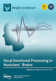
Cover Story (view full-size image): Musicians outperform non-musicians in vocal emotion recognition, but the underlying brain mechanisms are still debated. Hence, we compared event-related potentials (ERPs) to acoustically manipulated voices between musicians and non-musicians. We used parameter-specific voice morphing to create and present vocal stimuli that conveyed happiness, fear, pleasure, or sadness, either in all acoustic cues or selectively in either pitch contour (F0) or timbre. Differences between musicians and non-musicians appeared only for a centro-parietal late positive potential (500–1000 ms), suggesting that musicality affects the manner in which listeners use acoustic voice cues during later, controlled aspects of emotion evaluation. View this paper
  • Issues are regarded as officially published after their release is announced to the table of contents alert mailing list.
  • You may sign up for e-mail alerts to receive table of contents of newly released issues.
  • PDF is the official format for papers published in both, html and pdf forms. To view the papers in pdf format, click on the "PDF Full-text" link, and use the free Adobe Reader to open them.
Order results
Result details
Section
Select all
Export citation of selected articles as:
16 pages, 724 KiB  
Article
The Comparison of Early Hemodynamic Response to Single-Pulse Transcranial Magnetic Stimulation following Inhibitory or Excitatory Theta Burst Stimulation on Motor Cortex
by Corina Gorban, Zhongxing Zhang, Armand Mensen and Ramin Khatami
Brain Sci. 2023, 13(11), 1609; https://doi.org/10.3390/brainsci13111609 - 20 Nov 2023
Cited by 1 | Viewed by 942
Abstract
We present a new study design aiming to enhance the understanding of the mechanism by which continuous theta burst stimulation (cTBS) or intermittent theta burst stimulation (iTBS) paradigms elicit cortical modulation. Using near-infrared spectroscopy (NIRS), we compared the cortical hemodynamics of the previously [...] Read more.
We present a new study design aiming to enhance the understanding of the mechanism by which continuous theta burst stimulation (cTBS) or intermittent theta burst stimulation (iTBS) paradigms elicit cortical modulation. Using near-infrared spectroscopy (NIRS), we compared the cortical hemodynamics of the previously inhibited (after cTBS) or excited (after iTBS) left primary motor cortex (M1) as elicited by single-pulse TMS (spTMS) in a cross-over design. Mean relative changes in hemodynamics within 6 s of the stimulus were compared using a two-sample t-test (p < 0.05) and linear mixed model between real and sham stimuli and between stimuli after cTBS and iTBS. Only spTMS after cTBS resulted in a significant increase (p = 0.04) in blood volume (BV) compared to baseline. There were no significant changes in other hemodynamic parameters (oxygenated/deoxygenated hemoglobin). spTMS after cTBS induced a larger increase in BV than spTMS after iTBS (p = 0.021) and sham stimulus after cTBS (p = 0.009). BV showed no significant difference between real and sham stimuli after iTBS (p = 0.37). The greater hemodynamic changes suggest increased vasomotor reactivity after cTBS compared to iTBS. In addition, cTBS could decrease lateral inhibition, allowing activation of surrounding areas after cTBS. Full article
Show Figures

Figure 1

11 pages, 1299 KiB  
Article
EEG Changes during Propofol Anesthesia Induction in Vegetative State Patients Undergoing Spinal Cord Stimulation Implantation Surgery
by Xuewei Qin, Xuanling Chen, Bo Wang, Xin Zhao, Yi Tang, Lan Yao, Zhenhu Liang, Jianghong He and Xiaoli Li
Brain Sci. 2023, 13(11), 1608; https://doi.org/10.3390/brainsci13111608 - 20 Nov 2023
Viewed by 1108
Abstract
Objective: To compare the EEG changes in vegetative state (VS) patients and non-craniotomy, non-vegetative state (NVS) patients during general anesthesia with low-dose propofol and to find whether it affects the arousal rate of VS patients. Methods: Seven vegetative state patients (VS group: five [...] Read more.
Objective: To compare the EEG changes in vegetative state (VS) patients and non-craniotomy, non-vegetative state (NVS) patients during general anesthesia with low-dose propofol and to find whether it affects the arousal rate of VS patients. Methods: Seven vegetative state patients (VS group: five with traumatic brain injury, two with ischemic–hypoxic VS) and five non-craniotomy, non-vegetative state patients (NVS group) treated in the Department of Neurosurgery, Peking University International Hospital from January to May 2022 were selected. All patients were induced with 0.5 mg/kg propofol, and the Bispectral Index (BIS) changes within 5 min after administration were observed. Raw EEG signals and perioperative EEG signals were collected and analyzed using EEGLAB in the MATLAB software environment, time–frequency spectrums were calculated, and EEG changes were analyzed using power spectrums. Results: There was no significant difference in the general data before surgery between the two groups (p > 0.05); the BIS reduction in the VS group was significantly greater than that in the NVS group at 1 min, 2 min, 3 min, 4 min, and 5 min after 0.5 mg/kg propofol induction (p < 0.05). Time–frequency spectrum analysis showed the following: prominent α band energy around 10 Hz and decreased high-frequency energy in the NVS group, decreased high-frequency energy and main energy concentrated below 10 Hz in traumatic brain injury VS patients, higher energy in the 10–20 Hz band in ischemic–hypoxic VS patients. The power spectrum showed that the brain electrical energy of the NVS group was weakened R5 min after anesthesia induction compared with 5 min before induction, mainly concentrated in the small wave peak after 10 Hz, i.e., the α band peak; the energy of traumatic brain injury VS patients was weakened after anesthesia induction, but no α band peak appeared; and in ischemic–hypoxic VS patients, there was no significant change in low-frequency energy after anesthesia induction, high-frequency energy was significantly weakened, and a clear α band peak appeared slightly after 10 Hz. Three months after the operation, follow-up visits were made to the VS group patients who had undergone SCS surgery. One patient with traumatic brain injury VS was diagnosed with MCS-, one patient with ischemic–hypoxic VS had increased their CRS-R score by 1 point, and the remaining five patients had no change in their CRS scores. Conclusions: Low doses of propofol cause great differences in the EEG of different types of VS patients, which may be the unique response of damaged nerve cell residual function to propofol, and these weak responses may also be the basis of brain recovery Full article
Show Figures

Figure 1

19 pages, 4137 KiB  
Review
Identifying the Phenotypes of Diffuse Axonal Injury Following Traumatic Brain Injury
by Justin L. Krieg, Anna V. Leonard, Renée J. Turner and Frances Corrigan
Brain Sci. 2023, 13(11), 1607; https://doi.org/10.3390/brainsci13111607 - 20 Nov 2023
Viewed by 1639
Abstract
Diffuse axonal injury (DAI) is a significant feature of traumatic brain injury (TBI) across all injury severities and is driven by the primary mechanical insult and secondary biochemical injury phases. Axons comprise an outer cell membrane, the axolemma which is anchored to the [...] Read more.
Diffuse axonal injury (DAI) is a significant feature of traumatic brain injury (TBI) across all injury severities and is driven by the primary mechanical insult and secondary biochemical injury phases. Axons comprise an outer cell membrane, the axolemma which is anchored to the cytoskeletal network with spectrin tetramers and actin rings. Neurofilaments act as space-filling structural polymers that surround the central core of microtubules, which facilitate axonal transport. TBI has differential effects on these cytoskeletal components, with axons in the same white matter tract showing a range of different cytoskeletal and axolemma alterations with different patterns of temporal evolution. These require different antibodies for detection in post-mortem tissue. Here, a comprehensive discussion of the evolution of axonal injury within different cytoskeletal elements is provided, alongside the most appropriate methods of detection and their temporal profiles. Accumulation of amyloid precursor protein (APP) as a result of disruption of axonal transport due to microtubule failure remains the most sensitive marker of axonal injury, both acutely and chronically. However, a subset of injured axons demonstrate different pathology, which cannot be detected via APP immunoreactivity, including degradation of spectrin and alterations in neurofilaments. Furthermore, recent work has highlighted the node of Ranvier and the axon initial segment as particularly vulnerable sites to axonal injury, with loss of sodium channels persisting beyond the acute phase post-injury in axons without APP pathology. Given the heterogenous response of axons to TBI, further characterization is required in the chronic phase to understand how axonal injury evolves temporally, which may help inform pharmacological interventions. Full article
(This article belongs to the Section Neurosurgery and Neuroanatomy)
Show Figures

Figure 1

39 pages, 2097 KiB  
Review
Bridging the Divide: Brain and Behavior in Developmental Language Disorder
by Noelle Abbott and Tracy Love
Brain Sci. 2023, 13(11), 1606; https://doi.org/10.3390/brainsci13111606 - 19 Nov 2023
Viewed by 2208
Abstract
Developmental language disorder (DLD) is a heterogenous neurodevelopmental disorder that affects a child’s ability to comprehend and/or produce spoken and/or written language, yet it cannot be attributed to hearing loss or overt neurological damage. It is widely believed that some combination of genetic, [...] Read more.
Developmental language disorder (DLD) is a heterogenous neurodevelopmental disorder that affects a child’s ability to comprehend and/or produce spoken and/or written language, yet it cannot be attributed to hearing loss or overt neurological damage. It is widely believed that some combination of genetic, biological, and environmental factors influences brain and language development in this population, but it has been difficult to bridge theoretical accounts of DLD with neuroimaging findings, due to heterogeneity in language impairment profiles across individuals and inconsistent neuroimaging findings. Therefore, the purpose of this overview is two-fold: (1) to summarize the neuroimaging literature (while drawing on findings from other language-impaired populations, where appropriate); and (2) to briefly review the theoretical accounts of language impairment patterns in DLD, with the goal of bridging the disparate findings. As will be demonstrated with this overview, the current state of the field suggests that children with DLD have atypical brain volume, laterality, and activation/connectivity patterns in key language regions that likely contribute to language difficulties. However, the precise nature of these differences and the underlying neural mechanisms contributing to them remain an open area of investigation. Full article
Show Figures

Figure 1

12 pages, 690 KiB  
Article
How Posture and Previous Sensorimotor Experience Influence Muscle Activity during Gait Imagery in Young Healthy Individuals
by Barbora Kolářová, Marek Tomsa, Petr Kolář, Hana Haltmar, Tereza Diatelová and Miroslav Janura
Brain Sci. 2023, 13(11), 1605; https://doi.org/10.3390/brainsci13111605 - 19 Nov 2023
Viewed by 1108
Abstract
This study explores how gait imagery (GI) influences lower-limb muscle activity with respect to posture and previous walking experience. We utilized surface electromyography (sEMG) in 36 healthy young individuals aged 24 (±1.1) years to identify muscle activity during a non-gait imagery task (non-GI), [...] Read more.
This study explores how gait imagery (GI) influences lower-limb muscle activity with respect to posture and previous walking experience. We utilized surface electromyography (sEMG) in 36 healthy young individuals aged 24 (±1.1) years to identify muscle activity during a non-gait imagery task (non-GI), as well as GI tasks before (GI-1) and after the execution of walking (GI-2), with assessments performed in both sitting and standing postures. The sEMG was recorded on both lower limbs on the tibialis anterior (TA) and on the gastrocnemius medialis (GM) for all tested tasks. As a result, a significant muscle activity decrease was found in the right TA for GI-1 compared to GI-2 in both sitting (p = 0.008) and standing (p = 0.01) positions. In the left TA, the activity decreased in the sitting posture during non-GI (p = 0.004) and GI-1 (p = 0.009) in comparison to GI-2. No differences were found for GM. The subjective level of imagination difficulty improved for GI-2 in comparison to GI-1 in both postures (p < 0.001). Previous sensorimotor experience with real gait execution and sitting posture potentiate TA activity decrease during GI. These findings contribute to the understanding of neural mechanisms beyond GI. Full article
Show Figures

Figure 1

15 pages, 2979 KiB  
Review
Neural Mechanisms of Neuro-Rehabilitation Using Transcranial Direct Current Stimulation (tDCS) over the Front-Polar Area
by Koji Ishikuro, Noriaki Hattori, Hironori Otomune, Kohta Furuya, Takeshi Nakada, Kenichiro Miyahara, Takashi Shibata, Kyo Noguchi, Satoshi Kuroda, Yuji Nakatsuji and Hisao Nishijo
Brain Sci. 2023, 13(11), 1604; https://doi.org/10.3390/brainsci13111604 - 18 Nov 2023
Viewed by 1537
Abstract
Transcranial direct current stimulation (tDCS) is a noninvasive brain stimulation (NIBS) technique that applies a weak current to the scalp to modulate neuronal excitability by stimulating the cerebral cortex. The technique can produce either somatic depolarization (anodal stimulation) or somatic hyperpolarization (cathodal stimulation), [...] Read more.
Transcranial direct current stimulation (tDCS) is a noninvasive brain stimulation (NIBS) technique that applies a weak current to the scalp to modulate neuronal excitability by stimulating the cerebral cortex. The technique can produce either somatic depolarization (anodal stimulation) or somatic hyperpolarization (cathodal stimulation), based on the polarity of the current used by noninvasively stimulating the cerebral cortex with a weak current from the scalp, making it a NIBS technique that can modulate neuronal excitability. Thus, tDCS has emerged as a hopeful clinical neuro-rehabilitation treatment strategy. This method has a broad range of potential uses in rehabilitation medicine for neurodegenerative diseases, including Parkinson’s disease (PD). The present paper reviews the efficacy of tDCS over the front-polar area (FPA) in healthy subjects, as well as patients with PD, where tDCS is mainly applied to the primary motor cortex (M1 area). Multiple evidence lines indicate that the FPA plays a part in motor learning. Furthermore, recent studies have reported that tDCS applied over the FPA can improve motor functions in both healthy adults and PD patients. We argue that the application of tDCS to the FPA promotes motor skill learning through its effects on the M1 area and midbrain dopamine neurons. Additionally, we will review other unique outcomes of tDCS over the FPA, such as effects on persistence and motivation, and discuss their underlying neural mechanisms. These findings support the claim that the FPA could emerge as a new key brain region for tDCS in neuro-rehabilitation. Full article
(This article belongs to the Section Neurorehabilitation)
Show Figures

Figure 1

15 pages, 1366 KiB  
Article
Cervico-Ocular and Vestibulo-Ocular Reflexes in Subclinical Neck Pain and Healthy Individuals: A Cross-Sectional Study
by Devonte Campbell, Bernadette Ann Murphy, James Burkitt, Nicholas La Delfa, Praveen Sanmugananthan, Ushani Ambalavanar and Paul Yielder
Brain Sci. 2023, 13(11), 1603; https://doi.org/10.3390/brainsci13111603 - 18 Nov 2023
Viewed by 1161
Abstract
Alterations in neck sensory input from recurrent neck pain (known as subclinical neck pain (SCNP)) result in disordered sensorimotor integration (SMI). The cervico-ocular (COR) and vestibulo-ocular (VOR) reflexes involve various neural substrates but are coordinated by the cerebellum and reliant upon proprioceptive feedback. [...] Read more.
Alterations in neck sensory input from recurrent neck pain (known as subclinical neck pain (SCNP)) result in disordered sensorimotor integration (SMI). The cervico-ocular (COR) and vestibulo-ocular (VOR) reflexes involve various neural substrates but are coordinated by the cerebellum and reliant upon proprioceptive feedback. Given that proprioception and cerebellar processing are impaired in SCNP, we sought to determine if COR or VOR gain is also altered. COR and VOR were assessed using an eye-tracking device in 20 SCNP (9 M and 11 F; 21.8 (SD = 2.35) years) and 17 control (7 M and 10 F; 22.40 (SD = 3.66) years) participants. COR gain (10 trials): A motorized chair rotated the trunk at a frequency of 0.04 Hz and an amplitude of 5° while participants gazed at a circular target that disappeared after three seconds. VOR gain (30 trials): Rapid bilateral head movements away from a disappearing circular target while eyes fixated on the last observed target. Independent t-tests on COR and VOR gain were performed. SCNP had a significantly larger COR gain (p = 0.006) and smaller VOR gain (p = 0.487) compared to healthy controls. The COR group differences suggest an association between proprioceptive feedback and SMI, indicating COR may be a sensitive marker of altered cerebellar processing. Full article
(This article belongs to the Section Sensory and Motor Neuroscience)
Show Figures

Figure 1

41 pages, 2259 KiB  
Systematic Review
Molecular Targeted Therapies in Glioblastoma Multiforme: A Systematic Overview of Global Trends and Findings
by Emir Begagić, Ragib Pugonja, Hakija Bečulić, Amila Čeliković, Lejla Tandir Lihić, Samra Kadić Vukas, Lejla Čejvan, Rasim Skomorac, Edin Selimović, Belma Jaganjac, Fatima Juković-Bihorac, Aldin Jusić and Mirza Pojskić
Brain Sci. 2023, 13(11), 1602; https://doi.org/10.3390/brainsci13111602 - 17 Nov 2023
Cited by 5 | Viewed by 1890
Abstract
This systematic review assesses current molecular targeted therapies for glioblastoma multiforme (GBM), a challenging condition with limited treatment options. Using PRISMA methodology, 166 eligible studies, involving 2526 patients (61.49% male, 38.51% female, with a male-to-female ratio of 1.59/1), were analyzed. In laboratory studies, [...] Read more.
This systematic review assesses current molecular targeted therapies for glioblastoma multiforme (GBM), a challenging condition with limited treatment options. Using PRISMA methodology, 166 eligible studies, involving 2526 patients (61.49% male, 38.51% female, with a male-to-female ratio of 1.59/1), were analyzed. In laboratory studies, 52.52% primarily used human glioblastoma cell cultures (HCC), and 43.17% employed animal samples (mainly mice). Clinical participants ranged from 18 to 100 years, with 60.2% using combined therapies and 39.8% monotherapies. Mechanistic categories included Protein Kinase Phosphorylation (41.6%), Cell Cycle-Related Mechanisms (18.1%), Microenvironmental Targets (19.9%), Immunological Targets (4.2%), and Other Mechanisms (16.3%). Key molecular targets included Epidermal Growth Factor Receptor (EGFR) (10.8%), Mammalian Target of Rapamycin (mTOR) (7.2%), Vascular Endothelial Growth Factor (VEGF) (6.6%), and Mitogen-Activated Protein Kinase (MEK) (5.4%). This review provides a comprehensive assessment of molecular therapies for GBM, highlighting their varied efficacy in clinical and laboratory settings, ultimately impacting overall and progression-free survival in GBM management. Full article
Show Figures

Figure 1

20 pages, 6471 KiB  
Article
Neural Coupling between Interhemispheric and Frontoparietal Functional Connectivity during Semantic Processing
by Takahiro Soshi
Brain Sci. 2023, 13(11), 1601; https://doi.org/10.3390/brainsci13111601 - 17 Nov 2023
Viewed by 860
Abstract
Interhemispheric and frontoparietal functional connectivity have been reported to increase during explicit information processing. However, it is unclear how and when interhemispheric and frontoparietal functional connectivity interact during explicit semantic processing. Here, we tested the neural coupling hypothesis that explicit semantic processing promotes [...] Read more.
Interhemispheric and frontoparietal functional connectivity have been reported to increase during explicit information processing. However, it is unclear how and when interhemispheric and frontoparietal functional connectivity interact during explicit semantic processing. Here, we tested the neural coupling hypothesis that explicit semantic processing promotes neural activity in the nondominant right hemispheric areas, owing to synchronization with enhanced frontoparietal functional connectivity at later processing stages. We analyzed electroencephalogram data obtained using a semantic priming paradigm, which comprised visual priming and target words successively presented under direct or indirect attention to semantic association. Scalp potential analysis demonstrated that the explicit processing of congruent targets reduced negative event-related potentials, as previously reported. Current source density analysis showed that explicit semantic processing activated the right temporal area during later temporal intervals. Subsequent dynamic functional connectivity and neural coupling analyses revealed that explicit semantic processing increased the correlation between right temporal source activities and frontoparietal functional connectivity in later temporal intervals. These findings indicate that explicit semantic processing increases neural coupling between the interhemispheric and frontoparietal functional connectivity during later processing stages. Full article
(This article belongs to the Section Neuropsychology)
Show Figures

Figure 1

11 pages, 3108 KiB  
Article
Decrease in Cerebral Blood Flow after Reoxygenation Is Associated with Neurological Syndrome Sequelae and Blood Pressure
by Yanqiu Liu, Fengjuan Yuan, Zhongwei Peng, Yadong Zhan, Jianzhong Lin, Ran Zhang and Jiaxing Zhang
Brain Sci. 2023, 13(11), 1600; https://doi.org/10.3390/brainsci13111600 - 17 Nov 2023
Viewed by 947
Abstract
Changes in cerebral blood flow (CBF) and regulation of cerebral circulation occur at high altitude (HA). However, the changes in CBF and their associations with neurological syndrome sequelae and blood pressure after subjects return to the lowlands remain unclear. In this study, the [...] Read more.
Changes in cerebral blood flow (CBF) and regulation of cerebral circulation occur at high altitude (HA). However, the changes in CBF and their associations with neurological syndrome sequelae and blood pressure after subjects return to the lowlands remain unclear. In this study, the subjects were 23 college students who were teaching at an altitude of 4300 m for 30 days. These subjects were studied before reaching the HA (Test 1), one week after returning to the lowlands (Test 2), and three months after returning to the lowlands (Test 3). Symptom scores for de-acclimatization syndrome were evaluated. Changes in CBF were measured using the magnetic resonance imaging arterial spin labeling (ASL) technique. Additionally, the velocity of CBF in the cerebral arteries was measured using a transcranial doppler (TCD). In Test 2 vs. Test 1, the peak systolic velocity and mean velocity in the basilar artery were significantly decreased. CBF exhibited significant decreases in the left putamen/cerebellum crus1/vermis and right thalamus/inferior temporal gyrus, while significant increases were observed in the left postcentral gyrus/precuneus and right middle cingulate gyrus/superior frontal gyrus. In Test 3 vs. Test 1, the basilar artery velocity returned to the baseline level, while CBF continued to decrease. The mean global CBF showed a decreasing trend from Test 1 to Test 3. Furthermore, the mean global CBF had a negative correlation with the systolic pressure, pulse pressure, and mean arterial pressure. The decrease in CBF after reoxygenation may underlie the neurological symptoms in subjects returning to the lowlands. Increased blood pressure could serve as a predictor of a decrease in CBF. Full article
(This article belongs to the Section Systems Neuroscience)
Show Figures

Figure 1

12 pages, 615 KiB  
Article
Gait Domains May Be Used as an Auxiliary Diagnostic Index for Alzheimer’s Disease
by Qi Duan, Yinuo Zhang, Weihao Zhuang, Wenlong Li, Jincai He, Zhen Wang and Haoran Cheng
Brain Sci. 2023, 13(11), 1599; https://doi.org/10.3390/brainsci13111599 - 17 Nov 2023
Viewed by 969
Abstract
Background: Alzheimer’s disease (AD) is a progressive neurodegenerative disorder with cognitive dysfunction and behavioral impairment. We aimed to use principal components factor analysis to explore the association between gait domains and AD under single and dual-task gait assessments. Methods: A total of 41 [...] Read more.
Background: Alzheimer’s disease (AD) is a progressive neurodegenerative disorder with cognitive dysfunction and behavioral impairment. We aimed to use principal components factor analysis to explore the association between gait domains and AD under single and dual-task gait assessments. Methods: A total of 41 AD participants and 41 healthy control (HC) participants were enrolled in our study. Gait parameters were measured using the JiBuEn® gait analysis system. The principal component method was used to conduct an orthogonal maximum variance rotation factor analysis of quantitative gait parameters. Multiple logistic regression was used to adjust for potential confounding or risk factors. Results: Based on the factor analysis, three domains of gait performance were identified both in the free walk and counting backward assessments: “rhythm” domain, “pace” domain and “variability” domain. Compared with HC, we found that the pace factor was independently associated with AD in two gait assessments; the variability factor was independently associated with AD only in the counting backwards assessment; and a statistical difference still remained after adjusting for age, sex and education levels. Conclusions: Our findings indicate that gait domains may be used as an auxiliary diagnostic index for Alzheimer’s disease. Full article
(This article belongs to the Section Neurodegenerative Diseases)
Show Figures

Figure 1

18 pages, 2320 KiB  
Article
Increased Inhibition May Contribute to Maintaining Normal Network Function in the Ventral Hippocampus of a Fmr1-Targeted Transgenic Rat Model of Fragile X Syndrome
by Leonidas J. Leontiadis, George Trompoukis, Panagiotis Felemegkas, Giota Tsotsokou, Athina Miliou and Costas Papatheodoropoulos
Brain Sci. 2023, 13(11), 1598; https://doi.org/10.3390/brainsci13111598 - 17 Nov 2023
Cited by 1 | Viewed by 1002
Abstract
A common neurobiological mechanism in several neurodevelopmental disorders, including fragile X syndrome (FXS), is alterations in the balance between excitation and inhibition in the brain. It is thought that in the hippocampus, as in other brain regions, FXS is associated with increased excitability [...] Read more.
A common neurobiological mechanism in several neurodevelopmental disorders, including fragile X syndrome (FXS), is alterations in the balance between excitation and inhibition in the brain. It is thought that in the hippocampus, as in other brain regions, FXS is associated with increased excitability and reduced inhibition. However, it is still not known whether these changes apply to both the dorsal and ventral hippocampus, which appear to be differently involved in neurodegenerative disorders. Using a Fmr1 knock-out (KO) rat model of FXS, we found increased neuronal excitability in both the dorsal and ventral KO hippocampus and increased excitatory synaptic transmission in the dorsal hippocampus. Interestingly, synaptic inhibition is significantly increased in the ventral but not the dorsal KO hippocampus. Furthermore, the ventral KO hippocampus displays increased expression of the α1GABAA receptor subtype and a remarkably reduced rate of epileptiform discharges induced by magnesium-free medium. In contrast, the dorsal KO hippocampus displays an increased rate of epileptiform discharges and similar expression of α1GABAA receptors compared with the dorsal WT hippocampus. Blockade of α5GABAA receptors by L-655,708 did not affect epileptiform discharges in any genotype or hippocampal segment, and the expression of α5GABAA receptors did not differ between WT and KO hippocampus. These results suggest that the increased excitability of the dorsal KO hippocampus contributes to its heightened tendency to epileptiform discharges, while the increased phasic inhibition in the Fmr1-KO ventral hippocampus may represent a homeostatic mechanism that compensates for the increased excitability reducing its vulnerability to epileptic activity. Full article
(This article belongs to the Collection Collection on Molecular and Cellular Neuroscience)
Show Figures

Figure 1

12 pages, 3182 KiB  
Article
Abnormal Topological Organization of White Matter Structural Networks in Normal Tension Glaucoma Revealed via Diffusion Tensor Tractography
by Yin Wang, Linying Guo, Rong Wang, Yuzhe Wang, Fei Duan, Yang Zhan, Jingfeng Cheng, Xinghuai Sun and Zuohua Tang
Brain Sci. 2023, 13(11), 1597; https://doi.org/10.3390/brainsci13111597 - 17 Nov 2023
Viewed by 824
Abstract
Background: Normal tension glaucoma (NTG) is considered a neurodegenerative disease with glaucomatous damage extending to diffuse brain areas. Therefore, this study aims to explore the abnormalities in the NTG structural network to help in the early diagnosis and course evaluation of NTG. Methods: [...] Read more.
Background: Normal tension glaucoma (NTG) is considered a neurodegenerative disease with glaucomatous damage extending to diffuse brain areas. Therefore, this study aims to explore the abnormalities in the NTG structural network to help in the early diagnosis and course evaluation of NTG. Methods: The structural networks of 46 NTG patients and 19 age- and sex-matched healthy controls were constructed using diffusion tensor imaging, followed by graph theory analysis and correlation analysis of small-world properties with glaucoma clinical indicators. In addition, the network-based statistical analysis (NBS) method was used to compare structural network connectivity differences between NTG patients and healthy controls. Results: Structural brain networks in both NTG and NC groups exhibited small-world properties. However, the small-world index in the severe NTG group was reduced and correlated with a mean deviation of the visual field (MDVF) and retinal nerve fiber layer (RNFL) thickness. When compared to healthy controls, degree centrality and nodal efficiency in visual brain areas were significantly decreased, and betweenness centrality and nodal local efficiency in both visual and nonvisual brain areas were also significantly altered in NTG patients (all p < 0.05, FDR corrected). Furthermore, NTG patients exhibited increased structural connectivity in the occipitotemporal area, with the left fusiform gyrus (FFG.L) as the hub (p < 0.05). Conclusions: NTG exhibited altered global properties and local properties of visual and cognitive-emotional brain areas, with enhanced structural connections within the occipitotemporal area. Moreover, the disrupted small-world properties of white matter might be imaging biomarkers for assessing NTG progression. Full article
(This article belongs to the Section Neurotechnology and Neuroimaging)
Show Figures

Graphical abstract

14 pages, 596 KiB  
Systematic Review
Effect of Chronic Ankle Instability on the Biomechanical Organization of Gait Initiation: A Systematic Review
by Mohammad Yousefi, Shaghayegh Zivari, Eric Yiou and Teddy Caderby
Brain Sci. 2023, 13(11), 1596; https://doi.org/10.3390/brainsci13111596 - 17 Nov 2023
Viewed by 1075
Abstract
This systematic review was conducted to provide an overview of the effects of chronic ankle instability (CAI) on the biomechanical organization of gait initiation. Gait initiation is a classical model used in the literature to investigate postural control in healthy and pathological individuals. [...] Read more.
This systematic review was conducted to provide an overview of the effects of chronic ankle instability (CAI) on the biomechanical organization of gait initiation. Gait initiation is a classical model used in the literature to investigate postural control in healthy and pathological individuals. PubMed, ScienceDirect, Scopus, Web of Science, and Google Scholar were searched for relevant articles. Eligible studies were screened and data extracted by two independent reviewers. An evaluation of the quality of the studies was performed using the Downs and Black checklist. A total of 878 articles were found in the initial search, but only six studies met the inclusion criteria. The findings from the literature suggest that CAI affects the characteristics of gait initiation. Specifically, individuals with CAI exhibit notable differences in reaction time, the spatiotemporal parameters of anticipatory postural adjustments (APAs) and step execution, ankle–foot kinematics, and muscle activation compared to healthy controls. In particular, the observed differences in APA patterns associated with gait initiation suggest the presence of supraspinal motor control alterations in individuals with CAI. These findings may provide valuable information for the rehabilitation of these patients. However, the limited evidence available calls for caution in interpreting the results and underscores the need for further research. Full article
(This article belongs to the Special Issue Advances in the Study of Anticipatory Postural Adjustments)
Show Figures

Figure 1

14 pages, 4541 KiB  
Article
Paraventricular Mast Cell-Derived Histamine Activates CRH Neurons to Mediate Adult Visceral Hypersensitivity Induced by Neonatal Maternal Separation
by Ziyang Chen, Tiantian Zhou, Yunfan Li, Tingting Li, Zhengnian Ding and Li Liu
Brain Sci. 2023, 13(11), 1595; https://doi.org/10.3390/brainsci13111595 - 16 Nov 2023
Viewed by 990
Abstract
Neonatal maternal separation (NMS) is an early-life stress (ELS) that can result in adult visceral hypersensitivity, which is usually manifested as chronic visceral pain. Although mast cells and corticotropin-releasing hormone (CRH) neurons are involved in stress response, whether there is an interaction between [...] Read more.
Neonatal maternal separation (NMS) is an early-life stress (ELS) that can result in adult visceral hypersensitivity, which is usually manifested as chronic visceral pain. Although mast cells and corticotropin-releasing hormone (CRH) neurons are involved in stress response, whether there is an interaction between mast cells and CRH neurons in hypothalamic paraventricular nucleus (PVN) during the ELS-induced visceral hypersensitivity remains elusive. Herein, we established an NMS model by separating neonatal mice from their mothers, and observed that these mice presented visceral hypersensitivity in adulthood, as indicated by elevated abdominal withdrawal reflex and lowered visceral pain threshold. The NMS-induced adult visceral hypersensitivity was accompanied by activation of mast cells and CRH neurons in PVN. Also, NMS increased the histamine content (an inflammatory mediator mainly released by mast cells) and histamine H2 receptor (H2R) expression of CRH neurons in PVN. Remarkably, intra-PVN administration with mast cell stabilizer attenuated the NMS-induced CRH neuronal activation and adult visceral pain, while histamine administration showed the opposite effects. Moreover, intra-PVN injection with H2R antagonist alleviated the NMS-induced CRH neuronal activation, PKA and CREB phosphorylation, and importantly, adult visceral pain. Together, our findings revealed a role of an interaction between paraventricular mast cells and CRH neurons in NMS-induced adult visceral hypersensitivity, thereby providing a perspective for the management of visceral pain. Full article
(This article belongs to the Section Molecular and Cellular Neuroscience)
Show Figures

Figure 1

15 pages, 290 KiB  
Review
Non-Invasive Systems Application in Traumatic Brain Injury Rehabilitation
by Livia Livinț Popa, Diana Chira, Ștefan Strilciuc and Dafin F. Mureșanu
Brain Sci. 2023, 13(11), 1594; https://doi.org/10.3390/brainsci13111594 - 15 Nov 2023
Cited by 1 | Viewed by 1776
Abstract
Traumatic brain injury (TBI) is a significant public health concern, often leading to long-lasting impairments in cognitive, motor and sensory functions. The rapid development of non-invasive systems has revolutionized the field of TBI rehabilitation by offering modern and effective interventions. This narrative review [...] Read more.
Traumatic brain injury (TBI) is a significant public health concern, often leading to long-lasting impairments in cognitive, motor and sensory functions. The rapid development of non-invasive systems has revolutionized the field of TBI rehabilitation by offering modern and effective interventions. This narrative review explores the application of non-invasive technologies, including electroencephalography (EEG), quantitative electroencephalography (qEEG), brain–computer interface (BCI), eye tracking, near-infrared spectroscopy (NIRS), functional near-infrared spectroscopy (fNIRS), magnetic resonance imaging (MRI), functional magnetic resonance imaging (fMRI), magnetoencephalography (MEG), and transcranial magnetic stimulation (TMS) in assessing TBI consequences, and repetitive transcranial magnetic stimulation (rTMS), low-level laser therapy (LLLT), neurofeedback, transcranial direct current stimulation (tDCS), transcranial alternative current stimulation (tACS) and virtual reality (VR) as therapeutic approaches for TBI rehabilitation. In pursuit of advancing TBI rehabilitation, this narrative review highlights the promising potential of non-invasive technologies. We emphasize the need for future research and clinical trials to elucidate their mechanisms of action, refine treatment protocols, and ensure their widespread adoption in TBI rehabilitation settings. Full article
(This article belongs to the Special Issue Reviews in Neural Engineering, Neuroergonomics and Neurorobotics)
11 pages, 944 KiB  
Article
Alzheimer’s Disease CSF Biomarkers as Possible Indicators of Tap-Test Response in Idiopathic Normal Pressure Hydrocephalus
by Efstratios-Stylianos Pyrgelis, George P. Paraskevas, Vasilios C. Constantinides, Fotini Boufidou, Myrto Papaioannou, Leonidas Stefanis and Elisabeth Kapaki
Brain Sci. 2023, 13(11), 1593; https://doi.org/10.3390/brainsci13111593 - 15 Nov 2023
Viewed by 962
Abstract
The aim of the present study is the evaluation of established Alzheimer’s disease (AD) cerebrospinal fluid (CSF) biomarkers in patients with idiopathic normal-pressure hydrocephalus (iNPH), both individually and as a total profile, and the investigation of their use as potential predictors of Tap-test [...] Read more.
The aim of the present study is the evaluation of established Alzheimer’s disease (AD) cerebrospinal fluid (CSF) biomarkers in patients with idiopathic normal-pressure hydrocephalus (iNPH), both individually and as a total profile, and the investigation of their use as potential predictors of Tap-test responsiveness. Fifty-three patients with iNPH participated in the study. Aβ42, Aβ40, total Tau and phospho-Tau proteins were measured in duplicate with double-sandwich ELISA assays. Clinical evaluation involved a 10 m timed walk test before an evacuative lumbar puncture (LP) and every 24 h for three consecutive days afterwards. Neuropsychological assessment involved a mini-mental state examination, frontal assessment battery, 5-word test and CLOX drawing test 1 and 2, which were also performed before and 48 h after LP. Response in the Tap-test was defined as a 20% improvement in gait and/or a 10% improvement in neuropsychological tests. The Aβ42/Aβ40 ratio was found to be significantly higher in Tap-test responders than non-responders. Total Tau and phospho-Tau CSF levels also differed significantly between these two groups, with Tap-test responders presenting with lower levels compared to non-responders. Regarding the AD CSF biomarker profile (decreased amyloid and increased Tau proteins levels), patients with a non-AD profile were more likely to have a positive response in the Tap-test than patients with an AD profile. Full article
(This article belongs to the Section Neurodegenerative Diseases)
Show Figures

Figure 1

13 pages, 2766 KiB  
Article
Neurocognitive Adaptations for Spatial Orientation and Navigation in Astronauts
by Ford Burles and Giuseppe Iaria
Brain Sci. 2023, 13(11), 1592; https://doi.org/10.3390/brainsci13111592 - 15 Nov 2023
Viewed by 1395
Abstract
Astronauts often face orientation challenges while on orbit, which can lead to operator errors in demanding spatial tasks. In this study, we investigated the impact of long-duration spaceflight on the neural processes supporting astronauts’ spatial orientation skills. Using functional magnetic resonance imaging (fMRI), [...] Read more.
Astronauts often face orientation challenges while on orbit, which can lead to operator errors in demanding spatial tasks. In this study, we investigated the impact of long-duration spaceflight on the neural processes supporting astronauts’ spatial orientation skills. Using functional magnetic resonance imaging (fMRI), we collected data from 16 astronauts six months before and two weeks after their International Space Station (ISS) missions while performing a spatial orientation task that requires generating a mental representation of one’s surroundings. During this task, astronauts exhibited a general reduction in neural activity evoked from spatial-processing brain regions after spaceflight. The neural activity evoked in the precuneus was most saliently reduced following spaceflight, along with less powerful effects observed in the angular gyrus and retrosplenial regions of the brain. Importantly, the reduction in precuneus activity we identified was not accounted for by changes in behavioral performance or changes in grey matter concentration. These findings overall show less engagement of explicitly spatial neurological processes at postflight, suggesting astronauts make use of complementary strategies to perform some spatial tasks as an adaptation to spaceflight. These preliminary findings highlight the need for developing countermeasures or procedures that minimize the detrimental effects of spaceflight on spatial cognition, especially in light of planned long-distance future missions. Full article
Show Figures

Figure 1

13 pages, 1309 KiB  
Review
Exploring Neurophysiological Mechanisms and Treatment Efficacies in Laryngeal Dystonia: A Transcranial Magnetic Stimulation Approach
by Maja Rogić Vidaković, Joško Šoda, Joshua Elan Kuluva, Braco Bošković, Krešimir Dolić and Ivana Gunjača
Brain Sci. 2023, 13(11), 1591; https://doi.org/10.3390/brainsci13111591 - 15 Nov 2023
Viewed by 1156
Abstract
Laryngeal dystonia (LD), known or termed as spasmodic dysphonia, is a rare movement disorder with an unknown cause affecting the intrinsic laryngeal muscles. Neurophysiological studies point to perturbed inhibitory processes, while conventional genetic studies reveal fragments of genetic architecture in LD. The study’s [...] Read more.
Laryngeal dystonia (LD), known or termed as spasmodic dysphonia, is a rare movement disorder with an unknown cause affecting the intrinsic laryngeal muscles. Neurophysiological studies point to perturbed inhibitory processes, while conventional genetic studies reveal fragments of genetic architecture in LD. The study’s aims are to (1) describe transcranial magnetic stimulation (TMS) methodology for studying the functional integrity of the corticospinal tract by stimulating the primary motor cortex (M1) for laryngeal muscle representation and recording motor evoked potentials (MEPs) from laryngeal muscles; (2) evaluate the results of TMS studies investigating the cortical silent period (cSP) in LD; and (3) present the standard treatments of LD, as well as the results of new theoretical views and treatment approaches like repetitive TMS and laryngeal vibration over the laryngeal muscles as the recent research attempts in treatment of LD. Neurophysiological findings point to a shortened duration of cSP in adductor LD and altered cSP duration in abductor LD individuals. Future TMS studies could further investigate the role of cSP in relation to standard laryngological measures and treatment options. A better understanding of the neurophysiological mechanisms might give new perspectives for the treatment of LD. Full article
Show Figures

Figure 1

12 pages, 1488 KiB  
Article
Automatic Diagnosis of Major Depressive Disorder Using a High- and Low-Frequency Feature Fusion Framework
by Junyu Wang, Tongtong Li, Qi Sun, Yuhui Guo, Jiandong Yu, Zhijun Yao, Ning Hou and Bin Hu
Brain Sci. 2023, 13(11), 1590; https://doi.org/10.3390/brainsci13111590 - 15 Nov 2023
Cited by 1 | Viewed by 1123
Abstract
Major Depressive Disorder (MDD) is a common mental illness resulting in immune disorders and even thoughts of suicidal behavior. Neuroimaging techniques serve as a quantitative tool for the assessment of MDD diagnosis. In the domain of computer-aided magnetic resonance imaging diagnosis, current research [...] Read more.
Major Depressive Disorder (MDD) is a common mental illness resulting in immune disorders and even thoughts of suicidal behavior. Neuroimaging techniques serve as a quantitative tool for the assessment of MDD diagnosis. In the domain of computer-aided magnetic resonance imaging diagnosis, current research predominantly focuses on isolated local or global information, often neglecting the synergistic integration of multiple data sources, thus potentially overlooking valuable details. To address this issue, we proposed a diagnostic model for MDD that integrates high-frequency and low-frequency information using data from diffusion tensor imaging (DTI), structural magnetic resonance imaging (sMRI), and functional magnetic resonance imaging (fMRI). First, we designed a meta-low-frequency encoder (MLFE) and a meta-high-frequency encoder (MHFE) to extract the low-frequency and high-frequency feature information from DTI and sMRI, respectively. Then, we utilized a multilayer perceptron (MLP) to extract features from fMRI data. Following the feature cross-fusion, we designed the ensemble learning threshold voting method to determine the ultimate diagnosis for MDD. The model achieved accuracy, precision, specificity, F1-score, MCC, and AUC values of 0.724, 0.750, 0.882, 0.600, 0.421, and 0.667, respectively. This approach provides new research ideas for the diagnosis of MDD. Full article
(This article belongs to the Special Issue Advances of AI in Neuroimaging)
Show Figures

Figure 1

12 pages, 1807 KiB  
Article
Role of Vagus Nerve Stimulation in Refractory and Super Refractory Status Epilepticus: A Pediatric Case Series
by Giulia Melinda Furlanis, Jacopo Favaro, Nicola Bresolin, Daniele Grioni, Valentina Baro, Alberto D’Amico, Stefano Sartori, Luca Denaro and Andrea Landi
Brain Sci. 2023, 13(11), 1589; https://doi.org/10.3390/brainsci13111589 - 14 Nov 2023
Viewed by 1159
Abstract
Background: Status epilepticus is a life-threatening condition that is defined as refractory (RSE) when the seizure activity continues despite treatment with benzodiazepine and a second appropriate treatment. Super refractory status epilepticus (SRSE) is an RSE that persists or recurs for ≥24 h. Few [...] Read more.
Background: Status epilepticus is a life-threatening condition that is defined as refractory (RSE) when the seizure activity continues despite treatment with benzodiazepine and a second appropriate treatment. Super refractory status epilepticus (SRSE) is an RSE that persists or recurs for ≥24 h. Few papers have reported the outcomes of pediatric patients affected by RSE and SRSE and treated with neuromodulation therapies. Vagus nerve stimulation (VNS) is an approved treatment for drug-resistant epilepsy. We present our findings of pediatric patients treated with VNS for RSE/SRSE. Methods: We present a case series of seven consecutive pediatric patients treated with VNS for SRSE since 2012 by a single surgeon in Monza and Padua. A rapid titration was started soon after implantation. We considered electroclinical data before and after VNS implantation and at the last follow-up. Results: We achieved the resolution of SRSE in five out of seven patients in a mean time of two weeks. At the last follow-up, these patients had a significant reduction of seizure burden without any relapse of SE. Discussion and Conclusions: Based on our limited findings, we discuss the potential role of VNS therapy in similar but distinct clinical contexts. For patients with drug-resistant epilepsy and RSE/SRSE, prompt VNS consideration is suggested, offering rapid responses and potentially reducing pharmacological load. Meanwhile, in NORSE/FIRES, we suggest early neuromodulation during the acute phase if standard treatments prove ineffective or not tolerated. This approach may leverage VNS’s potential anti-inflammatory effects and neuromodulation, enhancing patient-specific treatments. Expanding case studies and prolonged follow-ups are recommended to strengthen these clinical insights. Full article
Show Figures

Figure 1

2 pages, 767 KiB  
Correction
Correction: Iannuzzi et al. Might Fibroblasts from Patients with Alzheimer’s Disease Reflect the Brain Pathology? A Focus on the Increased Phosphorylation of Amyloid Precursor Protein Tyr682 Residue. Brain Sci. 2021, 11, 103
by Filomena Iannuzzi, Vincenza Frisardi, Lucio Annunziato and Carmela Matrone
Brain Sci. 2023, 13(11), 1588; https://doi.org/10.3390/brainsci13111588 - 13 Nov 2023
Viewed by 614
Abstract
In the original article [...] Full article
Show Figures

Figure 1

22 pages, 2216 KiB  
Article
Multilingual Language Diversity Protects Native Language Production under Different Control Demands
by Keyi Kang, Yumeng Xiao, Hanxiang Yu, Michele T. Diaz and Haoyun Zhang
Brain Sci. 2023, 13(11), 1587; https://doi.org/10.3390/brainsci13111587 - 13 Nov 2023
Viewed by 1020
Abstract
The use of multiple languages has been found to influence individuals’ cognitive abilities. Although some studies have also investigated the effect of multilingualism on non-native language proficiency, fewer studies have focused on how multilingual experience affects native language production. This study investigated the [...] Read more.
The use of multiple languages has been found to influence individuals’ cognitive abilities. Although some studies have also investigated the effect of multilingualism on non-native language proficiency, fewer studies have focused on how multilingual experience affects native language production. This study investigated the effect of multilingualism on native language production, specifically examining control demands through a semantic Go/No-Go picture naming task. The multilingual experience was quantified using language entropy, which measures the uncertainty and diversity of language use. Control demands were achieved by manipulating the proportion of Go (i.e., naming) trials in different conditions. Results showed that as control demands increased, multilingual individuals exhibited poorer behavioral performance and greater brain activation throughout the brain. Moreover, more diverse language use was associated with higher accuracy in naming and more interconnected brain networks with greater involvement of domain-general neural resources and less domain-specific neural resources. Notably, the varied and balanced use of multiple languages enabled multilingual individuals to respond more efficiently to increased task demands during native language production. Full article
Show Figures

Figure 1

12 pages, 269 KiB  
Article
Movement Sonification Techniques to Improve Balance in Parkinson’s Disease: A Pilot Randomized Controlled Trial
by Alfredo Raglio, Beatrice De Maria, Monica Parati, Andrea Giglietti, Stefano Premoli, Stefano Salvaderi, Daniele Molteni, Simona Ferrante and Laura Adelaide Dalla Vecchia
Brain Sci. 2023, 13(11), 1586; https://doi.org/10.3390/brainsci13111586 - 12 Nov 2023
Cited by 1 | Viewed by 1175
Abstract
Background: Movement sonification has been recently introduced into the field of neuromotor rehabilitation alongside Neurologic Music Therapy and music-based interventions. This study introduces the use of musical auditory cues encompassing the melodic-harmonic aspect of music. Methods: Nineteen patients with Parkinson’s disease were randomly [...] Read more.
Background: Movement sonification has been recently introduced into the field of neuromotor rehabilitation alongside Neurologic Music Therapy and music-based interventions. This study introduces the use of musical auditory cues encompassing the melodic-harmonic aspect of music. Methods: Nineteen patients with Parkinson’s disease were randomly assigned to the experimental (n = 10) and control (n = 9) groups and underwent thrice-weekly sessions of the same gait training program, with or without sonification. Functional and motor parameters, as well as fatigue, quality of life, and the impact of intervention on patients’ well-being, were assessed at baseline (PRE), the end of treatment (POST), and at follow-up (FU). Between-group differences were assessed for each outcome measure using linear mixed-effects models. The outcome measure was entered as the dependent variable, group and time as fixed effects, and time by group as the interaction effect. Results: Mini BESTest and Dynamic Gait Index scores significantly improved in the experimental group (p = 0.01 and p = 0.03, respectively) from PRE to FU, demonstrating a significant impact of the sonification treatment on balance. No other significant differences were observed in the outcome measures. Conclusions: Larger sample sizes are needed to confirm the effectiveness of sonification approaches in Parkinson’s disease, as well as in other neurological disorders. Full article
(This article belongs to the Special Issue At the Frontiers of Neurorehabilitation: Series II)
12 pages, 291 KiB  
Opinion
Posturography Approaches: An Insightful Window to Explore the Role of the Brain in Socio-Affective Processes
by Harold Mouras, Alexandre Vonesch, Karina Lebel, Guillaume Léonard and Thierry Lelard
Brain Sci. 2023, 13(11), 1585; https://doi.org/10.3390/brainsci13111585 - 12 Nov 2023
Viewed by 954
Abstract
A significant amount of research has highlighted the importance of a motor component in the brain’s processing of emotional, motivational and social information. Posturography has emerged as an interesting way to assess motor correlates associated with this process. In this review, we highlight [...] Read more.
A significant amount of research has highlighted the importance of a motor component in the brain’s processing of emotional, motivational and social information. Posturography has emerged as an interesting way to assess motor correlates associated with this process. In this review, we highlight recent results within the functional context of painful stimulus perception and discuss the interest in broadening the use of posturography to other motivational and societal functional contexts. Although characterized by significant feasibility, the single measurement of the COP’s anteroposterior displacement presents limitations for attesting approach–avoidance behavior towards a visual target. Here, we discuss a number of methodological avenues that could go some way towards overcoming these limitations. Full article
(This article belongs to the Section Social Cognitive and Affective Neuroscience)
19 pages, 1774 KiB  
Systematic Review
A Review on Motor Imagery with Transcranial Alternating Current Stimulation: Bridging Motor and Cognitive Welfare for Patient Rehabilitation
by Rosary Yuting Lim, Kai Keng Ang, Effie Chew and Cuntai Guan
Brain Sci. 2023, 13(11), 1584; https://doi.org/10.3390/brainsci13111584 - 12 Nov 2023
Viewed by 1092
Abstract
Research has shown the effectiveness of motor imagery in patient motor rehabilitation. Transcranial electrical stimulation has also demonstrated to improve patient motor and non-motor performance. However, mixed findings from motor imagery studies that involved transcranial electrical stimulation suggest that current experimental protocols can [...] Read more.
Research has shown the effectiveness of motor imagery in patient motor rehabilitation. Transcranial electrical stimulation has also demonstrated to improve patient motor and non-motor performance. However, mixed findings from motor imagery studies that involved transcranial electrical stimulation suggest that current experimental protocols can be further improved towards a unified design for consistent and effective results. This paper aims to review, with some clinical and neuroscientific findings from literature as support, studies of motor imagery coupled with different types of transcranial electrical stimulation and their experiments onhealthy and patient subjects. This review also includes the cognitive domains of working memory, attention, and fatigue, which are important for designing consistent and effective therapy protocols. Finally, we propose a theoretical all-inclusive framework that synergizes the three cognitive domains with motor imagery and transcranial electrical stimulation for patient rehabilitation, which holds promise of benefiting patients suffering from neuromuscular and cognitive disorders. Full article
Show Figures

Figure 1

15 pages, 2013 KiB  
Article
EEG-Based Target Detection Using an RSVP Paradigm under Five Levels of Weak Hidden Conditions
by Jinling Lian, Xin Qiao, Yuwei Zhao, Siwei Li, Changyong Wang and Jin Zhou
Brain Sci. 2023, 13(11), 1583; https://doi.org/10.3390/brainsci13111583 - 12 Nov 2023
Viewed by 1131
Abstract
Although target detection based on electroencephalogram (EEG) signals has been extensively investigated recently, EEG-based target detection under weak hidden conditions remains a problem. In this paper, we proposed a rapid serial visual presentation (RSVP) paradigm for target detection corresponding to five levels of [...] Read more.
Although target detection based on electroencephalogram (EEG) signals has been extensively investigated recently, EEG-based target detection under weak hidden conditions remains a problem. In this paper, we proposed a rapid serial visual presentation (RSVP) paradigm for target detection corresponding to five levels of weak hidden conditions quantitively based on the RGB color space. Eighteen subjects participated in the experiment, and the neural signatures, including P300 amplitude and latency, were investigated. Detection performance was evaluated under five levels of weak hidden conditions using the linear discrimination analysis and support vector machine classifiers on different channel sets. The experimental results showed that, compared with the benchmark condition, (1) the P300 amplitude significantly decreased (8.92 ± 1.24 μV versus 7.84 ± 1.40 μV, p = 0.021) and latency was significantly prolonged (582.39 ± 25.02 ms versus 643.83 ± 26.16 ms, p = 0.028) only under the weakest hidden condition, and (2) the detection accuracy decreased by less than 2% (75.04 ± 3.24% versus 73.35 ± 3.15%, p = 0.029) with a more than 90% reduction in channel number (62 channels versus 6 channels), determined using the proposed channel selection method under the weakest hidden condition. Our study can provide new insights into target detection under weak hidden conditions based on EEG signals with a rapid serial visual presentation paradigm. In addition, it may expand the application of brain–computer interfaces in EEG-based target detection areas. Full article
Show Figures

Figure 1

15 pages, 1320 KiB  
Article
Abnormalities of Hippocampal Subfield and Amygdalar Nuclei Volumes and Clinical Correlates in Behavioral Variant Frontotemporal Dementia with Obsessive–Compulsive Behavior—A Pilot Study
by Mu-N Liu, Li-Yu Hu, Chia-Fen Tsai, Chen-Jee Hong, Yuan-Hwa Chou, Chiung-Chih Chang, Kai-Chun Yang, Zi-Hong You and Chi Ieong Lau
Brain Sci. 2023, 13(11), 1582; https://doi.org/10.3390/brainsci13111582 - 11 Nov 2023
Cited by 1 | Viewed by 1199
Abstract
(1) Background: The hippocampus (HP) and amygdala are essential structures in obsessive–compulsive behavior (OCB); however, the specific role of the HP in patients with behavioral variant frontotemporal dementia (bvFTD) and OCB remains unclear. (2) Objective: We investigated the alterations of hippocampal and amygdalar [...] Read more.
(1) Background: The hippocampus (HP) and amygdala are essential structures in obsessive–compulsive behavior (OCB); however, the specific role of the HP in patients with behavioral variant frontotemporal dementia (bvFTD) and OCB remains unclear. (2) Objective: We investigated the alterations of hippocampal and amygdalar volumes in patients with bvFTD and OCB and assessed the correlations of clinical severity with hippocampal subfield and amygdalar nuclei volumes in bvFTD patients with OCB. (3) Materials and methods: Eight bvFTD patients with OCB were recruited and compared with eight age- and sex-matched healthy controls (HCs). Hippocampal subfield and amygdalar nuclei volumes were analyzed automatically using a 3T magnetic resonance image and FreeSurfer v7.1.1. All participants completed the Yale–Brown Obsessive–Compulsive Scale (Y-BOCS), Neuropsychiatric Inventory (NPI), and Frontal Behavioral Inventory (FBI). (4) Results: We observed remarkable reductions in bilateral total hippocampal volumes. Compared with the HCs, reductions in the left hippocampal subfield volume over the cornu ammonis (CA)1 body, CA2/3 body, CA4 body, granule cell layer, and molecular layer of the dentate gyrus (GC-ML-DG) body, molecular layer of the HP body, and hippocampal tail were more obvious in patients with bvFTD and OCB. Right subfield volumes over the CA1 body and molecular layer of the HP body were more significantly reduced in bvFTD patients with OCB than in those in HCs. We observed no significant difference in amygdalar nuclei volume between the groups. Among patients with bvFTD and OCB, Y-BOCS score was negatively correlated with left CA2/3 body volume (τb = −0.729, p < 0.001); total NPI score was negatively correlated with left GC-ML-DG body (τb = −0.648, p = 0.001) and total bilateral hippocampal volumes (left, τb = −0.629, p = 0.002; right, τb = −0.455, p = 0.023); and FBI score was negatively correlated with the left molecular layer of the HP body (τb = −0.668, p = 0.001), CA4 body (τb = −0.610, p = 0.002), and hippocampal tail volumes (τb = −0.552, p < 0.006). Mediation analysis confirmed these subfield volumes as direct biomarkers for clinical severity, independent of medial and lateral orbitofrontal volumes. (5) Conclusions: Alterations in hippocampal subfield volumes appear to be crucial in the pathophysiology of OCB development in patients with bvFTD. Full article
(This article belongs to the Special Issue New Advances in Alzheimer’s Disease and Other Associated Diseases)
Show Figures

Figure 1

16 pages, 1921 KiB  
Article
Effects of Physical Activity on Cognitive Functioning: The Role of Cognitive Reserve and Active Aging
by Giulia D’Aurizio, Fabiana Festucci, Ilaria Di Pompeo, Daniela Tempesta and Giuseppe Curcio
Brain Sci. 2023, 13(11), 1581; https://doi.org/10.3390/brainsci13111581 - 11 Nov 2023
Viewed by 1310
Abstract
Background: The increase in average life expectancy necessitates the identification of possible mechanisms capable of promoting “active aging” to ensure adequate levels of global functioning. Numerous studies show that regular physical activity promotes, even in the elderly, a state of functional psychophysical well-being [...] Read more.
Background: The increase in average life expectancy necessitates the identification of possible mechanisms capable of promoting “active aging” to ensure adequate levels of global functioning. Numerous studies show that regular physical activity promotes, even in the elderly, a state of functional psychophysical well-being capable of slowing down age-related cognitive decline. This study aimed to clarify whether, and how, the intensity of physical activity can modulate cognitive and executive skills by influencing specific psychological variables. Methods: Our sample consisted of 151 senior subjects divided into hikers (HIK), gentle gymnastics (GYM), and sedentary (SED), who practice intense, moderate, and reduced physical activity, respectively. A battery of psychological questionnaires was administrated to evaluate attentional skills, decision-making, the ability to implement targeted behaviors, perceived self-efficacy, and psychophysical well-being. We included: the Mini-Mental State Examination, Cognitive Reserve Index Questionnaire, General Self-Efficacy Scale, Letter Cancellation Test, Everyday Competence Questionnaire, and Geriatric Depression Scale (GDS). Results: Comparisons between the scores reported by the three groups showed that the HIK group differs from the others with respect to most of the measurements, presenting better mood and cognitive performance, and a specific psychological profile. On the contrary, the GYM group appeared to have a greater affinity with the SED group than with the HIK group, both cognitively and psychologically. Conclusions: Types of physical activity, as well as the intensity and frequency with which they are practiced, are factors that promote an active aging process, protecting the psychophysical well-being and overall cognitive functioning of the elderly. Full article
Show Figures

Figure 1

15 pages, 1216 KiB  
Review
The Role of Cisternostomy and Cisternal Drainage in the Treatment of Aneurysmal Subarachnoid Hemorrhage: A Comprehensive Review
by Alberto Vandenbulcke, Mahmoud Messerer, Roy T. Daniel and Giulia Cossu
Brain Sci. 2023, 13(11), 1580; https://doi.org/10.3390/brainsci13111580 - 11 Nov 2023
Cited by 2 | Viewed by 1052
Abstract
Aneurysmal subarachnoid hemorrhage (aSAH) provokes a cascade reaction that is responsible for early and delayed brain injuries mediated by intracranial hypertension, hydrocephalus, cerebral vasospasm (CV), and delayed cerebral ischemia (DCI), which result in increased morbidity and mortality. During open microsurgical repair, cisternal access [...] Read more.
Aneurysmal subarachnoid hemorrhage (aSAH) provokes a cascade reaction that is responsible for early and delayed brain injuries mediated by intracranial hypertension, hydrocephalus, cerebral vasospasm (CV), and delayed cerebral ischemia (DCI), which result in increased morbidity and mortality. During open microsurgical repair, cisternal access is achieved essentially to gain proximal vascular control and aneurysm exposition. Cisternostomy also allows brain relaxation, removal of cisternal clots, and restoration of the CSF dynamics through the communication between the anterior and posterior circulation cisterns and the ventricular system, with the opening of the Membrane of Liliequist and lamina terminalis, respectively. Continuous postoperative CSF drainage through a cisternal drain (CD) is a valuable option for treating acute hydrocephalus and intracranial hypertension. Moreover, it efficiently removes the blood and toxic degradation products, with a potential benefit on CV, DCI, and shunt-dependent hydrocephalus. Finally, the CD is an effective pathway to administer vasoactive, fibrinolytic, and anti-oxidant agents and shows promising results in decreasing CV and DCI rates while minimizing systemic effects. We performed a comprehensive review to establish the adjuvant role of cisternostomy and CD performed in cases of direct surgical repair for ruptured intracranial aneurysms and their role in the prevention and treatment of aSAH complications. Full article
(This article belongs to the Special Issue Cerebrovascular Neurosurgery)
Show Figures

Figure 1

Previous Issue
Next Issue
Back to TopTop