New Diagnostic and Therapeutic Developments in Eye Diseases
A project collection of Life (ISSN 2075-1729). This project collection belongs to the section "Medical Research".
Papers displayed on this page all arise from the same project. Editorial decisions were made independently of project staff and handled by the Editor-in-Chief or qualified Editorial Board members.
Viewed by 30168
Share This Project Collection
Editors
 Dr. Claudio Iovino
Dr. Claudio Iovino
E-Mail
Website
Guest Editor
Multidisciplinary Department of Medical, Surgical and Dental Sciences, University of Campania Luigi Vanvitelli, 80131 Naples, Italy
Interests: retina; angio-OCT; OCT; angiography
Project Overview
Dear Colleagues,
Ophthalmology has been at the forefront of medical innovation due to its unique demand for device-assisted diagnostics and microsurgery. The recent technological developments have radically impacted the daily practice of ophthalmologists thanks to the advent of novel diagnostic and therapeutic tools that facilitate an early diagnosis, allowing a better management of ocular disorders. The use of modern devices does not only permit the earlier detection of eye diseases but allows ophthalmologists to offer more individualized treatment to their patients.
In this Special Issue, we are looking for research including new findings in the fields of diagnostic and therapeutic developments in eye diseases. Our goal is to discuss, amongst others, diagnostic imaging, molecular-pathology diagnosis (biomarkers), artificial-intelligence diagnosis, and innovative treatment options in the field of ophthalmology.
Primarily, we welcome submissions of high-quality original research articles showing new developments and innovative findings about these topics. Secondarily, we will consider high-interest review articles and case series of exceptional merit.
Dr. Claudio Iovino
Dr. Michele Lanza
Dr. Jay Chhablani
Guest Editors
Manuscript Submission Information
Manuscripts should be submitted online at www.mdpi.com by registering and logging in to this website. Once you are registered, click here to go to the submission form. Manuscripts can be submitted until the deadline. All submissions that pass pre-check are peer-reviewed. Accepted papers will be published continuously in the journal (as soon as accepted) and will be listed together on the collection website. Research articles, review articles as well as short communications are invited. For planned papers, a title and short abstract (about 100 words) can be sent to the Editorial Office for announcement on this website.
Submitted manuscripts should not have been published previously, nor be under consideration for publication elsewhere (except conference proceedings papers). All manuscripts are thoroughly refereed through a single-blind peer-review process. A guide for authors and other relevant information for submission of manuscripts is available on the Instructions for Authors page. Life is an international peer-reviewed open access monthly journal published by MDPI.
Please visit the Instructions for Authors page before submitting a manuscript.
The Article Processing Charge (APC) for publication in this open access journal is 2600 CHF (Swiss Francs).
Submitted papers should be well formatted and use good English. Authors may use MDPI's
English editing service prior to publication or during author revisions.
Keywords
- ocular imaging
- eye surgery
- retina
- cornea
- glaucoma
- refractive surgery
- OCT
- OCTA
Published Papers (19 papers)
Open AccessArticle
Optical and Clinical Outcomes of an Isofocal Intraocular Lens vs. a Monofocal Standard Lens
by
Lidia Pérez-Sanz, Veronica Gonzalez-Fernandez, José Antonio Gómez-Pedrero, César Albarrán-Diego, María García-Montero and Nuria Garzón
Cited by 1 | Viewed by 884
Abstract
The aim of this study is to evaluate the results obtained on the optical bench and clinically with an isofocal lens (ISOPure, BVI medical, Belgium) to compare them to a standard monofocal one (MicroPure, BVI medical, Belgium). To do so, we have combined
[...] Read more.
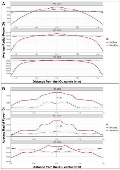
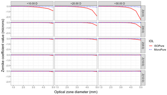
The aim of this study is to evaluate the results obtained on the optical bench and clinically with an isofocal lens (ISOPure, BVI medical, Belgium) to compare them to a standard monofocal one (MicroPure, BVI medical, Belgium). To do so, we have combined laboratory investigation and a prospective, comparative, and randomized clinical study. First, we have measured the wavefront of the two models studied using a NIMO TR1504 (Lambda-X, Belgium) deflectometer for three nominal powers: +10.00, +20.00 and +30.00 D. In the randomized study with 48 patients, half of them implanted with ISOPure and the other with MicroPure, we have measured visual acuities and contrast sensitivity under photopic and mesopic conditions. The optical bench results show that the isofocal lens presented higher power than the monofocal one, at the lens center, due to the spherical aberration (coefficients Z(4,0), Z(6,0) and Z(8,0)) induced by the greater asphericity of its design. The addition obtained depended on the nominal power, from +1.00 to +1.50 D. The results of the clinical study showed that the ISOPure lens presented better visual outcomes, which were statistically significant, at intermediate distance compared to the MicroPure lens (
p-values of 0.014 and 0.022 for 80 and 60 cm, respectively) without decreasing the contrast sensitivity. Clinical outcomes were not affected by pupillary size. In conclusion, due to the increase in power at the lens center due to its highly aspherical design, the isofocal lens evaluated showed better intermediate vision than the monofocal one.
Full article
►▼
Show Figures
Open AccessArticle
Progression of Rare Inherited Retinal Dystrophies May Be Monitored by Adaptive Optics Imaging
by
Katarzyna Samelska, Jacek Paweł Szaflik, Barbara Śmigielska and Anna Zaleska-Żmijewska
Cited by 1 | Viewed by 815
Abstract
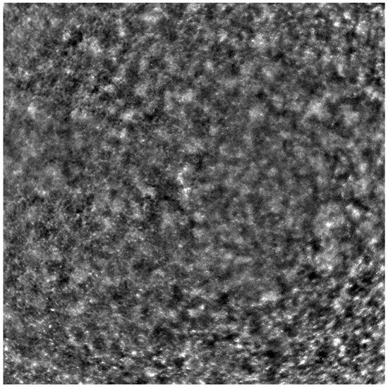
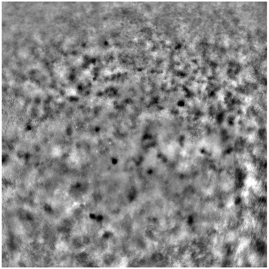
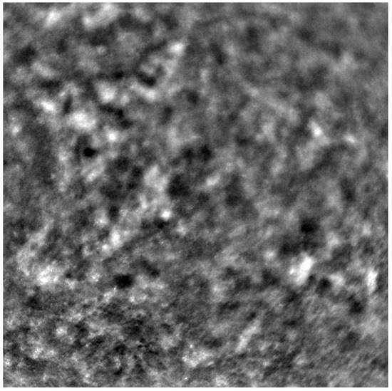
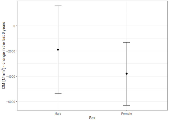
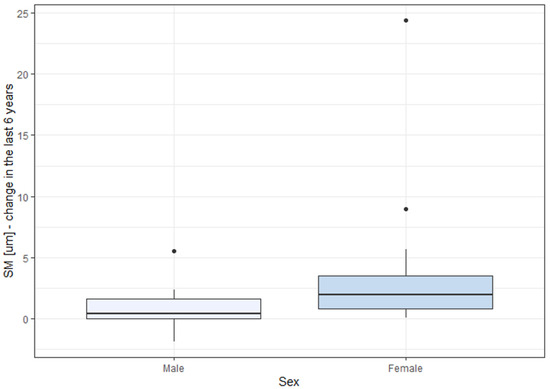
Inherited retinal dystrophies (IRDs) are bilateral genetic conditions of the retina, leading to irreversible vision loss. This study included 55 eyes afflicted with IRDs affecting the macula. The diseases examined encompassed Stargardt disease (STGD), cone dystrophy (CD), and cone–rod dystrophy (CRD) using adaptive
[...] Read more.
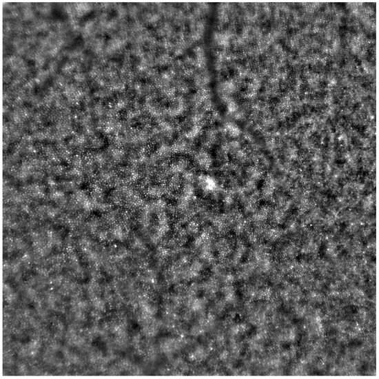
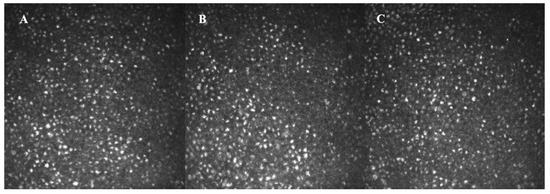
Inherited retinal dystrophies (IRDs) are bilateral genetic conditions of the retina, leading to irreversible vision loss. This study included 55 eyes afflicted with IRDs affecting the macula. The diseases examined encompassed Stargardt disease (STGD), cone dystrophy (CD), and cone–rod dystrophy (CRD) using adaptive optics (Rtx1™; Imagine Eyes, Orsay, France). Adaptive optics facilitate high-quality visualisation of retinal microstructures, including cones. Cone parameters, such as cone density (DM), cone spacing (SM), and regularity (REG), were analysed. The best corrected visual acuity (BCVA) was assessed as well. Examinations were performed twice over a 6-year observation period. A significant change was observed in DM (
/mm
vs. 10,073.42/mm
,
0.001) and SM (9.83
m vs. 12.16
m,
0.001) during the follow-up. BCVA deterioration was also significant (
vs.
,
p = 0.001), albeit uncorrelated with the change in cone parameters. No significant difference in REG was detected between the initial examination and the follow-up (
p = 0.089).
Full article
►▼
Show Figures
Open AccessReview
Sinonasal Orbital Apex Syndrome, Horner Syndrome and Pterygopalatine Fossa Infection: A Case Report and Mini-Review
by
Gregorio Benites, Jure Urbančič, Carolina Bardales and Domen Vozel
Viewed by 1591
Abstract
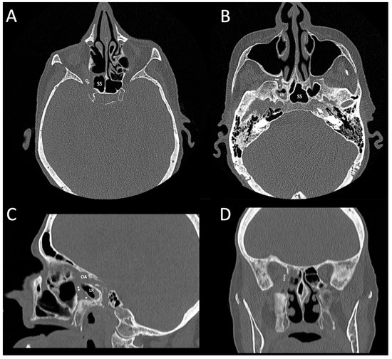
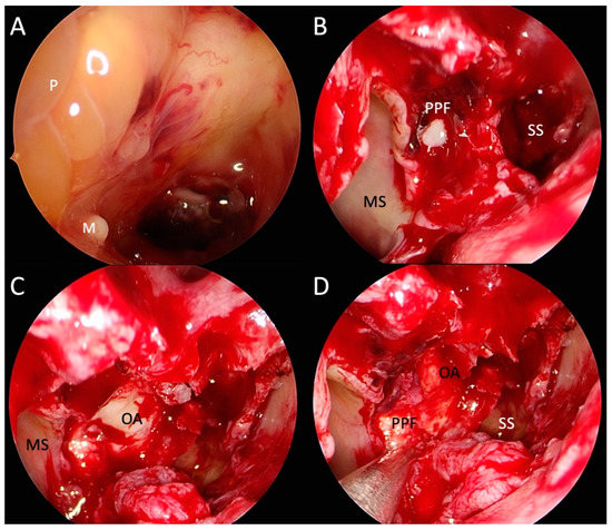
This paper presents a literature review and a case of an 83-year-old otherwise healthy female patient with a history of recent syncope, a sudden-onset right-sided temporal headache, diplopia, and vision loss. An exam revealed right-sided upper eyelid ptosis, myosis, vision loss, ophthalmoplegia, and
[...] Read more.
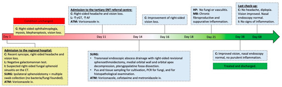
This paper presents a literature review and a case of an 83-year-old otherwise healthy female patient with a history of recent syncope, a sudden-onset right-sided temporal headache, diplopia, and vision loss. An exam revealed right-sided upper eyelid ptosis, myosis, vision loss, ophthalmoplegia, and a positive relative afferent pupillary defect on the right eye. CT showed sphenoid sinus opacification, eroded lateral sinus wall, Vidian canal, disease extension to the posterior ethmoid air cells, orbital apex, medial orbital wall, and pterygopalatine fossa. An orbital apex syndrome (Jacod’s syndrome), Horner syndrome, and pterygopalatine fossa infection were diagnosed due to the acute invasive fungal sinusitis developed from a sphenoid sinus fungal ball. The patient was treated with antimicrobial therapy and transnasal endoscopic surgery twice to decompress the orbital apex, drain the abscess and obtain specimens for analysis. The right-sided ptosis, visual loss, ophthalmoplegia, and headache resolved entirely. No immune or comorbid diseases were identified, microbiological and histopathological analyses were negative, and MRI could not be performed on the presented patient. For that reason, the diagnostic procedure was non-standard. Nevertheless, the treatment outcome of this vision and life-threatening disease was satisfactory. Treating the fungal ball in an older or immunocompromised patient is essential to prevent invasive fungal rhinosinusitis and fatal complications.
Full article
►▼
Show Figures
Open AccessArticle
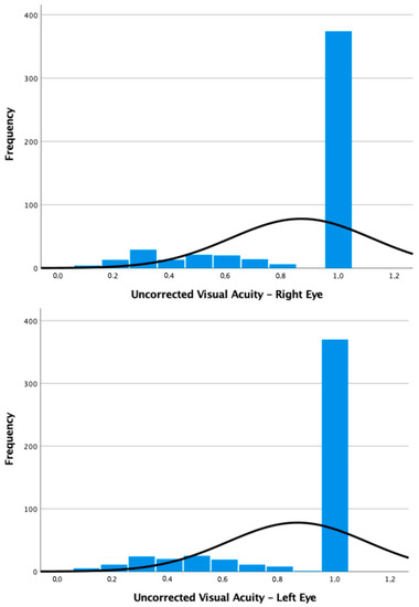
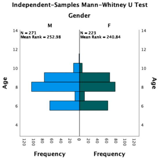
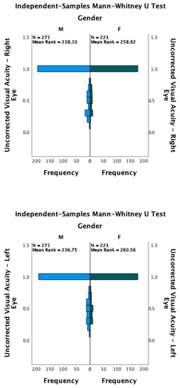
Analysis of Refractive Errors in a Large Italian Cohort of Pediatric Subjects Post the COVID-19 Pandemic
by
Michele Lanza, Adriano Ruggiero, Matteo Ruggiero, Clemente Maria Iodice and Francesca Simonelli
Viewed by 1241
Abstract
Background: The prevalence of refractive errors has sharply risen over recent decades. Despite the established role of genetics in the onset and progression of such conditions, the environment was also shown to play a pivotal role. Indeed, the COVID-19 pandemic has majorly impacted
[...] Read more.
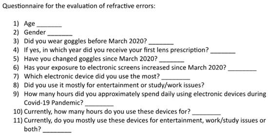
Background: The prevalence of refractive errors has sharply risen over recent decades. Despite the established role of genetics in the onset and progression of such conditions, the environment was also shown to play a pivotal role. Indeed, the COVID-19 pandemic has majorly impacted people’s lifestyles and healthy habits, especially among the youth, which might have led to a significant increase in this trend. Therefore, the aim of this study was to investigate the actual prevalence of refractive errors in a large cohort of pediatric patients. Methods: A large cohort of 496 participants was screened through anamnesis, a non-cycloplegic autorefractometry, a corrected and uncorrected visual acuity assessment, and a questionnaire and was retrospectively evaluated. Results: Overall, refractive errors were present in 25.1% of eyes, of which 14.6% were diagnosed with myopia/myopic astigmatism and 10.5% with hyperopia/hyperopic astigmatism. Among the patients enrolled, 298 (60%) had their eyes checked one year earlier or before and 122 (25%) had never had ophthalmological consultations; a total of 105 (21%) needed glasses and 34 (7%) required a change in their previous prescription. A substantial increase in daily electronic device screen exposure was declared by 426 patients (87.6%). Conclusions: Pediatric patients appear to have a higher prevalence of refractive errors than before.
Full article
►▼
Show Figures
Open AccessArticle
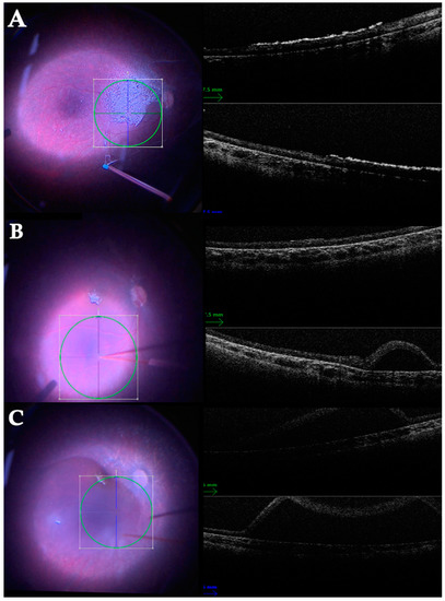
Central Serous Chorioretinopathy by Autofluorescence, Enface and SLO–Retromode Imaging
by
Maria Cristina Savastano, Claudia Fossataro, Riccardo Sadun, Andrea Scupola, Maria Grazia Sammarco, Clara Rizzo, Pia Clara Pafundi and Stanislao Rizzo
Cited by 1 | Viewed by 994
Abstract
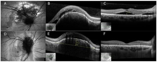
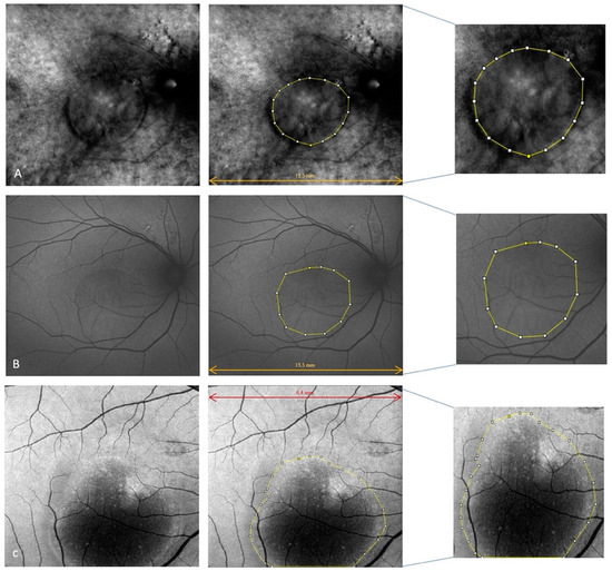
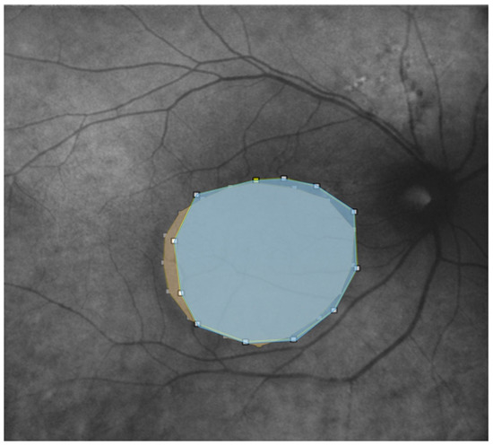
The aim of our study was to investigate the clinical features of central serous chorioretinopathy (CSC) with autofluorescence (AF), retromode (RM), and enface imaging. This retrospective study was conducted at Fondazione Policlinico Universitario A. Gemelli, IRCCS, Rome (Italy), between September and December 2022.
[...] Read more.
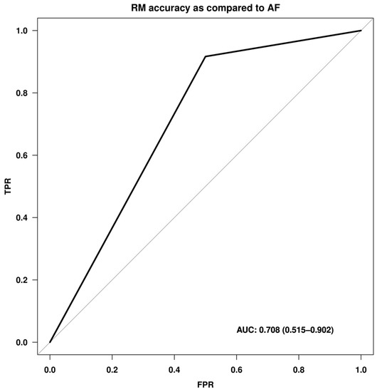
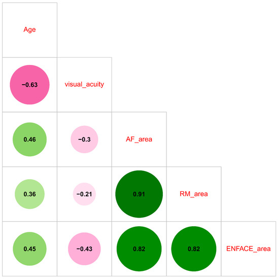
The aim of our study was to investigate the clinical features of central serous chorioretinopathy (CSC) with autofluorescence (AF), retromode (RM), and enface imaging. This retrospective study was conducted at Fondazione Policlinico Universitario A. Gemelli, IRCCS, Rome (Italy), between September and December 2022. Each patient underwent a complete ophthalmological examination, which included optical coherence tomography (OCT), enface image analysis, AF, and RM imaging. We further evaluated the presence and area of extension of serous retinal detachment and retinal pigment epithelium (RPE) atrophy through AF, RM, and enface imaging. We included 32 eyes from 27 patients (mean age: 52.7 ± 13.3 years). The median AF area was 19.5 mm
2 (IQR 6.1–29.3), while the median RM area was 12.3 mm
2 (IQR 8.1–30.8), and the median enface area was 9.3 mm
2 (IQR 4.8–18.6). RPE atrophy was diagnosed in 26 cases (81.3%) with RM imaging and in 75% of cases with AF. No difference emerged between AF and RM analysis in the detection of central serous detachment in CSC. However, RM imaging showed a high specificity (91.7%) and negative predictive value (84.6%) to detect RPE changes when compared to the AF standard-of-care technique. Thus, RM imaging could be considered an adjunctive imaging method in CSC.
Full article
►▼
Show Figures
Open AccessArticle
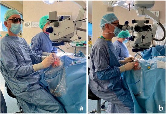
Three-Dimensional Visualization System for Vitreoretinal Surgery: Results from a Monocentric Experience and Comparison with Conventional Surgery
by
Fabrizio Giansanti, Cristina Nicolosi, Daniela Bacherini, Federica Soloperto, Federica Sarati, Dario Giattini and Giulio Vicini
Cited by 2 | Viewed by 1542
Abstract
Purpose: To describe the experience of our centre (Careggi University Hospital, Florence, Italy) in using a heads-up three-dimensional (3D) surgical viewing system in vitreoretinal surgery, making a comparison with the conventional microscope surgery. Methods: We retrospectively analyzed data taken from 240 patients (240
[...] Read more.
Purpose: To describe the experience of our centre (Careggi University Hospital, Florence, Italy) in using a heads-up three-dimensional (3D) surgical viewing system in vitreoretinal surgery, making a comparison with the conventional microscope surgery. Methods: We retrospectively analyzed data taken from 240 patients (240 eyes) with surgical macular diseases (macular hole and epiretinal membrane), retinal detachment or vitreous hemorrhage who underwent vitreoretinal surgeries, by means of the NGENUITY 3D Visualization System (Alcon Laboratories Inc., Fort Worth, TX, USA), in comparison with 210 patients (210 eyes) who underwent vitreoretinal surgeries performed using a conventional microscope. All surgeries were performed with standardized procedures by the same surgeons. We analyzed data over a follow-up period of 6 months, comparing the surgical outcomes (best-corrected visual acuity, anatomical success rate and postoperative complication rate) between the two groups. Results: the 3D group included 74 patients with retinal detachment, 78 with epiretinal membrane, 64 with macular hole and 24 with vitreous hemorrhage. There were no significant differences in the demographic and clinical characteristics between the 3D group and the conventional group. We found no significant differences in outcome measures at three and six months follow-up between the two groups (
p-value ≥ 0.05 for all comparisons). Surgery durations were similar between the two groups. Conclusions: In our experience, a heads-up 3D surgical viewing system provided comparable functional and anatomical outcomes in comparison with conventional microscope surgery, proving to be a valuable tool for vitreoretinal surgery in the treatment of different retinal diseases.
Full article
►▼
Show Figures
Open AccessReview
Pars Plana Vitrectomy in Inherited Retinal Diseases: A Comprehensive Review of the Literature
by
Claudio Iovino, Andrea Rosolia, Luciana Damiano, Clemente Maria Iodice, Valentina Di Iorio, Francesco Testa and Francesca Simonelli
Viewed by 1539
Abstract
Inherited retinal diseases (IRDs) are a group of clinically and genetically heterogeneous disorders that may be complicated by several vitreoretinal conditions requiring a surgical approach. Pars plana vitrectomy (PPV) stands as a valuable treatment option in these cases, but its application in eyes
[...] Read more.
Inherited retinal diseases (IRDs) are a group of clinically and genetically heterogeneous disorders that may be complicated by several vitreoretinal conditions requiring a surgical approach. Pars plana vitrectomy (PPV) stands as a valuable treatment option in these cases, but its application in eyes with such severely impaired chorioretinal architectures remains controversial. Furthermore, the spreading of gene therapy and the increasing use of retinal prostheses will end up in a marked increase in demand for PPV surgery for IRD patients. The retinal degeneration that typically affects patients with hereditary retinal disorders may influence the execution of the surgery and the expected results. Considering the importance of PPV application in IRD-related complications, it is fundamental to try to understand from the literature what is adequate and safe in posterior eye segment surgery. Use of dyes, light toxicity, and risk of wounding scar development have always been themes that discourage the execution of vitreoretinal surgery in already impaired eyes. Therefore, this review aims to comprehensively summarize all PPV applications in different IRDs, highlighting the favorable results as well as the potential precautions to consider when performing vitreoretinal surgery in these eyes.
Full article
►▼
Show Figures
Open AccessArticle
Micropulse Transscleral Cyclophotocoagulation Results in Secondary Glaucoma
by
Zsuzsa Szilagyi, Kinga Kranitz, Zoltan Zsolt Nagy and Zsuzsa Recsan
Viewed by 1437
Abstract
The aim of this study was to analyze the long-term outcome of first session of micropulse transscleral cyclophotocoagulation (MP-CPC) for refractory glaucoma developed after vitreoretinal surgery combined with silicone oil implantation. The inclusion criteria of this consecutive case series were: patients with secondary
[...] Read more.
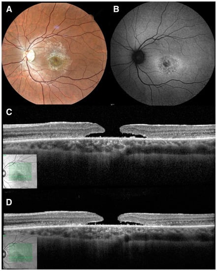
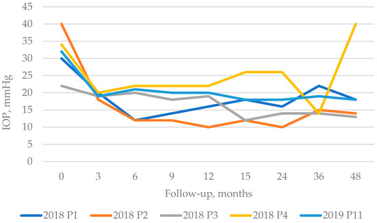
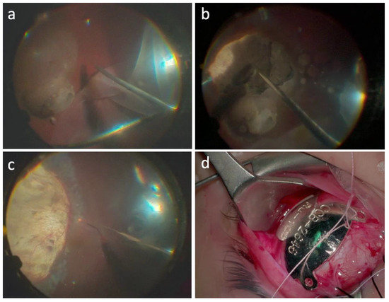
The aim of this study was to analyze the long-term outcome of first session of micropulse transscleral cyclophotocoagulation (MP-CPC) for refractory glaucoma developed after vitreoretinal surgery combined with silicone oil implantation. The inclusion criteria of this consecutive case series were: patients with secondary glaucoma in the refractory stage who underwent MP-CPC between 2018 and 2021, vitreoretinal surgery combined with silicon oil implantation, and at least a 24-month follow-up period after MP-CPC. Success was defined as the baseline eye pressure reduced at least 20%, and it should be ranged between 10 to 20 mmHg without further MP-CPC at the end of the follow-up. For this retrospective study, 11 eyes of 11 patients were selected. The reduction in IOP was found to be significant (
p = 0.004) at the end of the follow-up time, and the success rate was 72% according to our results. The change in the number of antiglaucoma agents in the administered eyedrops was not significant compared to the baseline values. At the end of the follow-up period the change in BCVA values was not significant (
p = 0.655). Our results confirm significant IOP lowering effect of this subthreshold method preserving visual performance safely even in eyes with previous vitrectomy surgery with a silicone oil implantation.
Full article
►▼
Show Figures
Open AccessReview
Nutritional Factors: Benefits in Glaucoma and Ophthalmologic Pathologies
by
Mutali Musa, Marco Zeppieri, George Nnamdi Atuanya, Ehimare S. Enaholo, Efioshiomoshi Kings Topah, Oluwasola Michael Ojo and Carlo Salati
Cited by 2 | Viewed by 3405
Abstract
Glaucoma is a chronic optic neuropathy that can lead to irreversible functional and morphological damage if left untreated. The gold standard therapeutic approaches in managing patients with glaucoma and limiting progression include local drops, laser, and/or surgery, which are all geared at reducing
[...] Read more.
Glaucoma is a chronic optic neuropathy that can lead to irreversible functional and morphological damage if left untreated. The gold standard therapeutic approaches in managing patients with glaucoma and limiting progression include local drops, laser, and/or surgery, which are all geared at reducing intraocular pressure (IOP). Nutrients, antioxidants, vitamins, organic compounds, and micronutrients have been gaining increasing interest in the past decade as integrative IOP-independent strategies to delay or halt glaucomatous retinal ganglion cell degeneration. In our minireview, we examine the various nutrients and compounds proposed in the current literature for the management of ophthalmology diseases, especially for glaucoma. With respect to each substance considered, this minireview reports the molecular and biological characteristics, neuroprotective activities, antioxidant properties, beneficial mechanisms, and clinical studies published in the past decade in the field of general medicine. This study highlights the potential benefits of these substances in glaucoma and other ophthalmologic pathologies. Nutritional supplementation can thus be useful as integrative IOP-independent strategies in the management of glaucoma and in other ophthalmologic pathologies. Large multicenter clinical trials based on functional and morphologic data collected over long follow-up periods in patients with IOP-independent treatments can pave the way for alternative and/or coadjutant therapeutic options in the management of glaucoma and other ocular pathologies.
Full article
►▼
Show Figures
Open AccessArticle
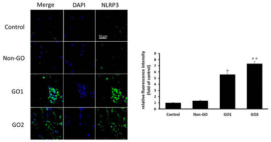
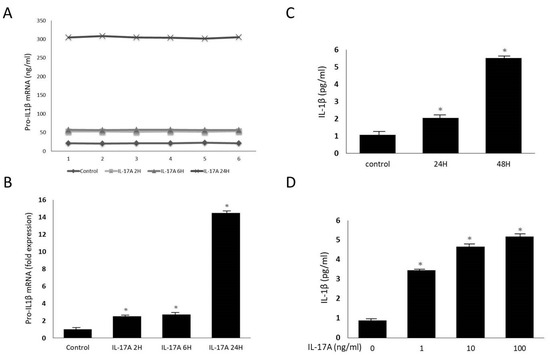
The Role of Interleukin-17A and NLRP3 Inflammasome in the Pathogenesis of Graves’ Ophthalmopathy
by
Chih-Chung Lin, Shu-Lang Liao and Yi-Hsuan Wei
Cited by 1 | Viewed by 1283
Abstract
The development of Graves’ ophthalmopathy (GO) is associated with autoimmune dysfunction. Recent studies have indicated that IL-17A, inflammasomes, and related cytokines may be involved in the etiology of GO. We sought to investigate the pathogenic role of IL-17A and NLRP3 inflammasomes in GO.
[...] Read more.
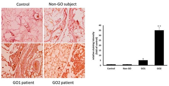
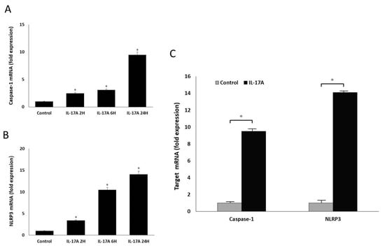
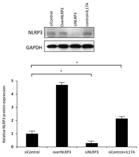
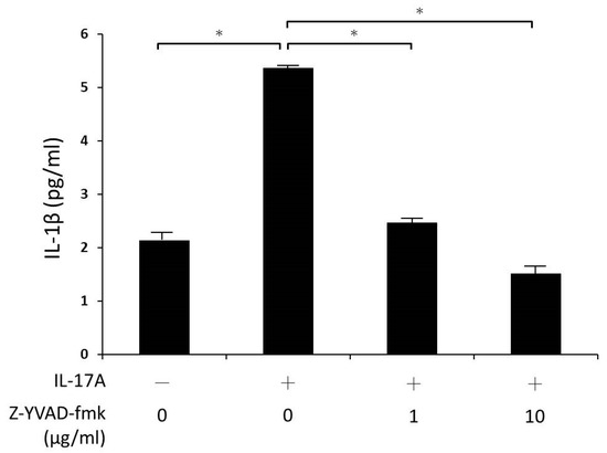
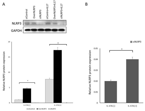
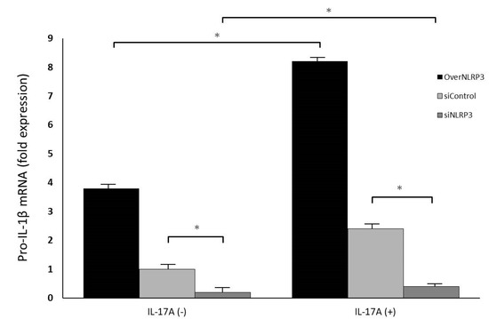
The development of Graves’ ophthalmopathy (GO) is associated with autoimmune dysfunction. Recent studies have indicated that IL-17A, inflammasomes, and related cytokines may be involved in the etiology of GO. We sought to investigate the pathogenic role of IL-17A and NLRP3 inflammasomes in GO. Orbital fat specimens were collected from 30 patients with GO and 30 non-GO controls. Immunohistochemical staining and orbital fibroblast cultures were conducted for both groups. IL-17A was added to the cell cultures, and cytokine expression, signaling pathways, and inflammasome mechanisms were investigated using reverse transcription polymerase chain reaction, enzyme-linked immunosorbent assay, Western blotting, and small interfering RNA (siRNA) methods. Immunohistochemical staining showed higher NLRP3 expression in GO orbital tissue than in non-GO controls. IL-17A upregulated pro-IL-1β mRNA levels and IL-1β protein levels in the GO group. Furthermore, IL-17A was confirmed to enhance caspase-1 and NLRP3 protein expression in orbital fibroblasts, suggesting NLRP3 inflammasome activation. Inhibiting caspase-1 activity could also decrease IL-1β secretion. In siRNA-transfected orbital fibroblasts, significantly decreased NLRP3 expression was observed, and IL-17A-mediated pro-IL-1β mRNA release was also downregulated. Our observations illustrate that IL-17A promotes IL-1β production from orbital fibroblasts via the NLRP3 inflammasome in GO, and cytokines subsequently released may induce more inflammation and autoimmunity.
Full article
►▼
Show Figures
Open AccessArticle
Anatomical and Functional Outcomes after Endoresection and Adjuvant Ruthenium Brachytherapy for Uveal Melanoma: A Single-Center Experience
by
Cinzia Mazzini, Giulio Vicini, Laura Di Leo, Daniela Massi, Stanislao Rizzo and Fabrizio Giansanti
Cited by 1 | Viewed by 1330
|
Correction
Abstract
Purpose: To evaluate the anatomical and functional outcomes of endoresection and adjuvant ruthenium (Ru)-106 brachytherapy for uveal melanoma (UM). Methods: Retrospective case series of 15 UM patients (15 eyes) treated at our center (Careggi University Hospital, Florence). Results: Six patients (40%) were male
[...] Read more.
Purpose: To evaluate the anatomical and functional outcomes of endoresection and adjuvant ruthenium (Ru)-106 brachytherapy for uveal melanoma (UM). Methods: Retrospective case series of 15 UM patients (15 eyes) treated at our center (Careggi University Hospital, Florence). Results: Six patients (40%) were male and nine were female (60%). The mean age of patients at the time of treatment was 61.6 years (±18.47). The mean BCVA at baseline was 20/76. In all cases UM originated from the choroid. The mean tumor thickness at baseline was 7.20 mm (±2.01), and the mean largest basal diameter was 11.24 mm (±2.20). A concurrent retinal detachment was diagnosed in 11 patients (73.3%). Two patients (13.3%) showed vitreous seeding at baseline. Eleven patients (73.3%) were treated with primary endoresection, while four patients (26.7%) were treated with a “salvage endoresection” after primary treatment failure (previous radiation treatment). The mean follow-up time was 29.9 months (±10.6). Thirteen out of fifteen patients were alive and showed no evidence of local recurrence or distance metastasis at the last follow-up visit. The treatment achieved local control of the disease in 14 out of 15 cases (93.3%). In one case, the patient underwent enucleation for disease recurrence. The overall survival rate at the end of the follow-up was 93.3%. The mean BCVA at last follow-up visit was 20/70. Treatment was well tolerated, without significant complications. Conclusions: Endoresection and adjuvant Ru-106 brachytherapy is a valuable conservative option for selected UM patients and can be used both as a primary treatment and as a salvage therapy. It can control melanoma and avoid enucleation, reduce radiation-related complications, and provide tumor tissue for chromosomal analysis and prognostic testing.
Full article
►▼
Show Figures
Open AccessBrief Report
Visual Telerehabilitation with Visually Impaired Children: From the Pandemic Emergency to a Stand-Alone Method
by
Giulia Perasso, Chiara Baghino, Elena Cocchi, Silvia Dini, Antonella Panizzi, Valentina Salvagno, Margherita Santarello and Aldo Vagge
Viewed by 988
Abstract
In the last two years, orthoptists have counteracted patient drop-out through visual telerehabilitation. Efforts were made to transfer the in-person visual rehabilitation setting to the telematic environment in response to the worldwide crisis. Nowadays, statistical evidence on the effects of visual telerehabilitation is
[...] Read more.
In the last two years, orthoptists have counteracted patient drop-out through visual telerehabilitation. Efforts were made to transfer the in-person visual rehabilitation setting to the telematic environment in response to the worldwide crisis. Nowadays, statistical evidence on the effects of visual telerehabilitation is still scarce. The present research is the first, in Italy, to offer a pre-post assessment of the impact of visual telerehabilitation. Twenty-four (n = 24) children (64% male, 14% monocles) aged 4 to 15 years (mean age = 9.21 years, SD = 3.36, mean residual vision 1.3/10) were randomly assigned to three different group types for rehabilitation: a telematic rehabilitation group (n = 7), a mixed rehabilitation group (n = 8), and an in-person rehabilitation group (n = 9). Each group underwent a six-week visual rehabilitation. Ergo-perimetric evaluation before and after the rehabilitation was administered to the three groups.
t-tests showed a significant improvement in ergo-perimetric outcomes in the visual telerehabilitation group (
p < 0.05) and in the mixed rehabilitation group (
p < 0.01), via a shortening of the response times. The findings suggest that visual telerehabilitation and mixed rehabilitation can lead to an ergo-perimetric improvement in visually impaired children within six weeks. Further research is needed, both to corroborate the findings with a larger sample size and to attain a follow-up measurement in order to clarify whether visual telerehabilitation could represent a stand-alone method.
Full article
►▼
Show Figures
Open AccessArticle
Is Erythrocyte Sedimentation Rate Necessary for the Initial Diagnosis of Giant Cell Arteritis?
by
Michael S. Hansen, Oliver N. Klefter, Lene Terslev, Mads R. Jensen, Jane M. Brittain, Uffe M. Døhn, Carsten Faber, Steffen Heegaard, Anne K. Wiencke, Yousif Subhi and Steffen Hamann
Viewed by 1673
Abstract
Giant cell arteritis (GCA) is an ophthalmological emergency that can be difficult to diagnose and prompt treatment is vital. We investigated the sequential diagnostic value for patients with suspected GCA using three biochemical measures as they arrive to the clinician: first, platelet count,
[...] Read more.
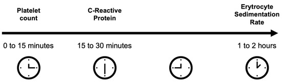
Giant cell arteritis (GCA) is an ophthalmological emergency that can be difficult to diagnose and prompt treatment is vital. We investigated the sequential diagnostic value for patients with suspected GCA using three biochemical measures as they arrive to the clinician: first, platelet count, then C-reactive protein (CRP), and lastly, erythrocyte sedimentation rate (ESR). This retrospective cross-sectional study of consecutive patients with suspected GCA investigated platelet count, CRP, and ESR using diagnostic test accuracy statistics and odds ratios (ORs) in a sequential fashion. The diagnosis was established by experts at follow-up, considering clinical findings and tests including temporal artery biopsy. A total of 94 patients were included, of which 37 (40%) were diagnosed with GCA. Compared with those without GCA, patients with GCA had a higher platelet count (
p < 0.001), CRP (
p < 0.001), and ESR (
p < 0.001). Platelet count demonstrated a low sensitivity (38%) and high specificity (88%); CRP, a high sensitivity (86%) and low specificity (56%); routine ESR, a high sensitivity (89%) and low specificity (47%); and age-adjusted ESR, a moderate sensitivity (65%) and moderate specificity (65%). Sequential analysis revealed that ESR did not provide additional value in evaluating risk of GCA. Initial biochemical evaluation can be based on platelet count and CRP, without waiting for ESR, which allows faster initial decision-making in GCA.
Full article
►▼
Show Figures
Open AccessArticle
Widefield and Ultra-Widefield Retinal Imaging: A Geometrical Analysis
by
Amedeo Lucente, Andrea Taloni, Vincenzo Scorcia and Giuseppe Giannaccare
Cited by 2 | Viewed by 2247
Abstract
Diabetic retinopathy (DR) often causes a wide range of lesions in the peripheral retina, which can be undetected when using a traditional fundus camera. Widefield (WF) and Ultra-Widefield (UWF) technologies aim to significantly expand the photographable retinal field. We conducted a geometrical analysis
[...] Read more.
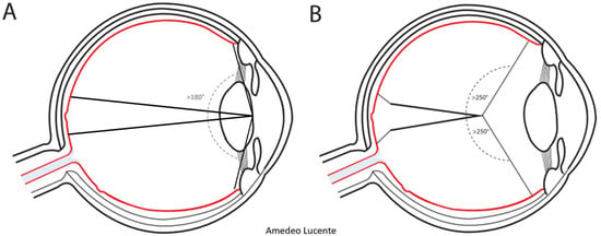
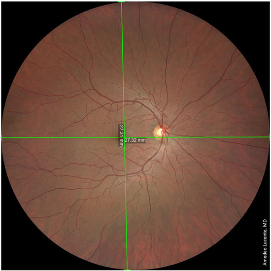
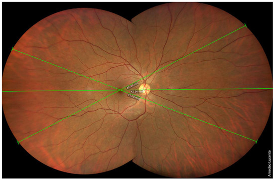
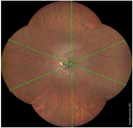
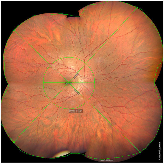
Diabetic retinopathy (DR) often causes a wide range of lesions in the peripheral retina, which can be undetected when using a traditional fundus camera. Widefield (WF) and Ultra-Widefield (UWF) technologies aim to significantly expand the photographable retinal field. We conducted a geometrical analysis to assess the field of view (FOV) of WF and UWF imaging, comparing it to the angular extension of the retina. For this task, we shot WF images using the Zeiss Clarus 500 fundus camera (Carl Zeiss Meditec, Jena, Germany). Approximating the ocular bulb to an ideal sphere, the angular extension of the theoretically photographable retinal surface was 242 degrees. Performing one shot, centered on the macula, it was possible to photograph a retinal surface of ~570 mm
2, with a FOV of 133 degrees. Performing four shots with automatic montage, we obtained a retinal surface area of ~1100 mm
2 and an FOV of 200 degrees. Finally, performing six shots with semi-automatic montage, we obtained a retinal surface area of ~1400 mm
2 and an FOV of 236.27 degrees, which is close to the entire surface of the retina. WF and UWF imaging allow the detailed visualization of the peripheral retina, with significant impact on the diagnosis and management of DR.
Full article
►▼
Show Figures
Open AccessReview
Ocular Surface Features in Patients with Parkinson Disease on and off Treatment: A Narrative Review
by
Matilde Buzzi, Giuseppe Giannaccare, Michela Cennamo, Federico Bernabei, Pierre-Raphael Rothschild, Aldo Vagge, Vincenzo Scorcia and Rita Mencucci
Cited by 2 | Viewed by 1545
Abstract
Parkinson disease (PD) is a progressive, neurodegenerative disease of the central nervous system. Visual disturbance is one of the most frequent nonmotor abnormalities referred to by patients suffering from PD at early stages. Furthermore, ocular surface alterations including mainly dry eye and blink
[...] Read more.
Parkinson disease (PD) is a progressive, neurodegenerative disease of the central nervous system. Visual disturbance is one of the most frequent nonmotor abnormalities referred to by patients suffering from PD at early stages. Furthermore, ocular surface alterations including mainly dry eye and blink reduction represent another common finding in patients with PD. Tears of PD patients show specific alterations related to protein composition, and in vivo confocal microscopy has demonstrated profound changes in different corneal layers in this setting. These changes can be attributed not only to the disease itself, but also to the medications used for its management. In particular, signs of corneal toxicity, both at epithelial and endothelial level, are well described in the literature in PD patients receiving amantadine. Management of PD patients from the ophthalmologist’s side requires knowledge of the common, but often underdiagnosed, ocular surface alterations as well as of the signs of drug toxicity. Furthermore, ocular surface biomarkers can be useful for the early diagnosis of PD as well as for monitoring the degree of neural degeneration over time.
Full article
►▼
Show Figures
Open AccessArticle
Half-Dose Photodynamic Therapy as a Novel Treatment Protocol for Circumscribed Choroidal Hemangioma
by
David Pérez-González, Michaella Goldstein, Matias Iglicki and Dinah Zur
Cited by 2 | Viewed by 1649
Abstract
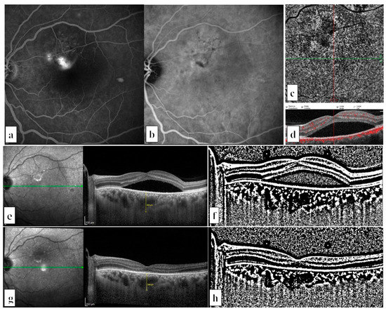
We present a case series of four patients with circumscribed choroidal hemangioma (CCH) treated with half-dose PDT, proposing this as a novel treatment protocol. Four patients with CCH were included, and then evaluated using multimodal imaging, including optical coherence tomography (OCT), fluorescein angiography,
[...] Read more.
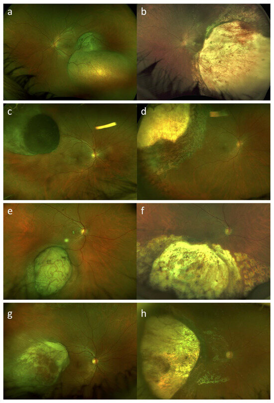
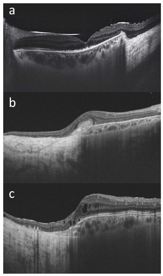
We present a case series of four patients with circumscribed choroidal hemangioma (CCH) treated with half-dose PDT, proposing this as a novel treatment protocol. Four patients with CCH were included, and then evaluated using multimodal imaging, including optical coherence tomography (OCT), fluorescein angiography, indocyanine green angiography, fundus autofluorescence, and ultrasound following treatment with half-dose and full-fluence PDT. Following half-dose PDT, all patients showed significant shrinkage of the hemangioma, functional improvement, and decrease of intra- and sub-retinal fluid. All patients remained stable after a single PDT treatment, with a follow-up of up to 60 months. No side effects were shown. This is the first report showing long term efficacy of half-dose PDT treatment in cases with CCH. The outcomes from this pilot study are comparable with results using full dose PDT protocols and it can be considered as a viable treatment option for CCH during the ongoing global verteporfin shortage.
Full article
►▼
Show Figures
Open AccessArticle
Long-Term Macular Vascular Changes after Primary Rhegmatogenous Retinal Detachment Surgery Resolved with Different Tamponade or Different Surgical Techniques
by
Matteo Gironi, Rossella D’Aloisio, Tommaso Verdina, Chiara Vivarelli, Riccardo Leonelli, Shaniko Kaleci, Lisa Toto and Rodolfo Mastropasqua
Cited by 3 | Viewed by 1351
Abstract
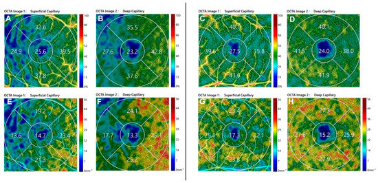
Background: The aim of this study was to assess long-term macular vascular changes and their correlation with functional recovery in patients successfully treated for Macula-ON and Macula-OFF rhegmatogenous retinal detachment (RRD). Methods: This retrospective observational study included 82 eyes of 82 patients who
[...] Read more.
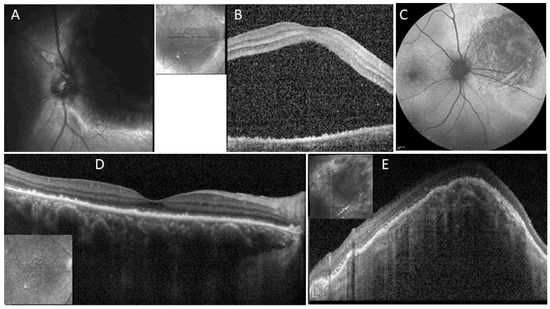
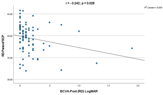
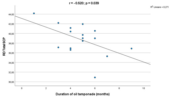
Background: The aim of this study was to assess long-term macular vascular changes and their correlation with functional recovery in patients successfully treated for Macula-ON and Macula-OFF rhegmatogenous retinal detachment (RRD). Methods: This retrospective observational study included 82 eyes of 82 patients who received primary successful retinal detachment surgery, 33 Macula-ON and 49 Macula-OFF. Superficial and deep capillary plexuses (SCP and DCP) were evaluated by optical coherence tomography angiography (OCTA), and were correlated with visual acuity (VA), surgical technique and tamponade at 12 months after surgery. The fellow eyes were used as controls. Results: At 12-month follow-up, there was a significant decrease in the vessel density (VD) in the SCP in the operated eyes compared to control eyes (
p < 0.05) in both the Macula-ON and Macula-OFF groups. Vessel length density (VLD) decrease in SCP was more extended in the Macula-OFF group. No difference in the DCP perfusion parameters was found, compared to controls. Subgroup analysis dependent on the type of surgery or tamponade showed no significant differences of VD and VLD. An inverse correlation was found between the SCP VD and the duration of silicone oil (SO) tamponade (
p = 0.039). A significant correlation was observed between parafoveal SCP VD and final best corrected visual acuity (BCVA) (
p = 0.028). The multivariate linear regression analysis showed that only the type of tamponade was significantly correlated with the final BCVA in the Macula-ON group (
p = 0.004). Conclusions: Our study described long-term perfusion changes in RRD after surgery, with lower SCP VD and VLD in the operated eyes compared to the fellow ones, not influenced by type of surgery or tamponade. The choice of tamponade and SO removal timing may affect functional outcomes, especially in Macula-ON RRD. In conclusion, such functional and perfusion changes can be considered biomarkers that highlight the relevance of careful management of this sight-threatening disease.
Full article
►▼
Show Figures
Open AccessArticle
Ocular Surface Changes Associated with Face Masks in Healthcare Personnel during COVID-19 Pandemic
by
Filippo Tatti, Lorenzo Mangoni, Simone Pirodda, Giuseppe Demarinis, Claudio Iovino, Emanuele Siotto Pintor, Germano Orrù, Luigi Isaia Lecca, Marcello Campagna, Gloria Denotti and Enrico Peiretti
Cited by 2 | Viewed by 1526
Abstract
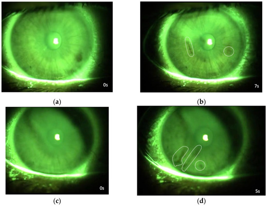
The aim of this study was to investigate ocular surface changes associated with face mask (FMs) use of healthcare personnel during the COVID-19 pandemic. We prospectively evaluated 200 eyes of 100 individuals during working hours and 40 eyes of 20 individuals during their
[...] Read more.
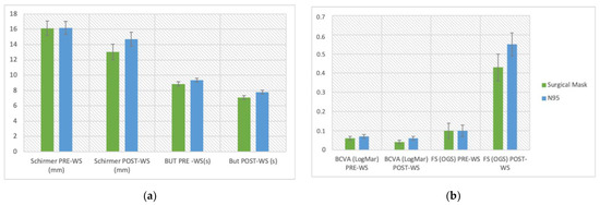
The aim of this study was to investigate ocular surface changes associated with face mask (FMs) use of healthcare personnel during the COVID-19 pandemic. We prospectively evaluated 200 eyes of 100 individuals during working hours and 40 eyes of 20 individuals during their rest days as a control group. Dry eye symptoms were assessed with the Ocular Surface Disease Index (OSDI) and McMonnies questionnaire. The clinical investigation included the best corrected visual acuity (BCVA), corneal fluorescein staining (FS), break-up time (BUT), and Schirmer test I before and after a 7-h work shift with a continuative use of surgical or N95 masks. The control group was evaluated similarly twice a day, at 8:00 a.m. and at 3:00 p.m.. In the study group, BCVA, FS, BUT, and Schirmer test were investigated and there was a significant negative variation at the end of the shift. On the contrary, the control group did not show significant variations of any clinical feature. Furthermore, no significant changes in clinical parameters were observed during the use of surgical or N95 masks. In conclusion, FMs continuative use resulted in daily ocular surface modifications specifically in healthcare personnel.
Full article
►▼
Show Figures
Open AccessArticle
A Comparative Study of Short-Term Vascular and Stromal Alterations of the Choroid Following Half-Fluence Photodynamic Therapy in Pachychoroid Neovasculopathy and Chronic Central Serous Chorioretinopathy
by
Özge Yanık, Sibel Demirel, Figen Batıoğlu and Emin Özmert
Cited by 3 | Viewed by 1286
Abstract
Background: Central serous chorioretinopathy (CSCR) and pachychoroid neovasculopathy (PNV) are among the pachychoroid spectrum diseases (PSDs). Half-fluence photodynamic therapy (hf-PDT) is one of the effective treatment methods for both diseases. The aim of this study was to compare the effect of hf-PDT on
[...] Read more.
Background: Central serous chorioretinopathy (CSCR) and pachychoroid neovasculopathy (PNV) are among the pachychoroid spectrum diseases (PSDs). Half-fluence photodynamic therapy (hf-PDT) is one of the effective treatment methods for both diseases. The aim of this study was to compare the effect of hf-PDT on the choroidal structure in CSCR and PNV. Methods: This study included 35 patients with chronic CSCR and 18 patients with PNV. The hf-PDT protocol was applied to all eyes. Before and 3 months after hf-PDT, enhanced-depth optical coherence tomography images were analyzed. The total choroidal area (CA), luminal area (LA), and stromal area (SA) were measured using ImageJ software. Results: Compared with baseline values, 3 months after hf-PDT, the mean CA reduced from 1.398 to 1.197 mm
2 (
p < 0.001) in the CSCR group and the total CA reduced from 1.050 to 1.000 mm
2 (
p < 0.021) in the PNV group. The mean percentage changes in CA, LA, and SA values were statistically higher in the chronic CSCR group (13.86%, 13.53%, and 14.11%, respectively) than those in the PNV group (4.61%, 4.02%, and 5.74%;
p = 0.001,
p = 0.002, and
p = 0.031, respectively). Conclusion: CSCR and PNV are thought to be PSDs. However, they differ in choroidal morphological response after hf-PDT, which might be a result of the different structural components of the PNV lesions.
Full article
►▼
Show Figures


