
Half-Dose Photodynamic Therapy as a Novel Treatment Protocol for Circumscribed Choroidal Hemangioma

Abstract
:1. Introduction
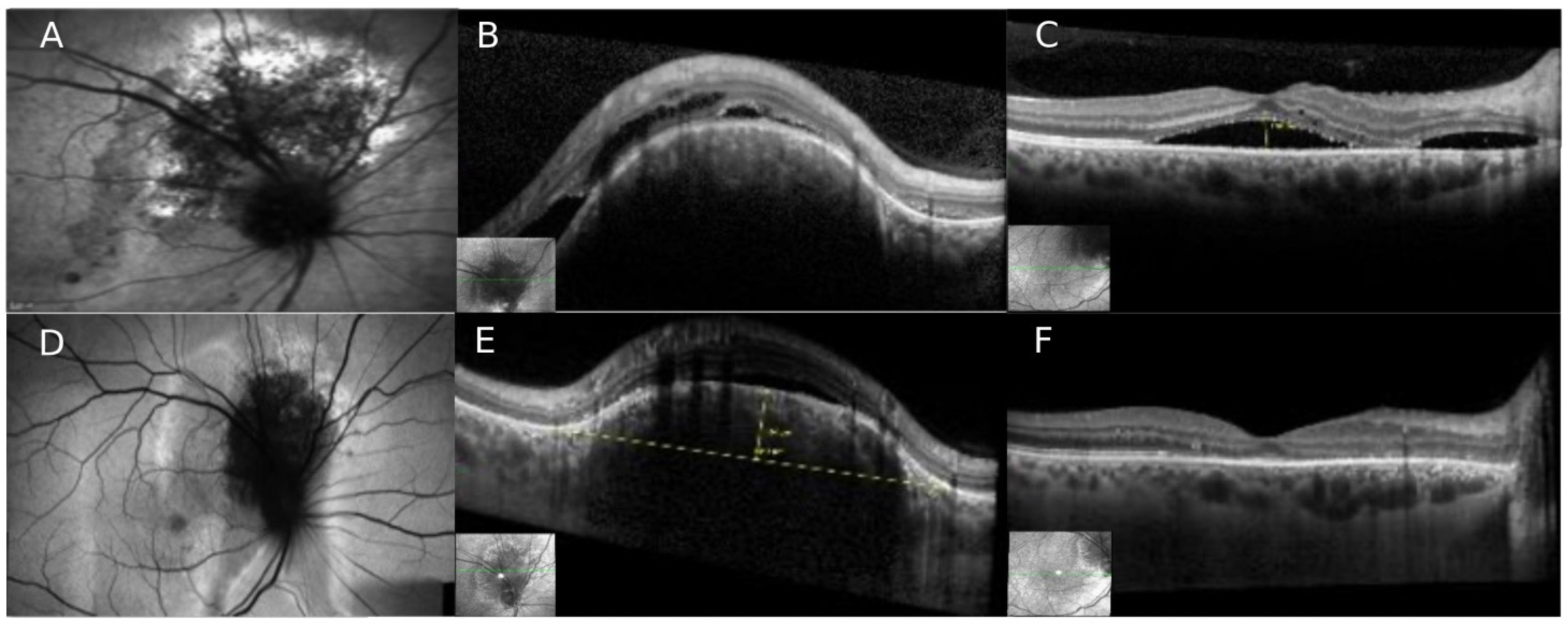
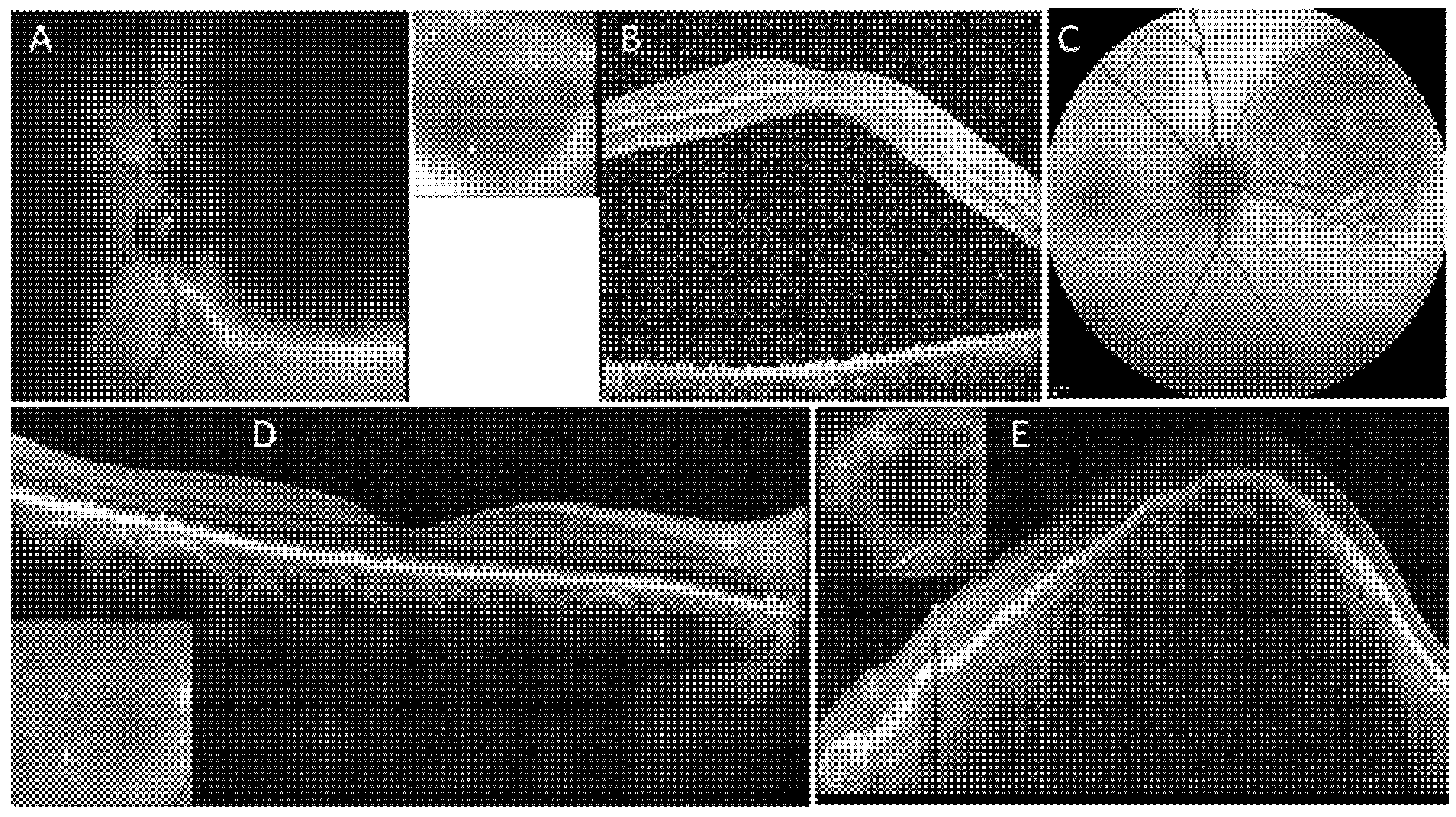
2. Case Number 1
3. Case Number 2
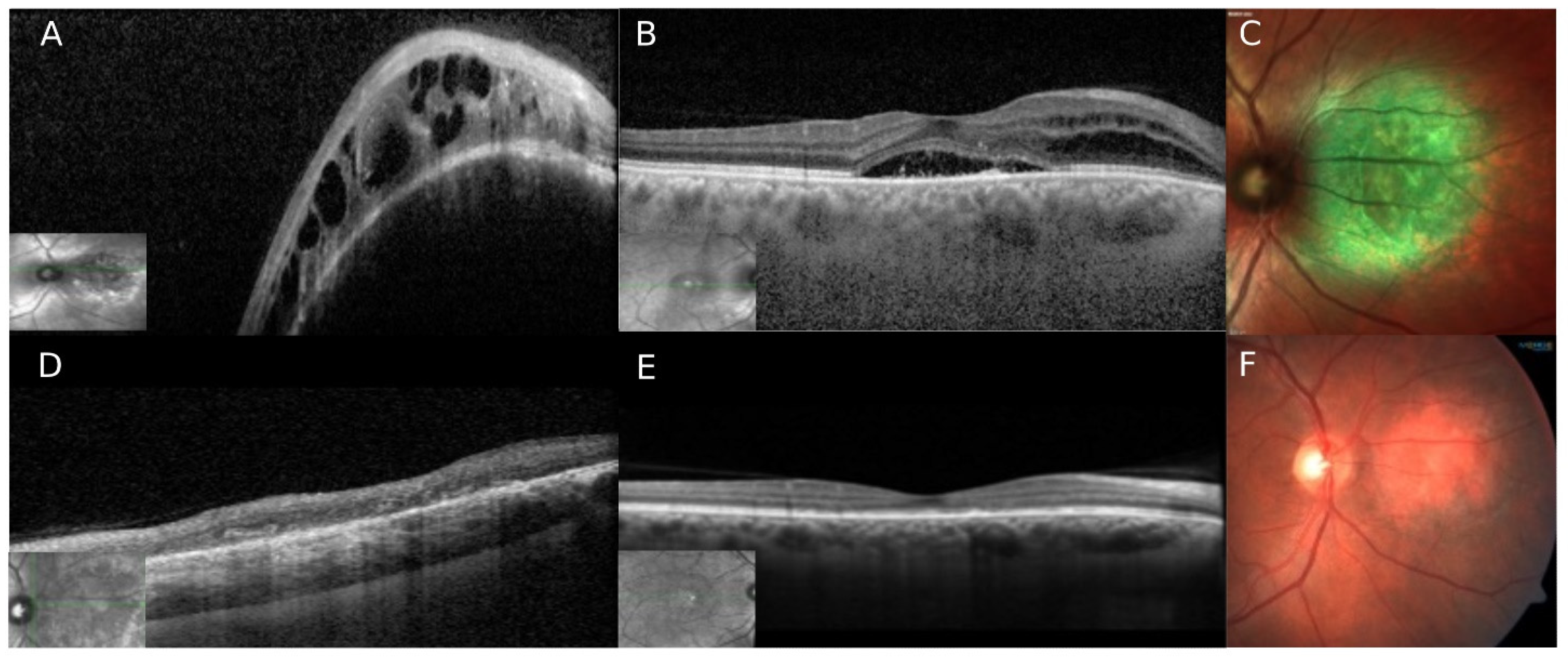
4. Case Number 3
5. Case Number 4
6. Discussion
Author Contributions
Funding
Institutional Review Board Statement
Informed Consent Statement
Conflicts of Interest
References
- Shields, C. Circumscribed Choroidal Hemangioma Clinical Manifestations and Factors Predictive of Visual Outcome in 200 Consecutive Cases. Ophthalmology 2001, 108, 2237–2248. [Google Scholar] [CrossRef]
- Tsipursky, M.S.; Golchet, P.R.; Jampol, L.M. Photodynamic Therapy of Choroidal Hemangioma in Sturge-Weber Syndrome, with a Review of Treatments for Diffuse and Circumscribed Choroidal Hemangiomas. Surv. Ophthalmol. 2011, 56, 68–85. [Google Scholar] [CrossRef]
- Newman, D.K. Photodynamic Therapy: Current Role in the Treatment of Chorioretinal Conditions. Eye 2016, 30, 202–210. [Google Scholar] [CrossRef] [Green Version]
- Jurklies, B.; Anastassiou, G.; Ortmans, S.; Schüler, A.; Schilling, H.; Schmidt-Erfurth, U.; Bornfeld, N. Photodynamic Therapy Using Verteporfin in Circumscribed Choroidal Haemangioma. Br. J. Ophthalmol. 2003, 87, 84–89. [Google Scholar] [CrossRef] [PubMed]
- Blasi, M.A.; Tiberti, A.C.; Scupola, A.; Balestrazzi, A.; Colangelo, E.; Valente, P.; Balestrazzi, E. Photodynamic Therapy with Verteporfin for Symptomatic Circumscribed Choroidal Hemangioma: Five-Year Outcomes. Ophthalmology 2010, 117, 1630–1637. [Google Scholar] [CrossRef]
- Isola, V.; Pece, A.; Parodi, M.B. Choroidal Ischemia After Photodynamic Therapy with Verteporfin for Choroidal Neovascularization. Am. J. Ophthalmol. 2006, 142, 680–683. [Google Scholar] [CrossRef]
- Reinke, M.H.; Canakis, C.; Husain, D.; Michaud, N.; Flotte, T.J.; Gragoudas, E.S.; Miller, J.W. Verteporfin Photodynamic Therapy Retreatment of Normal Retina and Choroid in the Cynomolgus Monkey. Ophthalmology 1999, 106, 1915–1923. [Google Scholar] [CrossRef]
- Borgia, F.; Giuffrida, R.; Caradonna, E.; Vaccaro, M.; Guarneri, F.; Cannavò, S. Early and Late Onset Side Effects of Photodynamic Therapy. Biomedicines 2018, 6, 12. [Google Scholar] [CrossRef] [Green Version]
- Hirami, Y.; Tsujikawa, A.; Otani, A.; Yodoi, Y.; Aikawa, H.; Mandai, M.; Yoshimura, N. Hemorrhagic complications after photodynamic therapy for polypoidal choroidal vasculopathy. Retina 2007, 27, 335–341. [Google Scholar] [CrossRef]
- Schmidt-Erfurth, U.; Michels, S.; Barbazetto, I.; Laqua, H. Photodynamic Effects on Choroidal Neovascularization and Physiological Choroid. Investig. Ophthalmol. Vis. Sci. 2002, 43, 830–841. [Google Scholar]
- Papastefanou, V.P.; Plowman, P.N.; Reich, E.; Pavlidou, E.; Restori, M.; Hungerford, J.L.; Arora, A.K.; Cohen, V.M.L.; Sagoo, M.S. Analysis of Long-Term Outcomes of Radiotherapy and Verteporfin Photodynamic Therapy for Circumscribed Choroidal Hemangioma. Ophthalmol. Retina 2018, 2, 842–857. [Google Scholar] [CrossRef] [PubMed]
- Shin, J.Y.; Woo, S.J.; Yu, H.G.; Park, K.H. Comparison of efficacy and safety between half-fluence and full-fluence photodynamic therapy for chronic central serous chorioretinopathy. Retina 2011, 31, 119–126. [Google Scholar] [CrossRef] [PubMed]
- Ziemssen, F.; Voelker, M.; Inhoffen, W.; Bartz-Schmidt, K.U.; Gelisken, F. Combined Treatment of a Juxtapapillary Retinal Capillary Haemangioma with Intravitreal Bevacizumab and Photodynamic Therapy. Eye 2007, 21, 1125–1126. [Google Scholar] [CrossRef] [PubMed]
- Shields, C.L.; Dalvin, L.A.; Lim, L.-A.S.; Chang, M.; Udyaver, S.; Mazloumi, M.; Vichitvejpaisal, P.; Su, G.L.; Florakis, E.; Mashayekhi, A.; et al. Circumscribed Choroidal Hemangioma: Visual Outcome in the Pre-Photodynamic Therapy Era versus Photodynamic Therapy Era in 458 Cases. Ophthalmol. Retina 2020, 4, 100–110. [Google Scholar] [CrossRef] [PubMed]
- Schmidt-Erfurth, U.; Hasan, T. Mechanisms of Action of Photodynamic Therapy with Verteporfin for the Treatment of Age-Related Macular Degeneration. Surv. Ophthalmol. 2000, 45, 195–214. [Google Scholar] [CrossRef]
- Miller, J.W. Photodynamic Therapy with Verteporfin for Choroidal Neovascularization Caused by Age-Related Macular Degeneration. Arch. Ophthalmol. 1999, 117, 1161. [Google Scholar] [CrossRef] [Green Version]
- Regillo, C.D. Update on Photodynamic Therapy. Curr. Opin. Ophthalmol. 2000, 11, 166–170. [Google Scholar] [CrossRef]
- Wong, I.Y.; Shi, X.; Gangwani, R.; Zhao, P.; Iu, L.P.; Li, Q.; Ng, A.; Li, X. 1-Year Results of Combined Half-Dose Photodynamic Therapy and Ranibizumab for Polypoidal Choroidal Vasculopathy. BMC Ophthalmol. 2015, 15, 66. [Google Scholar] [CrossRef] [Green Version]
- Ngo, W.K.; Chee, W.K.; Tan, C.S.; Lim, T.H. Comparing Efficacy of Reduced-Fluence and Standard-Fluence Photodynamic Therapy in the Treatment of Polypoidal Choroidal Vasculopathy. BMC Ophthalmol. 2020, 20, 150. [Google Scholar] [CrossRef] [Green Version]
- Sirks, M.J.; van Dijk, E.H.C.; Rosenberg, N.; Hollak, C.E.M.; Aslanis, S.; Cheung, C.M.G.; Chowers, I.; Eandi, C.M.; Freund, K.B.; Holz, F.G.; et al. Clinical Impact of the Worldwide Shortage of Verteporfin (Visudyne®) on Ophthalmic Care. Acta Ophthalmol. 2022, 100, e1522–e1532. [Google Scholar] [CrossRef]
- Uetani, R.; Ito, Y.; Oiwa, K.; Ishikawa, K.; Terasaki, H. Half-Dose vs One-Third-Dose Photodynamic Therapy for Chronic Central Serous Chorioretinopathy. Eye 2012, 26, 640–649. [Google Scholar] [CrossRef] [PubMed]
- Schlötzer-Schrehardt, U.; Viestenz, A.; Naumann, G.O.; Laqua, H.; Michels, S.; Schmidt-Erfurth, U. Dose-Related Structural Effects of Photodynamic Therapy on Choroidal and Retinal Structures of Human Eyes. Graefe’s Arch. Clin. Exp. Ophthalmol. 2002, 240, 748–757. [Google Scholar] [CrossRef] [PubMed]


Publisher’s Note: MDPI stays neutral with regard to jurisdictional claims in published maps and institutional affiliations. |
© 2022 by the authors. Licensee MDPI, Basel, Switzerland. This article is an open access article distributed under the terms and conditions of the Creative Commons Attribution (CC BY) license (https://creativecommons.org/licenses/by/4.0/).
Share and Cite
Pérez-González, D.; Goldstein, M.; Iglicki, M.; Zur, D. Half-Dose Photodynamic Therapy as a Novel Treatment Protocol for Circumscribed Choroidal Hemangioma. Life 2022, 12, 1748. https://doi.org/10.3390/life12111748
Pérez-González D, Goldstein M, Iglicki M, Zur D. Half-Dose Photodynamic Therapy as a Novel Treatment Protocol for Circumscribed Choroidal Hemangioma. Life. 2022; 12(11):1748. https://doi.org/10.3390/life12111748
Chicago/Turabian StylePérez-González, David, Michaella Goldstein, Matias Iglicki, and Dinah Zur. 2022. "Half-Dose Photodynamic Therapy as a Novel Treatment Protocol for Circumscribed Choroidal Hemangioma" Life 12, no. 11: 1748. https://doi.org/10.3390/life12111748