
Nutritional Factors: Benefits in Glaucoma and Ophthalmologic Pathologies
 ,
,  , , , and
Abstract
:1. Introduction
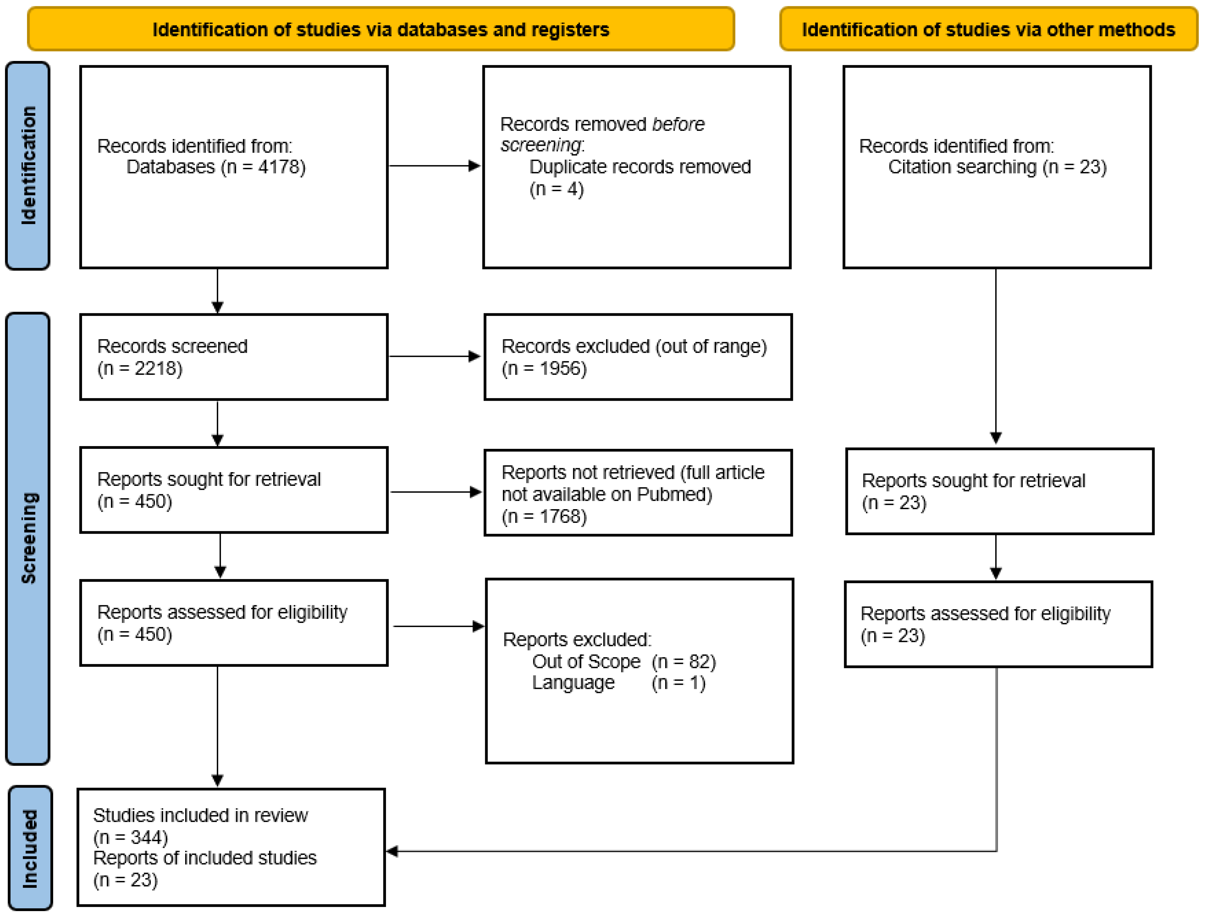
2. Materials and Methods
3. Results
3.1. Glutathione
3.2. Minocycline
3.3. Spermidine
3.4. Fisetin
3.5. Omega-3
- DHA {cis-4,cis-7,cis-10,cis-13,cis-16,cis-19- docosahexaenoic acid};
- DPA {cis-7,cis-10,cis-13,cis-16,cis-19- docosapentaenoic acid};
- EPA {cis-5,cis-8,cis-11,cis-14,cis-17- eicosapentaenoic acid}; as well as
- SDA: stearidonic acid {cis-6,cis-9,cis-12,cis-15- octadecatetraenoic acid} and;
- Short-chain ALA: alpha-linolenic acid {cis-9,cis-12,cis-15-octadecatrienoic acid}.
3.6. Rapamycin
3.7. Metformin
3.8. Alpha-Ketoglutarate
3.9. Vitamin B3 (Niacin)
3.10. Vitamin D
3.11. Zeaxanthin
3.12. Lutein
3.13. Resveratrol
3.14. Pyruvate
3.15. Vitamin A
3.16. Vitamin B1
3.17. Vitamin B2 (Riboflavin)
3.18. Vitamin B9
Vitamin B12 (Cobalamin)
3.19. Vitamin C
3.20. Vitamin E
3.21. Citicoline
3.22. Quercetin
3.23. Eyebright
4. Discussion

| Name | IUPAC | Sources | Chemical Structure |
|---|---|---|---|
| Glutathione | (2S)-2-amino-5-[[(2R)-1-(carboxymethylamino)-1-oxo-3-sulfanylpropan-2-yl]amino]-5-oxopentanoic acid | Avocados, okra, spinach |  [373] |
| Minocycline | (2E,4S,4aR,5aS,12aR)-2-(Amino-hydroxy-methylidene)-4,7-bis(dimethylamino)-10,11,12a-trihydroxy-4a,5,5a,6-tetrahydro-4H-tetracene-1,3,12-trione | Tetracycline antibiotic |  [374] |
| Spermidine | N-(3-Aminopropyl)butane-1,4-diamine | Soy protein, legumes, grain |  [375] |
| Fisetin | 2-(3,4-Dihydroxyphenyl)-3,7-dihydroxychromen-4-one | Apples, onions, cucumbers |  [376] |
| Omega-3s | (4Z,7Z,10Z,13Z,16Z,19Z)-docosa-4,7,10,13,16,19-hexaenoic acid;(5Z,8Z,11Z,14Z,17Z)-icosa-5,8,11,14,17-pentaenoic acid;(9Z,12Z,15Z)-octadeca-9,12,15-trienoic acid | Cod liver oil, salmon, mackerel, flaxseed |  [377] |
| Rapamycin | (1R,9S,12S,15R,16E,18R,19R,21R,23S,24E,26E,28E,30S,32S,35R)-1,18-dihydroxy-12-[(2R)-1-[(1S,3R,4R)-4-hydroxy-3-methoxycyclohexyl]propan-2-yl]-19,30-dimethoxy-15,17,21,23,29,35-hexamethyl-11,36-dioxa-4-azatricyclo[30.3.1.04,9]hexatriaconta-16,24,26,28-tetraene-2,3,10,14,20-pentone | Macrolide compound |  [378] |
| Metformin | 3-(diaminomethylidene)-1,1-dimethylguanidine | French lilac |  [379] |
| Alpha ketoglutarate | 2-Oxopentanedioic acid | Metabolite |  [380] |
| Vitamin B3 (niacin) | pyridine-3-carboxylic acid | Meats, nuts, legumes, bananas |  [381] |
| Vitamin D | (3β,5Z,7E,22E)-9,10-secoergosta-5,7,10(19),22-tetraen-3-ol | Sardines, tuna fish |  [382] |
| Lutein | β,ε-carotene-3,3’-diol | Green leafy vegetables such as kale and spinach |  [383] |
| Zeaxanthin | β,β-carotene-3,3′-diol | Green leafy vegetables such as broccoli and spinach |  [384] |
| Resveratrol | 3,5,4′-Trihydroxystilbene | Grapes and berries |  [385] |
| Pyruvate | 2-oxopropanoic acid | Metabolite |  [386] |
| Vitamin A (retinol) | (2E,4E,6E,8E)-3,7-dimethyl-9-(2,6,6-trimethylcyclohexen-1-yl)nona-2,4,6,8-tetraen-1-ol | Liver, eggs |  [387] |
| Vitamin B1 (thiamin) | 2-[3-[(4-amino-2-methylpyrimidin-5-yl)methyl]-4-methyl-1,3-thiazol-3-ium-5-yl]ethanol | Wholegrain, fish |  [388] |
| Vitamin B2 (riboflavin) | 7,8-dimethyl-10-[(2S,3S,4R)-2,3,4,5-tetrahydroxypentyl]benzo[g]pteridine-2,4-dione | Dairy, cheese, lean beef |  [389] |
| Vitamin B6 (folate) | (2S)-2-[[4-[(2-amino-4-oxo-3H-pteridin-6-yl)methylamino]benzoyl]amino]pentanedioic acid | Green leafy vegetables, fortified cereals |  [390] |
| Vitamin B12 (cobalamin) | cobalt(3+);[(2R,3S,4R,5S)-5-(5,6-dimethylbenzimidazol-1-yl)-4-hydroxy-2-(hydroxymethyl)oxolan-3-yl] [(2R)-1-[3-[(1R,2R,3R,4Z,7S,9Z,12S,13S,14Z,17S,18S,19R)-2,13,18-tris(2-amino-2-oxoethyl)-7,12,17-tris(3-amino-3-oxopropyl)-3,5,8,8,13,15,18,19-octamethyl-2,7,12,17-tetrahydro-1H-corrin-21-id-3-yl]propanoylamino]propan-2-yl] phosphate;cyanide | Seafood and dairy products |  [391] |
| Vitamin C (ascorbate) | (2R)-2-[(1S)-1,2-dihydroxyethyl]-3,4-dihydroxy-2H-furan-5-one | Citrus fruits |  [392] |
| Vitamin E | (2R)-2,5,7,8-tetramethyl-2-[(4R,8R)-4,8,12-trimethyltridecyl]-3,4-dihydrochromen-6-ol | Wheat germ oil; sunflower, safflower, and soybean oil; sunflower seeds, almonds; peanuts, peanut butter. |  [393] |
| Citicoline | [[(2R,3S,4R,5R)-5-(4-amino-2-oxopyrimidin-1-yl)-3,4-dihydroxyoxolan-2-yl]methoxy-hydroxyphosphoryl] 2-(trimethylazaniumyl)ethyl phosphate | Organ meats; cauliflower, broccoli |  [394] |
| Quercetin | 2-(3,4-dihydroxyphenyl)-3,5,7-trihydroxychromen-4-one | Citrus, red wine, apples, onions, sage, parsley |  [395] |
Author Contributions
Funding
Institutional Review Board Statement
Informed Consent Statement
Data Availability Statement
Conflicts of Interest
References
- Thomas, S.; Hodge, W.; Malvankar-Mehta, M. The Cost-Effectiveness Analysis of Teleglaucoma Screening Device. PLoS ONE 2015, 10, e0137913. [Google Scholar] [CrossRef] [PubMed]
- McMonnies, C.W. Glaucoma history and risk factors. J. Optom. 2017, 10, 71–78. [Google Scholar] [CrossRef] [PubMed] [Green Version]
- Skorin, L., Jr.; Knutson, R. Ophthalmic Diseases in Patients With Obstructive Sleep Apnea. J. Am. Osteopath. Assoc. 2016, 116, 522–529. [Google Scholar] [CrossRef] [PubMed] [Green Version]
- Emanuel, M.E.; Gedde, S.J. Indications for a systemic work-up in glaucoma. Can. J. Ophthalmol. 2014, 49, 506–511. [Google Scholar] [CrossRef]
- Zeppieri, M. Pigment dispersion syndrome: A brief overview. J. Clin. Transl. Res. 2022, 8, 344–350. [Google Scholar]
- Brusini, P.; Tosoni, C.; Zeppieri, M. Canaloplasty in Corticosteroid-Induced Glaucoma. Preliminary Results. J. Clin. Med. 2018, 7, 31. [Google Scholar] [CrossRef] [Green Version]
- Zeppieri, M.; Gurnani, B. Applanation Tonometry; StatPearls: Treasure Island, FL, USA, 2022. [Google Scholar]
- Bader, J.; Zeppieri, M.; Havens, S.J. Tonometry; StatPearls: Treasure Island, FL, USA, 2022. [Google Scholar]
- Rein, D.B.; Zhang, P.; Wirth, K.E.; Lee, P.P.; Hoerger, T.J.; McCall, N.; Klein, R.; Tielsch, J.M.; Vijan, S.; Saaddine, J. The economic burden of major adult visual disorders in the United States. Arch. Ophthalmol. 2006, 124, 1754–1760. [Google Scholar] [CrossRef] [Green Version]
- Varma, R.; Lee, P.P.; Goldberg, I.; Kotak, S. An assessment of the health and economic burdens of glaucoma. Am. J. Ophthalmol. 2011, 152, 515–522. [Google Scholar] [CrossRef] [Green Version]
- Morris, A.L.; Mohiuddin, S.S. Biochemistry, Nutrients; StatPearls: Treasure Island, FL, USA, 2022. [Google Scholar]
- Huang, L.; Drake, V.J.; Ho, E. Zinc. Adv. Nutr. 2015, 6, 224–226. [Google Scholar] [CrossRef] [Green Version]
- Santiago, P. Ferrous versus ferric oral iron formulations for the treatment of iron deficiency: A clinical overview. Sci. World J. 2012, 2012, 846824. [Google Scholar] [CrossRef]
- Christakos, S.; Dhawan, P.; Verstuyf, A.; Verlinden, L.; Carmeliet, G. Vitamin D: Metabolism, Molecular Mechanism of Action, and Pleiotropic Effects. Physiol. Rev. 2016, 96, 365–408. [Google Scholar] [CrossRef] [Green Version]
- Farzan, M.; Farzan, M.; Shahrani, M.; Navabi, S.P.; Vardanjani, H.R.; Amini-Khoei, H.; Shabani, S. Neuroprotective properties of Betulin, Betulinic acid, and Ursolic acid as triterpenoids derivatives: A comprehensive review of mechanistic studies. Nutr. Neurosci. 2023, 1–18. [Google Scholar] [CrossRef]
- Smith, T.J.; McClung, J.P. Nutrition, Immune Function, and Infectious Disease. Med. J. 2021, PB 8-21-01/02/03, 133–136. [Google Scholar]
- Psihogios, A.; Madampage, C.; Faught, B.E. Contemporary nutrition-based interventions to reduce risk of infection among elderly long-term care residents: A scoping review. PLoS ONE 2022, 17, e0272513. [Google Scholar] [CrossRef]
- Cassotta, M.; Forbes-Hernandez, T.Y.; Calderon Iglesias, R.; Ruiz, R.; Elexpuru Zabaleta, M.; Giampieri, F.; Battino, M. Links between Nutrition, Infectious Diseases, and Microbiota: Emerging Technologies and Opportunities for Human-Focused Research. Nutrients 2020, 12, 1827. [Google Scholar] [CrossRef]
- Plaza-Diaz, J. Nutrition, Microbiota and Noncommunicable Diseases. Nutrients 2020, 12, 1971. [Google Scholar] [CrossRef]
- Vaz-Rodrigues, R.; Mazuecos, L.; Villar, M.; Urra, J.M.; Gortazar, C.; de la Fuente, J. Serum biomarkers for nutritional status as predictors in COVID-19 patients before and after vaccination. J. Funct. Foods 2023, 101, 105412. [Google Scholar] [CrossRef]
- Boadi-Kusi, S.B.; Asiamah, E.; Ocansey, S.; Abu, S.L. Nutrition knowledge and dietary patterns in ophthalmic patients. Clin. Exp. Optom. 2021, 104, 78–84. [Google Scholar] [CrossRef]
- Tang, D.; Mitchell, P.; Liew, G.; Burlutsky, G.; Flood, V.; Gopinath, B. Evaluation of a Novel Tool for Screening Inadequate Food Intake in Age-Related Macular Degeneration Patients. Nutrients 2019, 11, 3031. [Google Scholar] [CrossRef] [Green Version]
- Dave, S.; Binns, A.; Vinuela-Navarro, V.; Callaghan, T. What Advice Is Currently Given to Patients with Age-Related Macular Degeneration (AMD) by Eyecare Practitioners, and How Effective Is It at Bringing about a Change in Lifestyle? A Systematic Review. Nutrients 2022, 14, 4652. [Google Scholar] [CrossRef]
- Rolando, M.; Barabino, S. Dry Eye Disease: What Is the Role of Vitamin D? Int. J. Mol. Sci. 2023, 24, 1458. [Google Scholar] [CrossRef] [PubMed]
- Page, M.J.; McKenzie, J.E.; Bossuyt, P.M.; Boutron IHoffmann, T.C.; Mulrow, C.D.; Shamseer, L.; Tetzlaff, J.M.; Akl, E.A.; Brennan, S.E.; Moher, D. The PRISMA 2020 statement: An updated guideline for reporting systematic reviews. Syst. Rev. 2021, 10, 89. [Google Scholar] [CrossRef] [PubMed]
- Park, M.H.; Moon, J. Circulating total glutathione in normal tension glaucoma patients: Comparison with normal control subjects. Korean J. Ophthalmol. 2012, 26, 84–91. [Google Scholar] [CrossRef] [PubMed] [Green Version]
- Yabana, T.; Sato, K.; Shiga, Y.; Himori, N.; Omodaka, K.; Nakazawa, T. The relationship between glutathione levels in leukocytes and ocular clinical parameters in glaucoma. PLoS ONE 2019, 14, e0227078. [Google Scholar] [CrossRef] [PubMed] [Green Version]
- Gherghel, D.; Mroczkowska, S.; Qin, L. Reduction in blood glutathione levels occurs similarly in patients with primary-open angle or normal tension glaucoma. Investig. Ophthalmol. Vis. Sci. 2013, 54, 3333–3339. [Google Scholar] [CrossRef]
- Levkovitch-Verbin, H.; Waserzoog, Y.; Vander, S.; Makarovsky, D.; Piven, I. Minocycline upregulates pro-survival genes and downregulates pro-apoptotic genes in experimental glaucoma. Graefes. Arch. Clin. Exp. Ophthalmol. 2014, 252, 761–772. [Google Scholar] [CrossRef]
- Downie, L.E.; Vingrys, A.J. Oral Omega-3 Supplementation Lowers Intraocular Pressure in Normotensive Adults. Transl. Vis. Sci. Technol. 2018, 7, 1. [Google Scholar] [CrossRef] [Green Version]
- Wang, F.; Ma, F.; Song, Y.; Li, N.; Li, X.; Pang, Y.; Hu, P.; Shao, A.; Deng, C.; Zhang, X. Topical administration of rapamycin promotes retinal ganglion cell survival and reduces intraocular pressure in a rat glaucoma model. Eur. J. Pharmacol. 2020, 884, 173369. [Google Scholar] [CrossRef]
- Xu, L.; Zhang, X.; Zhao, Y.; Gang, X.; Zhou, T.; Han, J.; Cao, Y.; Qi, B.; Song, S.; Wang, X.; et al. Metformin protects trabecular meshwork against oxidative injury via activating integrin/ROCK signals. eLife 2023, 12, e81198. [Google Scholar] [CrossRef]
- Gaynon, M.W.; Paulus, Y.M.; Rahimy, E.; Alexander, J.L.; Mansour, S.E. Effect of oral niacin on central retinal vein occlusion. Graefes. Arch. Clin. Exp. Ophthalmol. 2017, 255, 1085–1092. [Google Scholar] [CrossRef]
- Biswal, M.R.; Justis, B.D.; Han, P.; Li, H.; Gierhart, D.; Dorey, C.K.; Lewin, A.S. Daily zeaxanthin supplementation prevents atrophy of the retinal pigment epithelium (RPE) in a mouse model of mitochondrial oxidative stress. PLoS ONE 2018, 13, e0203816. [Google Scholar] [CrossRef] [Green Version]
- Schnebelen-Berthier, C.; Acar, N.; Simon, E.; Thabuis, C.; Bourdillon, A.; Mathiaud, A.; Dauchet, L.; Delcourt, C.; Benlian, P.; Crochet, M.; et al. The ALGOVUE Clinical Trial: Effects of the Daily Consumption of Eggs Enriched with Lutein and Docosahexaenoic Acid on Plasma Composition and Macular Pigment Optical Density. Nutrients 2021, 13, 3347. [Google Scholar] [CrossRef]
- Zhang, L.; Guo, K.; Tian, Q.; Ye, J.; Ding, Z.; Zhou, Q.; Li, X.; Zhou, Z.; Yang, L. Serum Metabolomics Reveals a Potential Benefit of Methionine in Type 1 Diabetes Patients with Poor Glycemic Control and High Glycemic Variability. Nutrients 2023, 15, 518. [Google Scholar] [CrossRef]
- Kong, Y.; Liu, P.K.; Li, Y.; Nolan, N.D.; Quinn, P.M.J.; Hsu, C.W.; Jenny, L.A.; Zhao, J.; Cui, X.; Chang, Y.J.; et al. HIF2alpha activation and mitochondrial deficit due to iron chelation cause retinal atrophy. EMBO Mol. Med. 2023, 15, e16525. [Google Scholar] [CrossRef]
- Hytti, M.; Piippo, N.; Korhonen, E.; Honkakoski, P.; Kaarniranta, K.; Kauppinen, A. Fisetin and luteolin protect human retinal pigment epithelial cells from oxidative stress-induced cell death and regulate inflammation. Sci. Rep. 2015, 5, 17645. [Google Scholar] [CrossRef] [Green Version]
- Zhao, L.; Wang, H.; Du, X. The therapeutic use of quercetin in ophthalmology: Recent applications. Biomed. Pharmacother. 2021, 137, 111371. [Google Scholar] [CrossRef]
- Gao, F.J.; Zhang, S.H.; Xu, P.; Yang, B.Q.; Zhang, R.; Cheng, Y.; Zhou, X.J.; Huang, W.J.; Wang, M.; Chen, J.Y.; et al. Quercetin Declines Apoptosis, Ameliorates Mitochondrial Function and Improves Retinal Ganglion Cell Survival and Function in In Vivo Model of Glaucoma in Rat and Retinal Ganglion Cell Culture In Vitro. Front. Mol. Neurosci. 2017, 10, 285. [Google Scholar] [CrossRef] [Green Version]
- Zhou, X.; Li, G.; Yang, B.; Wu, J. Quercetin Enhances Inhibitory Synaptic Inputs and Reduces Excitatory Synaptic Inputs to OFF- and ON-Type Retinal Ganglion Cells in a Chronic Glaucoma Rat Model. Front. Neurosci. 2019, 13, 672. [Google Scholar] [CrossRef]
- Pizzorno, J. Glutathione! Integr. Med. 2014, 13, 8–12. [Google Scholar]
- Pompella, A.; Visvikis, A.; Paolicchi, A.; De Tata, V.; Casini, A.F. The changing faces of glutathione, a cellular protagonist. Biochem. Pharmacol. 2003, 66, 1499–1503. [Google Scholar] [CrossRef]
- Julius, M.; Lang, C.A.; Gleiberman, L.; Harburg, E.; DiFranceisco, W.; Schork, A. Glutathione and morbidity in a community-based sample of elderly. J. Clin. Epidemiol. 1994, 47, 1021–1026. [Google Scholar] [CrossRef] [PubMed] [Green Version]
- Sekhar, K.R.; Hanna, D.N.; Cyr, S.; Baechle, J.J.; Kuravi, S.; Balusu, R.; Rathmell, K.; Baregamian, N. Glutathione peroxidase 4 inhibition induces ferroptosis and mTOR pathway suppression in thyroid cancer. Sci. Rep. 2022, 12, 19396. [Google Scholar] [CrossRef] [PubMed]
- Singh, B.; Eshaghian, E.; Chuang, J.; Covasa, M. Do Diet and Dietary Supplements Mitigate Clinical Outcomes in COVID-19? Nutrients 2022, 14, 1909. [Google Scholar] [CrossRef] [PubMed]
- Weschawalit, S.; Thongthip, S.; Phutrakool, P.; Asawanonda, P. Glutathione and its antiaging and antimelanogenic effects. Clin. Cosmet. Investig. Dermatol. 2017, 10, 147–153. [Google Scholar] [CrossRef] [Green Version]
- Sreekumar, P.G.; Ferrington, D.A.; Kannan, R. Glutathione Metabolism and the Novel Role of Mitochondrial GSH in Retinal Degeneration. Antioxidants 2021, 10, 661. [Google Scholar] [CrossRef]
- Garrido-Mesa, N.; Zarzuelo, A.; Galvez, J. Minocycline: Far beyond an antibiotic. Br. J. Pharmacol. 2013, 169, 337–352. [Google Scholar] [CrossRef] [Green Version]
- Rogers, R.L.; Perkins, J. Skin and soft tissue infections. Prim. Care 2006, 33, 697–710. [Google Scholar] [CrossRef]
- Colton, B.; McConeghy, K.W.; Schreckenberger, P.C.; Danziger, L.H.I.V. minocycline revisited for infections caused by multidrug-resistant organisms. Am. J. Health Syst. Pharm. 2016, 73, 279–285. [Google Scholar] [CrossRef]
- Xu, L.; Fagan, S.C.; Waller, J.L.; Edwards, D.; Borlongan, C.V.; Zheng, J.; Hill, W.D.; Feuerstein, G.; Hess, D.C. Low dose intravenous minocycline is neuroprotective after middle cerebral artery occlusion-reperfusion in rats. BMC Neurol. 2004, 4, 7. [Google Scholar] [CrossRef] [Green Version]
- Garner, S.E.; Eady, A.; Bennett, C.; Newton, J.N.; Thomas, K.; Popescu, C.M. Minocycline for acne vulgaris: Efficacy and safety. Cochrane Database Syst. Rev. 2012, 2012, CD002086. [Google Scholar] [CrossRef]
- Grotegut, P.; Perumal, N.; Kuehn, S.; Smit, A.; Dick, H.B.; Grus, F.H.; Joachim, S.C. Minocycline reduces inflammatory response and cell death in a S100B retina degeneration model. J. Neuroinflamm. 2020, 17, 375. [Google Scholar] [CrossRef]
- Tsai, T.; Joachim, S.C. Glaucoma-like damage induced by S100B injection is accompanied by microglial response. Neural. Regen. Res. 2022, 17, 572–574. [Google Scholar]
- Kuehn, S.; Meissner, W.; Grotegut, P.; Theiss, C.; Dick, H.B.; Joachim, S.C. Intravitreal S100B Injection Leads to Progressive Glaucoma Like Damage in Retina and Optic Nerve. Front. Cell. Neurosci. 2018, 12, 312. [Google Scholar] [CrossRef] [Green Version]
- Soda, K. Overview of Polyamines as Nutrients for Human Healthy Long Life and Effect of Increased Polyamine Intake on DNA Methylation. Cells 2022, 11, 164. [Google Scholar] [CrossRef]
- Madeo, F.; Hofer, S.J.; Pendl, T.; Bauer, M.A.; Eisenberg, T.; Carmona-Gutierrez, D.; Kroemer, G. Nutritional Aspects of Spermidine. Annu. Rev. Nutr. 2020, 40, 135–159. [Google Scholar] [CrossRef]
- Madeo, F.; Bauer, M.A.; Carmona-Gutierrez, D.; Kroemer, G. Spermidine: A physiological autophagy inducer acting as an anti-aging vitamin in humans? Autophagy 2019, 15, 165–168. [Google Scholar] [CrossRef]
- Hofer, S.J.; Liang, Y.; Zimmermann, A.; Schroeder, S.; Dengjel, J.; Kroemer, G.; Eisenberg, T.; Sigrist, S.J.; Madeo, F. Spermidine-induced hypusination preserves mitochondrial and cognitive function during aging. Autophagy 2021, 17, 2037–2039. [Google Scholar] [CrossRef] [PubMed]
- Brito, S.; Heo, H.; Cha, B.; Lee, S.H.; Chae, S.; Lee, M.G.; Kwak, B.M.; Bin, B.H. A systematic exploration reveals the potential of spermidine for hypopigmentation treatment through the stabilization of melanogenesis-associated proteins. Sci. Rep. 2022, 12, 14478. [Google Scholar] [CrossRef]
- Lefevre, P.L.; Palin, M.F.; Murphy, B.D. Polyamines on the reproductive landscape. Endocr. Rev. 2011, 32, 694–712. [Google Scholar] [CrossRef] [Green Version]
- Madeo, F.; Eisenberg, T.; Pietrocola, F.; Kroemer, G. Spermidine in health and disease. Science 2018, 359, eaan2788. [Google Scholar] [CrossRef] [Green Version]
- Yousefi-Manesh, H.; Shirooie, S.; Noori, T.; Sheibani, M.; Tavangar, S.M.; Hemmati, S.; Sadeghi, M.A.; Akbarniakhaky, H.; Mohammadi, Z.; Foroutani, L.; et al. Spermidine reduced neuropathic pain in chronic constriction injury-induced peripheral neuropathy in rats. Fundam. Clin. Pharmacol. 2023. [Google Scholar] [CrossRef] [PubMed]
- Han, W.; Li, H.; Chen, B. Research Progress and Potential Applications of Spermidine in Ocular Diseases. Pharmaceutics 2022, 14, 1500. [Google Scholar] [CrossRef] [PubMed]
- Leruez, S.; Marill, A.; Bresson, T.; de Saint Martin, G.; Buisset, A.; Muller, J.; Tessier, L.; Gadras, C.; Verny, C.; Gohier, P.; et al. A Metabolomics Profiling of Glaucoma Points to Mitochondrial Dysfunction, Senescence, and Polyamines Deficiency. Investig. Ophthalmol. Vis. Sci. 2018, 59, 4355–4361. [Google Scholar] [CrossRef] [Green Version]
- Buisset, A.; Gohier, P.; Leruez, S.; Muller, J.; Amati-Bonneau, P.; Lenaers, G.; Bonneau, D.; Simard, G.; Procaccio, V.; Annweiler, C.; et al. Metabolomic Profiling of Aqueous Humor in Glaucoma Points to Taurine and Spermine Deficiency: Findings from the Eye-D Study. J. Proteome Res. 2019, 18, 1307–1315. [Google Scholar] [CrossRef]
- Lillo, A.; Marin, S.; Serrano-Marin, J.; Binetti, N.; Navarro, G.; Cascante, M.; Sanchez-Naves, J.; Franco, R. Targeted Metabolomics Shows That the Level of Glutamine, Kynurenine, Acyl-Carnitines and Lysophosphatidylcholines Is Significantly Increased in the Aqueous Humor of Glaucoma Patients. Front. Med. 2022, 9, 935084. [Google Scholar] [CrossRef]
- Wang, Y.; Hou, X.W.; Liang, G.; Pan, C.W. Metabolomics in Glaucoma: A Systematic Review. Investig. Ophthalmol. Vis. Sci. 2021, 62, 9. [Google Scholar] [CrossRef]
- Yousefzadeh, M.J.; Zhu, Y.; McGowan, S.J.; Angelini, L.; Fuhrmann-Stroissnigg, H.; Xu, M.; Ling, Y.Y.; Melos, K.I.; Pirtskhalava, T.; Inman, C.L.; et al. Fisetin is a senotherapeutic that extends health and lifespan. EBioMedicine 2018, 36, 18–28. [Google Scholar] [CrossRef] [Green Version]
- Syed, D.N.; Adhami, V.M.; Khan, M.I.; Mukhtar, H. Inhibition of Akt/mTOR signaling by the dietary flavonoid fisetin. Anticancer Agents Med. Chem. 2013, 13, 995–1001. [Google Scholar] [CrossRef]
- Salerno, S.; Da Settimo, F.; Taliani, S.; Simorini, F.; La Motta, C.; Fornaciari, G.; Marini, A.M. Recent advances in the development of dual topoisomerase I and II inhibitors as anticancer drugs. Curr. Med. Chem. 2010, 17, 4270–4290. [Google Scholar] [CrossRef]
- Wyld, L.; Bellantuono, I.; Tchkonia, T.; Morgan, J.; Turner, O.; Foss, F.; George, J.; Danson, S.; Kirkland, J.L. Senescence and Cancer: A Review of Clinical Implications of Senescence and Senotherapies. Cancers 2020, 12, 2134. [Google Scholar] [CrossRef]
- Chou, R.H.; Hsieh, S.C.; Yu, Y.L.; Huang, M.H.; Huang, Y.C.; Hsieh, Y.H. Fisetin inhibits migration and invasion of human cervical cancer cells by down-regulating urokinase plasminogen activator expression through suppressing the p38 MAPK-dependent NF-kappaB signaling pathway. PLoS ONE 2013, 8, e71983. [Google Scholar] [CrossRef] [PubMed] [Green Version]
- Rengarajan, T.; Yaacob, N.S. The flavonoid fisetin as an anticancer agent targeting the growth signaling pathways. Eur. J. Pharmacol. 2016, 789, 8–16. [Google Scholar] [CrossRef]
- Pal, H.C.; Pearlman, R.L.; Afaq, F. Fisetin and Its Role in Chronic Diseases. Adv. Exp. Med. Biol. 2016, 928, 213–244. [Google Scholar]
- Khan, N.; Syed, D.N.; Ahmad, N.; Mukhtar, H. Fisetin: A dietary antioxidant for health promotion. Antioxid. Redox Signal. 2013, 19, 151–162. [Google Scholar] [CrossRef]
- Kubina, R.; Krzykawski, K.; Kabala-Dzik, A.; Wojtyczka, R.D.; Chodurek, E.; Dziedzic, A. Fisetin, a Potent Anticancer Flavonol Exhibiting Cytotoxic Activity against Neoplastic Malignant Cells and Cancerous Conditions: A Scoping, Comprehensive Review. Nutrients 2022, 14, 2604. [Google Scholar] [CrossRef]
- Lall, R.K.; Adhami, V.M.; Mukhtar, H. Dietary flavonoid fisetin for cancer prevention and treatment. Mol. Nutr. Food Res. 2016, 60, 1396–1405. [Google Scholar] [CrossRef]
- Noh, E.M.; Park, Y.J.; Kim, J.M.; Kim, M.S.; Kim, H.R.; Song, H.K.; Hong, O.Y.; So, H.S.; Yang, S.H.; Kim, J.S.; et al. Fisetin regulates TPA-induced breast cell invasion by suppressing matrix metalloproteinase-9 activation via the PKC/ROS/MAPK pathways. Eur. J. Pharmacol. 2015, 764, 79–86. [Google Scholar] [CrossRef]
- Tsiklauri, L.; An, G.; Ruszaj, D.M.; Alaniya, M.; Kemertelidze, E.; Morris, M.E. Simultaneous determination of the flavonoids robinin and kaempferol in human breast cancer cells by liquid chromatography-tandem mass spectrometry. J. Pharm. Biomed. Anal. 2011, 55, 109–113. [Google Scholar] [CrossRef]
- Ying, T.H.; Yang, S.F.; Tsai, S.J.; Hsieh, S.C.; Huang, Y.C.; Bau, D.T.; Hsieh, Y.H. Fisetin induces apoptosis in human cervical cancer HeLa cells through ERK1/2-mediated activation of caspase-8-/caspase-3-dependent pathway. Arch. Toxicol. 2012, 86, 263–273. [Google Scholar] [CrossRef]
- Chen, Y.; Wu, Q.; Song, L.; He, T.; Li, Y.; Li, L.; Su, W.; Liu, L.; Qian, Z.; Gong, C. Polymeric micelles encapsulating fisetin improve the therapeutic effect in colon cancer. ACS Appl. Mater. Interfaces 2015, 7, 534–542. [Google Scholar] [CrossRef]
- Khan, N.; Asim, M.; Afaq, F.; Abu Zaid, M.; Mukhtar, H. A novel dietary flavonoid fisetin inhibits androgen receptor signaling and tumor growth in athymic nude mice. Cancer Res. 2008, 68, 8555–8563. [Google Scholar] [CrossRef] [PubMed] [Green Version]
- Lai, M.; Lan, C.; Zhong, J.; Wu, L.; Lin, C. Fisetin Prevents Angiogenesis in Diabetic Retinopathy by Downregulating VEGF. J. Ophthalmol. 2023, 2023, 7951928. [Google Scholar] [CrossRef]
- Li, L.; Qin, J.; Fu, T.; Shen, J. Fisetin rescues retinal functions by suppressing inflammatory response in a DBA/2J mouse model of glaucoma. Doc. Ophthalmol. 2019, 138, 125–135. [Google Scholar] [CrossRef] [PubMed]
- Wang, K.; Hu, D.N.; Lin, H.W.; Yang, W.E.; Hsieh, Y.H.; Chien, H.W.; Yang, S.F. Fisetin induces apoptosis through mitochondrial apoptosis pathway in human uveal melanoma cells. Environ. Toxicol. 2018, 33, 527–534. [Google Scholar] [CrossRef]
- Kan, E.; Kilickan, E.; Ayar, A.; Colak, R. Effects of two antioxidants; alpha-lipoic acid and fisetin against diabetic cataract in mice. Int. Ophthalmol. 2015, 35, 115–120. [Google Scholar] [CrossRef]
- Abete, P.; Testa, G.; Galizia, G.; Della-Morte, D.; Cacciatore, F.; Rengo, F. PUFA for human health: Diet or supplementation? Curr. Pharm. Des. 2009, 15, 4186–4190. [Google Scholar] [CrossRef]
- Rodriguez-Leyva, D.; Dupasquier, C.M.; McCullough, R.; Pierce, G.N. The cardiovascular effects of flaxseed and its omega-3 fatty acid, alpha-linolenic acid. Can. J. Cardiol. 2010, 26, 489–496. [Google Scholar] [CrossRef] [Green Version]
- Ishihara, T.; Yoshida, M.; Arita, M. Omega-3 fatty acid-derived mediators that control inflammation and tissue homeostasis. Int. Immunol. 2019, 31, 559–567. [Google Scholar] [CrossRef] [Green Version]
- Arnold, C.; Konkel, A.; Fischer, R.; Schunck, W.H. Cytochrome P450-dependent metabolism of omega-6 and omega-3 long-chain polyunsaturated fatty acids. Pharmacol. Rep. 2010, 62, 536–547. [Google Scholar] [CrossRef]
- Gioxari, A.; Kaliora, A.C.; Marantidou, F.; Panagiotakos, D.P. Intake of omega-3 polyunsaturated fatty acids in patients with rheumatoid arthritis: A systematic review and meta-analysis. Nutrition 2018, 45, 114–124.e4. [Google Scholar] [CrossRef]
- Backes, J.; Anzalone, D.; Hilleman, D.; Catini, J. The clinical relevance of omega-3 fatty acids in the management of hypertriglyceridemia. Lipids Health Dis. 2016, 15, 118. [Google Scholar] [CrossRef] [PubMed] [Green Version]
- Larrieu, T.; Laye, S. Food for Mood: Relevance of Nutritional Omega-3 Fatty Acids for Depression and Anxiety. Front. Physiol. 2018, 9, 1047. [Google Scholar] [CrossRef] [PubMed] [Green Version]
- Weir, N.L.; Guan, W.; Karger, A.B.; Klein, B.; Meuer, S.; Cotch, M.F.; Guo, X.; Li, X.; Tan, J.; Genter, P.; et al. Omega-3 fatty acids are associated with decreased presence and severity of diabetic retinopathy: A Combined Analysis of MESA and GOLDR Cohorts. Retina 2023, 10, 1097. [Google Scholar] [CrossRef] [PubMed]
- Britten-Jones, A.C.; Craig, J.P.; Downie, L.E. Omega-3 polyunsaturated fatty acids and corneal nerve health: Current evidence and future directions. Ocul. Surf. 2023, 27, 1–12. [Google Scholar] [CrossRef]
- Britten-Jones, A.C.; Craig, J.P.; Anderson, A.J.; Downie, L.E. Association between systemic omega-3 polyunsaturated fatty acid levels, and corneal nerve structure and function. Eye 2022. [Google Scholar] [CrossRef]
- Dietrich-Ntoukas, T. Chronic Blepharitis. Klin. Monbl. Augenheilkd. 2022, 239, 1381–1393. [Google Scholar]
- Tellez-Vazquez, J. Omega-3 fatty acid supplementation improves dry eye symptoms in patients with glaucoma: Results of a prospective multicenter study. Clin. Ophthalmol. 2016, 10, 617–626. [Google Scholar] [CrossRef] [Green Version]
- Kaercher, T.; Messmer, E.M.; Berninger, T.; Huber-van der Velden, K.K.; Geiger, R.; Cipriano-Bonvin, P.; Jacobi, C. Topical Omega-3 Polyunsaturated Fatty Acids for the Treatment of Dry Eye—Results from a Pilot Randomized Controlled Masked-Observer Study. Clin. Ophthalmol. 2022, 16, 4021–4031. [Google Scholar] [CrossRef]
- Pameijer, E.M.; Heus, P.; Damen, J.A.A.; Spijker, R.; Hooft, L.; Ringens, P.J.; Imhof, S.M.; van Leeuwen, R. What did we learn in 35 years of research on nutrition and supplements for age-related macular degeneration: A systematic review. Acta Ophthalmol. 2022, 100, e1541–e1552. [Google Scholar] [CrossRef]
- Csader, S.; Korhonen, S.; Kaarniranta, K.; Schwab, U. The Effect of Dietary Supplementations on Delaying the Progression of Age-Related Macular Degeneration: A Systematic Review and Meta-Analysis. Nutrients 2022, 14, 4273. [Google Scholar] [CrossRef]
- Carre, C.; Baudin, F.; Buteau, B.; Martine, L.; Gregoire, S.; Vasku, G.; Berdeaux, O.; Beduneau, A.; Pellequer, Y.; Jamoussi, J.; et al. Effects of topical docosahexaenoic acid on postoperative fibrosis in an animal model of glaucoma filtration surgery. Acta Ophthalmol. 2023, 101, e61–e68. [Google Scholar] [CrossRef]
- Nguyen, C.T.; Vingrys, A.J.; Bui, B.V. Dietary omega-3 deficiency and IOP insult are additive risk factors for ganglion cell dysfunction. J. Glaucoma 2013, 22, 269–277. [Google Scholar] [CrossRef]
- Sacca, S.C.; Vernazza, S.; Iorio, E.L.; Tirendi, S.; Bassi, A.M.; Gandolfi, S.; Izzotti, A. Molecular changes in glaucomatous trabecular meshwork. Correlations with retinal ganglion cell death and novel strategies for neuroprotection. Prog. Brain Res. 2020, 256, 151–188. [Google Scholar]
- Pinazo-Duran, M.D.; Shoaie-Nia, K.; Zanon-Moreno, V.; Sanz-Gonzalez, S.M.; Del Castillo, J.B.; Garcia-Medina, J.J. Strategies to Reduce Oxidative Stress in Glaucoma Patients. Curr. Neuropharmacol. 2018, 16, 903–918. [Google Scholar] [CrossRef]
- Medori, M.C.; Naureen, Z.; Dhuli, K.; Placidi, G.; Falsini, B.; Bertelli, M. Dietary supplements in retinal diseases, glaucoma, and other ocular conditions. J. Prev. Med. Hyg. 2022, 63 (Suppl. 3), E189–E199. [Google Scholar]
- Dziedziak, J.; Kasarello, K.; Cudnoch-Jedrzejewska, A. Dietary Antioxidants in Age-Related Macular Degeneration and Glaucoma. Antioxidants 2021, 10, 1743. [Google Scholar] [CrossRef]
- Kalogerou, M.; Kolovos, P.; Prokopiou, E.; Papagregoriou, G.; Deltas, C.; Malas, S.; Georgiou, T. Omega-3 fatty acids protect retinal neurons in the DBA/2J hereditary glaucoma mouse model. Exp. Eye Res. 2018, 167, 128–139. [Google Scholar] [CrossRef]
- Kalogerou, M.; Ioannou, S.; Kolovos, P.; Prokopiou, E.; Potamiti, L.; Kyriacou, K.; Panagiotidis, M.; Ioannou, M.; Fella, E.; Worth, E.P.; et al. Omega-3 fatty acids promote neuroprotection, decreased apoptosis and reduced glial cell activation in the retina of a mouse model of OPA1-related autosomal dominant optic atrophy. Exp. Eye Res. 2022, 215, 108901. [Google Scholar] [CrossRef]
- Georgiou, T.; Wen, Y.T.; Chang, C.H.; Kolovos, P.; Kalogerou, M.; Prokopiou, E.; Neokleous, A.; Huang, C.T.; Tsai, R.K. Neuroprotective Effects of Omega-3 Polyunsaturated Fatty Acids in a Rat Model of Anterior Ischemic Optic Neuropathy. Investig. Ophthalmol. Vis. Sci. 2017, 58, 1603–1611. [Google Scholar] [CrossRef] [Green Version]
- Maiese, K. Taking aim at Alzheimer’s disease through the mammalian target of rapamycin. Ann. Med. 2014, 46, 587–596. [Google Scholar] [CrossRef] [Green Version]
- Phillips, E.J.; Simons, M.J.P. Rapamycin not dietary restriction improves resilience against pathogens: A meta-analysis. Geroscience 2023, 45, 1263–1270. [Google Scholar] [CrossRef] [PubMed]
- Baroja-Mazo, A.; Revilla-Nuin, B.; Ramirez, P.; Pons, J.A. Immunosuppressive potency of mechanistic target of rapamycin inhibitors in solid-organ transplantation. World J. Transplant. 2016, 6, 183–192. [Google Scholar] [CrossRef] [PubMed]
- Liu, X.; Ye, M.; Ma, L. The emerging role of autophagy and mitophagy in tauopathies: From pathogenesis to translational implications in Alzheimer’s disease. Front. Aging Neurosci. 2022, 14, 1022821. [Google Scholar] [CrossRef] [PubMed]
- Lieberthal, W.; Levine, J.S. The role of the mammalian target of rapamycin (mTOR) in renal disease. J. Am. Soc. Nephrol. 2009, 20, 2493–2502. [Google Scholar] [CrossRef] [Green Version]
- Wu, L.Z.; Weng, Y.Q.; Ling, Y.X.; Zhou, S.J.; Ding, X.K.; Wu, S.Q.; Yu, K.; Jiang, S.F.; Chen, Y. A Web of Science-based scientometric analysis about mammalian target of rapamycin signaling pathway in kidney disease from 1986 to 2020. Transl. Androl. Urol. 2021, 10, 1006–1017. [Google Scholar] [CrossRef]
- Curatolo, P. Mechanistic target of rapamycin (mTOR) in tuberous sclerosis complex-associated epilepsy. Pediatr. Neurol. 2015, 52, 281–289. [Google Scholar] [CrossRef]
- Fang, J.; Pan, L.; Gu, Q.X.; Juengpanich, S.; Zheng, J.H.; Tong, C.H.; Wang, Z.Y.; Nan, J.J.; Wang, Y.F. Scientometric analysis of mTOR signaling pathway in liver disease. Ann. Transl. Med. 2020, 8, 93. [Google Scholar] [CrossRef]
- Zhang, M.; Chong, K.K.; Chen, Z.Y.; Guo, H.; Liu, Y.F.; Kang, Y.Y.; Li, Y.J.; Shi, T.T.; Lai, K.K.; He, M.Q.; et al. Rapamycin improves Graves’ orbitopathy by suppressing CD4+ cytotoxic T lymphocytes. J. Clin. Investig. 2023, 8, e160377. [Google Scholar] [CrossRef]
- Chang, S.; Perry, J.D.; Kosmorsky, G.S.; Braun, W.E. Rapamycin for treatment of refractory dysthyroid compressive optic neuropathy. Ophthalmic Plast. Reconstr. Surg. 2007, 23, 225–226. [Google Scholar] [CrossRef]
- Roos, J.C.P.; Murthy, R. Sirolimus (rapamycin) for the targeted treatment of the fibrotic sequelae of Graves’ orbitopathy. Eye 2019, 33, 679–682. [Google Scholar] [CrossRef] [Green Version]
- Kwon, Y.S.; Hong, H.S.; Kim, J.C.; Shin, J.S.; Son, Y. Inhibitory effect of rapamycin on corneal neovascularization in vitro and in vivo. Investig. Ophthalmol. Vis. Sci. 2005, 46, 454–460. [Google Scholar] [CrossRef] [Green Version]
- Wang, Y.D.; Su, Y.J.; Li, J.Y.; Yao, X.C.; Liang, G.J. Rapamycin, a mTOR inhibitor, induced growth inhibition in retinoblastoma Y79 cell via down-regulation of Bmi-1. Int. J. Clin. Exp. Pathol. 2015, 8, 5182–5188. [Google Scholar]
- Wang, Y.; Fung, N.S.K.; Lam, W.C.; Lo, A.C.Y. mTOR Signalling Pathway: A Potential Therapeutic Target for Ocular Neurodegenerative Diseases. Antioxidants 2022, 11, 1304. [Google Scholar] [CrossRef]
- Merle, D.A.; Provenzano, F.; Jarboui, M.A.; Kilger, E.; Clark, S.J.; Deleidi, M.; Armento, A.; Ueffing, M. mTOR Inhibition via Rapamycin Treatment Partially Reverts the Deficit in Energy Metabolism Caused by FH Loss in RPE Cells. Antioxidants 2021, 10, 1944. [Google Scholar] [CrossRef]
- Kida, T.; Oku, H.; Osuka, S.; Horie, T.; Ikeda, T. Hyperglycemia-induced VEGF and ROS production in retinal cells is inhibited by the mTOR inhibitor, rapamycin. Sci. Rep. 2021, 11, 1885. [Google Scholar] [CrossRef]
- Gidfar, S.; Milani, F.Y.; Milani, B.Y.; Shen, X.; Eslani, M.; Putra, I.; Huvard, M.J.; Sagha, H.; Djalilian, A.R. Rapamycin Prolongs the Survival of Corneal Epithelial Cells in Culture. Sci. Rep. 2017, 7, 40308. [Google Scholar] [CrossRef] [Green Version]
- Harder, J.M.; Guymer, C.; Wood, J.P.M.; Daskalaki, E.; Chidlow, G.; Zhang, C.; Balasubramanian, R.; Cardozo, B.H.; Foxworth, N.E.; Deering, K.E.; et al. Disturbed glucose and pyruvate metabolism in glaucoma with neuroprotection by pyruvate or rapamycin. Proc. Natl. Acad. Sci. USA 2020, 117, 33619–33627. [Google Scholar] [CrossRef]
- Li, N.; Wang, F.; Zhang, Q.; Jin, M.; Lu, Y.; Chen, S.; Guo, C.; Zhang, X. Rapamycin mediates mTOR signaling in reactive astrocytes and reduces rsetinal ganglion cell loss. Exp. Eye Res. 2018, 176, 10–19. [Google Scholar] [CrossRef]
- Asani, B.; Siedlecki, J.; Wertheimer, C.; Liegl, R.; Wolf, A.; Ohlmann, A.; Priglinger, S.; Priglinger, C. Anti-angiogenic properties of rapamycin on human retinal pericytes in an in vitro model of neovascular AMD via inhibition of the mTOR pathway. BMC Ophthalmol. 2022, 22, 138. [Google Scholar] [CrossRef]
- Hunter, R.W.; Hughey, C.C.; Lantier, L.; Sundelin, E.I.; Peggie, M.; Zeqiraj, E.; Sicheri, F.; Jessen, N.; Wasserman, D.H.; Sakamoto, K. Metformin reduces liver glucose production by inhibition of fructose-1-6-bisphosphatase. Nat. Med. 2018, 24, 1395–1406. [Google Scholar] [CrossRef] [Green Version]
- Apolzan, J.W.; Venditti, E.M.; Edelstein, S.L.; Knowler, W.C.; Dabelea, D.; Boyko, E.J.; Pi-Sunyer, X.; Kalyani, R.R.; Franks, P.W.; Srikanthan, P.; et al. Long-Term Weight Loss With Metformin or Lifestyle Intervention in the Diabetes Prevention Program Outcomes Study. Ann. Intern. Med. 2019, 170, 682–690. [Google Scholar] [CrossRef] [PubMed]
- Sun, Y.; Fang, K.; Hu, X.; Yang, J.; Jiang, Z.; Feng, L.; Li, R.; Rao, Y.; Shi, S.; Dong, C. NIR-light-controlled G-quadruplex hydrogel for synergistically enhancing photodynamic therapy via sustained delivery of metformin and catalase-like activity in breast cancer. Mater. Today Bio 2022, 16, 100375. [Google Scholar] [CrossRef] [PubMed]
- Faria, J.; Negalha, G.; Azevedo, A.; Martel, F. Metformin and Breast Cancer: Molecular Targets. J. Mammary Gland. Biol. Neoplasia 2019, 24, 111–123. [Google Scholar] [CrossRef] [PubMed]
- Dodd, J.M.; Grivell, R.M.; Deussen, A.R.; Hague, W.M. Metformin for women who are overweight or obese during pregnancy for improving maternal and infant outcomes. Cochrane Database Syst. Rev. 2018, 7, CD010564. [Google Scholar] [CrossRef] [PubMed]
- Calzia, D.; Degan, P.; Caicci, F.; Bruschi, M.; Manni, L.; Ramenghi, L.A.; Candiano, G.; Traverso, C.E.; Panfoli, I. Modulation of the rod outer segment aerobic metabolism diminishes the production of radicals due to light absorption. Free Radic. Biol. Med. 2018, 117, 110–118. [Google Scholar] [CrossRef] [Green Version]
- Kim, A.H.; Kolesnikova, M.; Ngo, W.K.; Tsang, S.H. Effects of medications on hypoxia-inducible factor in the retina: A review. Clin. Exp. Ophthalmol. 2023, 51, 205–216. [Google Scholar] [CrossRef]
- Bailey, C.J. Metformin, historical overview. Diabetologia 2017, 60, 1566–1576. [Google Scholar] [CrossRef] [Green Version]
- Maleskic, S.; Kusturica, J.; Gusic, E.; Rakanovic-Todic, M.; Secic, D.; Burnazovic-Ristic, L.; Kulo, A. Metformin use associated with protective effects for ocular complications in patients with type 2 diabetes—Observational study. Acta Med. Acad. 2017, 46, 116–123. [Google Scholar] [CrossRef]
- Asahi, M.G.; Avaylon, J.; Wallsh, J.; Gallemore, R.P. Emerging biological therapies for the treatment of age-related macular degeneration. Expert. Opin. Emerg. Drugs 2021, 26, 193–207. [Google Scholar] [CrossRef]
- Gokhale, K.M.; Adderley, N.J.; Subramanian, A.; Lee, W.H.; Han, D.; Coker, J.; Braithwaite, T.; Denniston, A.K.; Keane, P.A.; Nirantharakumar, K. Metformin and risk of age-related macular degeneration in individuals with type 2 diabetes: A retrospective cohort study. Br. J. Ophthalmol. 2022. [Google Scholar] [CrossRef]
- Gyanwali, B.; Lim, Z.X.; Soh, J.; Lim, C.; Guan, S.P.; Goh, J.; Maier, A.B.; Kennedy, B.K. Alpha-Ketoglutarate dietary supplementation to improve health in humans. Trends Endocrinol. Metab. 2022, 33, 136–146. [Google Scholar] [CrossRef]
- Liu, S.; He, L.; Yao, K. The Antioxidative Function of Alpha-Ketoglutarate and Its Applications. BioMed Res. Int. 2018, 2018, 3408467. [Google Scholar] [CrossRef]
- Wang, Y.; Deng, P.; Liu, Y.; Wu, Y.; Chen, Y.; Guo, Y.; Zhang, S.; Zheng, X.; Zhou, L.; Liu, W.; et al. Alpha-ketoglutarate ameliorates age-related osteoporosis via regulating histone methylations. Nat. Commun. 2020, 11, 5596. [Google Scholar] [CrossRef]
- An, D.; Zeng, Q.; Zhang, P.; Ma, Z.; Zhang, H.; Liu, Z.; Li, J.; Ren, H.; Xu, D. Alpha-ketoglutarate ameliorates pressure overload-induced chronic cardiac dysfunction in mice. Redox Biol. 2021, 46, 102088. [Google Scholar] [CrossRef]
- Tran, T.Q.; Hanse, E.A.; Habowski, A.N.; Li, H.; Ishak Gabra, M.B.; Yang, Y.; Lowman, X.H.; Ooi, A.M.; Liao, S.Y.; Edwards, R.A.; et al. α-Ketoglutarate attenuates Wnt signaling and drives differentiation in colorectal cancer. Nat. Cancer 2020, 1, 345–358. [Google Scholar] [CrossRef]
- Guo, J.; Xiao, F.; Ren, W.; Zhu, Y.; Du, Q.; Li, Q.; Li, X. Circular Ribonucleic Acid circFTO Promotes Angiogenesis and Impairs Blood-Retinal Barrier Via Targeting the miR-128-3p/Thioredoxin Interacting Protein Axis in Diabetic Retinopathy. Front. Mol. Biosci. 2021, 8, 685466. [Google Scholar] [CrossRef]
- Betto, R.M.; Diamante, L.; Perrera, V.; Audano, M.; Rapelli, S.; Lauria, A.; Incarnato, D.; Arboit, M.; Pedretti, S.; Rigoni, G.; et al. Metabolic control of DNA methylation in naive pluripotent cells. Nat. Genet. 2021, 53, 215–229. [Google Scholar] [CrossRef]
- Luong, K.V.; Nguyen, L.T. The impact of thiamine treatment in the diabetes mellitus. J. Clin. Med. Res. 2012, 4, 153–160. [Google Scholar] [CrossRef] [Green Version]
- Varma, S.D.; Hegde, K.R. Effect of alpha-ketoglutarate against selenite cataract formation. Exp. Eye Res. 2004, 79, 913–918. [Google Scholar] [CrossRef]
- Julius, U. Niacin as antidyslipidemic drug. Can. J. Physiol. Pharmacol. 2015, 93, 1043–1054. [Google Scholar] [CrossRef]
- Xu, X.J.; Jiang, G.S. Niacin-respondent subset of schizophrenia—A therapeutic review. Eur. Rev. Med. Pharmacol. Sci. 2015, 19, 988–997. [Google Scholar] [PubMed]
- Charng, J.; Ansari, A.S.; Bondonno, N.P.; Hunter, M.L.; O’Sullivan, T.A.; Louca, P.; Hammond, C.J.; Mackey, D.A. Association between dietary niacin and retinal nerve fibre layer thickness in healthy eyes of different ages. Clin. Exp. Ophthalmol. 2022, 50, 736–744. [Google Scholar] [CrossRef] [PubMed]
- Domanico, D.; Verboschi, F.; Altimari, S.; Zompatori, L.; Vingolo, E.M. Ocular Effects of Niacin: A Review of the Literature. Med. Hypothesis Discov. Innov. Ophthalmol. 2015, 4, 64–71. [Google Scholar] [PubMed]
- Jeon, S.M.; Shin, E.A. Exploring vitamin D metabolism and function in cancer. Exp. Mol. Med. 2018, 50, 1–14. [Google Scholar] [CrossRef] [Green Version]
- De Nicolo, A.; Cusato, J.; Bezzio, C.; Saibeni, S.; Vernero, M.; Disabato, M.; Caviglia, G.P.; Ianniello, A.; Manca, A.; D’Avolio, A.; et al. Possible Impact of Vitamin D Status and Supplementation on SARS-CoV-2 Infection Risk and COVID-19 Symptoms in a Cohort of Patients with Inflammatory Bowel Disease. Nutrients 2022, 15, 169. [Google Scholar] [CrossRef]
- White, J.H. Emerging Roles of Vitamin D-Induced Antimicrobial Peptides in Antiviral Innate Immunity. Nutrients 2022, 14, 284. [Google Scholar] [CrossRef]
- Chai, X.; Jin, Y.; Wei, Y.; Yang, R. The effect of vitamin D supplementation on glycemic status and C-reactive protein levels in type 2 diabetic patients with ischemic heart disease: A protocol for systematic review and meta-analysis. Medicine 2022, 101, e32254. [Google Scholar] [CrossRef]
- Kurian, S.J.; Miraj, S.S.; Benson, R.; Munisamy, M.; Saravu, K.; Rodrigues, G.S.; Rao, M. Vitamin D Supplementation in Diabetic Foot Ulcers: A Current Perspective. Curr. Diabetes Rev. 2021, 17, 512–521. [Google Scholar] [CrossRef]
- Sutedja, E.K.; Arianto, T.R.; Lesmana, R.; Suwarsa, O.; Setiabudiawan, B. The Chemoprotective Role of Vitamin D in Skin Cancer: A Systematic Review. Cancer Manag. Res. 2022, 14, 3551–3565. [Google Scholar] [CrossRef]
- Cheng, Y.C.; Huang, Y.C.; Huang, W.L. The effect of vitamin D supplement on negative emotions: A systematic review and meta-analysis. Depress. Anxiety 2020, 37, 549–564. [Google Scholar] [CrossRef]
- Chan, H.N.; Zhang, X.J.; Ling, X.T.; Bui, C.H.; Wang, Y.M.; Ip, P.; Chu, W.K.; Chen, L.J.; Tham, C.C.; Yam, J.C.; et al. Vitamin D and Ocular Diseases: A Systematic Review. Int. J. Mol. Sci. 2022, 23, 4226. [Google Scholar] [CrossRef]
- Arikan, S.; Kamis, F. Effect of vitamin D deficiency on spatial contrast sensitivity function. Clin. Exp. Optom. 2022, 105, 733–739. [Google Scholar] [CrossRef]
- Akkaya, S.; Ulusoy, D.M. Serum Vitamin D Levels in Patients with Keratoconus. Ocul. Immunol. Inflamm. 2020, 28, 348–353. [Google Scholar] [CrossRef]
- Juturu, V.; Bowman, J.P.; Deshpande, J. Overall skin tone and skin-lightening-improving effects with oral supplementation of lutein and zeaxanthin isomers: A double-blind, placebo-controlled clinical trial. Clin. Cosmet. Investig. Dermatol. 2016, 9, 325–332. [Google Scholar] [CrossRef] [Green Version]
- Murillo, A.G.; Hu, S.; Fernandez, M.L. Zeaxanthin: Metabolism, Properties, and Antioxidant Protection of Eyes, Heart, Liver, and Skin. Antioxidants 2019, 8, 390. [Google Scholar] [CrossRef] [Green Version]
- Lima, V.C.; Rosen, R.B.; Farah, M. Macular pigment in retinal health and disease. Int. J. Retin. Vitr. 2016, 2, 19. [Google Scholar] [CrossRef] [Green Version]
- Li, X.; Zhang, P.; Li, H.; Yu, H.; Xi, Y. The Protective Effects of Zeaxanthin on Amyloid-beta Peptide 1-42-Induced Impairment of Learning and Memory Ability in Rats. Front. Behav. Neurosci. 2022, 16, 912896. [Google Scholar] [CrossRef]
- Kishimoto, Y.; Taguchi, C.; Saita, E.; Suzuki-Sugihara, N.; Nishiyama, H.; Wang, W.; Masuda, Y.; Kondo, K. Additional consumption of one egg per day increases serum lutein plus zeaxanthin concentration and lowers oxidized low-density lipoprotein in moderately hypercholesterolemic males. Food Res. Int. 2017, 99 Pt 2, 944–949. [Google Scholar] [CrossRef]
- Cougnard-Gregoire, A.; Merle, B.M.J.; Aslam, T.; Seddon, J.M.; Aknin, I.; Klaver, C.C.W.; Garhofer, G.; Layana, A.G.; Minnella, A.M.; Silva, R.; et al. Blue Light Exposure: Ocular Hazards and Prevention-A Narrative Review. Ophthalmol. Ther. 2023, 12, 755–788. [Google Scholar] [CrossRef]
- Manikandan, R.; Thiagarajan, R.; Goutham, G.; Arumugam, M.; Beulaja, M.; Rastrelli, L.; Skalicka-Wozniak, K.; Habtemariam, S.; Orhan, I.E.; Nabavi, S.F.; et al. Zeaxanthin and ocular health, from bench to bedside. Fitoterapia 2016, 109, 58–66. [Google Scholar] [CrossRef]
- Chew, E.Y.; Clemons, T.E.; Agron, E.; Domalpally, A.; Keenan, T.D.L.; Vitale, S.; Weber, C.; Smith, D.C.; Christen, W.; AREDS2 Research Group; et al. Long-term Outcomes of Adding Lutein/Zeaxanthin and omega-3 Fatty Acids to the AREDS Supplements on Age-Related Macular Degeneration Progression: AREDS2 Report 28. JAMA Ophthalmol. 2022, 140, 692–698. [Google Scholar] [CrossRef] [PubMed]
- Akuffo, K.O.; Nolan, J.M.; Howard, A.N.; Moran, R.; Stack, J.; Klein, R.; Klein, B.E.; Meuer, S.M.; Sabour-Pickett, S.; Thurnham, D.I.; et al. Sustained supplementation and monitored response with differing carotenoid formulations in early age-related macular degeneration. Eye 2015, 29, 902–912. [Google Scholar] [CrossRef] [PubMed] [Green Version]
- Lem, D.W.; Gierhart, D.L.; Davey, P.G. Carotenoids in the Management of Glaucoma: A Systematic Review of the Evidence. Nutrients 2021, 13, 1949. [Google Scholar] [CrossRef] [PubMed]
- Sanz-Gonzalez, S.M.; Raga-Cervera, J.; Aguirre Lipperheide, M.; Zanon-Moreno, V.; Chiner, V.; Ramirez, A.I.; Pinazo-Duran, M.D. Effect of an oral supplementation with a formula containing R-lipoic acid in glaucoma patients. Arch. Soc. Esp. Oftalmol. 2020, 95, 120–129. [Google Scholar] [CrossRef]
- Aldhabaan, W.; Al-Zomia, A.S.; Lahiq, L.A.; Alqahtani, M.; Al-Qahtani, S.; Aljohani, S.; Al-Mufarrih, T.; Alshahrani, Y.S. Impact of Food Habits on Cataract Development Among Adults in Aseer Region, Saudi Arabia: A Retrospective Study. Cureus 2022, 14, e24878. [Google Scholar] [CrossRef]
- Neelissen, J.; Leanderson, P.; Jonasson, L.; Chung, R.W.S. The Effects of Dairy and Plant-Based Liquid Components on Lutein Liberation in Spinach Smoothies. Nutrients 2023, 15, 779. [Google Scholar] [CrossRef]
- Eom, J.W.; Lim, J.W.; Kim, H. Lutein Induces Reactive Oxygen Species-Mediated Apoptosis in Gastric Cancer AGS Cells via NADPH Oxidase Activation. Molecules 2023, 28, 1178. [Google Scholar] [CrossRef]
- Higuera-Coelho, R.A.; Basanta, M.F.; Rossetti, L.; Perez, C.D.; Rojas, A.M.; Fissore, E.N. Antioxidant pectins from eggplant (Solanum melongena) fruit exocarp, calyx and flesh isolated through high-power ultrasound and sodium carbonate. Food Chem. 2023, 412, 135547. [Google Scholar] [CrossRef]
- Koraneeyakijkulchai, I.; Phumsuay, R.; Thiyajai, P.; Tuntipopipat, S.; Muangnoi, C. Anti-Inflammatory Activity and Mechanism of Sweet Corn Extract on Il-1beta-Induced Inflammation in a Human Retinal Pigment Epithelial Cell Line (ARPE-19). Int. J. Mol. Sci. 2023, 24, 2462. [Google Scholar] [CrossRef]
- Mahmassani, H.A.; Switkowski, K.M.; Johnson, E.J.; Scott, T.M.; Rifas-Shiman, S.L.; Oken, E.; Jacques, P.F. Early Childhood Lutein and Zeaxanthin Intake Is Positively Associated with Early Childhood Receptive Vocabulary and Mid-Childhood Executive Function But No Other Cognitive or Behavioral Outcomes in Project Viva. J. Nutr. 2022, 152, 2555–2564. [Google Scholar] [CrossRef]
- Li, J.; Abdel-Aal, E.M. Dietary Lutein and Cognitive Function in Adults: A Meta-Analysis of Randomized Controlled Trials. Molecules 2021, 26, 5794. [Google Scholar] [CrossRef]
- Gopal, S.S.; Sukhdeo, S.V.; Vallikannan, B.; Ponesakki, G. Lutein ameliorates high-fat diet-induced obesity, fatty liver, and glucose intolerance in C57BL/6J mice. Phytother. Res. 2023, 37, 329–341. [Google Scholar] [CrossRef]
- Murphy, C.H.; Duggan, E.; Davis, J.; O’Halloran, A.M.; Knight, S.P.; Kenny, R.A.; McCarthy, S.N.; Romero-Ortuno, R. Plasma lutein and zeaxanthin concentrations associated with musculoskeletal health and incident frailty in The Irish Longitudinal Study on Ageing (TILDA). Exp. Gerontol. 2023, 171, 112013. [Google Scholar] [CrossRef]
- Zhang, Y.; Ding, H.; Xu, L.; Zhao, S.; Hu, S.; Ma, A.; Ma, Y. Lutein Can Alleviate Oxidative Stress, Inflammation, and Apoptosis Induced by Excessive Alcohol to Ameliorate Reproductive Damage in Male Rats. Nutrients 2022, 14, 2835. [Google Scholar] [CrossRef]
- Chen, W.; Zhang, H.; Liu, G.; Kang, J.; Wang, B.; Wang, J.; Li, J.; Wang, H. Lutein attenuated methylglyoxal-induced oxidative damage and apoptosis in PC12 cells via the PI3K/Akt signaling pathway. J. Food Biochem. 2022, 46, e14382. [Google Scholar] [CrossRef]
- Orhan, C.; Erten, F.; Er, B.; Tuzcu, M.; Sahin, N.; Durmaz Kursun, O.E.; Juturu, V.; Sahin, K. Lutein/zeaxanthin isomers regulate neurotrophic factors and synaptic plasticity in trained rats. Turk. J. Med. Sci. 2021, 51, 2167–2176. [Google Scholar] [CrossRef]
- Wu, W.; Li, Y.; Wu, Y.; Zhang, Y.; Wang, Z.; Liu, X. Lutein suppresses inflammatory responses through Nrf2 activation and NF-kappaB inactivation in lipopolysaccharide-stimulated BV-2 microglia. Mol. Nutr. Food Res. 2015, 59, 1663–1673. [Google Scholar] [CrossRef]
- Pap, R.; Pandur, E.; Janosa, G.; Sipos, K.; Nagy, T.; Agocs, A.; Deli, J. Lutein Decreases Inflammation and Oxidative Stress and Prevents Iron Accumulation and Lipid Peroxidation at Glutamate-Induced Neurotoxicity. Antioxidants 2022, 11, 2269. [Google Scholar] [CrossRef]
- Algan, A.H.; Gungor-Ak, A.; Karatas, A. Nanoscale Delivery Systems of Lutein: An Updated Review from a Pharmaceutical Perspective. Pharmaceutics 2022, 14, 1852. [Google Scholar] [CrossRef]
- Obana, A.; Gohto, Y.; Asaoka, R.; Gellermann, W.; Bernstein, P.S. Lutein and Zeaxanthin Distribution in the Healthy Macula and Its Association with Various Demographic Factors Examined in Pseudophakic Eyes. Antioxidants 2021, 10, 1857. [Google Scholar] [CrossRef]
- Hayashi, R.; Hayashi, S.; Machida, S. Changes in Aqueous Humor Lutein Levels of Patients with Cataracts after a 6-Week Course of Lutein-Containing Antioxidant Supplementation. Curr. Eye Res. 2022, 47, 1016–1023. [Google Scholar] [CrossRef] [PubMed]
- Mrowicka, M.; Mrowicki, J.; Kucharska, E.; Majsterek, I. Lutein and Zeaxanthin and Their Roles in Age-Related Macular Degeneration-Neurodegenerative Disease. Nutrients 2022, 14, 827. [Google Scholar] [CrossRef] [PubMed]
- Fung, F.K.; Law, B.Y.; Lo, A.C. Lutein Attenuates Both Apoptosis and Autophagy upon Cobalt (II) Chloride-Induced Hypoxia in Rat Muller Cells. PLoS ONE 2016, 11, e0167828. [Google Scholar] [CrossRef] [PubMed] [Green Version]
- Fiod Riccio, B.V.; Fonseca-Santos, B.; Colerato Ferrari, P.; Chorilli, M. Characteristics, Biological Properties and Analytical Methods of Trans-Resveratrol: A Review. Crit. Rev. Anal. Chem. 2020, 50, 339–358. [Google Scholar] [CrossRef]
- Gambini, J.; Ingles, M.; Olaso, G.; Lopez-Grueso, R.; Bonet-Costa, V.; Gimeno-Mallench, L.; Mas-Bargues, C.; Abdelaziz, K.M.; Gomez-Cabrera, M.C.; Vina, J.; et al. Properties of Resveratrol: In Vitro and In Vivo Studies about Metabolism, Bioavailability, and Biological Effects in Animal Models and Humans. Oxid. Med. Cell Longev. 2015, 2015, 837042. [Google Scholar] [CrossRef] [Green Version]
- Tessitore, L.; Davit, A.; Sarotto, I.; Caderni, G. Resveratrol depresses the growth of colorectal aberrant crypt foci by affecting bax and p21(CIP) expression. Carcinogenesis 2000, 21, 1619–1622. [Google Scholar] [CrossRef] [Green Version]
- Liu, Y.; Liu, G. Isorhapontigenin and resveratrol suppress oxLDL-induced proliferation and activation of ERK1/2 mitogen-activated protein kinases of bovine aortic smooth muscle cells. Biochem. Pharmacol. 2004, 67, 777–785. [Google Scholar] [CrossRef]
- Yang, T.; Wang, L.; Zhu, M.; Zhang, L.; Yan, L. Properties and molecular mechanisms of resveratrol: A review. Pharmazie 2015, 70, 501–506. [Google Scholar]
- Zykova, T.A.; Zhu, F.; Zhai, X.; Ma, W.Y.; Ermakova, S.P.; Lee, K.W.; Bode, A.M.; Dong, Z. Resveratrol directly targets COX-2 to inhibit carcinogenesis. Mol. Carcinog. 2008, 47, 797–805. [Google Scholar] [CrossRef] [Green Version]
- Salehi, B.; Mishra, A.P.; Nigam, M.; Sener, B.; Kilic, M.; Sharifi-Rad, M.; Fokou, P.V.T.; Martins, N.; Sharifi-Rad, J. Resveratrol: A Double-Edged Sword in Health Benefits. Biomedicines 2018, 6, 91. [Google Scholar] [CrossRef] [Green Version]
- Angellotti, G.; Di Prima, G.; Belfiore, E.; Campisi, G.; De Caro, V. Chemopreventive and Anticancer Role of Resveratrol against Oral Squamous Cell Carcinoma. Pharmaceutics 2023, 15, 275. [Google Scholar] [CrossRef]
- Varoni, E.M.; Lo Faro, A.F.; Sharifi-Rad, J.; Iriti, M. Anticancer Molecular Mechanisms of Resveratrol. Front. Nutr. 2016, 3, 8. [Google Scholar] [CrossRef] [Green Version]
- van Ginkel, P.R.; Sareen, D.; Subramanian, L.; Walker, Q.; Darjatmoko, S.R.; Lindstrom, M.J.; Kulkarni, A.; Albert, D.M.; Polans, A.S. Resveratrol inhibits tumor growth of human neuroblastoma and mediates apoptosis by directly targeting mitochondria. Clin. Cancer Res. 2007, 13, 5162–5169. [Google Scholar] [CrossRef] [Green Version]
- Bola, C.; Bartlett, H.; Eperjesi, F. Resveratrol and the eye: Activity and molecular mechanisms. Graefes Arch. Clin. Exp. Ophthalmol. 2014, 252, 699–713. [Google Scholar] [CrossRef] [Green Version]
- Vin, A.P.; Hu, H.; Zhai, Y.; Von Zee, C.L.; Logeman, A.; Stubbs, E.B., Jr.; Perlman, J.I.; Bu, P. Neuroprotective effect of resveratrol prophylaxis on experimental retinal ischemic injury. Exp. Eye Res. 2013, 108, 72–75. [Google Scholar] [CrossRef]
- Luna, C.; Li, G.; Liton, P.B.; Qiu, J.; Epstein, D.L.; Challa, P.; Gonzalez, P. Resveratrol prevents the expression of glaucoma markers induced by chronic oxidative stress in trabecular meshwork cells. Food Chem. Toxicol. 2009, 47, 198–204. [Google Scholar] [CrossRef] [Green Version]
- Pirhan, D.; Yuksel, N.; Emre, E.; Cengiz, A.; Kursat Yildiz, D. Riluzole- and Resveratrol-Induced Delay of Retinal Ganglion Cell Death in an Experimental Model of Glaucoma. Curr. Eye Res. 2016, 41, 59–69. [Google Scholar] [CrossRef]
- King, R.E.; Kent, K.D.; Bomser, J.A. Resveratrol reduces oxidation and proliferation of human retinal pigment epithelial cells via extracellular signal-regulated kinase inhibition. Chem. Biol. Interact. 2005, 151, 143–149. [Google Scholar] [CrossRef]
- Kim, Y.H.; Kim, Y.S.; Roh, G.S.; Choi, W.S.; Cho, G.J. Resveratrol blocks diabetes-induced early vascular lesions and vascular endothelial growth factor induction in mouse retinas. Acta Ophthalmol. 2012, 90, e31–e37. [Google Scholar] [CrossRef]
- Li, C.; Wang, L.; Huang, K.; Zheng, L. Endoplasmic reticulum stress in retinal vascular degeneration: Protective role of resveratrol. Investig. Ophthalmol. Vis. Sci. 2012, 53, 3241–3249. [Google Scholar] [CrossRef] [Green Version]
- Losso, J.N.; Truax, R.E.; Richard, G. trans-resveratrol inhibits hyperglycemia-induced inflammation and connexin downregulation in retinal pigment epithelial cells. J. Agric. Food Chem. 2010, 58, 8246–8252. [Google Scholar] [CrossRef] [PubMed]
- Doganay, S.; Borazan, M.; Iraz, M.; Cigremis, Y. The effect of resveratrol in experimental cataract model formed by sodium selenite. Curr. Eye Res. 2006, 31, 147–153. [Google Scholar] [CrossRef] [PubMed]
- Tang, B.L. Targeting the Mitochondrial Pyruvate Carrier for Neuroprotection. Brain Sci. 2019, 9, 238. [Google Scholar] [CrossRef] [Green Version]
- Zhou, F.Q. Pyruvate as a Potential Beneficial Anion in Resuscitation Fluids. Front. Med. 2022, 9, 905978. [Google Scholar] [CrossRef] [PubMed]
- Gray, L.R.; Tompkins, S.C.; Taylor, E.B. Regulation of pyruvate metabolism and human disease. Cell. Mol. Life Sci. 2014, 71, 2577–2604. [Google Scholar] [CrossRef] [Green Version]
- de Andrade, P.B.; Casimir, M.; Maechler, P. Mitochondrial activation and the pyruvate paradox in a human cell line. FEBS Lett. 2004, 578, 224–228. [Google Scholar] [CrossRef] [Green Version]
- Halestrap, A.P.; Denton, R.M. Specific inhibition of pyruvate transport in rat liver mitochondria and human erythrocytes by alpha-cyano-4-hydroxycinnamate. Biochem. J. 1974, 138, 313–316. [Google Scholar] [CrossRef]
- Halestrap, A.P. The mitochondrial pyruvate carrier. Kinetics and specificity for substrates and inhibitors. Biochem. J. 1975, 148, 85–96. [Google Scholar] [CrossRef] [Green Version]
- Nakamichi, N.; Kambe, Y.; Oikawa, H.; Ogura, M.; Takano, K.; Tamaki, K.; Inoue, M.; Hinoi, E.; Yoneda, Y. Protection by exogenous pyruvate through a mechanism related to monocarboxylate transporters against cell death induced by hydrogen peroxide in cultured rat cortical neurons. J. Neurochem. 2005, 93, 84–93. [Google Scholar] [CrossRef]
- Zhou, F.Q. Advantages of pyruvate over lactate in peritoneal dialysis solutions. Acta Pharmacol. Sin. 2001, 22, 385–392. [Google Scholar]
- Wu, Y.T.; Wu, Z.L.; Jiang, X.F.; Li, S.; Zhou, F.Q. Pyruvate improves neutrophilic nitric oxide generation in peritoneal dialysis solutions. Artif. Organs. 2005, 29, 976–980. [Google Scholar] [CrossRef]
- Andrabi, S.A.; Umanah, G.K.; Chang, C.; Stevens, D.A.; Karuppagounder, S.S.; Gagne, J.P.; Poirier, G.G.; Dawson, V.L.; Dawson, T.M. Poly(ADP-ribose) polymerase-dependent energy depletion occurs through inhibition of glycolysis. Proc. Natl. Acad. Sci. USA 2014, 111, 10209–10214. [Google Scholar] [CrossRef] [Green Version]
- De Moraes, C.G.; John, S.W.M.; Williams, P.A.; Blumberg, D.M.; Cioffi, G.A.; Liebmann, J.M. Nicotinamide and Pyruvate for Neuroenhancement in Open-Angle Glaucoma: A Phase 2 Randomized Clinical Trial. JAMA Ophthalmol. 2022, 140, 11–18. [Google Scholar] [CrossRef]
- Brusini, P.; Salvetat, M.L.; Zeppieri, M. It Is All about Pressure. J. Clin. Med. 2022, 11, 3640. [Google Scholar] [CrossRef]
- Harvey, B.K.; Shen, H.; Chen, G.J.; Yoshida, Y.; Wang, Y. Midkine and retinoic acid reduce cerebral infarction induced by middle cerebral artery ligation in rats. Neurosci. Lett. 2004, 369, 138–141. [Google Scholar] [CrossRef]
- Bastos Maia, S.; Rolland Souza, A.S.; Costa Caminha, M.F.; Lins da Silva, S.; Callou Cruz, R.; Carvalho Dos Santos, C.; Batista Filho, M. Vitamin A and Pregnancy: A Narrative Review. Nutrients 2019, 11, 681. [Google Scholar] [CrossRef] [Green Version]
- Zasada, M.; Budzisz, E. Retinoids: Active molecules influencing skin structure formation in cosmetic and dermatological treatments. Postepy. Dermatol. Alergol. 2019, 36, 392–397. [Google Scholar] [CrossRef] [Green Version]
- Hyung, S.J.; Deroo, S.; Robinson, C.V. Retinol and retinol-binding protein stabilize transthyretin via formation of retinol transport complex. ACS Chem. Biol. 2010, 5, 1137–1146. [Google Scholar] [CrossRef]
- Duester, G. Retinoic acid synthesis and signaling during early organogenesis. Cell 2008, 134, 921–931. [Google Scholar] [CrossRef] [Green Version]
- Liden, M.; Eriksson, U. Understanding retinol metabolism: Structure and function of retinol dehydrogenases. J. Biol. Chem. 2006, 281, 13001–13004. [Google Scholar] [CrossRef] [Green Version]
- Love, S. Oxidative stress in brain ischemia. Brain Pathol. 1999, 9, 119–131. [Google Scholar] [CrossRef] [PubMed]
- Sato, Y.; Meller, R.; Yang, T.; Taki, W.; Simon, R.P. Stereo-selective neuroprotection against stroke with vitamin A derivatives. Brain Res. 2008, 1241, 188–192. [Google Scholar] [CrossRef] [PubMed] [Green Version]
- Scientific Committee of Consumer Safety; Rousselle, C. Opinion of the Scientific Committee on Consumer Safety (SCCS)—Final version of the Opinion on Vitamin A (retinol, retinyl acetate and retinyl palmitate) in cosmetic products. Regul. Toxicol. Pharmacol. 2017, 84, 102–104. [Google Scholar] [CrossRef] [PubMed]
- Das, B.C.; Dasgupta, S.; Ray, S.K. Potential therapeutic roles of retinoids for prevention of neuroinflammation and neurodegeneration in Alzheimer’s disease. Neural. Regen. Res. 2019, 14, 1880–1892. [Google Scholar]
- Wirth, J.P.; Petry, N.; Tanumihardjo, S.A.; Rogers, L.M.; McLean, E.; Greig, A.; Garrett, G.S.; Klemm, R.D.; Rohner, F. Vitamin A Supplementation Programs and Country-Level Evidence of Vitamin A Deficiency. Nutrients 2017, 9, 190. [Google Scholar] [CrossRef] [Green Version]
- Thirunavukarasu, A.J.; Ross, A.C.; Gilbert, R.M. Vitamin A, systemic T-cells, and the eye: Focus on degenerative retinal disease. Front. Nutr. 2022, 9, 914457. [Google Scholar] [CrossRef]
- Latt, N.; Dore, G. Thiamine in the treatment of Wernicke encephalopathy in patients with alcohol use disorders. Intern. Med. J. 2014, 44, 911–915. [Google Scholar] [CrossRef]
- Rauchhaus, M.; Koloczek, V.; Volk, H.; Kemp, M.; Niebauer, J.; Francis, D.P.; Coats, A.J.; Anker, S.D. Inflammatory cytokines and the possible immunological role for lipoproteins in chronic heart failure. Int. J. Cardiol. 2000, 76, 125–133. [Google Scholar] [CrossRef]
- Day, E.; Bentham, P.W.; Callaghan, R.; Kuruvilla, T.; George, S. Thiamine for prevention and treatment of Wernicke-Korsakoff Syndrome in people who abuse alcohol. Cochrane Database Syst. Rev. 2013, 2013, CD004033. [Google Scholar] [CrossRef] [Green Version]
- Rabbani, N.; Thornalley, P.J. Emerging role of thiamine therapy for prevention and treatment of early-stage diabetic nephropathy. Diabetes Obes. Metab. 2011, 13, 577–583. [Google Scholar] [CrossRef]
- Costantini, A.; Pala, M.I.; Tundo, S.; Matteucci, P. High-dose thiamine improves the symptoms of fibromyalgia. BMJ Case Rep. 2013, 2013, bcr2013009019. [Google Scholar] [CrossRef] [Green Version]
- Shi, C.; Wang, P.; Airen, S.; Brown, C.; Liu, Z.; Townsend, J.H.; Wang, J.; Jiang, H. Nutritional and medical food therapies for diabetic retinopathy. Eye Vis. 2020, 7, 33. [Google Scholar] [CrossRef]
- Kuzniarz, M.; Mitchell, P.; Cumming, R.G.; Flood, V.M. Use of vitamin supplements and cataract: The Blue Mountains Eye Study. Am. J. Ophthalmol. 2001, 132, 19–26. [Google Scholar] [CrossRef]
- Lee, J.Y.; Kim, J.M.; Lee, K.Y.; Kim, B.; Lee, M.Y.; Park, K.H. Relationships between Obesity, Nutrient Supply and Primary Open Angle Glaucoma in Koreans. Nutrients 2020, 12, 878. [Google Scholar] [CrossRef] [Green Version]
- Wangsuwan, S.; Meephansan, J. Comparative Study Of Photodynamic Therapy With Riboflavin-Tryptophan Gel And 13% 5-Aminolevulinic Acid In The Treatment Of Mild To Moderate Acne Vulgaris. Clin. Cosmet. Investig. Dermatol. 2019, 12, 805–814. [Google Scholar] [CrossRef] [Green Version]
- Baquerizo Nole, K.L.; Yim, E.; Keri, J.; Feldman, S.R. The role of vitamins and minerals in hair loss: A review. Dermatol. Ther. 2019, 9, 51–70. [Google Scholar]
- Wang, Y.; Qin, C.; Jin, J.; Ma, Y.; Li, S.; Zhang, X.; Pan, G.; Wang, X.; Li, X. Riboflavin inhibits the growth of human breast cancer cells by inducing apoptosis and autophagy. Food Funct. 2019, 10, 1725–1735. [Google Scholar]
- Bhide, S.V.; Gharpure, A.S. Ameliorative effect of riboflavin on chemotherapy and radiation-induced oral mucositis in head and neck cancer patients: A randomized controlled study. Eur. J. Cancer Care 2019, 28, e13090. [Google Scholar]
- Kim, J.H.; Nam, W.H.; Yoo, Y.S. Riboflavin and its role in age-related macular degeneration: A review of current evidence. Molecules 2018, 23, 2367. [Google Scholar]
- Raiskup, F.; Terai, N.; Velika, V.; Sporl, E. [Corneal Cross-Linking with Riboflavin and UVA in Keratoconus]. Klin. Monbl. Augenheilkd. 2016, 233, 938–944. [Google Scholar]
- Naderi, N.; House, J.D. Recent Developments in Folate Nutrition. Adv. Food Nutr. Res. 2018, 83, 195–213. [Google Scholar] [PubMed]
- Blancquaert, D.; Van Daele, J.; Strobbe, S.; Kiekens, F.; Storozhenko, S.; De Steur, H.; Gellynck, X.; Lambert, W.; Stove, C.; Van Der Straeten, D. Improving folate (vitamin B9) stability in biofortified rice through metabolic engineering. Nat. Biotechnol. 2015, 33, 1076–1078. [Google Scholar] [CrossRef] [PubMed]
- Ferrazzi, E.; Tiso, G.; Di Martino, D. Folic acid versus 5-methyl tetrahydrofolate supplementation in pregnancy. Eur. J. Obstet. Gynecol. Reprod. Biol. 2020, 253, 312–319. [Google Scholar] [CrossRef] [PubMed]
- Fenech, M. Folate (vitamin B9) and vitamin B12 and their function in the maintenance of nuclear and mitochondrial genome integrity. Mutat. Res. 2012, 733, 21–33. [Google Scholar] [CrossRef] [PubMed]
- Guilland, J.C.; Aimone-Gastin, I. Vitamin B9. Rev. Prat. 2013, 63, 1079, 1081–1084. [Google Scholar]
- Shulpekova, Y.; Nechaev, V.; Kardasheva, S.; Sedova, A.; Kurbatova, A.; Bueverova, E.; Kopylov, A.; Malsagova, K.; Dlamini, J.C.; Ivashkin, V. The Concept of Folic Acid in Health and Disease. Molecules 2021, 26, 3731. [Google Scholar] [CrossRef]
- Bottiglieri, T. Folate, vitamin B(1)(2), and S-adenosylmethionine. Psychiatr. Clin. N. Am. 2013, 36, 1–13. [Google Scholar] [CrossRef]
- Liu, L.; Liu, S.; Wang, C.; Guan, W.; Zhang, Y.; Hu, W.; Zhang, L.; He, Y.; Lu, J.; Li, T.; et al. Folate Supplementation for Methotrexate Therapy in Patients With Rheumatoid Arthritis: A Systematic Review. J. Clin. Rheumatol. 2019, 25, 197–202. [Google Scholar] [CrossRef]
- Valentin, M.; Coste Mazeau, P.; Zerah, M.; Ceccaldi, P.F.; Benachi, A.; Luton, D. Acid folic and pregnancy: A mandatory supplementation. Ann. Endocrinol. 2018, 79, 91–94. [Google Scholar] [CrossRef]
- Sahin, M.; Tutuncu, N.B.; Ertugrul, D.; Tanaci, N.; Guvener, N.D. Effects of metformin or rosiglitazone on serum concentrations of homocysteine, folate, and vitamin B12 in patients with type 2 diabetes mellitus. J. Diabetes Complicat. 2007, 21, 118–123. [Google Scholar] [CrossRef]
- Moya-Alvarez, V.; Ouedraogo, S.; Accrombessi, M.; Cot, M. High folate levels are not associated with increased malaria risk but with reduced anaemia rates in the context of high-dosed folate supplements and intermittent preventive treatment against malaria in pregnancy with sulphadoxine-pyrimethamine in Benin. Trop. Med. Int. Health 2018, 23, 582–588. [Google Scholar] [CrossRef]
- Zoia, A.; Morris, J.S.; Drigo, M.; Ventura, L.; McBrearty, A.R. Low serum folate concentrations in dogs with non-associative immune-mediated haemolytic anaemia. Vet. Rec. 2022, 190, e946. [Google Scholar] [CrossRef]
- Nnajekwu, U.C.; Nnajekwu, C.O.; Onukwuli, V.O.; Chukwu, B.F.; Ikefuna, A.N.; Emodi, I.J. Folate levels in children with sickle cell anaemia on folic acid supplementation in steady state and crises at a tertiary hospital in Enugu, Nigeria: A prospective, comparative study. Malawi Med. J. 2022, 34, 132–137. [Google Scholar] [CrossRef]
- Qin, X.; Spence, J.D.; Li, J.; Zhang, Y.; Li, Y.; Sun, N.; Liang, M.; Song, Y.; Zhang, Y.; Wang, B.; et al. Interaction of serum vitamin B(12) and folate with MTHFR genotypes on risk of ischemic stroke. Neurology 2020, 94, e1126–e1136. [Google Scholar] [CrossRef]
- Elsherbiny, N.M.; Sharma, I.; Kira, D.; Alhusban, S.; Samra, Y.A.; Jadeja, R.; Martin, P.; Al-Shabrawey, M.; Tawfik, A. Homocysteine Induces Inflammation in Retina and Brain. Biomolecules 2020, 10, 393. [Google Scholar] [CrossRef] [Green Version]
- Guo, J.; Ni, S.; Li, Q.; Wang, J.Z.; Yang, Y. Folate/Vitamin B Alleviates Hyperhomocysteinemia-Induced Alzheimer-Like Pathologies in Rat Retina. Neurosci. Bull. 2019, 35, 325–335. [Google Scholar] [CrossRef]
- Kazantzis, D.; Theodossiadis, P.; Kroupis, C.; Theodossiadis, G.; Chatziralli, I. Vitamin B12 and Folate as Risk Factors for Retinal Vein Occlusion: A Meta-Analysis. Klin. Monbl. Augenheilkd. 2022, 239, 709–716. [Google Scholar] [CrossRef]
- Napal Lecumberri, J.J.; Gonzalez Bores, P.; Cuesta Marin, A.; Caballero Avendano, F.A.; Olmos Martinez, J.M.; Hernandez, J.L. Lipid profile and serum folate, vitamin B(12) and homocysteine levels in patients with retinal vein occlusion. Clin. Investig. Arterioscler. 2021, 33, 169–174. [Google Scholar]
- Golnik, K.C.; Newman, S.A. Anterior ischemic optic neuropathy associated with macrocytic anemia. J. Clin. Neuroophthalmol. 1990, 10, 244–247. [Google Scholar]
- Chaloulis, S.K.; Tsaousis, K.T. Cystoid Macular Edema Associated With Venous Stasis in a Patient With Previously Undiagnosed Hyperhomocysteinemia. Cureus 2021, 13, e20782. [Google Scholar] [CrossRef]
- Sodi, A.; Giambene, B.; Marcucci, R.; Sofi, F.; Fedi, S.; Abbate, R.; Prisco, D.; Menchini, U. Atherosclerotic and thrombophilic risk factors in patients with ischemic central retinal vein occlusion. Retina 2011, 31, 724–729. [Google Scholar] [CrossRef] [PubMed]
- Roedl, J.B.; Bleich, S.; Reulbach, U.; Rejdak, R.; Naumann, G.O.; Kruse, F.E.; Schlotzer-Schrehardt, U.; Kornhuber, J.; Junemann, A.G. Vitamin deficiency and hyperhomocysteinemia in pseudoexfoliation glaucoma. J. Neural Transm. 2007, 114, 571–575. [Google Scholar] [CrossRef] [PubMed]
- Li, J.; Xu, F.; Zeng, R.; Gong, H.; Lan, Y. Plasma Homocysteine, Serum Folic Acid, Serum Vitamin B12, Serum Vitamin B6, MTHFR, and Risk of Normal-Tension Glaucoma. J. Glaucoma. 2016, 25, e94–e98. [Google Scholar] [CrossRef] [PubMed]
- Pandit, J.C.; McNally, T.W.; Hasan, H.; Pandit, R.L. Visual loss and optic neuropathy in a patient with Klinefelter’s syndrome, open-angle glaucoma, vitamin B(12) (cobalamin) and folate deficiency. BMJ Case Rep. 2022, 15, e244373. [Google Scholar] [CrossRef] [PubMed]
- Atalay, K.; Savur, F.G.; Kirgiz, A.; Kaldirim, H.E.; Zengi, O. Serum levels of thyroid hormone, vitamin D, vitamin B12, folic acid, C-reactive protein, and hemoglobin in Pseudoexfoliation and primary open angle Glaucoma. J. Fr. Ophtalmol. 2019, 42, 730–738. [Google Scholar] [CrossRef]
- Herrmann, W.; Obeid, R. Cobalamin deficiency. Subcell. Biochem. 2012, 56, 301–322. [Google Scholar]
- Guilland, J.C.; Aimone-Gastin, I. [Vitamin B12 (cobalamin)]. Rev. Prat. 2013, 63, 1085–1087, 1089–1090. [Google Scholar]
- Socha, D.S.; DeSouza, S.I.; Flagg, A.; Sekeres, M.; Rogers, H.J. Severe megaloblastic anemia: Vitamin deficiency and other causes. Cleve. Clin. J. Med. 2020, 87, 153–164. [Google Scholar] [CrossRef] [Green Version]
- Shipton, M.J.; Thachil, J. Vitamin B12 deficiency—A 21st century perspective. Clin. Med. 2015, 15, 145–150. [Google Scholar] [CrossRef] [Green Version]
- Cavalcoli, F.; Zilli, A.; Conte, D.; Massironi, S. Micronutrient deficiencies in patients with chronic atrophic autoimmune gastritis: A review. World J. Gastroenterol. 2017, 23, 563–572. [Google Scholar] [CrossRef]
- Gasmi, A.; Bjorklund, G.; Mujawdiya, P.K.; Semenova, Y.; Peana, M.; Dosa, A.; Piscopo, S.; Gasmi Benahmed, A.; Costea, D.O. Micronutrients deficiences in patients after bariatric surgery. Eur. J. Nutr. 2022, 61, 55–67. [Google Scholar] [CrossRef]
- Gallagher, N.D. Importance of vitamin B12 and folate metabolism in malabsorption. Clin. Gastroenterol. 1983, 12, 437–441. [Google Scholar] [CrossRef]
- Vaqar, S.; Shackelford, K. Pernicious Anemia; StatPearls: Treasure Island, FL, USA, 2022. [Google Scholar]
- Infante, M.; Leoni, M.; Caprio, M.; Fabbri, A. Long-term metformin therapy and vitamin B12 deficiency: An association to bear in mind. World J. Diabetes 2021, 12, 916–931. [Google Scholar] [CrossRef]
- Rizzo, G.; Lagana, A.S.; Rapisarda, A.M.; La Ferrera, G.M.; Buscema, M.; Rossetti, P.; Nigro, A.; Muscia, V.; Valenti, G.; Sapia, F.; et al. Vitamin B12 among Vegetarians: Status, Assessment and Supplementation. Nutrients 2016, 8, 767. [Google Scholar] [CrossRef] [Green Version]
- Santra, G.; Paul, R.; Ghosh, S.K.; Chakraborty, D.; Das, S.; Pradhan, S.; Das, A. Generalised hyperpigmentation in vitamin B12 deficiency. J. Assoc. Physicians India 2014, 62, 714–716. [Google Scholar]
- Boughanem, H.; Hernandez-Alonso, P.; Tinahones, A.; Babio, N.; Salas-Salvado, J.; Tinahones, F.J.; Macias-Gonzalez, M. Association between Serum Vitamin B12 and Global DNA Methylation in Colorectal Cancer Patients. Nutrients 2020, 12, 3567. [Google Scholar] [CrossRef]
- Chen, Y.L.; Yang, S.S.; Peng, H.C.; Hsieh, Y.C.; Chen, J.R.; Yang, S.C. Folate and vitamin B12 improved alcohol-induced hyperhomocysteinemia in rats. Nutrition 2011, 27, 1034–1039. [Google Scholar] [CrossRef]
- Kang, J.H.; Loomis, S.J.; Wiggs, J.L.; Willett, W.C.; Pasquale, L.R. A prospective study of folate, vitamin B(6), and vitamin B(1)(2) intake in relation to exfoliation glaucoma or suspected exfoliation glaucoma. JAMA Ophthalmol. 2014, 132, 549–559. [Google Scholar] [CrossRef] [Green Version]
- Xu, F.; Zhang, L.; Li, M. Plasma homocysteine, serum folic acid, serum vitamin B12, serum vitamin B6, MTHFR and risk of pseudoexfoliation glaucoma: A meta-analysis. Graefes Arch. Clin. Exp. Ophthalmol. 2012, 250, 1067–1074. [Google Scholar] [CrossRef]
- Jefferis, J.M.; Hickman, S.J. Treatment and Outcomes in Nutritional Optic Neuropathy. Curr. Treat. Options Neurol. 2019, 21, 5. [Google Scholar] [CrossRef]
- Chavala, S.H.; Kosmorsky, G.S.; Lee, M.K.; Lee, M.S. Optic neuropathy in vitamin B12 deficiency. Eur. J. Intern. Med. 2005, 16, 447–448. [Google Scholar] [CrossRef] [PubMed]
- Ata, F.; Bint, I.B.A.; Javed, S.; Shabir Chaudhry, H.; Sharma, R.; Fatima Malik, R.; Choudry, H.; Bhaskaran Kartha, A. Optic neuropathy as a presenting feature of vitamin B-12 deficiency: A systematic review of literature and a case report. Ann. Med. Surg. 2020, 60, 316–322. [Google Scholar] [CrossRef] [PubMed]
- Petramfar, P.; Hosseinzadeh, F.; Mohammadi, S.S. Pseudo-Foster Kennedy Syndrome as a Rare Presentation of Vitamin B12 Deficiency. Iran. Red. Crescent Med. J. 2016, 18, e24610. [Google Scholar] [CrossRef] [PubMed] [Green Version]
- Musa, M.; Aluyi-Osa, G.; Zeppieri, M. Foster Kennedy Syndrome (FKS): A Case Report. Clin. Pract. 2022, 12, 527–532. [Google Scholar] [CrossRef]
- Musa, M.J.; Zeppieri, M. Foster Kennedy Syndrome; StatPearls: Treasure Island, FL, USA, 2022. [Google Scholar]
- Zibold, J.; von Livonius, B.; Kolarova, H.; Rudolph, G.; Priglinger, C.S.; Klopstock, T.; Catarino, C.B. Vitamin B12 in Leber hereditary optic neuropathy mutation carriers: A prospective cohort study. Orphanet. J. Rare Dis. 2022, 17, 310. [Google Scholar] [CrossRef]
- De Tullio, M.C. Beyond the antioxidant: The double life of vitamin C. Subcell. Biochem. 2012, 56, 49–65. [Google Scholar]
- Valdes, F. Vitamin C. Actas Dermosifiliogr. 2006, 97, 557–568. [Google Scholar] [CrossRef]
- Institute of Medicine (US) Panel on Dietary Antioxidants and Related Compounds. Dietary Reference Intakes for Vitamin C, Vitamin E, Selenium, and Carotenoids 2000; National Academies Press (US): Washington, WA, USA, 2000. [Google Scholar]
- Krecak, I.; Babic, G.; Skelin, M. Scurvy. Acta Dermatovenerol. Croat. 2022, 30, 59–60. [Google Scholar]
- Magiorkinis, E.; Beloukas, A.; Diamantis, A. Scurvy: Past, present and future. Eur. J. Intern. Med. 2011, 22, 147–152. [Google Scholar] [CrossRef]
- Maekawa, T.; Miyake, T.; Tani, M.; Uemoto, S. Diverse antitumor effects of ascorbic acid on cancer cells and the tumor microenvironment. Front. Oncol. 2022, 12, 981547. [Google Scholar] [CrossRef]
- Gokce, N.; Basgoz, N.; Kenanoglu, S.; Akalin, H.; Ozkul, Y.; Ergoren, M.C.; Beccari, T.; Bertelli, M.; Dundar, M. An overview of the genetic aspects of hair loss and its connection with nutrition. J. Prev. Med. Hyg. 2022, 63 (Suppl. 3), E228–E238. [Google Scholar]
- Zhou, X.; Shi, H.; Zhu, S.; Wang, H.; Sun, S. Effects of vitamin E and vitamin C on male infertility: A meta-analysis. Int. Urol. Nephrol. 2022, 54, 1793–1805. [Google Scholar] [CrossRef]
- Buhling, K.; Schumacher, A.; Eulenburg, C.Z.; Laakmann, E. Influence of oral vitamin and mineral supplementation on male infertility: A meta-analysis and systematic review. Reprod. Biomed. Online 2019, 39, 269–279. [Google Scholar] [CrossRef] [Green Version]
- Maarouf, M.; Vaughn, A.R.; Shi, V.Y. Topical micronutrients in atopic dermatitis-An evidence-based review. Dermatol. Ther. 2018, 31, e12659. [Google Scholar] [CrossRef]
- Lim, J.C.; Caballero Arredondo, M.; Braakhuis, A.J.; Donaldson, P.J. Vitamin C and the Lens: New Insights into Delaying the Onset of Cataract. Nutrients 2020, 12, 3142. [Google Scholar] [CrossRef]
- Thiagarajan, R.; Manikandan, R. Antioxidants and cataract. Free Radic. Res. 2013, 47, 337–345. [Google Scholar] [CrossRef]
- Gujral, G.S.; Askari, S.N.; Ahmad, S.; Zakir, S.M.; Saluja, K. Topical vitamin C, vitamin E, and acetylcysteine as corneal wound healing agents: A comparative study. Indian J. Ophthalmol. 2020, 68, 2935–2939. [Google Scholar]
- Sant, D.W.; Camarena, V.; Mustafi, S.; Li, Y.; Wilkes, Z.; Van Booven, D.; Wen, R.; Wang, G. Ascorbate Suppresses VEGF Expression in Retinal Pigment Epithelial Cells. Investig. Ophthalmol. Vis. Sci. 2018, 59, 3608–3618. [Google Scholar] [CrossRef] [Green Version]
- Yuki, K.; Murat, D.; Kimura, I.; Ohtake, Y.; Tsubota, K. Reduced-serum vitamin C and increased uric acid levels in normal-tension glaucoma. Graefes Arch. Clin. Exp. Ophthalmol. 2010, 248, 243–248. [Google Scholar] [CrossRef]
- Zanon-Moreno, V.; Ciancotti-Olivares, L.; Asencio, J.; Sanz, P.; Ortega-Azorin, C.; Pinazo-Duran, M.D.; Corella, D. Association between a SLC23A2 gene variation, plasma vitamin C levels, and risk of glaucoma in a Mediterranean population. Mol. Vis. 2011, 17, 2997–3004. [Google Scholar]
- Zanon-Moreno, V.; Ortega-Azorin, C.; Asensio-Marquez, E.M.; Garcia-Medina, J.J.; Pinazo-Duran, M.D.; Coltell, O.; Ordovas, J.M.; Corella, D. A Multi-Locus Genetic Risk Score for Primary Open-Angle Glaucoma (POAG) Variants Is Associated with POAG Risk in a Mediterranean Population: Inverse Correlations with Plasma Vitamin C and E Concentrations. Int. J. Mol. Sci. 2017, 18, 2302. [Google Scholar] [CrossRef] [PubMed] [Green Version]
- Han, F.F.; Fu, X.X. Vitamin intake and glaucoma risk: A systematic review and meta-analysis. J. Fr. Ophtalmol. 2022, 45, 519–528. [Google Scholar] [CrossRef] [PubMed]
- Abraham, A.; Kattoor, A.J.; Saldeen, T.; Mehta, J.L. Vitamin E and its anticancer effects. Crit. Rev. Food Sci. Nutr. 2019, 59, 2831–2838. [Google Scholar] [CrossRef] [PubMed]
- Clarke, M.W.; Burnett, J.R.; Croft, K.D. Vitamin E in human health and disease. Crit. Rev. Clin. Lab. Sci. 2008, 45, 417–450. [Google Scholar] [CrossRef]
- Lewis, E.D.; Meydani, S.N.; Wu, D. Regulatory role of vitamin E in the immune system and inflammation. IUBMB Life 2019, 71, 487–494. [Google Scholar] [CrossRef]
- Rizvi, S.; Raza, S.T.; Ahmed, F.; Ahmad, A.; Abbas, S.; Mahdi, F. The role of vitamin e in human health and some diseases. Sultan Qaboos Univ. Med. J. 2014, 14, e157–e165. [Google Scholar]
- Gonzalez-Correa, J.A.; Arrebola, M.M.; Guerrero, A.; Munoz-Marin, J.; Ruiz-Villafranca, D.; Sanchez de La Cuesta, F.; De La Cruz, J.P. Influence of vitamin E on the antiplatelet effect of acetylsalicylic acid in human blood. Platelets 2005, 16, 171–179. [Google Scholar] [CrossRef]
- Kemnic, T.R.; Coleman, M. Vitamin E Deficiency; StatPearls: Treasure Island, FL, USA, 2022. [Google Scholar]
- Calder, P.C.; Ortega, E.F.; Meydani, S.N.; Adkins, Y.; Stephensen, C.B.; Thompson, B.; Zwickey, H. Nutrition, Immunosenescence, and Infectious Disease: An Overview of the Scientific Evidence on Micronutrients and on Modulation of the Gut Microbiota. Adv. Nutr. 2022, 13, S1–S26. [Google Scholar] [CrossRef]
- Wu, D.; Meydani, S.N. Age-associated changes in immune function: Impact of vitamin E intervention and the underlying mechanisms. Endocr. Metab. Immune Disord. Drug Targets 2014, 14, 283–289. [Google Scholar] [CrossRef]
- Meydani, S.N.; Han, S.N.; Wu, D. Vitamin E and immune response in the aged: Molecular mechanisms and clinical implications. Immunol. Rev. 2005, 205, 269–284. [Google Scholar] [CrossRef]
- Ghamari, K.; Kashani, L.; Jafarinia, M.; Tadayon Najafabadi, B.; Shokraee, K.; Esalatmanesh, S.; Akhondzadeh, S. Vitamin E and ginseng supplementation to enhance female sexual function: A randomized, double-blind, placebo-controlled, clinical trial. Women Health 2020, 60, 1164–1173. [Google Scholar] [CrossRef]
- Keen, M.A.; Hassan, I. Vitamin E in dermatology. Indian Dermatol. Online J. 2016, 7, 311–315. [Google Scholar] [CrossRef]
- Mohd Zaffarin, A.S.; Ng, S.F.; Ng, M.H.; Hassan, H.; Alias, E. Pharmacology and Pharmacokinetics of Vitamin E: Nanoformulations to Enhance Bioavailability. Int. J. Nanomed. 2020, 15, 9961–9974. [Google Scholar] [CrossRef]
- Herrera, E.; Barbas, C. Vitamin E: Action, metabolism and perspectives. J. Physiol. Biochem. 2001, 57, 43–56. [Google Scholar] [CrossRef]
- Tanito, M. Reported evidence of vitamin E protection against cataract and glaucoma. Free Radic. Biol. Med. 2021, 177, 100–119. [Google Scholar] [CrossRef]
- Mathew, M.C.; Ervin, A.M.; Tao, J.; Davis, R.M. Antioxidant vitamin supplementation for preventing and slowing the progression of age-related cataract. Cochrane Database Syst. Rev. 2012, 6, CD004567. [Google Scholar] [CrossRef] [Green Version]
- Li, L.; Jin, R.; Li, Y.; Yoon, H.S.; Yoon, H.J.; Yoon, K.C. Effects of eye drops containing a mixture of 3% diquafosol sodium and tocopherol acetate (vitamin E) on the ocular surface of murine dry eye. Cutan. Ocul. Toxicol. 2021, 40, 350–358. [Google Scholar] [CrossRef]
- Dixon, P.; Ghosh, T.; Mondal, K.; Konar, A.; Chauhan, A.; Hazra, S. Controlled delivery of pirfenidone through vitamin E-loaded contact lens ameliorates corneal inflammation. Drug Deliv. Transl. Res. 2018, 8, 1114–1126. [Google Scholar] [CrossRef]
- Hsu, K.H.; Carbia, B.E.; Plummer, C.; Chauhan, A. Dual drug delivery from vitamin E loaded contact lenses for glaucoma therapy. Eur. J. Pharm. Biopharm. 2015, 94, 312–321. [Google Scholar] [CrossRef]
- Sekar, P.; Chauhan, A. Effect of vitamin-E integration on delivery of prostaglandin analogs from therapeutic lenses. J. Colloid. Interface Sci. 2019, 539, 457–467. [Google Scholar] [CrossRef]
- Ko, M.L.; Peng, P.H.; Hsu, S.Y.; Chen, C.F. Dietary deficiency of vitamin E aggravates retinal ganglion cell death in experimental glaucoma of rats. Curr. Eye Res. 2010, 35, 842–849. [Google Scholar] [CrossRef] [PubMed]
- Secades, J.J.; Frontera, G. CDP-choline: Pharmacological and clinical review. Methods Find Exp. Clin. Pharmacol. 1995, 17 (Suppl. B), 1–54. [Google Scholar] [PubMed]
- Conant, R.; Schauss, A.G. Therapeutic applications of citicoline for stroke and cognitive dysfunction in the elderly: A review of the literature. Altern. Med. Rev. 2004, 9, 17–31. [Google Scholar] [PubMed]
- Wahyuningsih, E.; Wigid, D.; Dewi, A.; Moehariadi, H.; Sujuti, H.; Anandita, N. The Effect of Citicoline on the Expression of Matrix Metalloproteinase-2 (MMP-2), Transforming Growth Factor-beta1 (TGF-beta1), and Ki-67, and on the Thickness of Scleral Tissue of Rat Myopia Model. Biomedicines 2022, 10, 2600. [Google Scholar] [CrossRef]
- Alvarez, X.A.; Mouzo, R.; Pichel, V.; Perez, P.; Laredo, M.; Fernandez-Novoa, L.; Corzo, L.; Zas, R.; Alcaraz, M.; Secades, J.J.; et al. Double-blind placebo-controlled study with citicoline in APOE genotyped Alzheimer’s disease patients. Effects on cognitive performance, brain bioelectrical activity and cerebral perfusion. Methods Find Exp. Clin. Pharmacol. 1999, 21, 633–644. [Google Scholar]
- Abushukur, Y.; Knackstedt, R. The Impact of Supplements on Recovery After Peripheral Nerve Injury: A Review of the Literature. Cureus 2022, 14, e25135. [Google Scholar] [CrossRef]
- Roberti, G.; Tanga, L.; Michelessi, M.; Quaranta, L.; Parisi, V.; Manni, G.; Oddone, F. Cytidine 5’-Diphosphocholine (Citicoline) in Glaucoma: Rationale of Its Use, Current Evidence and Future Perspectives. Int. J. Mol. Sci. 2015, 16, 28401–28417. [Google Scholar] [CrossRef] [Green Version]
- van der Merwe, Y.; Murphy, M.C.; Sims, J.R.; Faiq, M.A.; Yang, X.L.; Ho, L.C.; Conner, I.P.; Yu, Y.; Leung, C.K.; Wollstein, G.; et al. Citicoline Modulates Glaucomatous Neurodegeneration Through Intraocular Pressure-Independent Control. Neurotherapeutics 2021, 18, 1339–1359. [Google Scholar] [CrossRef]
- Grieb, P.; Rejdak, R. Pharmacodynamics of citicoline relevant to the treatment of glaucoma. J. Neurosci. Res. 2002, 67, 143–148. [Google Scholar] [CrossRef] [Green Version]
- Iulia, C.; Ruxandra, T.; Costin, L.B.; Liliana-Mary, V. Citicoline—A neuroprotector with proven effects on glaucomatous disease. Rom. J. Ophthalmol. 2017, 61, 152–158. [Google Scholar]
- Parisi, V.; Oddone, F.; Ziccardi, L.; Roberti, G.; Coppola, G.; Manni, G. Citicoline and Retinal Ganglion Cells: Effects on Morphology and Function. Curr. Neuropharmacol. 2018, 16, 919–932. [Google Scholar] [CrossRef]
- Bodor, N.; Buchwald, P. Ophthalmic drug design based on the metabolic activity of the eye: Soft drugs and chemical delivery systems. AAPS J. 2005, 7, E820–E833. [Google Scholar] [CrossRef] [Green Version]
- Coroi, M.C.; Bungau, S.; Tit, M. Preservatives from the Eye Drops and the Ocular Surface. Rom. J. Ophthalmol. 2015, 59, 2–5. [Google Scholar]
- Baranowski, P.; Karolewicz, B.; Gajda, M.; Pluta, J. Ophthalmic drug dosage forms: Characterisation and research methods. Sci. World J. 2014, 2014, 861904. [Google Scholar] [CrossRef] [Green Version]
- Glevitzky, I.; Dumitrel, A.G.; levitzky, M.; Pasca, B.; Otrisal, P.; Bungau, S.; Cioca, G.; Pantis, C.; Popa, M. Statistical analysis of the relationship between antioxidant activity and the structure of flavonoid compounds. Rev. Chim. 2019, 70, 3103–3107. [Google Scholar] [CrossRef]
- Bors, W.; Michel, C. Chemistry of the antioxidant effect of polyphenols. Ann. N. Y. Acad. Sci. 2002, 957, 57–69. [Google Scholar] [CrossRef]
- Lin, C.W.; Hou, W.C.; Shen, S.C.; Juan, S.H.; Ko, C.H.; Wang, L.M.; Chen, Y.C. Quercetin inhibition of tumor invasion via suppressing PKC delta/ERK/AP-1-dependent matrix metalloproteinase-9 activation in breast carcinoma cells. Carcinogenesis 2008, 29, 1807–1815. [Google Scholar] [CrossRef] [Green Version]
- Natesan, S.; Pandian, S.; Ponnusamy, C.; Palanichamy, R.; Muthusamy, S.; Kandasamy, R. Co-encapsulated resveratrol and quercetin in chitosan and peg modified chitosan nanoparticles: For efficient intra ocular pressure reduction. Int. J. Biol. Macromol. 2017, 104 Pt B, 1837–1845. [Google Scholar] [CrossRef]
- Bungau, S.; Abdel-Daim, M.M.; Tit, D.M.; Ghanem, E.; Sato, S.; Maruyama-Inoue, M.; Yamane, S.; Kadonosono, K. Health Benefits of Polyphenols and Carotenoids in Age-Related Eye Diseases. Oxid. Med. Cell. Longev. 2019, 2019, 9783429. [Google Scholar] [CrossRef]
- Mantravadi, A.V.; Vadhar, N. Glaucoma. Prim. Care 2015, 42, 437–449. [Google Scholar] [CrossRef]
- Conlon, R.; Saheb, H.; Ahmed, I.I. Glaucoma treatment trends: A review. Can. J. Ophthalmol. 2017, 52, 114–124. [Google Scholar] [CrossRef] [PubMed]
- Weinreb, R.N.; Aung, T.; Medeiros, F.A. The pathophysiology and treatment of glaucoma: A review. JAMA 2014, 311, 1901–1911. [Google Scholar] [CrossRef] [PubMed] [Green Version]
- He, S.; Stankowska, D.L.; Ellis, D.Z.; Krishnamoorthy, R.R.; Yorio, T. Targets of Neuroprotection in Glaucoma. J. Ocul. Pharmacol. Ther. 2018, 34, 85–106. [Google Scholar] [CrossRef] [PubMed]
- Qian, Y.; Wang, L.; Qiang, X.; Weng, H.; Jiang, J.; Che, X.; Li, Q.; Wang, Z. Choroidal and retinal thickness in patients with vitamin C deficiency using swept-source optical coherence tomography. BMC Ophthalmol. 2022, 22, 311. [Google Scholar] [CrossRef] [PubMed]
- Moreno-Montanes, J.; Gandara, E.; Moreno-Galarraga, L.; Hershey, M.S.; Lopez-Gil, J.F.; Kales, S.; Bes-Rastrollo, M.; Martinez-Gonzalez, M.A.; Fernandez-Montero, A. ACE-Vitamin Index and Risk of Glaucoma: The SUN Project. Nutrients 2022, 14, 5129. [Google Scholar] [CrossRef]
- Marino, P.F.; Rossi, G.C.M.; Campagna, G.; Capobianco, D.; Costagliola, C.; On Behalf Of Qualicos Study, G. Effects of Citicoline, Homotaurine, and Vitamin E on Contrast Sensitivity and Visual-Related Quality of Life in Patients with Primary Open-Angle Glaucoma: A Preliminary Study. Molecules 2020, 25, 5614. [Google Scholar] [CrossRef]
- Leffler, C.T.; Schwartz, S.G.; Davenport, B.; Randolph, J.; Busscher, J.; Hadi, T. Enduring influence of elizabethan ophthalmic texts of the 1580s: Bailey, grassus, and guillemeau. Open Ophthalmol. J. 2014, 8, 12–18. [Google Scholar] [CrossRef] [Green Version]
- Paduch, R.; Wozniak, A.; Niedziela, P.; Rejdak, R. Assessment of eyebright (Euphrasia officinalis L.) extract activity in relation to human corneal cells using in vitro tests. Balkan Med. J. 2014, 31, 29–36. [Google Scholar] [CrossRef]
- Porchezhian, E.; Ansari, S.H.; Shreedharan, N.K. Antihyperglycemic activity of Euphrasia officinale leaves. Fitoterapia 2000, 71, 522–526. [Google Scholar] [CrossRef]
- Blazics, B.; Alberti, A.; Kery, A. [Antioxidant activity of different phenolic fractions separated from Euphrasia rostkoviana Hayne]. Acta Pharm. Hung. 2009, 79, 11–16. [Google Scholar]
- Stoss, M.; Michels, C.; Peter, E.; Beutke, R.; Gorter, R.W. Prospective cohort trial of Euphrasia single-dose eye drops in conjunctivitis. J. Altern. Complement. Med. 2000, 6, 499–508. [Google Scholar] [CrossRef]
- Dolgova, I.G.; Malishevskaia, T.N.; Shatskikh, S.V.; Lazareva, T.P.; Ampilova, T.P.; Nemtsova, I.V.; Dorkina, I.L.; Antipina, N.A.; Kalinina, O.N. Efficacy of vitamin mineral complex “Focus forte” in combined treatment of primary open-angle glaucoma and age-related macular degeneration. Vestn. Oftalmol. 2013, 129, 74–78, 80. [Google Scholar]
- Cammalleri, M.; Dal Monte, M.; Amato, R.; Bagnoli, P.; Rusciano, D. A Dietary Combination of Forskolin with Homotaurine, Spearmint and B Vitamins Protects Injured Retinal Ganglion Cells in a Rodent Model of Hypertensive Glaucoma. Nutrients 2020, 12, 1189. [Google Scholar] [CrossRef] [Green Version]
- Gazzard, G.; Konstantakopoulou, E.; Garway-Heath, D.; Adeleke, M.; Vickerstaff, V.; Ambler, G.; Hunter, R.; Bunce, C.; Nathwani, N.; Barton, K.; et al. Laser in Glaucoma and Ocular Hypertension (LiGHT) Trial: Six-Year Results of Primary Selective Laser Trabeculoplasty versus Eye Drops for the Treatment of Glaucoma and Ocular Hypertension. Ophthalmology 2023, 130, 139–151. [Google Scholar] [CrossRef]
- Petrillo, F.; Chianese, A.; De Bernardo, M.; Zannella, C.; Galdiero, M.; Reibaldi, M.; Avitabile, T.; Boccia, G.; Galdiero, M.; Rosa, N.; et al. Inhibitory Effect of Ophthalmic Solutions against SARS-CoV-2: A Preventive Action to Block the Viral Transmission? Microorganisms 2021, 9, 1550. [Google Scholar] [CrossRef]
- National Center for Biotechnology Information. PubChem Substance Record for SID 160963492, Glutathione. 2023. Available online: https://pubchem.ncbi.nlm.nih.gov/substance/160963492 (accessed on 7 April 2023).
- National Center for Biotechnology Information. PubChem Compound Summary for CID 54675783, Minocycline. 2023. Available online: https://pubchem.ncbi.nlm.nih.gov/compound/Minocycline (accessed on 7 April 2023).
- National Center for Biotechnology Information. PubChem Compound Summary for CID 1102, Spermidine. 2023. Available online: https://pubchem.ncbi.nlm.nih.gov/compound/Spermidine (accessed on 7 April 2023).
- National Center for Biotechnology Information. PubChem Compound Summary for CID 5281614, Fisetin. 2023. Available online: https://pubchem.ncbi.nlm.nih.gov/compound/Fisetin (accessed on 7 April 2023).
- National Center for Biotechnology Information. PubChem Compound Summary for CID 56842239, Omega-3 Fatty Acids. 2023. Available online: https://pubchem.ncbi.nlm.nih.gov/compound/56842239 (accessed on 7 April 2023).
- National Center for Biotechnology Information. PubChem Compound Summary for CID 118701644, Rapamycin. 2023. Available online: https://pubchem.ncbi.nlm.nih.gov/compound/Rapamycin-_TN (accessed on 7 April 2023).
- National Center for Biotechnology Information. PubChem Compound Summary for CID 4091, Metformin. 2023. Available online: https://pubchem.ncbi.nlm.nih.gov/compound/Metformin (accessed on 7 April 2023).
- National Center for Biotechnology Information. PubChem Compound Summary for CID 51, 2-Oxoglutaric Acid. 2023. Available online: https://pubchem.ncbi.nlm.nih.gov/compound/2-Oxoglutaric-acid (accessed on 7 April 2023).
- National Center for Biotechnology Information. PubChem Compound Summary for CID 938, Nicotinic Acid. 2023. Available online: https://pubchem.ncbi.nlm.nih.gov/compound/Nicotinic-acid (accessed on 7 April 2023).
- National Center for Biotechnology Information. PubChem Compound Summary for CID 5280795, Cholecalciferol. 2023. Available online: https://pubchem.ncbi.nlm.nih.gov/compound/Cholecalciferol (accessed on 7 April 2023).
- National Center for Biotechnology Information. PubChem Compound Summary for CID 5281243, Lutein. 2023. Available online: https://pubchem.ncbi.nlm.nih.gov/compound/Lutein (accessed on 7 April 2023).
- National Center for Biotechnology Information. PubChem Compound Summary for CID 5280899, Zeaxanthin. 2023. Available online: https://pubchem.ncbi.nlm.nih.gov/compound/Zeaxanthin (accessed on 7 April 2023).
- National Center for Biotechnology Information. PubChem Compound Summary for CID 445154, Resveratrol. 2023. Available online: https://pubchem.ncbi.nlm.nih.gov/compound/Resveratrol (accessed on 7 April 2023).
- National Center for Biotechnology Information. PubChem Compound Summary for CID 107735, Pyruvate. 2023. Available online: https://pubchem.ncbi.nlm.nih.gov/compound/Pyruvate (accessed on 7 April 2023).
- National Center for Biotechnology Information. PubChem Compound Summary for CID 445354, Retinol. 2023. Available online: https://pubchem.ncbi.nlm.nih.gov/compound/Retinol (accessed on 7 April 2023).
- National Center for Biotechnology Information. PubChem Compound Summary for CID 1130, Thiamine. 2023. Available online: https://pubchem.ncbi.nlm.nih.gov/compound/Thiamine (accessed on 7 April 2023).
- National Center for Biotechnology Information. PubChem Compound Summary for CID 493570, Riboflavin. 2023. Available online: https://pubchem.ncbi.nlm.nih.gov/compound/Riboflavin (accessed on 7 April 2023).
- National Center for Biotechnology Information. PubChem Compound Summary for CID 135398658, Folic Acid. 2023. Available online: https://pubchem.ncbi.nlm.nih.gov/compound/Folic-Acid (accessed on 7 April 2023).
- National Center for Biotechnology Information. PubChem Compound Summary for CID 46853873, Cobalamin. 2023. Available online: https://pubchem.ncbi.nlm.nih.gov/compound/Cobalamin (accessed on 7 April 2023).
- National Center for Biotechnology Information. PubChem Compound Summary for CID 54670067, Ascorbic Acid. 2023. Available online: https://pubchem.ncbi.nlm.nih.gov/compound/Ascorbic-Acid (accessed on 7 April 2023).
- National Center for Biotechnology Information. PubChem Compound Summary for CID 14985, Vitamin E. 2023. Available online: https://pubchem.ncbi.nlm.nih.gov/compound/Vitamin-E (accessed on 7 April 2023).
- National Center for Biotechnology Information. PubChem Compound Summary for CID 13804, Citicoline. 2023. Available online: https://pubchem.ncbi.nlm.nih.gov/compound/Citicoline (accessed on 7 April 2023).
- National Center for Biotechnology Information. PubChem Compound Summary for CID 5280343, Quercetin. 2023. Available online: https://pubchem.ncbi.nlm.nih.gov/compound/Quercetin (accessed on 7 April 2023).

| Nutrient | Methodology | Subjects | Number of Participants | Result | Reference |
|---|---|---|---|---|---|
| Glutathione | Case-control | Human | 49 (19NTG, 30 normals) | Glutathione levels were statistically the same in the two groups | [26] |
| Gluthathione | Case-control | Human | 77 (56 OAG, 21 normals) | A lower redox index was seen in the AOG group as compared with the normals | [27] |
| Gluthathione | Case-control | Human | 113 (34 OAG, 30 NTG, 53 normals) | POAG and NTG group had significantly lower GSH levels | [28] |
| Minocycline | Experimental/Cohort | Rodent | 159 rats | Minocycline 22 mg/kg upregulated survival genes in rat retina | [29] |
| Omega-3 | Randomized control trial | Human | 105 adults (all without diagnosis of glaucoma) | IOP reduced after 3 months of oral omega-3 supplementation in normotensive patients | [30] |
| Rapamycin | Experimental | Murine | Unspecified | Decreased intraocular pressure via aqueous ROCK inhibition following topical application | [31] |
| Metformin | Experimental | Mice | 44 mice | Parenteral administration of rapamycin diminished free radicals in the trabecular meshwork | [32] |
| Niacin | Prospective | Human | 50 patients with CRVO and macula edema | Niacin supplementation may play a vital role in improving structural and anatomical function in eyes with CRVO. | [33] |
| Zeaxanthin | Experimental | Murine | 7 mice | Zeaxanthin supplementation protected RPE cells from mitochondrial oxidative stress | [34] |
| Lutein | Experimental | Human | 99 volunteers | Lutein supplementation resulted in a significant improvement in macular pigment optical density. | [35] |
| Spermidine | Experimental | Human | 17 patients with high glycemic variability (GV), 16 patients with low GV, and 21 normal patients for validation | Spermidine was significantly positively associated with glucose coefficient of variation (CV); hence, it plays some part in glucose regulation | [36] |
| AKG | Experimental | Human | 4 | AKG protected retinal pigment epithelium cells by rescuing mitochondrial function | [37] |
| Fisetin | Experimental | In vitro Human RPE Cells | - | Fisetin and luteolin protected ARPE-19 cells from oxidative stress-induced cell death | [38] |
| Vitamin C | Observational | Human in vivo tissue | 138 individuals split into 69 normals and 69 individuals with established Vitamin C deficiency | There was a statistically reduced thickness of the retina and choroid in the group with Vitamin C deficiency as compared with the normals | [39] |
| Vitamin A, C, and E | Observational | Human in vivo ocular tissue | 18,669 individuals who participated in the SUN project | A combination of these three nutrients was associated with a reduced risk of glaucoma | [40] |
| Citicoline, Homotaurine, Vitamin E | Experimental | Human | 109 subjects | Contrast sensitivity scores and quality of life indices improved with nutrient supplementation | [41] |
Disclaimer/Publisher’s Note: The statements, opinions and data contained in all publications are solely those of the individual author(s) and contributor(s) and not of MDPI and/or the editor(s). MDPI and/or the editor(s) disclaim responsibility for any injury to people or property resulting from any ideas, methods, instructions or products referred to in the content. |
© 2023 by the authors. Licensee MDPI, Basel, Switzerland. This article is an open access article distributed under the terms and conditions of the Creative Commons Attribution (CC BY) license (https://creativecommons.org/licenses/by/4.0/).
Share and Cite
Musa, M.; Zeppieri, M.; Atuanya, G.N.; Enaholo, E.S.; Topah, E.K.; Ojo, O.M.; Salati, C. Nutritional Factors: Benefits in Glaucoma and Ophthalmologic Pathologies. Life 2023, 13, 1120. https://doi.org/10.3390/life13051120
Musa M, Zeppieri M, Atuanya GN, Enaholo ES, Topah EK, Ojo OM, Salati C. Nutritional Factors: Benefits in Glaucoma and Ophthalmologic Pathologies. Life. 2023; 13(5):1120. https://doi.org/10.3390/life13051120
Chicago/Turabian StyleMusa, Mutali, Marco Zeppieri, George Nnamdi Atuanya, Ehimare S. Enaholo, Efioshiomoshi Kings Topah, Oluwasola Michael Ojo, and Carlo Salati. 2023. "Nutritional Factors: Benefits in Glaucoma and Ophthalmologic Pathologies" Life 13, no. 5: 1120. https://doi.org/10.3390/life13051120