
Three-Dimensional Visualization System for Vitreoretinal Surgery: Results from a Monocentric Experience and Comparison with Conventional Surgery
 , ,
, ,
Abstract
:1. Introduction
2. Materials and Methods
2.1. Subjects
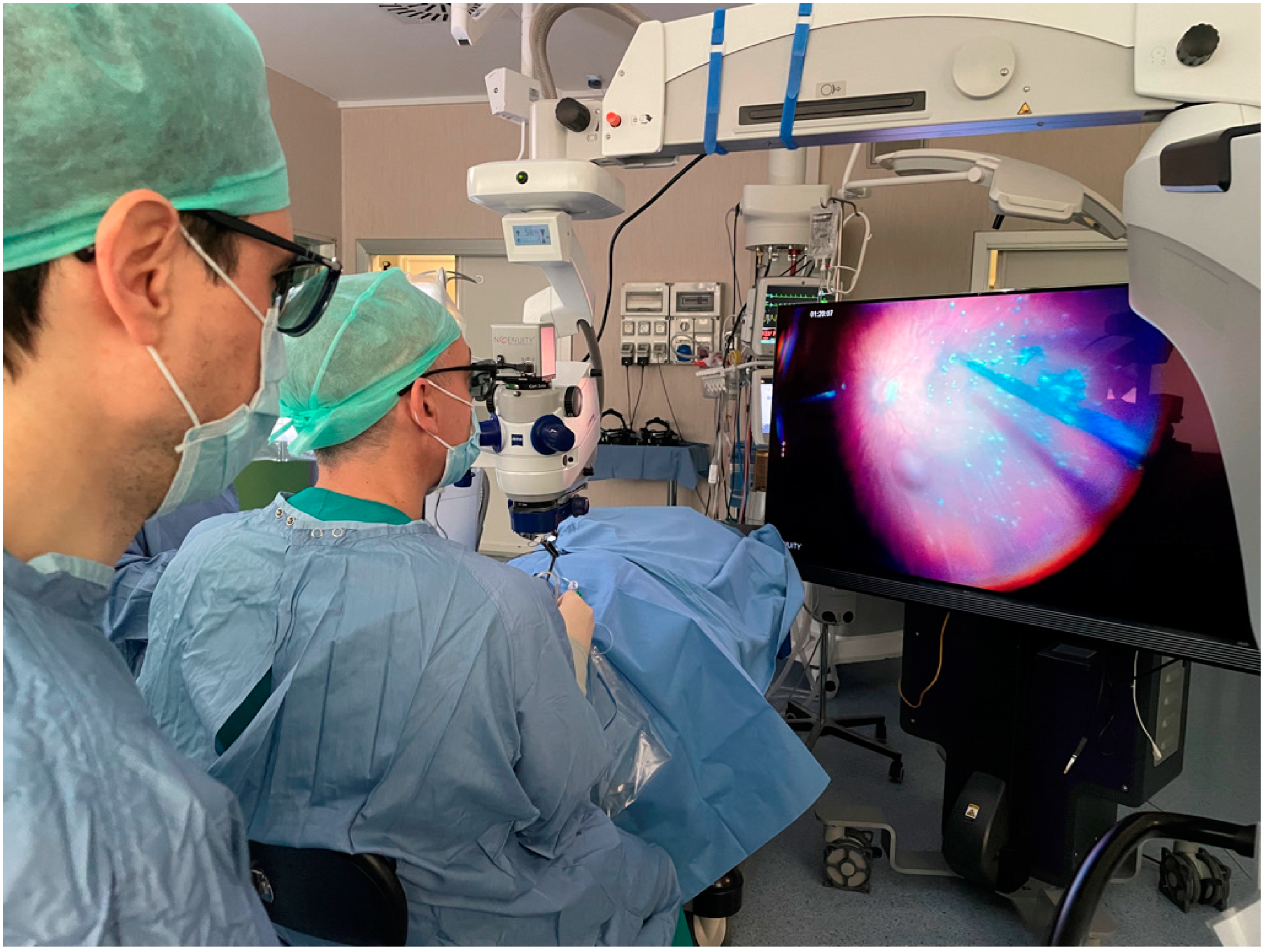
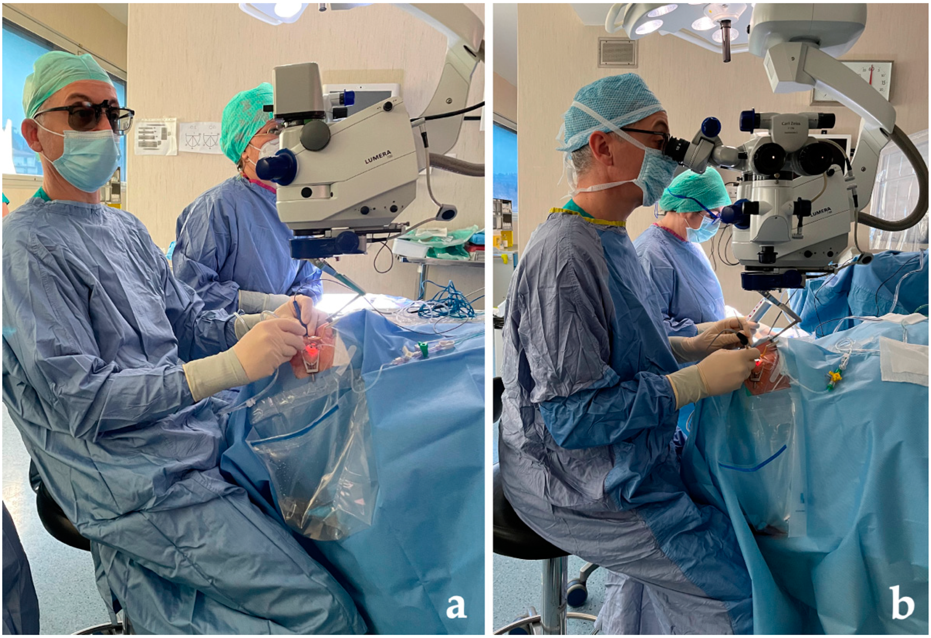
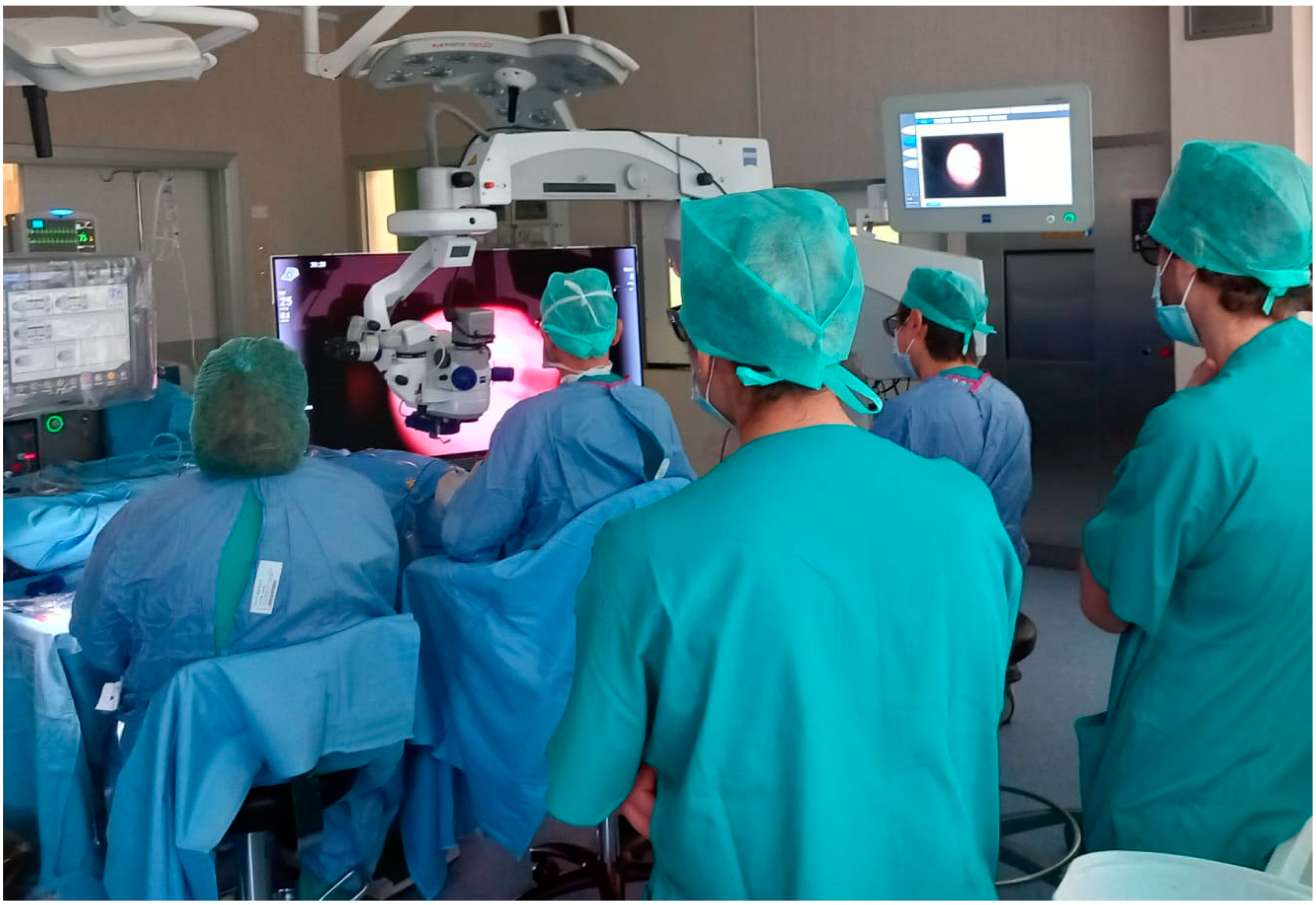
2.2. Methodology
2.3. Analysis
3. Results
4. Discussion
5. Conclusions
Author Contributions
Funding
Institutional Review Board Statement
Informed Consent Statement
Data Availability Statement
Conflicts of Interest
References
- Eckardt, C.; Paulo, E.B. Heads-up surgery for vitreoretinal procedures: An experimental and clinical study. Retina 2016, 36, 137–147. [Google Scholar] [CrossRef] [PubMed]
- Figueroa, M.S. 3D vitrectomy. Is it really useful? Arch. Soc. Esp. Oftalmol. 2017, 92, 249–250. [Google Scholar] [CrossRef] [PubMed]
- Moura-Coelho, N.; Henriques, J.; Nascimento, J.; Dutra-Medeiros, M. Three-Dimensional Display Systems in Ophthalmic Surgery—A Review. Eur. Ophthalmic Rev. 2019, 13, 31. [Google Scholar] [CrossRef]
- Liu, J.; Wu, D.; Ren, X.; Li, X. Clinical Experience of Using the NGENUITY Three-Dimensional Surgery System in Ophthalmic Surgical Procedures. Acta Ophthalmol. 2021, 99, e101–e108. [Google Scholar] [CrossRef] [PubMed]
- Rizzo, S.; Abbruzzese, G.; Savastano, A.; Giansanti, F.; Caporossi, T.; Barca, F.; Faraldi, F.; Virgili, G. 3D SURGICAL VIEWING SYSTEM IN OPHTHALMOLOGY: Perceptions of the Surgical Team. Retina 2018, 38, 857–861. [Google Scholar] [CrossRef]
- Weinstock, R.J.; Diakonis, V.F.; Schwartz, A.J.; Weinstock, A.J. Heads-up Cataract Surgery: Complication Rates, Surgical Duration, and Comparison with Traditional Microscopes. J. Refract. Surg. 2019, 35, 318–322. [Google Scholar] [CrossRef]
- Wang, K.; Song, F.; Zhang, L.; Xu, J.; Zhong, Y.; Lu, B.; Yao, K. Three-Dimensional Heads-up Cataract Surgery Using Femtosecond Laser: Efficiency, Efficacy, Safety, and Medical Education-A Randomized Clinical Trial. Transl. Vis. Sci. Technol. 2021, 10, 4. [Google Scholar] [CrossRef]
- Rosenberg, E.D.; Nuzbrokh, Y.; Sippel, K.C. Efficacy of 3D Digital Visualization in Minimizing Coaxial Illumination and Phototoxic Potential in Cataract Surgery: Pilot Study. J. Cataract Refract. Surg. 2021, 47, 291–296. [Google Scholar] [CrossRef]
- Qian, Z.; Wang, H.; Fan, H.; Lin, D.; Li, W. Three-Dimensional Digital Visualization of Phacoemulsification and Intraocular Lens Implantation. Indian J. Ophthalmol. 2019, 67, 341–343. [Google Scholar] [CrossRef] [PubMed]
- Nariai, Y.; Horiguchi, M.; Mizuguchi, T.; Sakurai, R.; Tanikawa, A. Comparison of Microscopic Illumination between a Three-Dimensional Heads-up System and Eyepiece in Cataract Surgery. Eur. J. Ophthalmol. 2021, 31, 1817–1821. [Google Scholar] [CrossRef] [PubMed]
- Kelkar, J.A.; Kelkar, A.S.; Bolisetty, M. Initial Experience with Three-Dimensional Heads-up Display System for Cataract Surgery—A Comparative Study. Indian J. Ophthalmol. 2021, 69, 2304–2309. [Google Scholar] [CrossRef]
- Del Turco, C.; D’Amico Ricci, G.; Dal Vecchio, M.; Bogetto, C.; Panico, E.; Giobbio, D.C.; Romano, M.R.; Panico, C.; la Spina, C. Heads-up 3D Eye Surgery: Safety Outcomes and Technological Review after 2 Years of Day-to-Day Use. Eur. J. Ophthalmol. 2022, 32, 1129–1135. [Google Scholar] [CrossRef] [PubMed]
- Berquet, F.; Henry, A.; Barbe, C.; Cheny, T.; Afriat, M.; Benyelles, A.K.; Bartolomeu, D.; Arndt, C. Comparing Heads-Up versus Binocular Microscope Visualization Systems in Anterior and Posterior Segment Surgeries: A Retrospective Study. Int. J. Ophthalmol. 2020, 243, 347–354. [Google Scholar] [CrossRef] [PubMed]
- Bedar, M.S.; Kellner, U. Digital 3D “Heads-up” Cataract Surgery: Safety Profile and Comparison with the Conventional Microscope System. Klin. Mon. Augenheilkd. 2022, 239, 991–995. [Google Scholar] [CrossRef]
- Bawankule, P.; Narnaware, S.; Chakraborty, M.; Raje, D.; Phusate, R.; Gupta, R.; Rewatkar, K.; Chivane, A.; Sontakke, S. Digitally Assisted Three-Dimensional Surgery—Beyond Vitreous. Indian J. Ophthalmol. 2021, 69, 1793–1800. [Google Scholar] [CrossRef]
- Kelkar, A.; Kelkar, J.; Chougule, Y.; Bolisetty, M.; Singhvi, P. Cognitive Workload, Complications and Visual Outcomes of Phacoemulsification Cataract Surgery: Three-Dimensional versus Conventional Microscope. Eur. J. Ophthalmol. 2022, 32, 2935–2941. [Google Scholar] [CrossRef] [PubMed]
- Agranat, J.S.; Miller, J.B.; Douglas, V.P.; Douglas, K.A.A.; Marmalidou, A.; Cunningham, M.A.; Houston, S.K., 3rd. The Scope Of Three-Dimensional Digital Visualization Systems In Vitreoretinal Surgery. Clin. Ophthalmol. 2019, 13, 2093–2096. [Google Scholar] [CrossRef]
- Bin Helayel, H.; Al-Mazidi, S.; Al Akeely, A. Can the Three-Dimensional Heads-Up Display Improve Ergonomics, Surgical Performance, and Ophthalmology Training Compared to Conventional Microscopy? Clin. Ophthalmol. 2021, 15, 679–686. [Google Scholar] [CrossRef]
- Coppola, M.; La Spina, C.; Rabiolo, A.; Querques, G.; Bandello, F. Heads-up 3D vision system for retinal detachment surgery. Int. J. Retina Vitreous. 2017, 3, 46. [Google Scholar] [CrossRef]
- Romano, M.R.; Cennamo, G.; Comune, C.; Cennamo, M.; Ferrara, M.; Rombetto, L.; Cennamo, G. Evaluation of 3D heads-up vitrectomy: Outcomes of psychometric skills testing and surgeon satisfaction. Eye 2018, 32, 1093–1098. [Google Scholar] [CrossRef]
- Talcott, K.E.; Adam, M.K.; Sioufi, K.; Aderman, C.M.; Ali, F.S.; Mellen, P.L.; Garg, S.J.; Hsu, J.; Ho, A.C. Comparison of a three-dimensional heads-up display surgical platform with a standard operating microscope for macular surgery. Ophthalmol. Retina 2019, 3, 244–251. [Google Scholar] [CrossRef] [PubMed]
- Palácios, R.M.; de Carvalho, A.C.M.; Maia, M.; Caiado, R.R.; Camilo, D.A.G.; Farah, M.E. An experimental and clinical study on the initial experiences of Brazilian vitreoretinal surgeons with heads-up surgery. Graefes Arch. Clin. Exp. Ophthalmol. 2019, 257, 473–483. [Google Scholar] [CrossRef] [PubMed]
- Kumar, A.; Hasan, N.; Kakkar, P.; Mutha, V.; Karthikeya, R.; Sundar, D.; Ravani, R. Comparison of clinical outcomes between ‘heads-up’ 3D viewing system and conventional microscope in macular hole surgeries: A pilot study. Indian J. Ophthalmol. 2018, 66, 1816–1819. [Google Scholar] [CrossRef] [PubMed]
- Zhang, T.; Tang, W.; Xu, G. Comparative analysis of three-dimensional heads-up vitrectomy and traditional microscopic vitrectomy for vitreoretinal diseases. Curr. Eye Res. 2019, 44, 1080–1086. [Google Scholar] [CrossRef] [PubMed]
- Palácios, R.M.; Kayat, K.V.; Morel, C.; Conrath, J.; Matonti, F.; Morin, B.; Farah, M.E.; Devin, F. Clinical study on the initial experiences of French vitreoretinal surgeons with heads-up surgery. Curr. Eye Res. 2020, 45, 1265–1272. [Google Scholar] [CrossRef]
- Kantor, P.; Matonti, F.; Varenne, F.; Sentis, V.; Pagot-Mathis, V.; Fournié, P.; Soler, V. Use of the heads-up NGENUITY 3D Visualization System for vitreoretinal surgery: A retrospective evaluation of outcomes in a French tertiary center. Sci. Rep. 2021, 11, 10031. [Google Scholar] [CrossRef] [PubMed]
- Zhao, X.Y.; Zhao, Q.; Li, N.N.; Meng, L.H.; Zhang, W.F.; Wang, E.Q.; Chen, Y.X. Surgery-related characteristics, efficacy, safety and surgical team satisfaction of three-dimensional heads-up system versus traditional microscopic equipment for various vitreoretinal diseases. Graefes Arch. Clin. Exp. Ophthalmol. 2023, 261, 669–679. [Google Scholar] [CrossRef]
- Nowomiejska, K.; Toro, M.D.; Bonfiglio, V.; Czarnek-Chudzik, A.; Brzozowska, A.; Torres, K.; Rejdak, R. Vitrectomy combined with cataract surgery for retinal detachment using a three-dimensional viewing system. J. Clin. Med. 2022, 11, 1788. [Google Scholar] [CrossRef]
- Zeng, R.; Feng, Y.; Begaj, T.; Baldwin, G.; Miller, J.B. Comparison of the Safety and Efficacy of a 3-Dimensional Heads-up Display vs a Standard Operating Microscope in Retinal Detachment Repair. J. Vitreoretin. Dis. 2023, 7, 97–102. [Google Scholar] [CrossRef]
- Asani, B.; Siedlecki, J.; Schworm, B.; Mayer, W.J.; Kreutzer, T.C.; Luft, N.; Priglinger, S.G. 3D Heads-Up Display vs. Standard Operating Microscope Vitrectomy for Rhegmatogenous Retinal Detachment. Front. Med. 2020, 7, 615515. [Google Scholar] [CrossRef]
- Parravano, M.; Giansanti, F.; Eandi, C.M.; Yap, Y.C.; Rizzo, S.; Virgili, G. Vitrectomy for idiopathic macular hole. Cochrane Database Syst. Rev. 2015, 2015, CD009080. [Google Scholar] [CrossRef] [PubMed]
- Guber, J.; Pereni, I.; Scholl, H.P.N.; Guber, I.; Haynes, R.J. Outcomes after epiretinal membrane surgery with or without internal limiting membrane peeling. Ophthalmol. Ther. 2019, 8, 297. [Google Scholar] [CrossRef] [PubMed]
- Palácios, R.M.; Maia, A.; Farah, M.E.; Maia, M. Learning curve of three-dimensional heads-up vitreoretinal surgery for treating macular holes: A prospective study. Int. Ophthalmol. 2019, 39, 2353–2359. [Google Scholar] [CrossRef] [PubMed]
- Freeman, W.R.; Chen, K.C.; Ho, J.; Chao, D.L.; Ferreyra, H.A.; Tripathi, A.B.; Nudleman, E.; Bartsch, D.-U. resolution, depth of field, and physician satisfaction during digitally assisted vitreoretinal surgery. Retina 2019, 39, 1768–1771. [Google Scholar] [CrossRef]
- Reinhard, E.; Heidrich, W.; Debevec, P.; Pattanaik, S.; Ward, G.; Myszkowski, K. High Dynamic Range Imaging: Acquisition, Display, and Image-Based Lighting, 2nd ed.; Morgan Kauffman Publisher: Burlington, MA, USA, 2010; pp. 91–117. [Google Scholar]
- Seetzen, H.; Heidrich, W.; Stuerzlinger, W.; Ward, G.; Whitehead, L.; Trentacoste, M.; Ghosh, A.; Vorozcovs, A. High dynamic range display systems. ACM Trans. Graph. 2004, 23, 760–768. [Google Scholar] [CrossRef]
- Franklin, A.J.; Sarangapani, R.; Yin, L.; Tripathi, B.; Riemann, C. Digital vs. Analog Surgical Visualization for Vitreoretinal Surgery. Retinal Physician. 2017. Available online: https://www.retinalphysician.com/issues/2017/may-2017/digital-vs-analog-surgical-visualization-for-vitre (accessed on 3 February 2023).
- Nakajima, K.; Inoue, M.; Mizuno, M.; Koto, T.; Ishida, T.; Ozawa, H.; Oshika, T. Effects of image-sharpening algorithm on surgical field visibility during 3D heads-up surgery for vitreoretinal diseases. Sci. Rep. 2023, 13, 2758. [Google Scholar] [CrossRef] [PubMed]
- Hyer, J.; Lee, R.M.; Chowdhury, H.R.; Smith, H.B.; Dhital, A.; Khandwala, M. National survey of back & neck pain amongst consultant ophthalmologists in the United Kingdom. Int. Ophthalmol. 2015, 35, 769–775. [Google Scholar]
- Nakajima, K.; Inoue, M.; Mizuno, M.; Koto, T.; Ishida, T.; Ozawa, H.; Oshika, T. Minimal endoillumination levels and display luminous emittance during three-dimensional heads-up vitreoretinal surgery. Retina 2017, 37, 1746–1749. [Google Scholar]
- Lu, E.S.; Reppucci, V.S.; Houston, S.K.S., 3rd; Kras, A.L.; Miller, J.B. Three-dimensional telesurgery and remote proctoring over a 5G network. Digit. J. Ophthalmol. 2021, 27, 38–43. [Google Scholar] [CrossRef]
- Seddon, I.A.; Rahimy, E.; Miller, J.B.; Charles, S.; Kitchens, J.; Houston, S.K., 3rd. Feasibility and Potential for Real-Time 3D Vitreoretinal Surgery Telementoring. Retina, 2022; Epub ahead of print. [Google Scholar] [CrossRef]


| 3D Group | Traditional Microscope Group | p-Value | |
|---|---|---|---|
| Number of patients, n | 240 | 210 | |
| Age, years (mean ± SD) | 59.3 ± 12.1 | 61.7 ±13.3 | 0.075 * |
| Sex, n (%) | 0.63 ** | ||
| Male | 149 (62.1%) | 135 (64.3%) | |
| Female | 91 (37.9%) | 75 (35.7%) | |
| Lens status, n (%) | 0.41 ** | ||
| Phakia | 144 (60%) | 111 (52.9%) | |
| Pseudophakia | 96 (40%) | 99 (47.1%) | |
| Indications, n (%) | 0.86 ** | ||
| Retinal detachment | 74 (30.8%) | 65 (30.9%) | |
| Idiopathic epiretinal membrane | 78 (32.5%) | 67 (31.9%) | |
| Macular hole | 64 (26.7%) | 52 (24.8%) | |
| Vitreous hemorrhage | 24 (10%) | 26 (12.4%) | |
| Macular hole diameter, µm (mean ± SD) | 374.2 ± 125.3 | 392.75 ± 139.3 | 0.45 * |
| Epiretinal baseline central macular thickness, µm (mean ± SD) | 438.75 ± 125.8 | 441.67 ± 81.2 | 0.52 * |
| Retinal detachment macular involvement, n (%) | 39 (52.7%) | 32 (49.2%) | 0.68 ** |
| Baseline decimal BCVA (mean) | 0.36 | 0.41 | 0.67 * |
| 3D Group (N = 240) | Traditional Microscope Group (N = 210) | p-Value | |
|---|---|---|---|
| Post-op decimal BCVA (mean) | 0.53 | 0.57 | 0.12 * |
| Surgery time, minutes (mean) | 60.7 | 61.0 | 0.46 * |
| Retinal detachment time, minutes (mean) | 69 | 67 | 0.26 * |
| Idiopathic epiretinal membrane time, minutes (mean) | 57.6 | 56.6 | 0.74 * |
| Macular hole time, minutes (mean) | 56.14 | 58.1 | 0.86 * |
| Vitreous hemorrhage time, minutes (mean) | 57.05 | 63.0 | 0.16 * |
| Surgical outcome *** | |||
| ERM removal, % | 100 | 100 | - |
| MH closure, % | 93.8 | 94.2 | 0.91 ** |
| Retinal reattachment, % | 93.2 | 90.8 | 0.59 ** |
| VH clearing, % | 95.8 | 96.1 | 0.95 ** |
| Authors | Number of Patients 3D Group/CM Group | Type of Treatment/ Surgical Indication | Outcome | Results |
|---|---|---|---|---|
| Kumar et al. [23] | 25/25 | PPV with multilayered inverted ILM membrane flap technique and 20% SF6 for FTMH | Pre- and postoperative BCVA, macular hole index, total surgical time, total ILM peel time, number of flap initiations, duration of Brilliant Blue G dye exposure, and illumination intensity | Comparable clinical outcomes. Illumination intensity of microscope and endoillumination were significantly less in the 3D group |
| Talcott et al. [21] | 23/16 | PPV for ERM and FTMH | Total operative time, macular peel time, surgeon rating of viewing system ease of use, minimum required endoillumination, intraoperative complication rate, and postoperative BCVA | No significant difference in overall operative time, but macular peel time was significantly longer using 3D HUD and associated with less ease of use. The minimum required endoillumination was significantly lower with 3D HUD. No significant differences in BCVA and complication occurrence |
| Zhang et al. [24] | 124/202 | PPV for RRD, FTMH, ERM, VH, VO, SOR, and MF | Pre- and postoperative BCVA, ERM removal VH clearing, MH closure, RD reattachment, MF resolution, SOR success, VO clearing, operation time, postoperative complications occurrence | Comparable visual and anatomical outcomes without a significant difference in the rate of complications |
| Palácios et al. [33] | 94/94 | PPV for RRD and MH | Surgeon preference was assessed using a questionnaire, anatomical success rate | Comparable anatomical outcomes |
| Asani et al. [30] | 70/70 | PPV for RRD | Primary retinal reattachment rate, rate of proliferative vitreoretinopathy, final BCVA, duration of surgery | Comparable anatomical and functional outcomes. Duration of surgery was significantly longer in the 3D group, an effect which, however, vanished after a “learning curve” of the first 35 eyes |
| Kantor et al. [26] | 131/96 | PPV for RRD, FTMH, and ERM | Primary endpoints: recurrence rates of RD, FTMH closure rates, reduction in central macular thickness in ERMs at 3 months after surgery. Secondary endpoints: surgery durations, 3-month postoperative BCVA | Comparable visual and anatomical outcomes |
| Zhao et al. [27] | 220/242 | PPV for RRD, TRD, FTMH, ERM, VMT, VH, VO, SOR, and MF | BCVA, primary anatomical success (varied according to the surgical indicators), general surgical duration, duration of specific surgical steps, perioperative complications, and satisfaction feedback from the surgical team | Comparable efficacy and safety. Shorter duration of ERM or ILM peeling for the 3D HUD group with significantly shorter general surgical duration for ERM and MH surgery. Better surgical team satisfaction |
| Nowomiejska et al. [28] | 26/56 | PPV combined with cataract surgery for RRD | BCVA, surgery duration, rate of postoperative complications | No significant differences in surgery duration, rate of complications, and functional results |
| Zeng et al. [29] | 50/138 | PPV alone or combined PPV and scleral buckle for RRD | Anatomic success rate, rate of postoperative proliferative vitreoretinopathy, surgery duration | Anatomical and functional outcomes and surgical efficiency comparable in the two groups |
Disclaimer/Publisher’s Note: The statements, opinions and data contained in all publications are solely those of the individual author(s) and contributor(s) and not of MDPI and/or the editor(s). MDPI and/or the editor(s) disclaim responsibility for any injury to people or property resulting from any ideas, methods, instructions or products referred to in the content. |
© 2023 by the authors. Licensee MDPI, Basel, Switzerland. This article is an open access article distributed under the terms and conditions of the Creative Commons Attribution (CC BY) license (https://creativecommons.org/licenses/by/4.0/).
Share and Cite
Giansanti, F.; Nicolosi, C.; Bacherini, D.; Soloperto, F.; Sarati, F.; Giattini, D.; Vicini, G. Three-Dimensional Visualization System for Vitreoretinal Surgery: Results from a Monocentric Experience and Comparison with Conventional Surgery. Life 2023, 13, 1289. https://doi.org/10.3390/life13061289
Giansanti F, Nicolosi C, Bacherini D, Soloperto F, Sarati F, Giattini D, Vicini G. Three-Dimensional Visualization System for Vitreoretinal Surgery: Results from a Monocentric Experience and Comparison with Conventional Surgery. Life. 2023; 13(6):1289. https://doi.org/10.3390/life13061289
Chicago/Turabian StyleGiansanti, Fabrizio, Cristina Nicolosi, Daniela Bacherini, Federica Soloperto, Federica Sarati, Dario Giattini, and Giulio Vicini. 2023. "Three-Dimensional Visualization System for Vitreoretinal Surgery: Results from a Monocentric Experience and Comparison with Conventional Surgery" Life 13, no. 6: 1289. https://doi.org/10.3390/life13061289