Next Issue
Volume 13, May
Previous Issue
Volume 13, March


Antioxidants, Volume 13, Issue 4 (April 2024) – 118 articles

Cover Story (view full-size image): 
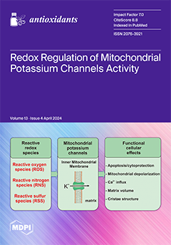
Redox reactions exert a profound influence on numerous cellular functions with mitochondria playing a central role in orchestrating these processes. This pivotal involvement arises from three primary factors: (1) the synthesis of reactive oxygen species (ROS) by mitochondria, (2) the presence of a substantial array of redox enzymes such as respiratory chain, and (3) the responsiveness of mitochondria to the cellular redox state. Within the inner mitochondrial membrane, a group of potassium channels, including ATP-regulated, large conductance calcium-activated, and voltage-regulated channels, is present. These mitochondrial potassium channels play a crucial role in conditions such as cytoprotection or ischemia/reperfusion injury, and they are regulated by various redox reactions. View this paper
  • Issues are regarded as officially published after their release is announced to the table of contents alert mailing list.
  • You may sign up for e-mail alerts to receive table of contents of newly released issues.
  • PDF is the official format for papers published in both, html and pdf forms. To view the papers in pdf format, click on the "PDF Full-text" link, and use the free Adobe Reader to open them.
Order results
Result details
Section
Select all
Export citation of selected articles as:
18 pages, 2650 KiB  
Article
Comparative Analysis of Polyphenolic Compounds in Different Amaranthus Species: Influence of Genotypes and Harvesting Year
by Jun-Hyoung Bang, Ick-Hyun Jo, Raveendar Sebastin, Won Tea Jeong, Sangtaek Oh, Tae-Young Heo, Jeehye Sung, Tae kyung Hyun, Yoon-Sup So, Ju-Kyung Yu, Amal Mohamed AlGarawi, Ashraf Atef Hatamleh, Gi-Ho Sung and Jong-Wook Chung
Antioxidants 2024, 13(4), 501; https://doi.org/10.3390/antiox13040501 - 22 Apr 2024
Viewed by 271
Abstract
Amaranth is a nutritionally valuable crop, as it contains phenolic acids and flavonoids, yielding diverse plant secondary metabolites (PSMs) like phytosterol, tocopherols, and carotenoids. This study explored the variations in the contents of seventeen polyphenolic compounds within the leaves of one hundred twenty [...] Read more.
Amaranth is a nutritionally valuable crop, as it contains phenolic acids and flavonoids, yielding diverse plant secondary metabolites (PSMs) like phytosterol, tocopherols, and carotenoids. This study explored the variations in the contents of seventeen polyphenolic compounds within the leaves of one hundred twenty Amaranthus accessions representing nine Amaranthus species. The investigation entailed the analysis of phenolic content across nine Amaranthus species, specifically A. hypochondriacus, A. cruentus, A. caudatus, A. tricolor, A. dubius, A. blitum, A. crispus, A. hybridus, and A. viridis, utilizing ultra performance liquid chromatography with photodiode array detection (UPLC-PDA). The results revealed significant differences in polyphenolic compounds among accessions in which rutin content was predominant in all Amaranthus species in both 2018 and 2019. Among the nine Amaranthus species, the rutin content ranged from 95.72 ± 199.17 μg g−1 (A. dubius) to 1485.09 ± 679.51 μg g−1 (A. viridis) in 2018 and from 821.59 ± 709.95 μg g−1 (A. tricolor) to 3166.52 ± 1317.38 μg g−1 (A. hypochondriacus) in 2019. Correlation analysis revealed, significant positive correlations between rutin and kaempferol-3-O-β-rutinoside (r = 0.93), benzoic acid and ferulic acid (r = 0.76), and benzoic acid and kaempferol-3-O-β-rutinoside (r = 0.76), whereas gallic acid showed consistently negative correlations with each of the 16 phenolic compounds. Wide variations were identified among accessions and between plants grown in the two years. The nine species and one hundred twenty Amaranthus accessions were clustered into six groups based on their seventeen phenolic compounds in each year. These findings contribute to expanding our understanding of the phytochemical traits of accessions within nine Amaranthus species, which serve as valuable resources for Amaranthus component breeding and functional material development. Full article
Show Figures

Figure 1

12 pages, 2158 KiB  
Communication
The Role of NOX2-Derived Reactive Oxygen Species in the Induction of Endothelin-Converting Enzyme-1 by Angiotensin II
by Michael Adu-Gyamfi, Claudia Goettsch, Julian Kamhieh-Milz, Lei Chen, Anna Maria Pfefferkorn, Anja Hofmann, Coy Brunssen, Gregor Müller, Thomas Walther, Muhammad Imtiaz Ashraf, Henning Morawietz, Janusz Witowski and Rusan Catar
Antioxidants 2024, 13(4), 500; https://doi.org/10.3390/antiox13040500 - 22 Apr 2024
Viewed by 278
Abstract
Endothelin-1 is a key regulator of vascular tone and blood pressure in health and disease. We have recently found that ET-1 production in human microvascular endothelial cells (HMECs) can be promoted by angiotensin II (Ang II) through a novel mechanism involving octamer-binding transcription [...] Read more.
Endothelin-1 is a key regulator of vascular tone and blood pressure in health and disease. We have recently found that ET-1 production in human microvascular endothelial cells (HMECs) can be promoted by angiotensin II (Ang II) through a novel mechanism involving octamer-binding transcription factor-1 (Oct-1), NADPH oxidase-2 (NOX2), and superoxide anions. As the formation of bioactive ET-1 also depends on endothelin-converting enzyme-1 (ECE-1), we investigated the transcriptional regulation of the ECE1 gene. We found that exposure of HMECs to Ang II resulted in a concentration- and time-dependent increase in ECE1 mRNA expression. Pharmacological inhibition of ECE-1 reduced Ang II-stimulated ET-1 release to baseline values. The effect of Ang II on ECE1 mRNA expression was associated with Oct-1 binding to the ECE1 promoter, resulting in its increased activity. Consequently, the Ang II-stimulated increase in ECE1 mRNA expression could be prevented by siRNA-mediated Oct-1 inhibition. It could also be abolished by silencing the NOX2 gene and neutralizing superoxide anions with superoxide dismutase. In mice fed a high-fat diet, cardiac expression of Ece1 mRNA increased in wild-type mice but not in Nox2-deficient animals. It can be concluded that Ang II engages Oct-1, NOX2, and superoxide anions to stimulate ECE1 expression in the endothelium. Full article
Show Figures

Figure 1

17 pages, 3608 KiB  
Article
Redox Regulation of LAT Enhances T Cell-Mediated Inflammation
by Jaime James, Ana Coelho, Gonzalo Fernandez Lahore, Clara M. Hernandez, Florian Forster, Bernard Malissen and Rikard Holmdahl
Antioxidants 2024, 13(4), 499; https://doi.org/10.3390/antiox13040499 - 22 Apr 2024
Viewed by 237
Abstract
The positional cloning of single nucleotide polymorphisms (SNPs) of the neutrophil cytosolic factor 1 (Ncf1) gene, advocating that a low oxidative burst drives autoimmune disease, demands an understanding of the underlying molecular causes. A cellular target could be T cells, which [...] Read more.
The positional cloning of single nucleotide polymorphisms (SNPs) of the neutrophil cytosolic factor 1 (Ncf1) gene, advocating that a low oxidative burst drives autoimmune disease, demands an understanding of the underlying molecular causes. A cellular target could be T cells, which have been shown to be regulated by reactive oxygen species (ROS). However, the pathways by which ROS mediate T cell signaling remain unclear. The adaptor molecule linker for activation of T cells (LAT) is essential for coupling T cell receptor-mediated antigen recognition to downstream responses, and it contains several cysteine residues that have previously been suggested to be involved in redox regulation. To address the possibility that ROS regulate T cell-dependent inflammation through LAT, we established a mouse strain with cysteine-to-serine mutations at positions 120 and 172 (LATSS). We found that redox regulation of LAT through C120 and C172 mediate its localization and phosphorylation. LATSS mice had reduced numbers of double-positive thymocytes and naïve peripheral T cells. Importantly, redox insensitivity of LAT enhanced T cell-dependent autoimmune inflammation in collagen-induced arthritis (CIA), a mouse model of rheumatoid arthritis (RA). This effect was reversed on an NCF1-mutated (NCF1m1j), ROS-deficient, background. Overall, our data show that LAT is redox-regulated, acts to repress T cell activation, and is targeted by ROS induced by NCF1 in antigen-presenting cells (APCs). Full article
Show Figures

Graphical abstract

24 pages, 9238 KiB  
Article
The Reducing Agent Dithiothreitol Modulates the Ventilatory Responses That Occur in Freely Moving Rats during and following a Hypoxic–Hypercapnic Challenge
by Paulina M. Getsy, Gregory A. Coffee, Walter J. May, Santhosh M. Baby, James N. Bates and Stephen J. Lewis
Antioxidants 2024, 13(4), 498; https://doi.org/10.3390/antiox13040498 - 22 Apr 2024
Viewed by 258
Abstract
The present study examined the hypothesis that changes in the oxidation–reduction state of thiol residues in functional proteins play a major role in the expression of the ventilatory responses in conscious rats that occur during a hypoxic–hypercapnic (HH) gas challenge and upon return [...] Read more.
The present study examined the hypothesis that changes in the oxidation–reduction state of thiol residues in functional proteins play a major role in the expression of the ventilatory responses in conscious rats that occur during a hypoxic–hypercapnic (HH) gas challenge and upon return to room air. A HH gas challenge in vehicle-treated rats elicited robust and sustained increases in minute volume (via increases in frequency of breathing and tidal volume), peak inspiratory and expiratory flows, and inspiratory and expiratory drives while minimally affecting the non-eupneic breathing index (NEBI). The HH-induced increases in these parameters, except for frequency of breathing, were substantially diminished in rats pre-treated with the potent and lipophilic disulfide-reducing agent, L,D-dithiothreitol (100 µmol/kg, IV). The ventilatory responses that occurred upon return to room air were also substantially different in dithiothreitol-treated rats. In contrast, pre-treatment with a substantially higher dose (500 µmol/kg, IV) of the lipophilic congener of the monosulfide, N-acetyl-L-cysteine methyl ester (L-NACme), only minimally affected the expression of the above-mentioned ventilatory responses that occurred during the HH gas challenge or upon return to room air. The effectiveness of dithiothreitol suggests that the oxidation of thiol residues occurs during exposure to a HH gas challenge and that this process plays an essential role in allowing for the expression of the post-HH excitatory phase in breathing. However, this interpretation is contradicted by the lack of effects of L-NACme. This apparent conundrum may be explained by the disulfide structure affording unique functional properties to dithiothreitol in comparison to monosulfides. More specifically, the disulfide structure may give dithiothreitol the ability to alter the conformational state of functional proteins while transferring electrons. It is also possible that dithiothreitol is simply a more efficient reducing agent following systemic injection, although one interpretation of the data is that the effects of dithiothreitol are not due to its reducing ability. Full article
Show Figures

Figure 1

17 pages, 12726 KiB  
Article
Protective and Regenerative Effects of Reconstituted HDL on Human Rotator Cuff Fibroblasts under Hypoxia: An In Vitro Study
by Ra Jeong Kim and Hyung Bin Park
Antioxidants 2024, 13(4), 497; https://doi.org/10.3390/antiox13040497 - 22 Apr 2024
Viewed by 271
Abstract
Hypoxia and hypo-high-density lipoproteinemia (hypo-HDLemia) are proposed risk factors for rotator cuff tear. HDL is recognized for its potential benefits in ischemia-driven angiogenesis and wound healing. Nevertheless, research on the potential benefits of reconstituted HDL (rHDL) on human rotator cuff fibroblasts (RCFs) under [...] Read more.
Hypoxia and hypo-high-density lipoproteinemia (hypo-HDLemia) are proposed risk factors for rotator cuff tear. HDL is recognized for its potential benefits in ischemia-driven angiogenesis and wound healing. Nevertheless, research on the potential benefits of reconstituted HDL (rHDL) on human rotator cuff fibroblasts (RCFs) under hypoxia is limited. This study investigates the cytoprotective and regenerative effects of rHDL, as well as N-acetylcysteine (NAC), vitamin C (Vit C), and HDL on human RCFs under hypoxic conditions. Sixth-passage human RCFs were divided into normoxia, hypoxia, and hypoxia groups pretreated with antioxidants (NAC, Vit C, rHDL, HDL). Hypoxia was induced by 1000 µM CoCl2. In the hypoxia group compared to the normoxia group, there were significant increases in hypoxia-inducible factor-1α (HIF-1α), heme oxygenase-1 (HO-1), and Bcl-2/E1B-19kDa interacting protein 3 (BNIP3) expressions, along with reduced cell viability, elevated reactive oxygen species (ROS) production, apoptosis rate, expressions of cleaved caspase-3, cleaved poly ADP-ribose polymerase-1 (PARP-1), vascular endothelial growth factors (VEGF), and matrix metalloproteinase-2 (MMP-2), as well as decreased collagen I and III production, and markedly lower cell proliferative activity (p ≤ 0.039). These responses were significantly mitigated by pretreatment with rHDL (p ≤ 0.046). This study suggests that rHDL can enhance cell proliferation and collagen I and III production while reducing apoptosis in human RCFs under hypoxic conditions. Full article
Show Figures

Figure 1

17 pages, 4094 KiB  
Article
An AlphaFold Structure Analysis of COQ2 as Key a Component of the Coenzyme Q Synthesis Complex
by María de los Ángeles Vargas-Pérez, Damien Paul Devos and Guillermo López-Lluch
Antioxidants 2024, 13(4), 496; https://doi.org/10.3390/antiox13040496 - 21 Apr 2024
Viewed by 656
Abstract
Coenzyme Q (CoQ) is a lipidic compound that is widely distributed in nature, with crucial functions in metabolism, protection against oxidative damage and ferroptosis and other processes. CoQ biosynthesis is a conserved and complex pathway involving several proteins. COQ2 is a member of [...] Read more.
Coenzyme Q (CoQ) is a lipidic compound that is widely distributed in nature, with crucial functions in metabolism, protection against oxidative damage and ferroptosis and other processes. CoQ biosynthesis is a conserved and complex pathway involving several proteins. COQ2 is a member of the UbiA family of transmembrane prenyltransferases that catalyzes the condensation of the head and tail precursors of CoQ, which is a key step in the process, because its product is the first intermediate that will be modified in the head by the next components of the synthesis process. Mutations in this protein have been linked to primary CoQ deficiency in humans, a rare disease predominantly affecting organs with a high energy demand. The reaction catalyzed by COQ2 and its mechanism are still unknown. Here, we aimed at clarifying the COQ2 reaction by exploring possible substrate binding sites using a strategy based on homology, comprising the identification of available ligand-bound homologs with solved structures in the Protein Data Bank (PDB) and their subsequent structural superposition in the AlphaFold predicted model for COQ2. The results highlight some residues located on the central cavity or the matrix loops that may be involved in substrate interaction, some of which are mutated in primary CoQ deficiency patients. Furthermore, we analyze the structural modifications introduced by the pathogenic mutations found in humans. These findings shed new light on the understanding of COQ2’s function and, thus, CoQ’s biosynthesis and the pathogenicity of primary CoQ deficiency. Full article
(This article belongs to the Special Issue The Ubiquitous and Multifaceted Coenzyme Q)
Show Figures

Figure 1

16 pages, 3190 KiB  
Article
Litchi Pericarp Extract Treats Type 2 Diabetes Mellitus by Regulating Oxidative Stress, Inflammatory Response, and Energy Metabolism
by Ziming Yang, Li Zhang, Jinlei Liu and Dianpeng Li
Antioxidants 2024, 13(4), 495; https://doi.org/10.3390/antiox13040495 - 21 Apr 2024
Viewed by 272
Abstract
Litchi pericarp is rich in polyphenols, and demonstrates significant biological activity. This study assessed the therapeutic effects of litchi pericarp extract (LPE) on type 2 diabetes mellitus in db/db mice. The results showed that LPE ameliorated symptoms of glucose metabolism disorder, oxidative stress, [...] Read more.
Litchi pericarp is rich in polyphenols, and demonstrates significant biological activity. This study assessed the therapeutic effects of litchi pericarp extract (LPE) on type 2 diabetes mellitus in db/db mice. The results showed that LPE ameliorated symptoms of glucose metabolism disorder, oxidative stress, inflammatory response, and insulin resistance in db/db mice. The mechanistic studies indicated that LPE activates adenosine 5‘-monophosphate (AMP)-activated protein kinase (AMPK) and suppresses the protein expression of phosphoenolpyruvate carboxykinase (PEPCK), thereby reducing hepatic gluconeogenesis. Additionally, LPE facilitates the translocation of nuclear factor erythroid2-related factor 2 (Nrf2) into the cell nucleus, initiating the transcription of antioxidant factors superoxide dismutase (SOD) and NAD(P)H: quinone oxidoreductase 1 (NQO1), which alleviate oxidative stress and reduce oxidative damage. Furthermore, LPE blocks nuclear factor kappa-B (NF-κB) nuclear translocation and subsequent inflammatory response initiation, thereby reducing inflammation. These findings indicate that LPE addresses type 2 diabetes mellitus by activating the AMPK energy metabolic pathway and regulating the Nrf2 oxidative stress and NF-κB inflammatory signaling pathways. Full article
(This article belongs to the Special Issue Natural Antioxidants and Metabolic Diseases)
Show Figures

Figure 1

20 pages, 4669 KiB  
Article
Cord Blood Adductomics Reveals Oxidative Stress Exposure Pathways of Bronchopulmonary Dysplasia
by Erika T. Lin, Yeunook Bae, Robert Birkett, Abhineet M. Sharma, Runze Zhang, Kathleen M. Fisch, William Funk and Karen K. Mestan
Antioxidants 2024, 13(4), 494; https://doi.org/10.3390/antiox13040494 - 20 Apr 2024
Viewed by 307
Abstract
Fetal and neonatal exposures to perinatal oxidative stress (OS) are key mediators of bronchopulmonary dysplasia (BPD). To characterize these exposures, adductomics is an exposure science approach that captures electrophilic addition products (adducts) in blood protein. Adducts are bound to the nucleophilic cysteine loci [...] Read more.
Fetal and neonatal exposures to perinatal oxidative stress (OS) are key mediators of bronchopulmonary dysplasia (BPD). To characterize these exposures, adductomics is an exposure science approach that captures electrophilic addition products (adducts) in blood protein. Adducts are bound to the nucleophilic cysteine loci of human serum albumin (HSA), which has a prolonged half-life. We conducted targeted and untargeted adductomics to test the hypothesis that adducts of OS vary with BPD. We studied 205 preterm infants (≤28 weeks) and 51 full-term infants from an ongoing birth cohort. Infant plasma was collected at birth (cord blood), 1-week, 1-month, and 36-weeks postmenstrual age. HSA was isolated from plasma, trypsin digested, and analyzed using high-performance liquid chromatography–mass spectrometry to quantify previously annotated (known) and unknown adducts. We identified 105 adducts in cord and postnatal blood. A total of 51 known adducts (small thiols, direct oxidation products, and reactive aldehydes) were increased with BPD. Postnatally, serial concentrations of several known OS adducts correlated directly with supplemental oxygen exposure. The application of large-scale adductomics elucidated OS-mediated pathways of BPD. This is the first study to investigate the “neonatal–perinatal exposome” and to identify oxidative stress-related exposure biomarkers that may inform antioxidant strategies to protect the health of future generations of infants. Full article
Show Figures

Figure 1

18 pages, 10149 KiB  
Article
Uncovering the Cardioprotective Potential of Diacerein in Doxorubicin Cardiotoxicity: Mitigating Ferritinophagy-Mediated Ferroptosis via Upregulating NRF2/SLC7A11/GPX4 Axis
by Rehab M. El-Gohary, Asmaa H. Okasha, Alaa H. Abd El-Azeem, Muhammad T. Abdel Ghafar, Sarah Ibrahim, Islam I. Hegab, Eman E. Farghal, Soha Abdel Fattah Shalaby, Ola A. Elshora, Aisha E. ElMehy, Amany Nagy Barakat, Basma Saed Amer, Fatma G. Sobeeh, Gehan H. AboEl-Magd and Asmaa A. Ghalwash
Antioxidants 2024, 13(4), 493; https://doi.org/10.3390/antiox13040493 - 20 Apr 2024
Viewed by 363
Abstract
Doxorubicin (DOX)-induced cardiotoxicity (DIC) is a life-threatening clinical issue with limited preventive approaches, posing a substantial challenge to cancer survivors. The anthraquinone diacerein (DCN) exhibits significant anti-inflammatory, anti-proliferative, and antioxidant actions. Its beneficial effects on DIC have yet to be clarified. Therefore, this [...] Read more.
Doxorubicin (DOX)-induced cardiotoxicity (DIC) is a life-threatening clinical issue with limited preventive approaches, posing a substantial challenge to cancer survivors. The anthraquinone diacerein (DCN) exhibits significant anti-inflammatory, anti-proliferative, and antioxidant actions. Its beneficial effects on DIC have yet to be clarified. Therefore, this study investigated DCN’s cardioprotective potency and its conceivable molecular targets against DIC. Twenty-eight Wister rats were assigned to CON, DOX, DCN-L/DOX, and DCN-H/DOX groups. Serum cardiac damage indices, iron assay, oxidative stress, inflammation, endoplasmic reticulum (ER) stress, apoptosis, ferritinophagy, and ferroptosis-related biomarkers were estimated. Nuclear factor E2-related factor 2 (NRF2) DNA-binding activity and phospho-p53 immunoreactivity were assessed. DCN administration effectively ameliorated DOX-induced cardiac cytomorphological abnormalities. Additionally, DCN profoundly combated the DOX-induced labile iron pool expansion alongside its consequent lethal lipid peroxide overproduction, whereas it counteracted ferritinophagy and enhanced iron storage. Indeed, DCN valuably reinforced the cardiomyocytes’ resistance to ferroptosis, mainly by restoring the NRF2/solute carrier family 7 member 11 (SLC7A11)/glutathione peroxidase 4 (GPX4) signaling axis. Furthermore, DCN abrogated the cardiac oxidative damage, inflammatory response, ER stress, and cardiomyocyte apoptosis elicited by DOX. In conclusion, for the first time, our findings validated DCN’s cardioprotective potency against DIC based on its antioxidant, anti-inflammatory, anti-ferroptotic, and anti-apoptotic imprint, chiefly mediated by the NRF2/SLC7A11/GPX4 axis. Accordingly, DCN could represent a promising therapeutic avenue for patients under DOX-dependent chemotherapy. Full article
(This article belongs to the Special Issue Iron Metabolism, Oxidative Stress and Cellular Dysfunction)
Show Figures

Figure 1

16 pages, 2648 KiB  
Article
Selenium-Enriched E. coli Bacteria Mitigate the Age-Associated Degeneration of Cholinergic Neurons in C. elegans
by Palina Zytner, Anne Kutschbach, Weiye Gong, Verena Alexia Ohse, Laura Taudte, Anna Patricia Kipp, Lars-Oliver Klotz, Josephine Priebs and Holger Steinbrenner
Antioxidants 2024, 13(4), 492; https://doi.org/10.3390/antiox13040492 - 20 Apr 2024
Viewed by 345
Abstract
Selenium (Se) is an essential trace element for humans and animals, but high-dose supplementation with Se compounds, most notably selenite, may exert cytotoxic and other adverse effects. On the other hand, bacteria, including Escherichia coli (E. coli), are capable of reducing [...] Read more.
Selenium (Se) is an essential trace element for humans and animals, but high-dose supplementation with Se compounds, most notably selenite, may exert cytotoxic and other adverse effects. On the other hand, bacteria, including Escherichia coli (E. coli), are capable of reducing selenite to red elemental Se that may serve as a safer Se source. Here, we examined how a diet of Se-enriched E. coli bacteria affected vital parameters and age-associated neurodegeneration in the model organism Caenorhabditis elegans (C. elegans). The growth of E. coli OP50 for 48 h in medium supplemented with 1 mM sodium selenite resulted in reddening of the bacterial culture, accompanied by Se accumulation in the bacteria. Compared to nematodes supplied with the standard E. coli OP50 diet, the worms fed on Se-enriched bacteria were smaller and slimmer, even though their food intake was not diminished. Nevertheless, given the choice, the nematodes preferred the standard diet. The fecundity of the worms was not affected by the Se-enriched bacteria, even though the production of progeny was somewhat delayed. The levels of the Se-binding protein SEMO-1, which serves as a Se buffer in C. elegans, were elevated in the group fed on Se-enriched bacteria. The occurrence of knots and ruptures within the axons of cholinergic neurons was lowered in aged nematodes provided with Se-enriched bacteria. In conclusion, C. elegans fed on Se-enriched E. coli showed less age-associated neurodegeneration, as compared to nematodes supplied with the standard diet. Full article
Show Figures

Figure 1

15 pages, 4149 KiB  
Article
Corilagin Inhibits Neutrophil Extracellular Trap Formation and Protects against Hydrochloric Acid/Lipopolysaccharide-Induced Acute Lung Injury in Mice by Suppressing the STAT3 and NOX2 Signaling Pathways
by Fu-Chao Liu, Huang-Ping Yu, Chia-Chih Liao, An-Hsun Chou and Hung-Chen Lee
Antioxidants 2024, 13(4), 491; https://doi.org/10.3390/antiox13040491 - 19 Apr 2024
Viewed by 223
Abstract
Acute lung injury (ALI) and its severe manifestation, acute respiratory distress syndrome (ARDS), are characterized by uncontrolled inflammatory responses, neutrophil activation and infiltration, damage to the alveolar capillary membrane, and diffuse alveolar injury. Neutrophil extracellular traps (NETs), formed by activated neutrophils, contribute significantly [...] Read more.
Acute lung injury (ALI) and its severe manifestation, acute respiratory distress syndrome (ARDS), are characterized by uncontrolled inflammatory responses, neutrophil activation and infiltration, damage to the alveolar capillary membrane, and diffuse alveolar injury. Neutrophil extracellular traps (NETs), formed by activated neutrophils, contribute significantly to various inflammatory disorders and can lead to tissue damage and organ dysfunction. Corilagin, a compound found in Phyllanthus urinaria, possesses antioxidative and anti-inflammatory properties. In this study, we investigated the protective effects and underlying mechanisms of corilagin in hydrochloric acid (HCl)/lipopolysaccharide (LPS)-induced lung injury. Mice received intraperitoneal administration of corilagin (2.5, 5, or 10 mg/kg) or an equal volume of saline 30 min after intratracheal HCl/LPS administration. After 20 h, lung tissues were collected for analysis. Corilagin treatment significantly mitigated lung injury, as evidenced by reduced inflammatory cell infiltration, decreased production of proinflammatory cytokines, and alleviated oxidative stress. Furthermore, corilagin treatment suppressed neutrophil elastase expression, reduced NET formation, and inhibited the expression of ERK, p38, AKT, STAT3, and NOX2. Our findings suggest that corilagin inhibits NET formation and protects against HCl/LPS-induced ALI in mice by modulating the STAT3 and NOX2 signaling pathways. Full article
(This article belongs to the Special Issue Antioxidant and Protective Effects of Plant Extracts)
Show Figures

Figure 1

15 pages, 2258 KiB  
Article
Targeted Metabolomics Highlights Dramatic Antioxidant Depletion, Increased Oxidative/Nitrosative Stress and Altered Purine and Pyrimidine Concentrations in Serum of Primary Myelofibrosis Patients
by Renata Mangione, Cesarina Giallongo, Andrea Duminuco, Enrico La Spina, Lucia Longhitano, Sebastiano Giallongo, Daniele Tibullo, Giuseppe Lazzarino, Miriam Wissam Saab, Arianna Sbriglione, Giuseppe A. Palumbo, Andrea Graziani, Amer M. Alanazi, Valentina Di Pietro, Barbara Tavazzi, Angela Maria Amorini and Giacomo Lazzarino
Antioxidants 2024, 13(4), 490; https://doi.org/10.3390/antiox13040490 - 19 Apr 2024
Viewed by 284
Abstract
To date, little is known concerning the circulating levels of biochemically relevant metabolites (antioxidants, oxidative/nitrosative stress biomarkers, purines, and pyrimidines) in patients with primary myelofibrosis (PMF), a rare form of myeloproliferative tumor causing a dramatic decrease in erythropoiesis and angiogenesis. In this study, [...] Read more.
To date, little is known concerning the circulating levels of biochemically relevant metabolites (antioxidants, oxidative/nitrosative stress biomarkers, purines, and pyrimidines) in patients with primary myelofibrosis (PMF), a rare form of myeloproliferative tumor causing a dramatic decrease in erythropoiesis and angiogenesis. In this study, using a targeted metabolomic approach, serum samples of 22 PMF patients and of 22 control healthy donors were analyzed to quantify the circulating concentrations of hypoxanthine, xanthine, uric acid (as representative purines), uracil, β-pseudouridine, uridine (as representative pyrimidines), reduced glutathione (GSH), ascorbic acid (as two of the main water-soluble antioxidants), malondialdehyde, nitrite, nitrate (as oxidative/nitrosative stress biomarkers) and creatinine, using well-established HPLC method for their determination. Results showed that PMF patients have dramatic depletions of both ascorbic acid and GSH (37.3- and 3.81-times lower circulating concentrations, respectively, than those recorded in healthy controls, p < 0.0001), accompanied by significant increases in malondialdehyde (MDA) and nitrite + nitrate (4.73- and 1.66-times higher circulating concentrations, respectively, than those recorded in healthy controls, p < 0.0001). Additionally, PMF patients have remarkable alterations of circulating purines, pyrimidines, and creatinine, suggesting potential mitochondrial dysfunctions causing energy metabolism imbalance and consequent increases in these cell energy-related compounds. Overall, these results, besides evidencing previously unknown serum metabolic alterations in PMF patients, suggest that the determination of serum levels of the aforementioned compounds may be useful to evaluate PMF patients on hospital admission for adjunctive therapies aimed at recovering their correct antioxidant status, as well as to monitor patients’ status and potential pharmacological treatments. Full article
(This article belongs to the Special Issue Redox Balance in Hematologic Diseases)
Show Figures

Figure 1

20 pages, 6613 KiB  
Article
Specific NOX4 Inhibition Preserves Mitochondrial Function and Dampens Kidney Dysfunction Following Ischemia–Reperfusion-Induced Kidney Injury
by Tomas A. Schiffer, Lucas Rannier Ribeiro Antonino Carvalho, Drielle Guimaraes, Ariela Boeder, Per Wikström and Mattias Carlström
Antioxidants 2024, 13(4), 489; https://doi.org/10.3390/antiox13040489 - 19 Apr 2024
Viewed by 264
Abstract
Background: Acute kidney injury (AKI) is a sudden episode of kidney failure which is frequently observed at intensive care units and related to high morbidity/mortality. Although AKI can have many different causes, ischemia–reperfusion (IR) injury is the main cause of AKI. Mechanistically, [...] Read more.
Background: Acute kidney injury (AKI) is a sudden episode of kidney failure which is frequently observed at intensive care units and related to high morbidity/mortality. Although AKI can have many different causes, ischemia–reperfusion (IR) injury is the main cause of AKI. Mechanistically, NADPH oxidases (NOXs) are involved in the pathophysiology contributing to oxidative stress following IR. Previous reports have indicated that knockout of NOX4 may offer protection in cardiac and brain IR, but there is currently less knowledge about how this could be exploited therapeutically and whether this could have significant protection in IR-induced AKI. Aim: To investigate the hypothesis that a novel and specific NOX4 inhibitor (GLX7013114) may have therapeutic potential on kidney and mitochondrial function in a mouse model of IR-induced AKI. Methods: Kidneys of male C57BL/6J mice were clamped for 20 min, and the NOX4 inhibitor (GLX7013114) was administered via osmotic minipump during reperfusion. Following 3 days of reperfusion, kidney function (i.e., glomerular filtration rate, GFR) was calculated from FITC-inulin clearance and mitochondrial function was assessed by high-resolution respirometry. Renal histopathological evaluations (i.e., hematoxylin–eosin) and TUNEL staining were performed for apoptotic evaluation. Results: NOX4 inhibition during reperfusion significantly improved kidney function, as evidenced by a better-maintained GFR (p < 0.05) and lower levels of blood urea nitrogen (p < 0.05) compared to untreated IR animals. Moreover, IR caused significant tubular injuries that were attenuated by simultaneous NOX4 inhibition (p < 0.01). In addition, the level of renal apoptosis was significantly reduced in IR animals with NOX4 inhibition (p < 0.05). These favorable effects of the NOX4 inhibitor were accompanied by enhanced Nrf2 Ser40 phosphorylation and conserved mitochondrial function, as evidenced by the better-preserved activity of all mitochondrial complexes. Conclusion: Specific NOX4 inhibition, at the time of reperfusion, significantly preserves mitochondrial and kidney function. These novel findings may have clinical implications for future treatments aimed at preventing AKI and related adverse events, especially in high-risk hospitalized patients. Full article
(This article belongs to the Special Issue Oxidative Stress in Renal Health)
Show Figures

Graphical abstract

38 pages, 2143 KiB  
Review
Clove Essential Oil: Chemical Profile, Biological Activities, Encapsulation Strategies, and Food Applications
by Rafael Liñán-Atero, Fatemeh Aghababaei, Samuel Rodríguez García, Zahra Hasiri, Dimitrios Ziogkas, Andres Moreno and Milad Hadidi
Antioxidants 2024, 13(4), 488; https://doi.org/10.3390/antiox13040488 - 19 Apr 2024
Viewed by 388
Abstract
Plants have proven to be important sources for discovering new compounds that are useful in the treatment of various diseases due to their phytoconstituents. Clove (Syzygium aromaticum L.), an aromatic plant widely cultivated around the world, has been traditionally used for food [...] Read more.
Plants have proven to be important sources for discovering new compounds that are useful in the treatment of various diseases due to their phytoconstituents. Clove (Syzygium aromaticum L.), an aromatic plant widely cultivated around the world, has been traditionally used for food preservation and medicinal purposes. In particular, clove essential oil (CEO) has attracted attention for containing various bioactive compounds, such as phenolics (eugenol and eugenol acetate), terpenes (β-caryophyllene and α-humulene), and hydrocarbons. These constituents have found applications in cosmetics, food, and medicine industries due to their bioactivity. Pharmacologically, CEO has been tested against a variety of parasites and pathogenic microorganisms, demonstrating antibacterial and antifungal properties. Additionally, many studies have also demonstrated the analgesic, antioxidant, anticancer, antiseptic, and anti-inflammatory effects of this essential oil. However, CEO could degrade for different reasons, impacting its quality and bioactivity. To address this challenge, encapsulation is viewed as a promising strategy that could prolong the shelf life of CEO, improving its physicochemical stability and application in various areas. This review examines the phytochemical composition and biological activities of CEO and its constituents, as well as extraction methods to obtain it. Moreover, encapsulation strategies for CEO and numerous applications in different food fields are also highlighted. Full article
(This article belongs to the Section Extraction and Industrial Applications of Antioxidants)
Show Figures

Figure 1

15 pages, 1630 KiB  
Article
Effects of Dietary Inosine 5′-Monophosphate Supplementation on the Growth Performance and Salinity and Oxidative Stress Resistance of Gibel Carp (Carassius auratus gibelio)
by Luohai Hua, Peiyu Zhang, Haokun Liu, Mingze Xin, Zhiwei Zhang, Dong Han, Zhimin Zhang, Xiaoming Zhu, Junyan Jin, Yunxia Yang and Shouqi Xie
Antioxidants 2024, 13(4), 487; https://doi.org/10.3390/antiox13040487 - 19 Apr 2024
Viewed by 254
Abstract
An 88-day feeding trial was conducted to evaluate the effects of dietary inosine 5′-monophosphate (5′-IMP) on the growth performance and salinity and oxidative stress resistance in the juvenile gibel carp CAS III (Carassius auratus gibelio; initial body weight: 7.48 g). Four [...] Read more.
An 88-day feeding trial was conducted to evaluate the effects of dietary inosine 5′-monophosphate (5′-IMP) on the growth performance and salinity and oxidative stress resistance in the juvenile gibel carp CAS III (Carassius auratus gibelio; initial body weight: 7.48 g). Four isonitrogenous and isoenergetic diets containing exogenous 5′-IMP were formulated. P1, P2, P3 and P4 were diets containing 5′-IMP at four concentrations (0, 1, 2 and 4 g kg−1). The four diets were randomly allotted to triplicate tanks in a recirculating system. After the feeding trial, six fish per tank were netted randomly and placed into 12‰ saline water to test their response to salinity stress. The results indicated that the feed conversion rate was enhanced by dietary supplementation with 5′-IMP. The appetite, plasma neuropeptide Y level and feeding rate of the P3 group were lower than those in the control treatment group. Dietary supplementation with 5′-IMP improved the osmoregulatory adaptation of gibel carp under acute salinity stress. Six hours after the salinity stress treatment, in the dietary 5′-IMP treatment group, the plasma cortisol and K+ concentrations were lower and the Na+/K+-ATPase activity was greater than that in the control group. Dietary supplementation with 5′-IMP promoted the expression of the glucocorticoid receptors NKA-α1b and NKCC and retarded the expression of Hsp70 in P4-treated gill filaments and kidneys. Dietary supplementation with 5′-IMP resulted in a stable oxidative-stress-resistant phenotype characterized by increased levels of cellular antioxidants, including SOD, catalase, glutathione peroxidase, glutathione reductase and MPO. The above results of the current study demonstrate that supplementation of 5′-IMP can promote feed utilization and have positive influences on the salinity and oxidative stress resistance of gibel carp. Full article
(This article belongs to the Special Issue Oxidative Stress and Nutrition in Aquatic Animals)
Show Figures

Figure 1

29 pages, 2729 KiB  
Review
Potential of Natural Phenolic Compounds against Doxorubicin-Induced Chemobrain: Biological and Molecular Mechanisms Involved
by Simona Serini and Gabriella Calviello
Antioxidants 2024, 13(4), 486; https://doi.org/10.3390/antiox13040486 - 18 Apr 2024
Viewed by 377
Abstract
Chemotherapy-induced cognitive impairment or “chemobrain” is a prevalent long-term complication of chemotherapy and one of the more devastating. Most of the studies performed so far to identify the cognitive dysfunctions induced by antineoplastic chemotherapies have been focused on treatment with anthracyclines, frequently administered [...] Read more.
Chemotherapy-induced cognitive impairment or “chemobrain” is a prevalent long-term complication of chemotherapy and one of the more devastating. Most of the studies performed so far to identify the cognitive dysfunctions induced by antineoplastic chemotherapies have been focused on treatment with anthracyclines, frequently administered to breast cancer patients, a population that, after treatment, shows a high possibility of long survival and, consequently, of chemobrain development. In the last few years, different possible strategies have been explored to prevent or reduce chemobrain induced by the anthracycline doxorubicin (DOX), known to promote oxidative stress and inflammation, which have been strongly implicated in the development of this brain dysfunction. Here, we have critically analyzed the results of the preclinical studies from the last few years that have evaluated the potential of phenolic compounds (PheCs), a large class of natural products able to exert powerful antioxidant and anti-inflammatory activities, in inhibiting DOX-induced chemobrain. Several PheCs belonging to different classes have been shown to be able to revert DOX-induced brain morphological damages and deficits associated with learning, memory, and exploratory behavior. We have analyzed the biological and molecular mechanisms implicated and suggested possible future perspectives in this research area. Full article
(This article belongs to the Special Issue Oxidative Stress and the Central Nervous System)
Show Figures

Figure 1

20 pages, 1317 KiB  
Review
Unlocking the Therapeutic Potential of Ellagic Acid for Non-Alcoholic Fatty Liver Disease and Non-Alcoholic Steatohepatitis
by Tharani Senavirathna, Armaghan Shafaei, Ricky Lareu and Lois Balmer
Antioxidants 2024, 13(4), 485; https://doi.org/10.3390/antiox13040485 - 18 Apr 2024
Viewed by 427
Abstract
Obesity is in epidemic proportions in many parts of the world, contributing to increasing rates of non-alcoholic fatty liver disease (NAFLD). NAFLD represents a range of conditions from the initial stage of fatty liver to non-alcoholic steatohepatitis (NASH), which can progress to severe [...] Read more.
Obesity is in epidemic proportions in many parts of the world, contributing to increasing rates of non-alcoholic fatty liver disease (NAFLD). NAFLD represents a range of conditions from the initial stage of fatty liver to non-alcoholic steatohepatitis (NASH), which can progress to severe fibrosis, through to hepatocellular carcinoma. There currently exists no treatment for the long-term management of NAFLD/NASH, however, dietary interventions have been investigated for the treatment of NASH, including several polyphenolic compounds. Ellagic acid is one such polyphenolic compound. Nutraceutical food abundant in ellagic acid undergoes initial hydrolysis to free ellagic acid within the stomach and small intestine. The proposed mechanism of action of ellagic acid extends beyond its initial therapeutic potential, as it is further broken down by the gut microbiome into urolithin. Both ellagic acid and urolithin have been found to alleviate oxidative stress, inflammation, and fibrosis, which are associated with NAFLD/NASH. While progress has been made in understanding the pharmacological and biological activity of ellagic acid and its involvement in NAFLD/NASH, it has yet to be fully elucidated. Thus, the aim of this review is to summarise the currently available literature elucidating the therapeutic potential of ellagic acid and its microbial-derived metabolite urolithin in NAFLD/NASH. Full article
(This article belongs to the Section Health Outcomes of Antioxidants and Oxidative Stress)
Show Figures

Figure 1

41 pages, 3286 KiB  
Review
Hormetic Nutrition and Redox Regulation in Gut–Brain Axis Disorders
by Maria Scuto, Francesco Rampulla, Giuseppe Maria Reali, Sestina Maria Spanò, Angela Trovato Salinaro and Vittorio Calabrese
Antioxidants 2024, 13(4), 484; https://doi.org/10.3390/antiox13040484 - 18 Apr 2024
Viewed by 509
Abstract
The antioxidant and anti-inflammatory effects of hormetic nutrition for enhancing stress resilience and overall human health have received much attention. Recently, the gut–brain axis has attracted prominent interest for preventing and therapeutically impacting neuropathologies and gastrointestinal diseases. Polyphenols and polyphenol-combined nanoparticles in synergy [...] Read more.
The antioxidant and anti-inflammatory effects of hormetic nutrition for enhancing stress resilience and overall human health have received much attention. Recently, the gut–brain axis has attracted prominent interest for preventing and therapeutically impacting neuropathologies and gastrointestinal diseases. Polyphenols and polyphenol-combined nanoparticles in synergy with probiotics have shown to improve gut bioavailability and blood–brain barrier (BBB) permeability, thus inhibiting the oxidative stress, metabolic dysfunction and inflammation linked to gut dysbiosis and ultimately the onset and progression of central nervous system (CNS) disorders. In accordance with hormesis, polyphenols display biphasic dose–response effects by activating at a low dose the Nrf2 pathway resulting in the upregulation of antioxidant vitagenes, as in the case of heme oxygenase-1 upregulated by hidrox® or curcumin and sirtuin-1 activated by resveratrol to inhibit reactive oxygen species (ROS) overproduction, microbiota dysfunction and neurotoxic damage. Importantly, modulation of the composition and function of the gut microbiota through polyphenols and/or probiotics enhances the abundance of beneficial bacteria and can prevent and treat Alzheimer’s disease and other neurological disorders. Interestingly, dysregulation of the Nrf2 pathway in the gut and the brain can exacerbate selective susceptibility under neuroinflammatory conditions to CNS disorders due to the high vulnerability of vagal sensory neurons to oxidative stress. Herein, we aimed to discuss hormetic nutrients, including polyphenols and/or probiotics, targeting the Nrf2 pathway and vitagenes for the development of promising neuroprotective and therapeutic strategies to suppress oxidative stress, inflammation and microbiota deregulation, and consequently improve cognitive performance and brain health. In this review, we also explore interactions of the gut–brain axis based on sophisticated and cutting-edge technologies for novel anti-neuroinflammatory approaches and personalized nutritional therapies. Full article
Show Figures

Figure 1

14 pages, 1742 KiB  
Article
Comparison of the Antioxidant Potency of Four Triterpenes of Centella asiatica against Oxidative Stress
by Jinyeong Lim, Hana Lee, Seonghwa Hong, Junsoo Lee and Younghwa Kim
Antioxidants 2024, 13(4), 483; https://doi.org/10.3390/antiox13040483 - 18 Apr 2024
Viewed by 350
Abstract
We comparatively evaluated the antioxidant properties of key triterpenes from Centella asiatica, including asiatic acid (AA), asiaticoside, madecassic acid, and madecassoside, in several cell types, including skin fibroblasts, macrophages, hepatocytes, and endothelial cells, under conditions promoting oxidative stress. AA conferred the highest [...] Read more.
We comparatively evaluated the antioxidant properties of key triterpenes from Centella asiatica, including asiatic acid (AA), asiaticoside, madecassic acid, and madecassoside, in several cell types, including skin fibroblasts, macrophages, hepatocytes, and endothelial cells, under conditions promoting oxidative stress. AA conferred the highest viability on Hs68 cells exposed to ultraviolet B (UVB) irradiation. Triterpene pretreatment attenuated the UVB-induced generation of reactive oxygen species (ROS) and malondialdehyde (MDA), as well as the UVB-induced depletion of glutathione (GSH) in skin fibroblasts. AA most potently inhibited UVB-induced MMP generation, resulting in increased intracellular collagen levels. Pretreatment with triterpenes, particularly AA, significantly improved cell viability and attenuated TBHP-induced levels of ROS, alanine aminotransferase, and aspartate aminotransferase in HepG2 cells. Triterpenes attenuated ROS levels and reduced MDA and GSH expression in EA.hy926 cells. In RAW264.7 macrophages, production of nitric oxide, tumor necrosis factor-α, and interleukin-6 (indicators of LPS-induced oxidative damage) was significantly reduced by treatment with any of the triterpenes. Statistical analyses of triterpene biological activities using principal component analysis and hierarchical clustering revealed that AA exerted the greatest overall influence and showed remarkable activity in Hs68 and HepG2 cells. Full article
(This article belongs to the Special Issue Antioxidant Capacity of Natural Products)
Show Figures

Figure 1

23 pages, 1102 KiB  
Article
Chemical Characterization and Biological Properties of Leguminous Honey
by Florinda Fratianni, Giuseppe Amato, Maria Neve Ombra, Vincenzo De Feo, Filomena Nazzaro and Beatrice De Giulio
Antioxidants 2024, 13(4), 482; https://doi.org/10.3390/antiox13040482 - 18 Apr 2024
Viewed by 427
Abstract
Honey can beneficially act against different human diseases, helping our body to improve its health. The aim of the present study was first to increase knowledge of some biochemical characteristics (amount and composition of polyphenols and volatile organic compounds, vitamin C content) of [...] Read more.
Honey can beneficially act against different human diseases, helping our body to improve its health. The aim of the present study was first to increase knowledge of some biochemical characteristics (amount and composition of polyphenols and volatile organic compounds, vitamin C content) of five Italian legume honeys (alfalfa, astragalus, carob, indigo, and sainfoin). Furthermore, we evaluated their potential health properties by studying their antioxidant and in vitro anti-inflammatory activities and in vitro inhibitory effects on three enzymes involved in neurodegenerative diseases (acetylcholinesterase, butyrylcholinesterase, and tyrosinase). Alfalfa honey showed the highest total polyphenol content (TPC) (408 μg g−1 of product). Indigo honey showed the lowest TPC (110 μg g−1 of product). The antioxidant activity was noteworthy, especially in the case of sainfoin honey (IC50 = 6.08 mg), which also exhibited excellent inhibitory action against butyrylcholinesterase (74%). Finally, the correlation between the biochemical and functional results allowed us to identify classes of molecules, or even single molecules, present in these five honeys, which are capable of influencing the properties indicated above. Full article
(This article belongs to the Special Issue Bee Products as a Source of Natural Antioxidants: Second Edition)
Show Figures

Figure 1

18 pages, 3912 KiB  
Article
NLRX1 Inhibits LPS-Induced Microglial Death via Inducing p62-Dependent HO-1 Expression, Inhibiting MLKL and Activating PARP-1
by Yu-Ling Huang, Duen-Yi Huang, Vladlen Klochkov, Chi-Ming Chan, Yuan-Shen Chen and Wan-Wan Lin
Antioxidants 2024, 13(4), 481; https://doi.org/10.3390/antiox13040481 - 17 Apr 2024
Viewed by 575
Abstract
The activation of microglia and the production of cytokines are key factors contributing to progressive neurodegeneration. Despite the well-recognized neuronal programmed cell death regulated by microglial activation, the death of microglia themselves is less investigated. Nucleotide-binding oligomerization domain, leucine-rich repeat-containing X1 (NLRX1) functions [...] Read more.
The activation of microglia and the production of cytokines are key factors contributing to progressive neurodegeneration. Despite the well-recognized neuronal programmed cell death regulated by microglial activation, the death of microglia themselves is less investigated. Nucleotide-binding oligomerization domain, leucine-rich repeat-containing X1 (NLRX1) functions as a scaffolding protein and is involved in various central nervous system diseases. In this study, we used the SM826 microglial cells to understand the role of NLRX1 in lipopolysaccharide (LPS)-induced cell death. We found LPS-induced cell death is blocked by necrostatin-1 and zVAD. Meanwhile, LPS can activate poly (ADP-ribose) polymerase-1 (PARP-1) to reduce DNA damage and induce heme oxygenase (HO)-1 expression to counteract cell death. NLRX1 silencing and PARP-1 inhibition by olaparib enhance LPS-induced SM826 microglial cell death in an additive manner. Less PARylation and higher DNA damage are observed in NLRX1-silencing cells. Moreover, LPS-induced HO-1 gene and protein expression through the p62-Keap1-Nrf2 axis are attenuated by NLRX1 silencing. In addition, the Nrf2-mediated positive feedback regulation of p62 is accordingly reduced by NLRX1 silencing. Of note, NLRX1 silencing does not affect LPS-induced cellular reactive oxygen species (ROS) production but increases mixed lineage kinase domain-like pseudokinase (MLKL) activation and cell necroptosis. In addition, NLRX1 silencing blocks bafilomycin A1-induced PARP-1 activation. Taken together, for the first time, we demonstrate the role of NLRX1 in protecting microglia from LPS-induced cell death. The underlying protective mechanisms of NLRX1 include upregulating LPS-induced HO-1 expression via Nrf2-dependent p62 expression and downstream Keap1-Nrf2 axis, mediating PARP-1 activation for DNA repair via ROS- and autophagy-independent pathway, and reducing MLKL activation. Full article
(This article belongs to the Special Issue Oxidative Stress and Nrf2-Mediated Cellular Inflammation)
Show Figures

Graphical abstract

16 pages, 1122 KiB  
Article
Impact of Adherence to the Mediterranean Diet on Antioxidant Status and Metabolic Parameters in NAFLD Patients: A 24-Month Lifestyle Intervention Study
by Maria Magdalena Quetglas-Llabrés, Margalida Monserrat-Mesquida, Cristina Bouzas, Silvia García, Emma Argelich, Miguel Casares, Lucía Ugarriza, Isabel Llompart, Josep A. Tur and Antoni Sureda
Antioxidants 2024, 13(4), 480; https://doi.org/10.3390/antiox13040480 - 17 Apr 2024
Viewed by 271
Abstract
Background: The Mediterranean Diet (MedDiet) is recognized as a healthy dietary pattern. Non-alcoholic fatty liver disease (NAFLD) is characterized by the excessive accumulation of fat in the liver. Objectives: To assess the antioxidant status in erythrocytes, plasma, and peripheral blood mononuclear cells (PBMCs) [...] Read more.
Background: The Mediterranean Diet (MedDiet) is recognized as a healthy dietary pattern. Non-alcoholic fatty liver disease (NAFLD) is characterized by the excessive accumulation of fat in the liver. Objectives: To assess the antioxidant status in erythrocytes, plasma, and peripheral blood mononuclear cells (PBMCs) of NAFLD patients following a 24-month lifestyle intervention based on the MedDiet. Adult patients (n = 40; aged 40–60 years) diagnosed with NAFLD by magnetic resonance imaging were divided into two groups based on their adherence to the MedDiet. Consumption was assessed using a validated 143-item semiquantitative Food Frequency Questionnaire. Anthropometrics, biochemistry parameters, intrahepatic fat contents (IFC), antioxidants, and inflammatory biomarkers were measured in plasma and erythrocytes before and after the intervention. Results: After the intervention, body mass index (BMI) and plasma levels of total cholesterol, low-density lipoprotein cholesterol (LDL-chol), triglycerides, malondialdehyde (MDA), and cytokeratin-18 (CK18) decreased, and high-density lipoprotein cholesterol (HDL-chol) increased. Participants with high adherence to MedDiet showed lower IFC, hepatic enzyme (AST, ALT, and GGT), glycemia, oxidase LDL (oxLDL) plasma levels, and erythrocyte MDA levels. Higher antioxidant activity (erythrocyte catalase-CAT, superoxide dismutase-SOD, glutathione peroxidase-GPx, glutathione reductase-GRd, and total glutathione-GSH as well as PBMCs-CAT gene expression) was observed in these patients, along with a reduction of PBMCs reactive oxygen species production and Toll-like receptor 4 (TLR4) expression. Inverse associations were observed between adherence to the MedDiet and BMI, glycemia, AST, IFC, and CK18 plasma levels and oxLDL, CAT, SOD, and GRd activities in erythrocytes. A significant linear regression was observed between adherence to the MedDiet and antioxidant score. Conclusions: Adherence to the MedDiet is associated with improved plasma and PBMC antioxidant and inflammatory biomarker profiles and high antioxidant defences in erythrocytes. Full article
(This article belongs to the Special Issue The Role of Oxidative Stress in Non-Alcoholic Fatty Liver Disease)
Show Figures

Graphical abstract

29 pages, 1913 KiB  
Review
Advances in Research on the Activity Evaluation, Mechanism and Structure-Activity Relationships of Natural Antioxidant Peptides
by Baoting Xu, Qin Dong, Changxia Yu, Hongyu Chen, Yan Zhao, Baosheng Zhang, Panling Yu and Mingjie Chen
Antioxidants 2024, 13(4), 479; https://doi.org/10.3390/antiox13040479 - 17 Apr 2024
Viewed by 318
Abstract
Antioxidant peptides are a class of biologically active peptides with low molecular weights and stable antioxidant properties that are isolated from proteins. In this review, the progress in research on the activity evaluation, action mechanism, and structure-activity relationships of natural antioxidant peptides are [...] Read more.
Antioxidant peptides are a class of biologically active peptides with low molecular weights and stable antioxidant properties that are isolated from proteins. In this review, the progress in research on the activity evaluation, action mechanism, and structure-activity relationships of natural antioxidant peptides are summarized. The methods used to evaluate antioxidant activity are mainly classified into three categories: in vitro chemical, in vitro cellular, and in vivo animal methods. Also, the biological effects produced by these three methods are listed: the scavenging of free radicals, chelation of metal ions, inhibition of lipid peroxidation, inhibition of oxidative enzyme activities, and activation of antioxidant enzymes and non-enzymatic systems. The antioxidant effects of natural peptides primarily consist of the regulation of redox signaling pathways, which includes activation of the Nrf2 pathway and the inhibition of the NF-κB pathway. The structure-activity relationships of the antioxidant peptides are investigated, including the effects of peptide molecular weight, amino acid composition and sequence, and secondary structure on antioxidant activity. In addition, four computer-assisted methods (molecular docking, molecular dynamics simulation, quantum chemical calculations, and the determination of quantitative structure-activity relationships) for analyzing the structure-activity effects of natural peptides are summarized. Thus, this review lays a theoretical foundation for the development of new antioxidants, nutraceuticals, and cosmetics. Full article
(This article belongs to the Special Issue Dietary Antioxidants and Cosmetics—2nd Edition)
Show Figures

Figure 1

17 pages, 8020 KiB  
Article
Bipiperidinyl Derivatives of Cannabidiol Enhance Its Antiproliferative Effects in Melanoma Cells
by Peihong Lyu, Huifang Li, Junzhao Wan, Ying Chen, Zhen Zhang, Panpan Wu, Yinsheng Wan, Navindra P. Seeram, Jean Christopher Chamcheu, Chang Liu and Hang Ma
Antioxidants 2024, 13(4), 478; https://doi.org/10.3390/antiox13040478 - 17 Apr 2024
Viewed by 338
Abstract
Cannabis and its major cannabinoid cannabidiol (CBD) are reported to exhibit anticancer activity against skin tumors. However, the cytotoxic effects of other minor cannabinoids and synthetic CBD derivatives in melanoma are not fully elucidated. Herein, the antiproliferative activity of a panel of phytocannabinoids [...] Read more.
Cannabis and its major cannabinoid cannabidiol (CBD) are reported to exhibit anticancer activity against skin tumors. However, the cytotoxic effects of other minor cannabinoids and synthetic CBD derivatives in melanoma are not fully elucidated. Herein, the antiproliferative activity of a panel of phytocannabinoids was screened against murine (B16F10) and human (A375) melanoma cells. CBD was the most cytotoxic natural cannabinoid with respective IC50 of 28.6 and 51.6 μM. Further assessment of the cytotoxicity of synthetic CBD derivatives in B16F10 cells identified two bipiperidinyl group-bearing derivatives (22 and 34) with enhanced cytotoxicity (IC50 = 3.1 and 8.5 μM, respectively). Furthermore, several cell death assays including flow cytometric (for apoptosis and ferroptosis) and lactate dehydrogenase (for pyroptosis) assays were used to characterize the antiproliferative activity of CBD and its bipiperidinyl derivatives. The augmented cytotoxicity of 22 and 34 in B16F10 cells was attributed to their capacity to promote apoptosis (as evidenced by increased apoptotic population). Taken together, this study supports the notion that CBD and its derivatives are promising lead compounds for cannabinoid-based interventions for melanoma management. Full article
(This article belongs to the Special Issue Natural Antioxidants: Multiple Mechanisms for Skin Protection)
Show Figures

Graphical abstract

21 pages, 7667 KiB  
Article
Single-Cell Profiling Reveals Immune-Based Mechanisms Underlying Tumor Radiosensitization by a Novel Mn Porphyrin Clinical Candidate, MnTnBuOE-2-PyP5+ (BMX-001)
by Sun Up Noh, Jinyeong Lim, Sung-Won Shin, Yeeun Kim, Woong-Yang Park, Ines Batinic-Haberle, Changhoon Choi and Won Park
Antioxidants 2024, 13(4), 477; https://doi.org/10.3390/antiox13040477 - 17 Apr 2024
Viewed by 293
Abstract
Manganese porphyrins reportedly exhibit synergic effects when combined with irradiation. However, an in-depth understanding of intratumoral heterogeneity and immune pathways, as affected by Mn porphyrins, remains limited. Here, we explored the mechanisms underlying immunomodulation of a clinical candidate, MnTnBuOE-2-PyP5+ (BMX-001, MnBuOE), using [...] Read more.
Manganese porphyrins reportedly exhibit synergic effects when combined with irradiation. However, an in-depth understanding of intratumoral heterogeneity and immune pathways, as affected by Mn porphyrins, remains limited. Here, we explored the mechanisms underlying immunomodulation of a clinical candidate, MnTnBuOE-2-PyP5+ (BMX-001, MnBuOE), using single-cell analysis in a murine carcinoma model. Mice bearing 4T1 tumors were divided into four groups: control, MnBuOE, radiotherapy (RT), and combined MnBuOE and radiotherapy (MnBuOE/RT). In epithelial cells, the epithelial–mesenchymal transition, TNF-α signaling via NF-кB, angiogenesis, and hypoxia-related genes were significantly downregulated in the MnBuOE/RT group compared with the RT group. All subtypes of cancer-associated fibroblasts (CAFs) were clearly reduced in MnBuOE and MnBuOE/RT. Inhibitory receptor–ligand interactions, in which epithelial cells and CAFs interacted with CD8+ T cells, were significantly lower in the MnBuOE/RT group than in the RT group. Trajectory analysis showed that dendritic cells maturation-associated markers were increased in MnBuOE/RT. M1 macrophages were significantly increased in the MnBuOE/RT group compared with the RT group, whereas myeloid-derived suppressor cells were decreased. CellChat analysis showed that the number of cell–cell communications was the lowest in the MnBuOE/RT group. Our study is the first to provide evidence for the combined radiotherapy with a novel Mn porphyrin clinical candidate, BMX-001, from the perspective of each cell type within the tumor microenvironment. Full article
(This article belongs to the Section Natural and Synthetic Antioxidants)
Show Figures

Figure 1

19 pages, 3926 KiB  
Article
Porcine Brain Enzyme Hydrolysate Enhances Immune Function and Antioxidant Defense via Modulation of Gut Microbiota in a Cyclophosphamide-Induced Immunodeficiency Model
by Yu Yue, Hye Jeong Yang, Ting Zhang, Chen Li, Min Jung Kim, Keun-Nam Kim and Sunmin Park
Antioxidants 2024, 13(4), 476; https://doi.org/10.3390/antiox13040476 - 17 Apr 2024
Viewed by 365
Abstract
This study examined how consuming porcine brain enzyme hydrolysate (PBEH) affects the immune function and composition of the gut microbiota in an immunodeficient animal model. Male Wistar rats aged 6 weeks were fed casein (control), 100 mg/kg body weight (BW), red ginseng extract [...] Read more.
This study examined how consuming porcine brain enzyme hydrolysate (PBEH) affects the immune function and composition of the gut microbiota in an immunodeficient animal model. Male Wistar rats aged 6 weeks were fed casein (control), 100 mg/kg body weight (BW), red ginseng extract (positive-control), and 6, 13, and 26 mg PBEH per kg BW (PBEH-L, PBEH-M, and PBEH-H, respectively) daily for 4 weeks. At 30 min after consuming assigned compounds, they were orally administered cyclophosphamide (CTX; 5 mg/kg BW), an immunosuppressive agent, to suppress the immune system by inhibiting the proliferation of lymphocytes. The normal-control rats were fed casein and water instead of CTX. Natural killer cell activity and splenocyte proliferation induced by 1 μg/mL lipopolysaccharide were lower in the control group than the normal-control group, and they significantly increased with PBEH consumption, particularly at high doses. The PBEH consumption increased dose-dependently in the Th1/Th2 ratio compared to the control. The lipid peroxide contents were lower in the PBEH group than in the control group. Moreover, PBEH m and PBEH-H consumption mitigated white pulp cell damage, reduced red pulp congestion, and increased spleen mast cells in the histological analysis. Intestinal microbiota composition demonstrated differences between the groups at the genus levels, with Akkermansia being more abundant in the control group than the normal-control group and the PBEH-H group showing a decrease. However, Bifidobacterium decreased in the control group but increased in the PBEH-H group. The β-diversity revealed distinct microbial communities of PBEH and positive-control groups compared to the control group (p < 0.05). The metagenome predictions revealed that PBEH-H influenced amino acid metabolism, antioxidant defense, insulin sensitivity, and longevity pathways. In conclusion, PBEH-H intake boosted immune responses and reduced lipid peroxides by modulating gut microbiota composition. These findings suggest that PBEH-H has the potential as a dietary supplement for improving immune function and gut health in individuals with immunodeficiency. Full article
Show Figures

Figure 1

33 pages, 2229 KiB  
Review
Potential New Therapies “ROS-Based” in CLL: An Innovative Paradigm in the Induction of Tumor Cell Apoptosis
by Raffaele Sciaccotta, Sebastiano Gangemi, Giuseppa Penna, Laura Giordano, Giovanni Pioggia and Alessandro Allegra
Antioxidants 2024, 13(4), 475; https://doi.org/10.3390/antiox13040475 - 17 Apr 2024
Viewed by 408
Abstract
Chronic lymphocytic leukemia, in spite of recent advancements, is still an incurable disease; the majority of patients eventually acquire resistance to treatment through relapses. In all subtypes of chronic lymphocytic leukemia, the disruption of normal B-cell homeostasis is thought to be mostly caused [...] Read more.
Chronic lymphocytic leukemia, in spite of recent advancements, is still an incurable disease; the majority of patients eventually acquire resistance to treatment through relapses. In all subtypes of chronic lymphocytic leukemia, the disruption of normal B-cell homeostasis is thought to be mostly caused by the absence of apoptosis. Consequently, apoptosis induction is crucial to the management of this illness. Damaged biological components can accumulate as a result of the oxidation of intracellular lipids, proteins, and DNA by reactive oxygen species. It is possible that cancer cells are more susceptible to apoptosis because of their increased production of reactive oxygen species. An excess of reactive oxygen species can lead to oxidative stress, which can harm biological elements like DNA and trigger apoptotic pathways that cause planned cell death. In order to upset the balance of oxidative stress in cells, recent therapeutic treatments in chronic lymphocytic leukemia have focused on either producing reactive oxygen species or inhibiting it. Examples include targets created in the field of nanomedicine, natural extracts and nutraceuticals, tailored therapy using biomarkers, and metabolic targets. Current developments in the complex connection between apoptosis, particularly ferroptosis and its involvement in epigenomics and alterations, have created a new paradigm. Full article
Show Figures

Figure 1

19 pages, 7583 KiB  
Article
S-Allylmercapto-N-Acetylcysteine (ASSNAC) Attenuates Osteoporosis in Ovariectomized (OVX) Mice
by Itay Bleichman, Sahar Hiram-Bab, Yankel Gabet and Naphtali Savion
Antioxidants 2024, 13(4), 474; https://doi.org/10.3390/antiox13040474 - 17 Apr 2024
Viewed by 320
Abstract
Osteoporosis is a bone-debilitating disease, demonstrating a higher prevalence in post-menopausal women due to estrogen deprivation. One of the main mechanisms underlying menopause-related bone loss is oxidative stress. S-allylmercapto-N-acetylcysteine (ASSNAC) is a nuclear factor erythroid 2-related factor 2 (Nrf2) activator [...] Read more.
Osteoporosis is a bone-debilitating disease, demonstrating a higher prevalence in post-menopausal women due to estrogen deprivation. One of the main mechanisms underlying menopause-related bone loss is oxidative stress. S-allylmercapto-N-acetylcysteine (ASSNAC) is a nuclear factor erythroid 2-related factor 2 (Nrf2) activator and cysteine supplier, previously shown to have anti-oxidation protective effects in cultured cells and animal models. Here, we studied the therapeutic potential of ASSNAC with and without Alendronate in ovariectomized (OVX) female mice. The experimental outcome included (i) femur and L3 lumbar vertebra morphometry via Micro-Computed Tomography (μCT); (ii) bone remodeling (formation vs. resorption); and (iii) oxidative stress markers in bone marrow (BM) cells. Four weeks after OVX, there was a significant bone loss that remained evident after 8 weeks, as demonstrated via µCT in the femur (cortical and trabecular bone compartments) and vertebra (trabecular bone). ASSNAC at a dose of 50 mg/Kg/day prevented bone loss after the four-week treatment but had no significant effect after 8 weeks, while ASSNAC at a dose of 20 mg/Kg/day significantly protected against bone loss after 8 weeks of treatment. Alendronate prevented ovariectomy-induced bone loss, and combining it with ASSNAC further augmented this effect. OVX mice demonstrated high serum levels of both C-terminal cross-linked telopeptides of type I collagen (CTX) (bone resorption) and procollagen I N-terminal propeptide (P1NP) (bone formation) after 2 weeks, and these returned to control levels after 8 weeks. Alendronate, ASSNAC and their combination decreased CTX and increased P1NP. Alendronate induced oxidative stress as reflected by decreased glutathione and increased malondialdehyde (MDA) levels, and combining it with ASSNAC partially attenuated these changes. These results portray the therapeutic potential of ASSNAC for the management of post-menopausal osteoporosis. Furthermore, ASSNAC ameliorates the Alendronate-associated oxidative stress, suggesting its potential to prevent Alendronate side effects as well as improve its bone-protective effect. Full article
Show Figures

Figure 1

14 pages, 4653 KiB  
Communication
Redox Regulation of Phosphatase and Tensin Homolog by Bicarbonate and Hydrogen Peroxide: Implication of Peroxymonocarbonate in Cell Signaling
by Vu Hoang Trinh, Jin-Myung Choi, Thang Nguyen Huu, Dhiraj Kumar Sah, Hyun-Joong Yoon, Sang-Chul Park, Yu-Seok Jung, Young-Keun Ahn, Kun-Ho Lee and Seung-Rock Lee
Antioxidants 2024, 13(4), 473; https://doi.org/10.3390/antiox13040473 - 17 Apr 2024
Viewed by 331
Abstract
Phosphatase and tensin homolog (PTEN) is a negative regulator of the phosphoinositide 3-kinases/protein kinase B (PI3K/AKT) signaling pathway. Notably, its active site contains a cysteine residue that is susceptible to oxidation by hydrogen peroxide (H2O2). This oxidation inhibits the [...] Read more.
Phosphatase and tensin homolog (PTEN) is a negative regulator of the phosphoinositide 3-kinases/protein kinase B (PI3K/AKT) signaling pathway. Notably, its active site contains a cysteine residue that is susceptible to oxidation by hydrogen peroxide (H2O2). This oxidation inhibits the phosphatase function of PTEN, critically contributing to the activation of the PI3K/AKT pathway. Upon the stimulation of cell surface receptors, the activity of NADPH oxidase (NOX) generates a transient amount of H2O2, serving as a mediator in this pathway by oxidizing PTEN. The mechanism underlying this oxidation, occurring despite the presence of highly efficient and abundant cellular oxidant-protecting and reducing systems, continues to pose a perplexing conundrum. Here, we demonstrate that the presence of bicarbonate (HCO3) promoted the rate of H2O2-mediated PTEN oxidation, probably through the formation of peroxymonocarbonate (HCO4), and consequently potentiated the phosphorylation of AKT. Acetazolamide (ATZ), a carbonic anhydrase (CA) inhibitor, was shown to diminish the oxidation of PTEN. Thus, CA can also be considered as a modulator in this context. In essence, our findings consolidate the crucial role of HCO3 in the redox regulation of PTEN by H2O2, leading to the presumption that HCO4 is a signaling molecule during cellular physiological processes. Full article
Show Figures

Figure 1

19 pages, 4353 KiB  
Article
Tributyrin Supplementation Rescues Chronic–Binge Ethanol-Induced Oxidative Stress in the Gut–Lung Axis in Mice
by Anthony Santilli, David Shapiro, Yingchun Han, Naseer Sangwan and Gail A. M. Cresci
Antioxidants 2024, 13(4), 472; https://doi.org/10.3390/antiox13040472 - 17 Apr 2024
Viewed by 661
Abstract
Excessive alcohol consumption increases the severity and worsens outcomes of pulmonary infections, often due to oxidative stress and tissue damage. While the mechanism behind this relationship is multifaceted, recent evidence suggests ethanol-induced changes to the gut microbiome impact the gut–lung axis. To assess [...] Read more.
Excessive alcohol consumption increases the severity and worsens outcomes of pulmonary infections, often due to oxidative stress and tissue damage. While the mechanism behind this relationship is multifaceted, recent evidence suggests ethanol-induced changes to the gut microbiome impact the gut–lung axis. To assess this, a chronic–binge ethanol feeding mouse model was used to determine how ethanol altered the gut microbiome, small intestinal epithelial barrier, and immune responses, as well as neutrophil abundance and oxidative stress in the lungs, and how supporting gut health with tributyrin supplementation during chronic–binge ethanol exposure affected these responses. We found that ethanol consumption altered gut bacterial taxa and metabolic processes, distorted small intestinal immune responses, and induced both bacteria and endotoxin translocation into the lymphatic and circulatory systems. These changes were associated with increased neutrophil (Ly6G) presence and markers of oxidative stress, lipocalin-2 and myeloperoxidase, in the lungs. Importantly, tributyrin supplementation during ethanol exposure rescued gut bacterial function (p < 0.05), small intestinal barrier integrity, and immune responses, as well as reducing both Ly6G mRNA (p < 0.05) and lipocalin-2 mRNA (p < 0.01) in the lungs. These data suggest ethanol-associated disruption of gut homeostasis influenced the health of the lungs, and that therapeutics supporting gut health may also support lung health. Full article
(This article belongs to the Special Issue Alcohol-Induced Oxidative Stress in Health and Disease)
Show Figures

Figure 1

Previous Issue
Next Issue
Back to TopTop