


Int. J. Mol. Sci. 2024, 25(2), 1268; https://doi.org/10.3390/ijms25021268 - 19 Jan 2024
Viewed by 725
Abstract
►
Show Figures
Nymphoides peltata has been used as a medicinal herb in traditional medicines to treat strangury, polyuria, and swelling. The phytochemical investigation of the MeOH extract of N. peltata roots led to the isolation of three iridoid glycosides and three coumarin glycoside derivatives, which
[...] Read more.
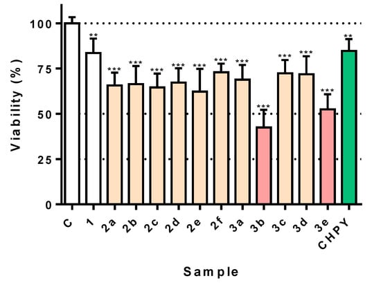
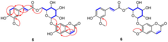
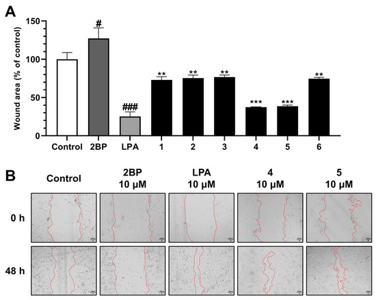
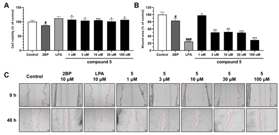
Nymphoides peltata has been used as a medicinal herb in traditional medicines to treat strangury, polyuria, and swelling. The phytochemical investigation of the MeOH extract of N. peltata roots led to the isolation of three iridoid glycosides and three coumarin glycoside derivatives, which were characterized as menthiafolin (1), threoninosecologanin (2), callicoside C (3), and scopolin (4), as well as two undescribed peltatamarins A (5) and B (6). The chemical structures of the undescribed compounds were determined by analyzing their 1 dimensional (D) and 2D nuclear magnetic resonance (NMR) spectra and using high-resolution (HR)-electrospray ionization mass spectroscopy (ESI-MS), along with the chemical reaction of acid hydrolysis. The wound healing activities of the isolated compounds 1–6 were evaluated using a HaCaT cell scratch test. Among the isolates, scopolin (4) and peltatamarin A (5) promoted HaCaT cell migration over scratch wounds, and compound 5 was the most effective. Furthermore, compound 5 significantly promoted cell migration without adversely affecting cell proliferation, even when treated at a high dose (100 μM). Our results demonstrate that peltatamarin A (5), isolated from N. peltata roots, has the potential for wound healing effects.
Full article
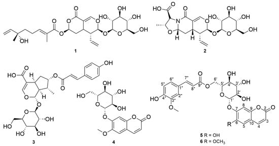
Figure 1


