Next Issue
Volume 12, May
Previous Issue
Volume 12, March


Pathogens, Volume 12, Issue 4 (April 2023) – 125 articles

Cover Story (view full-size image): 
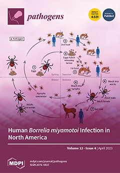
Borrelia miyamotoi is an emerging pathogen that is transmitted by the same hard-bodied (ixodid) ticks that transmit Borrelia burgdorferi, the causative agent of Lyme disease. Human cases have been reported in North America, Europe, and Asia. In North America, B. miyamotoi infection is widespread in Ixodes ticks in the Northeastern, Northern Midwestern, and Far Western United States and in Canada. In endemic areas, human B. miyamotoi seroprevalence averages from 1 to 3% of the population, compared with 15 to 20% for B. burgdorferi. The most common clinical manifestations of B. miyamotoi infection are fever, fatigue, headache, chills, myalgia, arthralgia, and nausea. Complications include relapsing fever and meningoencephalitis. Diagnosis is confirmed by PCR or blood smear. Antibiotic therapy is the same as for Lyme disease. View this paper
  • Issues are regarded as officially published after their release is announced to the table of contents alert mailing list.
  • You may sign up for e-mail alerts to receive table of contents of newly released issues.
  • PDF is the official format for papers published in both, html and pdf forms. To view the papers in pdf format, click on the "PDF Full-text" link, and use the free Adobe Reader to open them.
Order results
Result details
Section
Select all
Export citation of selected articles as:
11 pages, 5113 KiB  
Article
Establishment of a Simple and Rapid Nucleic Acid Detection Method for Hookworm Identification
by Xin Ding, Yougui Yang, Yingshu Zhang, Qiang Zhang, Fanzhen Mao and Yang Dai
Pathogens 2023, 12(4), 630; https://doi.org/10.3390/pathogens12040630 - 21 Apr 2023
Viewed by 1231
Abstract
Hookworm infection is one of the most common neglected tropical diseases and is mainly found in tropical and subtropical areas. Two species of human hookworm are distributed in China, i.e., Ancylostoma duodenale (AD) and Necator americanus (NA). Background: Traditional microscopic technology such as [...] Read more.
Hookworm infection is one of the most common neglected tropical diseases and is mainly found in tropical and subtropical areas. Two species of human hookworm are distributed in China, i.e., Ancylostoma duodenale (AD) and Necator americanus (NA). Background: Traditional microscopic technology such as the Kato-Katz method is not suitable for hookworm diagnosis due to the rapid degeneration of fragile hookworm eggs or for species identification of hookworm infection. The aim of the present study was to establish and evaluate a novel nucleic acid detection method based on recombinase-aided isothermal amplification (RAA) for the detection of hookworm infections and species identification. Methods: Based on the specific target gene sequences of hookworms (5.8S rRNA for AD and ITS2 for NA, respectively), we designed and synthesized amplification primers and fluorescence probes referring to the principle of the fluorescence recombinase-aided amplification (RAA) technique. Results: Each assay provided specific amplification of larval DNA from AD and NA by fluorescence RAA, and the detection limits in plasmids reached 102 copies and 10 copies, respectively. Genomic DNA of two hookworm species was successfully detected at a concentration of 0.1 pg/μL, revealing a high detection sensitivity. No positive amplification occurred for genomic DNA from crossed hookworm species and genomic DNA from Cryptosporidium, Giardia lamblia, Strongyloides stercoralis, Schistosoma japonicum, Ascaris lumbricoides, and Clonorchis sinensis, revealing a satisfactory specificity. Fecal sample detection results demonstrated a similar efficacy to the Kato-Katz method; however, it had a greater sensitivity than the larvae culture method. Conclusion: A simple and rapid nucleic acid method was successfully established based on RAA, which improved the detection efficacy and species identification for human hookworm infections. Full article
(This article belongs to the Special Issue State-of-Art and Perspectives of Parasites in China)
Show Figures

Figure 1

13 pages, 5379 KiB  
Review
Mechanism and Modulation of SidE Family Proteins in the Pathogenesis of Legionella pneumophila
by Yongchao Xie, Yi Zhang, Yong Wang and Yue Feng
Pathogens 2023, 12(4), 629; https://doi.org/10.3390/pathogens12040629 - 21 Apr 2023
Cited by 1 | Viewed by 1319
Abstract
Legionella pneumophila is the causative agent of Legionnaires’ disease, causing fever and lung infection, with a death rate up to 15% in severe cases. In the process of infection, Legionella pneumophila secretes over 330 effectors into host cell via the Dot/Icm type IV secretion [...] Read more.
Legionella pneumophila is the causative agent of Legionnaires’ disease, causing fever and lung infection, with a death rate up to 15% in severe cases. In the process of infection, Legionella pneumophila secretes over 330 effectors into host cell via the Dot/Icm type IV secretion system to modulate multiple host cellular physiological processes, thereby changing the environment of the host cell and promoting the growth and propagation of the bacterium. Among these effector proteins, SidE family proteins from Legionella pneumophila catalyze a non-canonical ubiquitination reaction, which combines mono-ADP-ribosylation and phosphodiesterase activities together to attach ubiquitin onto substrates. Meanwhile, the activity of SidE family proteins is also under multiple modulations by other effectors. Herein we summarize the key insights into recent studies in this area, emphasizing the tight link between the modular structure of SidE family proteins and the pathogen virulence as well as the fundamental mechanism and modulation network for further extensive research. Full article
(This article belongs to the Special Issue ADP-Ribosylation in Pathogens)
Show Figures

Figure 1

15 pages, 6114 KiB  
Article
Viability of African Swine Fever Virus with the Shallow Burial with Carbon Carcass Disposal Method
by Hoang Minh Duc, Mark Hutchinson, Gary A. Flory, Pham Hong Ngan, Hoang Minh Son, Le Van Hung, Tran Thi Khanh Hoa, Nguyen Thi Lan, Truong Quang Lam, Dale Rozeboom, Marta D. Remmenga, Matthew Vuolo, Robert Miknis, Amira Burns and Renée Flory
Pathogens 2023, 12(4), 628; https://doi.org/10.3390/pathogens12040628 - 21 Apr 2023
Viewed by 1901
Abstract
African swine fever (ASF) is a highly contagious swine disease with high mortality. In many countries, culling pigs infected and exposed to the ASF virus is mandatory to control the disease, which poses a real challenge in the disposal of large numbers of [...] Read more.
African swine fever (ASF) is a highly contagious swine disease with high mortality. In many countries, culling pigs infected and exposed to the ASF virus is mandatory to control the disease, which poses a real challenge in the disposal of large numbers of carcasses during ASF outbreaks. Shallow burial with carbon (SBC) Thanks ew mortality disposal method developed from deep burial and composting. The present study investigates the effectiveness of SBC in disposing of ASF virus-infected pigs. The real-time PCR results showed that DNA of the ASF virus was still detected in bone marrow samples on day 56, while the virus isolation test revealed that the infectious ASF virus was destroyed in both spleen and bone marrow samples on day 5. Interestingly, decomposition was found to occur rapidly in these shallow burial pits. On day 144, only large bones were found in the burial pit. In general, the results of this study indicated that SBC is a potential method for the disposal of ASF-infected carcasses; however, further studies are needed to provide more scientific evidence for the efficacy of SBC in different environment conditions. Full article
(This article belongs to the Special Issue An Update on African Swine Fever)
Show Figures

Figure 1

13 pages, 305 KiB  
Review
Familial Hypercholesterolemia and Acute Coronary Syndromes: The Microbiota–Immunity Axis in the New Diagnostic and Prognostic Frontiers
by Andrea Piccioni, Elena Niccolai, Gloria Rozzi, Giacomo Spaziani, Christian Zanza, Marcello Candelli, Marcello Covino, Antonio Gasbarrini, Francesco Franceschi and Amedeo Amedei
Pathogens 2023, 12(4), 627; https://doi.org/10.3390/pathogens12040627 - 21 Apr 2023
Cited by 3 | Viewed by 1882
Abstract
Familial hypercholesterolemia is a common genetic disorder with a propensity towards early onset of atherosclerotic cardiovascular disease (CVD). The main goal of therapy is to reduce the LDL cholesterol and the current treatment generally consists of statin, ezetimibe and PCSK9 inhibitors. Unfortunately, lowering [...] Read more.
Familial hypercholesterolemia is a common genetic disorder with a propensity towards early onset of atherosclerotic cardiovascular disease (CVD). The main goal of therapy is to reduce the LDL cholesterol and the current treatment generally consists of statin, ezetimibe and PCSK9 inhibitors. Unfortunately, lowering LDL cholesterol may be difficult for many reasons such as the variation of response to statin therapy among the population or the high cost of some therapies (i.e., PCSK9 inhibitors). In addition to conventional therapy, additional strategies may be used. The gut microbiota has been recently considered to play a part in chronic systemic inflammation and hence in CVD. Several studies, though they are still preliminary, consider dysbiosis a risk factor for various CVDs through several mechanisms. In this review, we provide an update of the current literature about the intricate relation between the gut microbiota and the familial hypercholesterolemia. Full article
(This article belongs to the Special Issue Gut Microbiome: Current Status and Future Perspectives)
17 pages, 2051 KiB  
Article
SARS-CoV-2 Variants by Whole-Genome Sequencing in a University Hospital in Bangkok: First to Third COVID-19 Waves
by Chayanee Setthapramote, Thanwa Wongsuk, Chuphong Thongnak, Uraporn Phumisantiphong, Tonsan Hansirisathit and Maytawan Thanunchai
Pathogens 2023, 12(4), 626; https://doi.org/10.3390/pathogens12040626 - 21 Apr 2023
Cited by 1 | Viewed by 1582
Abstract
Background: Multiple severe acute respiratory syndrome coronavirus 2 (SARS-CoV-2) variants emerged globally during the recent coronavirus disease (COVID-19) pandemic. From April 2020 to April 2021, Thailand experienced three COVID-19 waves, and each wave was driven by different variants. Therefore, we aimed to analyze [...] Read more.
Background: Multiple severe acute respiratory syndrome coronavirus 2 (SARS-CoV-2) variants emerged globally during the recent coronavirus disease (COVID-19) pandemic. From April 2020 to April 2021, Thailand experienced three COVID-19 waves, and each wave was driven by different variants. Therefore, we aimed to analyze the genetic diversity of circulating SARS-CoV-2 using whole-genome sequencing analysis. Methods: A total of 33 SARS-CoV-2 positive samples from three consecutive COVID-19 waves were collected and sequenced by whole-genome sequencing, of which, 8, 10, and 15 samples were derived from the first, second, and third waves, respectively. The genetic diversity of variants in each wave and the correlation between mutations and disease severity were explored. Results: During the first wave, A.6, B, B.1, and B.1.375 were found to be predominant. The occurrence of mutations in these lineages was associated with low asymptomatic and mild symptoms, providing no transmission advantage and resulting in extinction after a few months of circulation. B.1.36.16, the predominant lineage of the second wave, caused more symptomatic COVID-19 cases and contained a small number of key mutations. This variant was replaced by the VOC alpha variant, which later became dominant in the third wave. We found that B.1.1.7 lineage-specific mutations were crucial for increasing transmissibility and infectivity, but not likely associated with disease severity. There were six additional mutations found only in severe COVID-19 patients, which might have altered the virus phenotype with an inclination toward more highly pathogenic SARS-CoV-2. Conclusion: The findings of this study highlighted the importance of whole-genome analysis in tracking newly emerging variants, exploring the genetic determinants essential for transmissibility, infectivity, and pathogenicity, and helping better understand the evolutionary process in the adaptation of viruses in humans. Full article
(This article belongs to the Topic Acute Respiratory Viruses Molecular Epidemiology)
Show Figures

Figure 1

10 pages, 854 KiB  
Article
Comparison of Antibody Isotype Response to Angiostrongylus cantonensis in Experimentally Infected Rats (Rattus norvegicus) Using Hawai’i 31 kDa Antigen in an Indirect ELISA
by Argon Steel, Lisa Kaluna, John Jacob and Susan Jarvi
Pathogens 2023, 12(4), 625; https://doi.org/10.3390/pathogens12040625 - 21 Apr 2023
Viewed by 1171
Abstract
Neuroangiostrongyliasis (NAS) is an emerging tropical disease in humans and some animals which is caused by infection with the parasitic nematode Angiostrongylus cantonensis. It is the leading cause of eosinophilic meningitis worldwide. Diagnoses in humans and susceptible animals are generally presumptive and [...] Read more.
Neuroangiostrongyliasis (NAS) is an emerging tropical disease in humans and some animals which is caused by infection with the parasitic nematode Angiostrongylus cantonensis. It is the leading cause of eosinophilic meningitis worldwide. Diagnoses in humans and susceptible animals are generally presumptive and easily confused with other central nervous system disorders. The 31 kDa antigen is currently the only NAS immunodiagnostic assay that has achieved 100% sensitivity. However, little is known about the humoral immune response against the 31 kDa antigen in NAS infections, which would be critical for widespread adoption of this assay. We used the Hawai’i 31 kDa isolate in an indirect ELISA assay to confirm the presence of immunoglobulin IgG, IgM, IgA, and IgE isotypes in six-week post-infection plasma from lab-reared rats infected with 50 live, third-stage, A. cantonensis larvae isolated from a wild Parmarion martensi semi-slug. Our results confirmed the presence of all four isotypes against the Hawaii 31 kDa isolate, with sensitivity ranging from 22–100%. The IgG isotype showed 100% sensitivity in detecting A. cantonensis infection, which validates the use of IgG indirect ELISA with 31 kDa antigen as an effective immunodiagnostic assay for rats six weeks post-infection. Given each isotype may be present at different times during NAS infections, our data provides preliminary information on the humoral immune response to A. cantonensis infection in lab-reared rats and serves as a baseline for future studies. Full article
(This article belongs to the Special Issue Rat Lungworm Disease)
Show Figures

Figure 1

8 pages, 651 KiB  
Viewpoint
Neuroangiostrongyliasis: Updated Provisional Guidelines for Diagnosis and Case Definitions
by Carlos Graeff-Teixeira, Kittisak Sawanyawisuth, Shan Lv, William Sears, Zhaily González Rodríguez, Hilda Hernández Álvarez, Pedro Casanova Arias, Leticia Karolini Walger Schultz, Alicia Rojas, John Jacob, Susan Jarvi and Kenton Kramer
Pathogens 2023, 12(4), 624; https://doi.org/10.3390/pathogens12040624 - 20 Apr 2023
Cited by 1 | Viewed by 1686
Abstract
Angiostrongylus cantonensis is the main causative agent for eosinophilic meningoencephalitis in humans. Larvae are rarely found in the cerebral spinal fluid (CSF). Consequently, serology and DNA detection represent important diagnostic tools. However, interpretation of the results obtained from these tools requires that more [...] Read more.
Angiostrongylus cantonensis is the main causative agent for eosinophilic meningoencephalitis in humans. Larvae are rarely found in the cerebral spinal fluid (CSF). Consequently, serology and DNA detection represent important diagnostic tools. However, interpretation of the results obtained from these tools requires that more extensive accuracy studies be conducted. The aim of the present study is to update guidelines for diagnosis and case definitions of neuroangiostrongyliasis (NA) as provided by a working group of a recently established International Network on Angiostrongyliasis. A literature review, a discussion regarding criteria and diagnostic categories, recommendations issued by health authorities in China and an expert panel in Hawaii (USA), and the experience of Thailand were considered. Classification of NA cases and corresponding criteria are proposed as follows: minor (exposure history, positive serology, and blood eosinophilia); major (headache or other neurological signs or symptoms, CSF eosinophilia); and confirmatory (parasite detection in tissues, ocular chambers, or CSF, or DNA detection by PCR and sequencing). In addition, diagnostic categories or suspected, probable, and confirmatory are proposed. Updated guidelines should improve clinical study design, epidemiological surveillance, and the proper characterization of biological samples. Moreover, the latter will further facilitate accuracy studies of diagnostic tools for NA to provide better detection and treatment. Full article
(This article belongs to the Special Issue Rat Lungworm Disease)
Show Figures

Graphical abstract

17 pages, 2005 KiB  
Review
Urinary Tract Infections: The Current Scenario and Future Prospects
by Giuseppe Mancuso, Angelina Midiri, Elisabetta Gerace, Maria Marra, Sebastiana Zummo and Carmelo Biondo
Pathogens 2023, 12(4), 623; https://doi.org/10.3390/pathogens12040623 - 20 Apr 2023
Cited by 33 | Viewed by 16502
Abstract
Urinary tract infections (UTIs) are among the most common bacterial infections worldwide, occurring in both community and healthcare settings. Although the clinical symptoms of UTIs are heterogeneous and range from uncomplicated (uUTIs) to complicated (cUTIs), most UTIs are usually treated empirically. Bacteria are [...] Read more.
Urinary tract infections (UTIs) are among the most common bacterial infections worldwide, occurring in both community and healthcare settings. Although the clinical symptoms of UTIs are heterogeneous and range from uncomplicated (uUTIs) to complicated (cUTIs), most UTIs are usually treated empirically. Bacteria are the main causative agents of these infections, although more rarely, other microorganisms, such as fungi and some viruses, have been reported to be responsible for UTIs. Uropathogenic Escherichia coli (UPEC) is the most common causative agent for both uUTIs and cUTIs, followed by other pathogenic microorganisms, such as Klebsiella pneumoniae, Proteus mirabilis, Enterococcus faecalis, and Staphylococcus spp. In addition, the incidence of UTIs caused by multidrug resistance (MDR) is increasing, resulting in a significant increase in the spread of antibiotic resistance and the economic burden of these infections. Here, we discuss the various factors associated with UTIs, including the mechanisms of pathogenicity related to the bacteria that cause UTIs and the emergence of increasing resistance in UTI pathogens. Full article
(This article belongs to the Special Issue Pathogens in 2023)
Show Figures

Figure 1

12 pages, 425 KiB  
Article
Feral Swine as Indirect Indicators of Environmental Anthrax Contamination and Potential Mechanical Vectors of Infectious Spores
by Rachel M. Maison, Maggie R. Priore, Vienna R. Brown, Michael J. Bodenchuk, Bradley R. Borlee, Richard A. Bowen and Angela M. Bosco-Lauth
Pathogens 2023, 12(4), 622; https://doi.org/10.3390/pathogens12040622 - 20 Apr 2023
Cited by 1 | Viewed by 1601
Abstract
Anthrax is a disease that affects livestock, wildlife, and humans worldwide; however, its relative impacts on these populations remain underappreciated. Feral swine (Sus scrofa) are relatively resistant to developing anthrax, and past serosurveys have alluded to their utility as sentinels, yet [...] Read more.
Anthrax is a disease that affects livestock, wildlife, and humans worldwide; however, its relative impacts on these populations remain underappreciated. Feral swine (Sus scrofa) are relatively resistant to developing anthrax, and past serosurveys have alluded to their utility as sentinels, yet empirical data to support this are lacking. Moreover, whether feral swine may assist in the dissemination of infectious spores is unknown. To address these knowledge gaps, we intranasally inoculated 15 feral swine with varying quantities of Bacillus anthracis Sterne 34F2 spores and measured the seroconversion and bacterial shedding over time. The animals also were inoculated either one or three times. The sera were evaluated by enzyme-linked immunosorbent assay (ELISA) for antibodies against B. anthracis, and nasal swabs were cultured to detect bacterial shedding from the nasal passages. We report that the feral swine developed antibody responses to B. anthracis and that the strength of the response correlated with the inoculum dose and the number of exposure events experienced. Isolation of viable bacteria from the nasal passages of the animals throughout the study period suggests that feral swine may assist in the spread of infectious spores on the landscape and have implications for the identification of environments contaminated with B. anthracis as well as the exposure risk to more susceptible hosts. Full article
(This article belongs to the Special Issue Pathogens in 2023)
Show Figures

Figure 1

15 pages, 5178 KiB  
Article
Characterization and Control of Dendrobium officinale Bud Blight Disease
by Jinzhao Zhang, Haodong Sha, Weiliang Chen and Bizeng Mao
Pathogens 2023, 12(4), 621; https://doi.org/10.3390/pathogens12040621 - 20 Apr 2023
Cited by 1 | Viewed by 1330
Abstract
Dendrobium officinale is an important traditional Chinese medicine (TCM). A disease causing bud blight in D. officinale appeared in 2021 in Yueqing city, Zhejiang Province, China. In this paper, 127 isolates were obtained from 61 plants. The isolates were grouped into 13 groups [...] Read more.
Dendrobium officinale is an important traditional Chinese medicine (TCM). A disease causing bud blight in D. officinale appeared in 2021 in Yueqing city, Zhejiang Province, China. In this paper, 127 isolates were obtained from 61 plants. The isolates were grouped into 13 groups based on collected areas and morphological observations. Four loci (ITS, LSU, tub2 and rpb2) of 13 representative isolates were sequenced and the isolates were identified by constructing phylogenetic trees with the multi-locus sequence analysis (MLSA) method. We found the disease to be associated with three strains: Ectophoma multirostrata, Alternaria arborescens and Stagonosporopsis pogostemonis, with isolates frequencies of 71.6%, 21.3% and 7.1%, respectively. All three strains are pathogenic to D. officinale. A. arborescens and S. pogostemonis isolated from D. officinale were reported for the first time. Iprodione (50%), 33.5% oxine-copper and Meitian (containing 75 g/L pydiflumetofen and 125 g/L difenoconazole) were chosen to control the dominant pathogen E. multirostrata, with an EC50 value of 2.10, 1.78 and 0.09 mg/L, respectively. All three fungicides exhibited an effective inhibition of activities to the growth of the dominant pathogen E. multirostrata on potato dextrose agar (PDA) plates, with Meitian showing the strongest inhibitory effect. We further found that Meitian can effectively control D. officinale bud blight disease in pot trial. Full article
Show Figures

Figure 1

16 pages, 2476 KiB  
Article
A Retrospective Assessment of Sputum Samples and Antimicrobial Resistance in COVID-19 Patients
by Talida Georgiana Cut, Adelina Mavrea, Alin Adrian Cumpanas, Dorin Novacescu, Cristian Iulian Oancea, Felix Bratosin, Adelina Raluca Marinescu, Ruxandra Laza, Alexandra Mocanu, Alexandru Silvius Pescariu, Diana Manolescu, Raluca Dumache, Alexandra Enache, Elena Hogea and Voichita Elena Lazureanu
Pathogens 2023, 12(4), 620; https://doi.org/10.3390/pathogens12040620 - 19 Apr 2023
Cited by 6 | Viewed by 1708
Abstract
Data on bacterial or fungal pathogens and their impact on the mortality rates of Western Romanian COVID-19 patients are scarce. As a result, the purpose of this research was to determine the prevalence of bacterial and fungal co- and superinfections in Western Romanian [...] Read more.
Data on bacterial or fungal pathogens and their impact on the mortality rates of Western Romanian COVID-19 patients are scarce. As a result, the purpose of this research was to determine the prevalence of bacterial and fungal co- and superinfections in Western Romanian adults with COVID-19, hospitalized in in-ward settings during the second half of the pandemic, and its distribution according to sociodemographic and clinical conditions. The unicentric retrospective observational study was conducted on 407 eligible patients. Expectorate sputum was selected as the sampling technique followed by routine microbiological investigations. A total of 31.5% of samples tested positive for Pseudomonas aeruginosa, followed by 26.2% having co-infections with Klebsiella pneumoniae among patients admitted with COVID-19. The third most common Pathogenic bacteria identified in the sputum samples was Escherichia coli, followed by Acinetobacter baumannii in 9.3% of samples. Commensal human pathogens caused respiratory infections in 67 patients, the most prevalent being Streptococcus penumoniae, followed by methicillin-sensitive and methicillin-resistant Staphylococcus aureus. A total of 53.4% of sputum samples tested positive for Candida spp., followed by 41.1% of samples with Aspergillus spp. growth. The three groups with positive microbial growth on sputum cultures had an equally proportional distribution of patients admitted to the ICU, with an average of 30%, compared with only 17.3% among hospitalized COVID-19 patients with negative sputum cultures (p = 0.003). More than 80% of all positive samples showed multidrug resistance. The high prevalence of bacterial and fungal co-infections and superinfections in COVID-19 patients mandates for strict and effective antimicrobial stewardship and infection control policies. Full article
(This article belongs to the Special Issue Epidemiology of Pathogenic Agents)
Show Figures

Figure 1

19 pages, 2394 KiB  
Review
Natural and Engineered Resistance Mechanisms in Plants against Phytoviruses
by Anik Majumdar, Abhishek Sharma and Rakesh Belludi
Pathogens 2023, 12(4), 619; https://doi.org/10.3390/pathogens12040619 - 19 Apr 2023
Cited by 1 | Viewed by 2012
Abstract
Plant viruses, as obligate intracellular parasites, rely exclusively on host machinery to complete their life cycle. Whether a virus is pathogenic or not depends on the balance between the mechanisms used by both plants and viruses during the intense encounter. Antiviral defence mechanisms [...] Read more.
Plant viruses, as obligate intracellular parasites, rely exclusively on host machinery to complete their life cycle. Whether a virus is pathogenic or not depends on the balance between the mechanisms used by both plants and viruses during the intense encounter. Antiviral defence mechanisms in plants can be of two types, i.e., natural resistance and engineered resistance. Innate immunity, RNA silencing, translational repression, autophagy-mediated degradation, and resistance to virus movement are the possible natural defence mechanisms against viruses in plants, whereas engineered resistance includes pathogen-derived resistance along with gene editing technologies. The incorporation of various resistance genes through breeding programmes, along with gene editing tools such as CRISPR/Cas technologies, holds great promise in developing virus-resistant plants. In this review, different resistance mechanisms against viruses in plants along with reported resistance genes in major vegetable crops are discussed. Full article
(This article belongs to the Special Issue Emerging and Re-emerging Plant Viruses in a Context of Global Change)
Show Figures

Figure 1

11 pages, 268 KiB  
Article
Enteric Pathogens Detected in Children under Five Years Old Admitted with Diarrhea in Moshi, Kilimanjaro, Tanzania
by Ephrasia A. Hugho, Happiness H. Kumburu, Nelson B. Amani, Bahati Mseche, Athanasia Maro, Lilian E. Ngowi, Yudathadei Kyara, Grace Kinabo, Kate M. Thomas, Eric R. Houpt, Jie Liu, Tine Hald and Blandina T. Mmbaga
Pathogens 2023, 12(4), 618; https://doi.org/10.3390/pathogens12040618 - 19 Apr 2023
Cited by 5 | Viewed by 1817
Abstract
Despite the availability and wide coverage of rotavirus vaccinations in Tanzania, there is still a significant number of diarrhea cases being reported, with some patients requiring hospital admission. We investigated diarrhea-causing pathogens and determined the effect of co-infection on clinical symptoms. Total nucleic [...] Read more.
Despite the availability and wide coverage of rotavirus vaccinations in Tanzania, there is still a significant number of diarrhea cases being reported, with some patients requiring hospital admission. We investigated diarrhea-causing pathogens and determined the effect of co-infection on clinical symptoms. Total nucleic acid was extracted from archived stool samples (N = 146) collected from children (0–59 months) admitted with diarrhea in health facilities in Moshi, Kilimanjaro. Pathogen detection was performed using the quantitative polymerase chain reaction with custom TaqMan Array cards. The Poisson model was used to determine the effect of co-infection on clinical presentation during admission. Of all the participants, 56.85% were from rural Moshi with a median age of 11.74 months (IQR: 7.41–19.09). Vomiting (88.36%) and a fever (60.27%) were the most frequent clinical manifestations. At least one diarrhea-associated pathogen was detected in 80.14% (n = 117) of the study population. The most prevalent pathogens were rotavirus 38.36% (n = 56), adenovirus 40/41 19.86% (n = 29), Shigella/EIEC 12.33% (n = 18), norovirus GII 11.44% (n = 17) and Cryptosporidium 9.59% (n = 14). Co-infections were detected in 26.03% of the study population (n = 38). The presence of multiple pathogens in the stool samples of children with diarrhea indicates poor sanitation and may have significant implications for disease management and patient outcomes. Full article
(This article belongs to the Special Issue Burden, Prevention, and Control of Enteric Viral Infections)
34 pages, 7584 KiB  
Review
Antifungal Compounds from Microbial Symbionts Associated with Aquatic Animals and Cellular Targets: A Review
by Madeleine Nina Love Ngo-Mback, Elisabeth Zeuko’o Menkem and Heather G. Marco
Pathogens 2023, 12(4), 617; https://doi.org/10.3390/pathogens12040617 - 18 Apr 2023
Cited by 1 | Viewed by 1493
Abstract
Fungal infections continue to be a serious public health problem, leading to an estimated 1.6 million deaths annually. It remains a major cause of mortality for people with a weak or affected immune system, such as those suffering from cancer under aggressive chemotherapies. [...] Read more.
Fungal infections continue to be a serious public health problem, leading to an estimated 1.6 million deaths annually. It remains a major cause of mortality for people with a weak or affected immune system, such as those suffering from cancer under aggressive chemotherapies. On the other hand, pathogenic fungi are counted among the most destructive factors affecting crops, causing a third of all food crop losses annually and critically affecting the worldwide economy and food security. However, the limited number currently available and the cytotoxicity of the conventional antifungal drugs, which are not yet properly diversified in terms of mode of action, in addition to resistance phenomena, make the search for new antifungals imperative to improve both human health and food protection. Symbiosis has been a crucial alternative for drug discovery, through which many antimicrobials have been discovered. This review highlights some antifungal models of a defensive symbiosis of microbial symbiont natural products derived from interacting with aquatic animals as one of the best opportunities. Some recorded compounds with supposed novel cell targets such as apoptosis could lead to the development of a multitherapy involving the mutual treatment of fungal infections and other metabolic diseases involving apoptosis in their pathogenesis pathways. Full article
3 pages, 200 KiB  
Editorial
Tsunami-Driven Spread of Toxoplasma gondii and Other Microbial Pathogens: Implications for Cetacean Health and Conservation
by Giovanni Di Guardo
Pathogens 2023, 12(4), 616; https://doi.org/10.3390/pathogens12040616 - 18 Apr 2023
Cited by 1 | Viewed by 929
Abstract
Six years have now gone by since Dr James T [...] Full article
(This article belongs to the Section Parasitic Pathogens)
12 pages, 3436 KiB  
Article
The Identification of Streptococcus pasteurianus Obtained from Six Regions in China by Multiplex PCR Assay and the Characteristics of Pathogenicity and Antimicrobial Resistance of This Zoonotic Pathogen
by Miaohang Ma, Shuoyue Wang, Xinchi Zhu, Xinchun Li, Yinli Bao, Xiang Chen and Zongfu Wu
Pathogens 2023, 12(4), 615; https://doi.org/10.3390/pathogens12040615 - 18 Apr 2023
Cited by 2 | Viewed by 1307
Abstract
Streptococcus pasteurianus is a zoonotic pathogen causing meningitis and bacteremia in animals and humans. A lack of accurate and convenient detection methods hinders preventing and controlling diseases caused by S. pasteurianus. Additionally, there is limited knowledge about its pathogenicity and antimicrobial resistance [...] Read more.
Streptococcus pasteurianus is a zoonotic pathogen causing meningitis and bacteremia in animals and humans. A lack of accurate and convenient detection methods hinders preventing and controlling diseases caused by S. pasteurianus. Additionally, there is limited knowledge about its pathogenicity and antimicrobial resistance characteristics, as there are only three complete genome sequences available. In this study, we established a multiplex PCR assay for the detection of S. pasteurianus, which was applied to six fecal samples from cattle with diarrhea and 285 samples from healthy pigs. Out of the samples tested, 24 were positive, including 5 from pig tonsils, 18 from pig hilar lymph nodes, and 1 from cattle feces. Two strains were isolated from positive samples, and their complete genomes were sequenced. The two strains were non-virulent in mice and multidrug-resistant by the antimicrobial susceptibility test. We first found the presence of genes tet(O/W/32/O) and lsa(E) in S. pasteurianus, leading to resistance to lincosamides and tetracyclines. The convenient and specific multiplex PCR assay provides essential technical support for epidemiological research, and the complete genome sequence of two non-virulent strains contributes to understanding this zoonotic bacterium’s genomic characteristics and pathogenesis. Full article
Show Figures

Figure 1

10 pages, 1055 KiB  
Article
Infectiousness of Asymptomatic Meriones shawi, Reservoir Host of Leishmania major
by Jovana Sadlova, Barbora Vojtkova, Tereza Lestinova, Tomas Becvar, Daniel Frynta, Kamal Eddine Benallal, Nalia Mekarnia, Zoubir Harrat and Petr Volf
Pathogens 2023, 12(4), 614; https://doi.org/10.3390/pathogens12040614 - 18 Apr 2023
Cited by 1 | Viewed by 1225
Abstract
Leishmaniases are neglected diseases caused by protozoans of the genus Leishmania that threaten millions of people worldwide. Cutaneous leishmaniasis (CL) caused by L. major is a typical zoonosis transmitted by phlebotomine sand flies and maintained in rodent reservoirs. The female sand fly was [...] Read more.
Leishmaniases are neglected diseases caused by protozoans of the genus Leishmania that threaten millions of people worldwide. Cutaneous leishmaniasis (CL) caused by L. major is a typical zoonosis transmitted by phlebotomine sand flies and maintained in rodent reservoirs. The female sand fly was assumed to become infected by feeding on the skin lesion of the host, and the relative contribution of asymptomatic individuals to disease transmission was unknown. In this study, we infected 32 Meriones shawi, North African reservoirs, with a natural dose of L. major obtained from the gut of infected sand flies. Skin manifestations appeared in 90% of the animals, and xenodiagnosis with the proven vector Phlebotomus papatasi showed transmissibility in 67% of the rodents, and 45% were repeatedly infectious to sand flies. Notably, the analysis of 113 xenodiagnostic trials with 2189 sand flies showed no significant difference in the transmissibility of animals in the asymptomatic and symptomatic periods; asymptomatic animals were infectious several weeks before the appearance of skin lesions and several months after their healing. These results clearly confirm that skin lesions are not a prerequisite for vector infection in CL and that asymptomatic animals are an essential source of L. major infection. These data are important for modeling the epidemiology of CL caused by L. major. Full article
Show Figures

Figure 1

6 pages, 492 KiB  
Communication
Reduced Cholesterol Levels during Acute Human Babesiosis
by Luis A. Marcos, Charles Kyriakos Vorkas, Inderjit Mann, Evan Garry, Pooja Lamba, Sophia K. Pham, Rachel Spector, Aikaterini Papamanoli, Sara Krivacsy, Michael Lum, Aleena Zahra, Wei Hou and Eric D. Spitzer
Pathogens 2023, 12(4), 613; https://doi.org/10.3390/pathogens12040613 - 18 Apr 2023
Viewed by 1204
Abstract
Background: Babesiosis, an intra-erythrocytic protozoan disease, is an emerging zoonotic parasitic disease worldwide. Cholesterol levels are correlated with severe infections, such as sepsis and COVID-19, and anecdotal reports suggest that high-density lipoprotein (HDL) cholesterol declines during acute babesiosis. Our aim was to describe [...] Read more.
Background: Babesiosis, an intra-erythrocytic protozoan disease, is an emerging zoonotic parasitic disease worldwide. Cholesterol levels are correlated with severe infections, such as sepsis and COVID-19, and anecdotal reports suggest that high-density lipoprotein (HDL) cholesterol declines during acute babesiosis. Our aim was to describe the cholesterol levels in patients with acute babesiosis diagnosed in an endemic area in New York, hypothesizing that HDL levels correlate with the severity of infection. Methods: We reviewed the medical records of adult patients with babesiosis diagnosed by identification of Babesia parasites on a thin blood smear and confirmed by polymerase chain reaction from 2013 to 2018, who also had available a lipid profile drawn at the time of clinical presentation. Additional lipid profile levels were considered as “baseline” if they were drawn within 2 months before or after the infection as part of routine care. Results: A total of 39 patients with babesiosis had a lipid profile drawn on presentation. The patients were divided into two groups for comparison based on the treating physician’s clinical decision: 33 patients who were admitted to the hospital and 8 patients who were evaluated as outpatients. A history of hypertension was more common in admitted patients (37% vs. 17%, p = 0.02). The median levels of low-density lipoprotein (LDL) and HDL were significantly reduced in admitted patients compared to non-admitted patients (46 vs. 76 mg/dL, p = 0.04; and 9 vs. 28.5 mg/dL, p = 0.03, respectively). In addition, LDL and HDL levels returned to baseline values following resolution of acute babesiosis. Conclusion: LDL and HDL levels are significantly reduced during acute babesiosis, suggesting that cholesterol depletion may predict disease severity. Pathogen and host factors may contribute to a reduction in serum cholesterol levels during acute babesiosis. Full article
Show Figures

Figure 1

14 pages, 1093 KiB  
Review
Clinical Evidence for the Use of Octenidine Dihydrochloride to Prevent Healthcare-Associated Infections and Decrease Staphylococcus aureus Carriage or Transmission—A Review
by Robin Köck, Luisa Denkel, Andrea T. Feßler, Rudolf Eicker, Alexander Mellmann, Stefan Schwarz, Christine Geffers, Nils-Olaf Hübner and Rasmus Leistner
Pathogens 2023, 12(4), 612; https://doi.org/10.3390/pathogens12040612 - 18 Apr 2023
Cited by 1 | Viewed by 2043
Abstract
Background: The antiseptic agent octenidine dihydrochloride (OCT) is used for skin preparation, for Staphylococcus aureus decolonization, and within bundles for the prevention of catheter-related or surgical site infections (SSIs). Here, we review the evidence for the effects of OCT from clinical studies. Methods: [...] Read more.
Background: The antiseptic agent octenidine dihydrochloride (OCT) is used for skin preparation, for Staphylococcus aureus decolonization, and within bundles for the prevention of catheter-related or surgical site infections (SSIs). Here, we review the evidence for the effects of OCT from clinical studies. Methods: Review of studies published in the Medline, Scopus, and Cochrane databases until August 2022, performed in clinical settings and reporting on effects of OCT on S. aureus carriage/transmission, SSI prevention, and prevention of intensive care unit (ICU)-related or catheter-related bloodstream and insertion site infections. Results: We included 31 articles. The success of S. aureus decolonization with OCT-containing therapies ranged between 6 and 87%. Single studies demonstrated that OCT application led to a reduction in S. aureus infections, acquisition, and carriage. No study compared OCT for skin preparation before surgical interventions to other antiseptics. Weak evidence for the use of OCT for pre-operative washing was found in orthopedic and cardiac surgery, if combined with other topical measures. Mostly, studies did not demonstrate that daily OCT bathing reduced ICU-/catheter-related bloodstream infections with one exception. Conclusions: There is a need to perform studies assessing the clinical use of OCT compared with other antiseptics with respect to its effectiveness to prevent nosocomial infections. Full article
Show Figures

Figure 1

7 pages, 372 KiB  
Article
Impact of the COVID-19 Pandemic on the Management of Staphylococcus aureus Bloodstream Infections in a Tertiary Care Hospital
by Christian W. Böing, Neele J. Froböse, Frieder Schaumburg and Stefanie Kampmeier
Pathogens 2023, 12(4), 611; https://doi.org/10.3390/pathogens12040611 - 18 Apr 2023
Cited by 1 | Viewed by 1085
Abstract
Staphylococcus aureus bacteremia (SAB) is associated with a high mortality rate. The clinical outcome of SAB patients highly depends on early diagnosis, adequate antibiotic therapy and source control. In the context of the COVID-19 pandemic, the health care system faced additional organizational challenges [...] Read more.
Staphylococcus aureus bacteremia (SAB) is associated with a high mortality rate. The clinical outcome of SAB patients highly depends on early diagnosis, adequate antibiotic therapy and source control. In the context of the COVID-19 pandemic, the health care system faced additional organizational challenges and the question arose whether structured screening and triaging for COVID-19 and shifting resources influence the management of SAB. Patients (n = 115) with SAB were enrolled in a retrospective comparative study with historical controls (March 2019–February 2021). The quality of SAB therapy was assessed with a point score, which included correct choice of antibiotic, adequate dosage of antibiotic, sufficient duration of therapy, early start of therapy after receipt of findings, focus search and taking control blood cultures 3–4 days after starting adequate antibiotic therapy. The quality of treatment before and after the onset of the COVID-19 pandemic were compared. No significant differences in the total score points were found between the pre-COVID-19 and COVID-19 cohort. All quality indicators, except the correct duration of antibiotic therapy, showed no significant differences in both cohorts. Furthermore, there were no significant differences in the outcome between both cohorts. The treatment quality of SAB therapy was comparable before and during the COVID-19 pandemic. Full article
(This article belongs to the Special Issue Recent Research on Hospital-Acquired Bloodstream Infections)
Show Figures

Figure 1

17 pages, 1183 KiB  
Review
Avian Influenza: Strategies to Manage an Outbreak
by Alison Simancas-Racines, Santiago Cadena-Ullauri, Patricia Guevara-Ramírez, Ana Karina Zambrano and Daniel Simancas-Racines
Pathogens 2023, 12(4), 610; https://doi.org/10.3390/pathogens12040610 - 17 Apr 2023
Cited by 2 | Viewed by 5183
Abstract
Avian influenza (AI) is a contagious disease among the poultry population with high avian mortality, which generates significant economic losses and elevated costs for disease control and outbreak eradication. AI is caused by an RNA virus part of the Orthomyxoviridae family; however, only [...] Read more.
Avian influenza (AI) is a contagious disease among the poultry population with high avian mortality, which generates significant economic losses and elevated costs for disease control and outbreak eradication. AI is caused by an RNA virus part of the Orthomyxoviridae family; however, only Influenzavirus A is capable of infecting birds. AI pathogenicity is based on the lethality, signs, and molecular characteristics of the virus. Low pathogenic avian influenza (LPAI) virus has a low mortality rate and ability to infect, whereas the highly pathogenic avian influenza (HPAI) virus can cross respiratory and intestinal barriers, diffuse to the blood, damage all tissues of the bird, and has a high mortality rate. Nowadays, avian influenza is a global public health concern due to its zoonotic potential. Wild waterfowl is the natural reservoir of AI viruses, and the oral–fecal path is the main transmission route between birds. Similarly, transmission to other species generally occurs after virus circulation in densely populated infected avian species, indicating that AI viruses can adapt to promote the spread. Moreover, HPAI is a notifiable animal disease; therefore, all countries must report infections to the health authorities. Regarding laboratory diagnoses, the presence of influenza virus type A can be identified by agar gel immunodiffusion (AGID), enzyme immunoassay (EIA), immunofluorescence assays, and enzyme-linked immunoadsorption assay (ELISAs). Furthermore, reverse transcription polymerase chain reaction is used for viral RNA detection and is considered the gold standard for the management of suspect and confirmed cases of AI. If there is suspicion of a case, epidemiological surveillance protocols must be initiated until a definitive diagnosis is obtained. Moreover, if there is a confirmed case, containment actions should be prompt and strict precautions must be taken when handling infected poultry cases or infected materials. The containment measures for confirmed cases include the sanitary slaughter of infected poultry using methods such as environment saturation with CO2, carbon dioxide foam, and cervical dislocation. For disposal, burial, and incineration, protocols should be followed. Lastly, disinfection of affected poultry farms must be carried out. The present review aims to provide an overview of the avian influenza virus, strategies for its management, the challenges an outbreak can generate, and recommendations for informed decision making. Full article
(This article belongs to the Special Issue Emerging and Re-emerging Viral Infectious Diseases)
Show Figures

Figure 1

15 pages, 705 KiB  
Article
Snapshot of Phenotypic and Molecular Virulence and Resistance Profiles in Multidrug-Resistant Strains Isolated in a Tertiary Hospital in Romania
by Bianca Simona Truşcă, Irina Gheorghe-Barbu, Marina Manea, Elvira Ianculescu, Ilda Czobor Barbu, Luminița Gabriela Măruțescu, Lia-Mara Dițu, Mariana-Carmen Chifiriuc and Veronica Lazăr
Pathogens 2023, 12(4), 609; https://doi.org/10.3390/pathogens12040609 - 17 Apr 2023
Cited by 3 | Viewed by 1517
Abstract
A current major healthcare problem is represented by antibiotic resistance, mainly due to multidrug resistant (MDR) Gram negative bacilli (GNB), because of their extended spread both in hospital facilities and in the community’s environment. The aim of this study was to investigate the [...] Read more.
A current major healthcare problem is represented by antibiotic resistance, mainly due to multidrug resistant (MDR) Gram negative bacilli (GNB), because of their extended spread both in hospital facilities and in the community’s environment. The aim of this study was to investigate the virulence traits of Klebsiella pneumoniae, Acinetobacter baumannii, and Pseudomonas aeruginosa MDR, XDR, and PDR strains isolated from various hospitalized patients. These GNB strains were investigated for the presence of soluble virulence factors (VF), such as hemolysins, lecithinase, amylase, lipase, caseinase, gelatinase, and esculin hydrolysis, as well as for the presence of virulence genes encoding for VF involved in adherence (TC, fimH, and fimA), biofilm formation (algD, ecpRAB, mrkA, mrkD, ompA, and epsA), tissue destruction (plcH and plcN), and in toxin production (cnfI, hlyA, hlyD, and exo complex). All P. aeruginosa strains produced hemolysins; 90% produced lecithinase; and 80% harbored algD, plcH, and plcN genes. The esculin hydrolysis was detected in 96.1% of the K. pneumoniae strains, whereas 86% of them were positive for the mrkA gene. All of the A. baumannii strains produced lecithinase and 80% presented the ompA gene. A significant association was found between the number of VF and the XDR strains, regardless of the isolation sources. This study opens new research perspectives related to bacterial fitness and pathogenicity, and it provides new insights regarding the connection between biofilm formation, other virulence factors, and antibiotic resistance. Full article
Show Figures

Figure 1

23 pages, 831 KiB  
Review
The Humanized Mouse Model: What Added Value Does It Offer for HIV Research?
by Luca Baroncini, Simon Bredl, Kadzioch P. Nicole and Roberto F. Speck
Pathogens 2023, 12(4), 608; https://doi.org/10.3390/pathogens12040608 - 17 Apr 2023
Cited by 4 | Viewed by 3248
Abstract
In the early 2000s, novel humanized mouse models based on the transplantation of human hematopoietic stem and progenitor cells (HSPCs) into immunocompromised mice were introduced (hu mice). The human HSPCs gave rise to a lymphoid system of human origin. The HIV research community [...] Read more.
In the early 2000s, novel humanized mouse models based on the transplantation of human hematopoietic stem and progenitor cells (HSPCs) into immunocompromised mice were introduced (hu mice). The human HSPCs gave rise to a lymphoid system of human origin. The HIV research community has greatly benefitted from these hu mice. Since human immunodeficiency virus (HIV) type 1 infection results in a high-titer disseminated HIV infection, hu mice have been of great value for all types of HIV research from pathogenesis to novel therapies. Since the first description of this new generation of hu mice, great efforts have been expended to improve humanization by creating other immunodeficient mouse models or supplementing mice with human transgenes to improve human engraftment. Many labs have their own customized hu mouse models, making comparisons quite difficult. Here, we discuss the different hu mouse models in the context of specific research questions in order to define which characteristics should be considered when determining which hu mouse model is appropriate for the question posed. We strongly believe that researchers must first define their research question and then determine whether a hu mouse model exists, allowing the research question to be studied. Full article
(This article belongs to the Special Issue Animal Models for Human Viruses)
Show Figures

Figure 1

30 pages, 13078 KiB  
Article
Oncolytic Rodent Protoparvoviruses Evade a TLR- and RLR-Independent Antiviral Response in Transformed Cells
by Assia Angelova, Kristina Pierrard, Claudia N. Detje, Estelle Santiago, Annabel Grewenig, Jürg P. F. Nüesch, Ulrich Kalinke, Guy Ungerechts, Jean Rommelaere and Laurent Daeffler
Pathogens 2023, 12(4), 607; https://doi.org/10.3390/pathogens12040607 - 17 Apr 2023
Cited by 2 | Viewed by 1550
Abstract
The oncolytic rodent protoparvoviruses (PVs) minute virus of mice (MVMp) and H-1 parvovirus (H-1PV) are promising cancer viro-immunotherapy candidates capable of both exhibiting direct oncolytic activities and inducing anticancer immune responses (AIRs). Type-I interferon (IFN) production is instrumental for the activation of an [...] Read more.
The oncolytic rodent protoparvoviruses (PVs) minute virus of mice (MVMp) and H-1 parvovirus (H-1PV) are promising cancer viro-immunotherapy candidates capable of both exhibiting direct oncolytic activities and inducing anticancer immune responses (AIRs). Type-I interferon (IFN) production is instrumental for the activation of an efficient AIR. The present study aims at characterizing the molecular mechanisms underlying PV modulation of IFN induction in host cells. MVMp and H-1PV triggered IFN production in semi-permissive normal mouse embryonic fibroblasts (MEFs) and human peripheral blood mononuclear cells (PBMCs), but not in permissive transformed/tumor cells. IFN production triggered by MVMp in primary MEFs required PV replication and was independent of the pattern recognition receptors (PRRs) Toll-like (TLR) and RIG-like (RLR) receptors. PV infection of (semi-)permissive cells, whether transformed or not, led to nuclear translocation of the transcription factors NFĸB and IRF3, hallmarks of PRR signaling activation. Further evidence showed that PV replication in (semi-)permissive cells resulted in nuclear accumulation of dsRNAs capable of activating mitochondrial antiviral signaling (MAVS)-dependent cytosolic RLR signaling upon transfection into naïve cells. This PRR signaling was aborted in PV-infected neoplastic cells, in which no IFN production was detected. Furthermore, MEF immortalization was sufficient to strongly reduce PV-induced IFN production. Pre-infection of transformed/tumor but not of normal cells with MVMp or H-1PV prevented IFN production by classical RLR ligands. Altogether, our data indicate that natural rodent PVs regulate the antiviral innate immune machinery in infected host cells through a complex mechanism. In particular, while rodent PV replication in (semi-)permissive cells engages a TLR-/RLR-independent PRR pathway, in transformed/tumor cells this process is arrested prior to IFN production. This virus-triggered evasion mechanism involves a viral factor(s), which exert(s) an inhibitory action on IFN production, particularly in transformed/tumor cells. These findings pave the way for the development of second-generation PVs that are defective in this evasion mechanism and therefore endowed with increased immunostimulatory potential through their ability to induce IFN production in infected tumor cells. Full article
(This article belongs to the Special Issue The Multifaceted Parvoviridae Family: From Pathogens to Therapeutics)
Show Figures

Figure 1

9 pages, 279 KiB  
Communication
Potential Inhibitory Effect of Miltefosine against Terbinafine-Resistant Trichophyton indotineae
by Iman Haghani, Javad Akhtari, Zahra Yahyazadeh, Amirreza Espahbodi, Firoozeh Kermani, Javad Javidnia, Mohammad Taghi Hedayati, Tahereh Shokohi, Hamid Badali, Ali Rezaei-Matehkolaei, Seyed Reza Aghili, Ahmed Al-Rawahi, Ahmed Al-Harrasi, Mahdi Abastabar and Abdullah M. S. Al-Hatmi
Pathogens 2023, 12(4), 606; https://doi.org/10.3390/pathogens12040606 - 17 Apr 2023
Cited by 5 | Viewed by 1340
Abstract
Several prolonged and significant outbreaks of dermatophytosis caused by Trichophyton indotineae, a new emerging terbinafine-resistant species, have been ongoing in India in recent years, and have since spread to various countries outside Asia. Miltefosine, an alkylphosphocholine, is the most recently approved drug [...] Read more.
Several prolonged and significant outbreaks of dermatophytosis caused by Trichophyton indotineae, a new emerging terbinafine-resistant species, have been ongoing in India in recent years, and have since spread to various countries outside Asia. Miltefosine, an alkylphosphocholine, is the most recently approved drug for the treatment of both visceral and cutaneous leishmaniasis. Miltefosine in vitro activity against terbinafine-resistant and susceptible T. mentagrophytes/T. interdigitale species complex, including T. indotineae, is limited. The current study aimed to assess miltefosine’s in vitro activity against dermatophyte isolates, which are the most common causes of dermatophytosis. Miltefosine, terbinafine, butenafine, tolnaftate, and itraconazole susceptibility testing was performed using Clinical and Laboratory Standards Institute broth microdilution methods (CLSI M38-A3) against 40 terbinafine-resistant T. indotineae isolates and 40 terbinafine-susceptible T. mentagrophytes/T. interdigitale species complex isolates. Miltefosine had MIC ranges of 0.063–0.5 µg/mL and 0.125–0.25 µg/mL against both terbinafine-resistant and susceptible isolates. In terbinafine-resistant isolates, the MIC50 and MIC90 were 0.125 µg/mL and 0.25 µg/mL, respectively, and 0.25 µg/mL in susceptible isolates. Miltefosine had statistically significant differences in MIC results when compared to other antifungal agents (p-value 0.05) in terbinafine-resistant strains. Accordingly, the findings suggest that miltefosine has a potential activity for treating infections caused by terbinafine-resistant T. indotineae. However, further studies are needed to determine how well this in vitro activity translates into in vivo efficacy. Full article
(This article belongs to the Special Issue Opportunistic Fungal Infections)
9 pages, 478 KiB  
Article
Debridement, Antibiotic Pearls, and Retention of the Implant (DAPRI) in the Treatment of Early Periprosthetic Joint Infections: A Consecutive Series
by Pier Francesco Indelli, Stefano Ghirardelli, Pieralberto Valpiana, Lorenzo Bini, Michele Festini and Ferdinando Iannotti
Pathogens 2023, 12(4), 605; https://doi.org/10.3390/pathogens12040605 - 16 Apr 2023
Cited by 5 | Viewed by 2087
Abstract
Introduction: Periprosthetic joint infections (PJI) represent a devastating consequence following total joint arthroplasty (TJA). In this study, the authors describe a modified surgical technique developed to enhance the classical irrigation and debridement procedure (DAIR) to improve the possibilities of retaining an acutely infected [...] Read more.
Introduction: Periprosthetic joint infections (PJI) represent a devastating consequence following total joint arthroplasty (TJA). In this study, the authors describe a modified surgical technique developed to enhance the classical irrigation and debridement procedure (DAIR) to improve the possibilities of retaining an acutely infected TJA. Materials and Methods: This technique, debridement antibiotic pearls and retention of the implant (DAPRI), aims to remove the intra-articular biofilm allowing a higher and prolonged local antibiotic concentration by using calcium sulphate antibiotic-added beads in a setting of acute (<4 weeks from symptoms onset) PJI with pathogen identification. The combination of three different surgical techniques (tumor-like synovectomy, argon beam/acetic acid application and chlorhexidine gluconate brushing) aims to remove the bacterial biofilm from the implant without explanting the original hardware. Results: In total, 62 patients met the acute infection criteria (<4 weeks of symptoms); there were 57 males and five females. The patients’ average age at the time of treatment was 71 years (62–77) and the average BMI was 37 kg/m2. The micro-organism, always identified through synovial fluid analysis (culture, multiplex PCR or Next Generation Sequencing), was an aerobic Gram + in 76% (S. Coag-Neg 41%; S. aureus 16%), Gram—in 10% (E. coli 4%) and anaerobic Gram + in 4%. The DAPRI treatment was performed at an average of 3 days from symptoms onset (1–7 days). All patients underwent a 12-week course of post-operative antibiotic therapy (6 weeks I.V. and 6 weeks oral). All patients were available at the 2-year minimum FU (24–84 months). A total of 48 (77.5%) patients were infection-free at the final FU, while 14 patients underwent 2-stage revision for PJI recurrence. In total, four patients (6.4%) had a prolonged drainage from the wound after placement of the calcium sulphate beads. Conclusions: This study suggests that the DAPRI technique could represent a valid alternative to the classic DAIR procedure. The current authors do not recommend this procedure outside of the main inclusive criteria (acute scenario micro-organism identification). Full article
(This article belongs to the Section Bacterial Pathogens)
Show Figures

Figure 1

12 pages, 3068 KiB  
Article
Development of a High-Throughput Urosepsis Mouse Model
by Roman Herout, Sreeparna Vappala, Sarah Hanstock, Igor Moskalev, Ben H. Chew, Jayachandran N. Kizhakkedathu and Dirk Lange
Pathogens 2023, 12(4), 604; https://doi.org/10.3390/pathogens12040604 - 15 Apr 2023
Cited by 3 | Viewed by 1336
Abstract
Murine sepsis models are typically polymicrobial, and are associated with high mortality. We aimed to develop a high-throughput murine model that mimics a slow-paced, monomicrobial sepsis originating from the urinary tract. A total of 23 male C57Bl/6 mice underwent percutaneous insertion of a [...] Read more.
Murine sepsis models are typically polymicrobial, and are associated with high mortality. We aimed to develop a high-throughput murine model that mimics a slow-paced, monomicrobial sepsis originating from the urinary tract. A total of 23 male C57Bl/6 mice underwent percutaneous insertion of a 4 mm catheter into the bladder using an ultrasound-guided method, previously developed by our group. The following day, Proteus mirabilis (PM) was introduced percutaneously in the bladder in three groups: g1—50 µL 1 × 10⁸ CFU/mL solution (n = 10); g2—50 µL 1 × 10⁷ CFU/mL solution (n = 10); and g3 (sham mice)—50 µL sterile saline (n = 3). On day 4, mice were sacrificed. The number of planktonic bacteria in urine, adherent to catheters, and adherent to/invaded into the bladder and spleen was assessed. Cell-free DNA, D-dimer, thrombin–antithrombin complex (TAT), and 32 pro-/anti-inflammatory cytokines/chemokines were quantified in the blood. All mice survived the 4 day postinterventional period. Mean weight loss was 11% in g1, 9% in g2, and 3% in the control mice. Mean urine CFU counts were highest in group 1. All catheters showed high catheter-adhered bacterial counts. Of the infected mice, 17/20 had CFU counts in the splenic tissue, indicating septicemia. Plasma levels of cell-free DNA, D-dimer, and the proinflammatory cytokines IFN-γ, IL-6, IP-10, MIG, and G-CSF were significantly elevated in infected mice versus controls. We present a reproducible, monomicrobial murine model of urosepsis that does not lead to rapid deterioration and death, and is useful for studying prolonged urosepsis. Full article
(This article belongs to the Special Issue Pathogenesis, Prophylaxis and Treatment of Uropathogenic Infections)
Show Figures

Figure 1

13 pages, 1269 KiB  
Article
Colonization with Escherichia coli ST131-H30R (H30R) Corresponds with Increased Serum Anti-O25 IgG Levels and Decreased TNFα and IL-10 Responsiveness to H30R
by Brian D. Johnston, Connie Clabots, Tricia Bender, Stephen B. Porter, Germie van den Dobbelsteen, Jan Poolman, Paul Thuras and James R. Johnson
Pathogens 2023, 12(4), 603; https://doi.org/10.3390/pathogens12040603 - 15 Apr 2023
Viewed by 1207
Abstract
An exceptional gut-colonizing ability may underlie the dramatic epidemiological success of the multidrug-resistant H30R subclone of Escherichia coli sequence type 131 (O25b:K+:H4). In order to inform the development of colonization-preventing measures, we studied systemic immune correlates of H30R intestinal colonization. Human [...] Read more.
An exceptional gut-colonizing ability may underlie the dramatic epidemiological success of the multidrug-resistant H30R subclone of Escherichia coli sequence type 131 (O25b:K+:H4). In order to inform the development of colonization-preventing measures, we studied systemic immune correlates of H30R intestinal colonization. Human volunteers’ fecal samples were screened for H30R by selective culture and PCR. Subjects were assessed by enzyme immunoassay for serum levels of anti-O25 IgG (representing H30R) and anti-O6 IgG (representing non-H30 E. coli generally), initially and for up to 14 months. Whole blood was tested for the antigen-stimulated release of IFNγ, TNFα, IL-4, IL-10, and IL-17 after incubation with E. coli strains JJ1886 (H30R; O25b:K+:H4) or CFT073 (non-H30; O6:K2:H1). Three main findings were obtained. First, H30R-colonized subjects had significantly higher anti-O25 IgG levels than controls, but similar anti-O6 IgG levels, suggesting an IgG response to H30R colonization. Second, anti-O25 and anti-O6 IgG levels were stable over time. Third, H30R-colonized subjects exhibited a lower TNFα and IL-10 release than controls in response to strain JJ1886 (H30R) relative to strain CFT073 (non-H30R), consistent with TNFα hypo-responsiveness to H30R possibly predisposing to H30R colonization. Thus, H30R-colonized hosts exhibit a sustained serum anti-O25 IgG response and an underlying deficit in TNFα responsiveness to H30R that could potentially be addressed for colonization prevention. Full article
Show Figures

Figure 1

14 pages, 2130 KiB  
Article
Increased Clinical Signs and Mortality in IFNAR(−/−) Mice Immunised with the Bluetongue Virus Outer-Capsid Proteins VP2 or VP5, after Challenge with an Attenuated Heterologous Serotype
by Houssam Attoui, Fauziah Mohd Jaafar, Baptiste Monsion, Bernard Klonjkowski, Elizabeth Reid, Petra C. Fay, Keith Saunders, George Lomonossoff, David Haig and Peter P. C. Mertens
Pathogens 2023, 12(4), 602; https://doi.org/10.3390/pathogens12040602 - 15 Apr 2023
Cited by 4 | Viewed by 1579
Abstract
Bluetongue is an economically important disease of domesticated and wild ruminants caused by bluetongue virus (BTV). There are at least 36 different serotypes of BTV (the identity of which is determined by its outer-capsid protein VP2), most of which are transmitted by Culicoides [...] Read more.
Bluetongue is an economically important disease of domesticated and wild ruminants caused by bluetongue virus (BTV). There are at least 36 different serotypes of BTV (the identity of which is determined by its outer-capsid protein VP2), most of which are transmitted by Culicoides biting midges. IFNAR(−/−) mice immunised with plant-expressed outer-capsid protein VP2 (rVP2) of BTV serotypes -1, -4 or -8, or the smaller outer-capsid protein rVP5 of BTV-10, or mock-immunised with PBS, were subsequently challenged with virulent strains of BTV-4 or BTV-8, or with an attenuated clone of BTV-1 (BTV-1RGC7). The mice that had received rVP2 generated a protective immune response against the homologous BTV serotype, reducing viraemia (as detected by qRT-PCR), the severity of clinical signs and mortality levels. No cross-serotype protection was observed after challenge with the heterologous BTV serotypes. However, the severity of clinical signs, viraemia and fatality levels after challenge with the attenuated strain of BTV-1 were all increased in mice immunised with rVP2 of BTV-4 and BTV-8, or with rVP5 of BTV10. The possibility is discussed that non-neutralising antibodies, reflecting serological relationships between the outer-capsid proteins of these different BTV serotypes, could lead to ‘antibody-dependent enhancement of infection’ (ADE). Such interactions could affect the epidemiology and emergence of different BTV strains in the field and would therefore be relevant to the design and implementation of vaccination campaigns. Full article
Show Figures

Figure 1

12 pages, 6120 KiB  
Communication
First Report on Detection and Complete Genomic Analysis of a Novel CRESS DNA Virus from Sea Turtles
by Kerry Gainor, Kimberly M. Stewart, Angela Picknell, Morgan Russ, Noah Makela, Kierra Watson, Diana M. Mancuso, Yashpal Singh Malik and Souvik Ghosh
Pathogens 2023, 12(4), 601; https://doi.org/10.3390/pathogens12040601 - 15 Apr 2023
Viewed by 1907
Abstract
To date, only a handful of viruses have been identified in sea turtles. Although eukaryotic circular Rep (replication initiation protein)-encoding single-stranded DNA (CRESS DNA) viruses have been reported from a wide variety of terrestrial species, and some of these viruses have been associated [...] Read more.
To date, only a handful of viruses have been identified in sea turtles. Although eukaryotic circular Rep (replication initiation protein)-encoding single-stranded DNA (CRESS DNA) viruses have been reported from a wide variety of terrestrial species, and some of these viruses have been associated with clinical conditions in certain animals, limited information is available on CRESS DNA viruses from marine life. The present study aimed to investigate the presence of CRESS DNA viruses in sea turtles. In the present study, two (samples T3 and T33) of the 34 cloacal samples from 31 sea turtles (found in ocean waters around the Caribbean Islands of St. Kitts and Nevis) tested positive for CRESS DNA viruses by a pan-rep nested PCR assay. The partial Rep sequence of T3 shared 75.78% of a deduced amino acid (aa) identity with that of a CRESS DNA virus (classified under family Circoviridae) from a mollusk. On the other hand, the complete genome (2428 bp) of T33 was determined by an inverse nested PCR assay. The genomic organization of T33 mirrored those of type II CRESS DNA viral genomes of cycloviruses, characterized by the putative “origin of replication” in the 5’-intergenic region, and the putative Capsid (Cap)- and Rep-encoding open reading frame on the virion-sense- and antisense-strand, respectively. The putative Rep (322 aa) of T33 retained the conserved “HUH endonuclease” and the “super 3 family helicase” domains and shared pairwise aa identities of ~57% with unclassified CRESS DNA viruses from benthic sediment and mollusks. Phylogenetically, the T33 Rep formed a distinct branch within an isolated cluster of unclassified CRESS DNA viruses. The putative Cap (370 aa) of T33 shared maximum pairwise aa identity of 30.51% with an unclassified CRESS DNA virus from a capybara. Except for a blood sample from T33 that tested negative for CRESS DNA viruses, other tissue samples were not available from the sea turtles. Therefore, we could not establish whether the T3 and T33 viral strains infected the sea turtles or were of dietary origin. To our knowledge, this is the first report on the detection of CRESS DNA viruses from sea turtles, adding yet another animal species to the rapidly expanding host range of these viruses. Complete genome analysis of T33 identified a novel, unclassified CRESS DNA virus, providing insights into the high genetic diversity between viruses within the phylum Cressdnaviricota. Considering that sea turtles are an at-risk species, extensive studies on virus discovery, surveillance, and pathogenesis in these marine animals are of the utmost importance. Full article
(This article belongs to the Special Issue Pathogens in 2023)
Show Figures

Figure 1

Previous Issue
Next Issue
Back to TopTop