Article

The Identification of Streptococcus pasteurianus Obtained from Six Regions in China by Multiplex PCR Assay and the Characteristics of Pathogenicity and Antimicrobial Resistance of This Zoonotic Pathogen

1
OIE Reference Lab for Swine Streptococcosis, College of Veterinary Medicine, Nanjing Agricultural University, Nanjing 210014, China
2
Engineering Research Center for the Prevention and Control of Animal Original Zoonosis, College of Life Science, Longyan University, Longyan 364012, China
3
Jiangsu Key Laboratory of Zoonosis, Yangzhou University, Yangzhou 225009, China
*
Authors to whom correspondence should be addressed.
Pathogens 2023, 12(4), 615; https://doi.org/10.3390/pathogens12040615
Submission received: 22 February 2023 / Revised: 13 April 2023 / Accepted: 14 April 2023 / Published: 18 April 2023

Abstract

:
Streptococcus pasteurianus is a zoonotic pathogen causing meningitis and bacteremia in animals and humans. A lack of accurate and convenient detection methods hinders preventing and controlling diseases caused by S. pasteurianus. Additionally, there is limited knowledge about its pathogenicity and antimicrobial resistance characteristics, as there are only three complete genome sequences available. In this study, we established a multiplex PCR assay for the detection of S. pasteurianus, which was applied to six fecal samples from cattle with diarrhea and 285 samples from healthy pigs. Out of the samples tested, 24 were positive, including 5 from pig tonsils, 18 from pig hilar lymph nodes, and 1 from cattle feces. Two strains were isolated from positive samples, and their complete genomes were sequenced. The two strains were non-virulent in mice and multidrug-resistant by the antimicrobial susceptibility test. We first found the presence of genes tet(O/W/32/O) and lsa(E) in S. pasteurianus, leading to resistance to lincosamides and tetracyclines. The convenient and specific multiplex PCR assay provides essential technical support for epidemiological research, and the complete genome sequence of two non-virulent strains contributes to understanding this zoonotic bacterium’s genomic characteristics and pathogenesis.

1. Introduction

Streptococcus pasteurianus was classified as Streptococcus bovis biotype II/2 [1,2] and later classified as a new species, S. pasteurianus, by phylogenetic tree analysis of gene sodA encoding manganese-dependent superoxide dismutase [3]. It is considered to be a zoonotic pathogen, causing urinary tract infection [4], endocarditis [5], meningitis [6,7], bacteremia [8,9], sepsis [9,10], and other symptoms [11,12,13,14] and even death in neonates, adults, the elderly, and immunocompromised patients. Furthermore, it may be associated with human gastrointestinal malignancy [15]. To date, 37 papers have reported cases of human infection caused by S. pasteurianus, occurring in 15 countries, namely 10 in America, 8 in Japan, 5 in China, 2 in Spain, 2 in France, and 1 in Argentina, Australia, Britain, Costa Rica, Korea, the Netherlands, Portugal, Thailand, Turkey, and India (further details provided in Table S1). Additionally, geese [16,17], ducks [18], turkeys [19], cattle [20], and emperor tamarin [21] are susceptible to S. pasteurianus infection, which can result in septicemia, meningitis, and other symptoms with a high mortality rate. Our group was the first to confirm that this bacterium can cause meningitis in pigs and is a new pathogen of swine streptococcosis [22]. Animal infections involve 6 species across 6 countries, spanning America, Austria, Brazil, Britain, China, and Italy (further details provided in Table S2). Currently, there are no available molecular typing and serotyping methods for S. pasteurianus. The transmission of this bacterium in animals remains uncertain. However, in humans, neonatal infections have been reported, which are likely caused by vertical transmission [23] and horizontal transmission [14,24]. A recent study indicated that a cluster of neonatal sepsis was caused by S. pasteurianus, possibly due to fecal–oral or contact transmission [25]. Overall, there are no clear reports on the mode of transmission of S. pasteurianus, nor are there any established management practices to reduce the risk of infection.
Due to its potential to cause severe infections in both humans and animals, there is a critical need to develop an accurate, rapid, and convenient detection method for S. pasteurianus. Biochemical tests are common methods to identify S. pasteurianus [7,9]. However, the biochemical reaction of bacteria from Streptococcus genus is active with significant differences between strains, and the phenotype of the biochemical reaction is unstable [26]. So, only the biochemical test is not accurate enough to identify S. pasteurianus. In 2002, Poyart et al. proposed that the sodA sequence can be used to identify S. pasteurianus, and sodA sequence identity (from positions 25 to 510) of S. pasteurianus strains should be ≥98.9% with that of type strain NCTC 13784 [3]. Although it is an accurate method for identifying S. pasteurianus, detecting a large number of clinical samples based on this method is inconvenient and time-consuming. In 2017, Hatrongjit et al. established a PCR assay for S. pasteurianus using the SGPB0680 gene encoding a cell wall surface protein as a species-specific gene [27]. However, our previous research found that some strains that contained this gene were not S. pasteurianus, indicating that this gene cannot accurately distinguish S. pasteurianus from its related species. So far, it is infeasible to identify S. pasteurianus based on one species-specific gene by PCR method. Thus, in addition to gene SGPB0680 (E8M05_RS04035 in strain WUSP067), two more genes, E8M05_RS05155 encoding a major facilitator superfamily (MFS) transporter and E8M05_RS06300 encoding a carboxylesterase family protein, were included, and strains with the presence of three genes were identified as S. pasteurianus [22]. However, the rapid and convenient detection method based on these three genes was not established, and no S. pasteurianus strain was isolated from pig tonsils or hilar lymph nodes in our previous study [22].
In this study, we established a multiplex PCR assay for this bacterium based on these three genes, evaluated the sensitivity and specificity of this assay, and applied the assay to detect S. pasteurianus from pig mesenteric lymph nodes, hilar lymph nodes, tonsils, and cattle feces. In addition, two S. pasteurianus strains were isolated from the above samples and sequenced, and the characteristics of the pathogenicity and antimicrobial resistance of these two strains were investigated.

2. Materials and Methods

2.1. Bacterial Strains and Culture Conditions

Information on strains is shown in Table 1. The S. pasteurianus, Streptococcus suis, Streptococcus equi subsp. zooepidemicus, Streptococcus agalactiae, Streptococcus pluranimalium, Enterococcus sp., and Streptococcus hyovaginalis were grown in Todd–Hewitt broth (THB, Hope, Qingdao, China) or agar medium at 37 °C. Escherichia coli was grown in Luria–Bertani medium (LB, Hope, Qingdao, China) at 37 °C. The Bacillus subtilis was grown in nutrient broth (NB, Hope, Qingdao, China) or agar medium at 30 °C. The Aeromonas hydrophila was grown in tryptic soy broth (TSB, Hope, Qingdao, China) or agar medium at 37 °C. The Klebsiella pneumoniae was grown in MacConkey agar (Hope, Qingdao, China) at 37 °C.

2.2. DNA Extraction

The Bacterial DNA Kit (TIANGEN, Beijing, China) was used to extract the bacterial genome, following the manufacturer’s guidelines. The DS-11+ Spectrophotometer (DeNovix Inc, Wilmington, DE, USA) was used to quantify DNA.

2.3. Multiplex PCR Assay

Primers for the multiplex PCR assay are shown in Table 2. The single PCR amplification was performed in 25 μL reactions, including 12.5 μL of 2 × Rapid Taq Master Mix (Vazyme, Nanjing, China), 1 μL of each primer, 9.5 μL of ddH2O, and 1 μL of the template, by an initial 3 min denaturation at 95 °C, followed by 30 cycles of 15 s at 95 °C, 15 s at 55 °C, and 12 s extension at 72 °C, with a final extension step of 5 min at 72 °C. Based on single PCR amplification conditions, the multiplex PCR assay was optimized by varying the ratio of each primer pair and the annealing temperature. The optimal reaction system is 12.5 μL of 2 × Rapid Taq Master Mix, 2.5 μL of 1-F/R, 0.5 μL of 2-F/R, 1.0 μL of 3-F/R, 3.5 μL of ddH2O, and 1 μL of the template. The optimal annealing temperature is 52.0 °C. The amplification product was analyzed on 1.5% agarose (Tsingke, Beijing, China) in 1 × TAE buffer, stained with GelstainRed (BioScience, Shanghai, China), and shown by Gel Doc XR+ (Bio-Rad, Hercules, CA, USA).

2.4. Sensitivity and Specificity Assays

Sensitivity and specificity tests were conducted to evaluate the performance of the multiplex PCR assay. For the sensitivity assay, bacteria from 5.46 × 10⁵ to 5.46 × 101 colonies forming unit (CFU), and DNA from 17.137 to 1.71376 × 10⁻⁴ ng, were used as templates for the multiplex PCR assay. For the specificity assay, eleven different bacteria, listed in Table 1, were used as templates, consisting of S. suis, and S. equi subsp. zooepidemicus, S. agalactiae, S. pluranimalium, B. subtilis, A. hydrophila, K. pneumoniae, S. dysgalactiae, E. coli, Enterococcus sp., and S. hyovaginalis.

2.5. Sample Processing and S. pasteurianus Isolation

The multiplex PCR assay was performed on six fecal samples obtained from cattle with diarrhea in Inner Mongolia. Moreover, the assay was used to detect the presence of S. pasteurianus in 285 samples obtained from healthy pigs. These samples comprised 50 tonsils from Chongqing, 50 hilar lymph nodes from Yunnan, 50 mesenteric lymph nodes from Jiangsu, 45 hilar lymph nodes and 30 tonsils from Guangxi, as well as 60 tonsils from Sichuan (Table 3). The cattle feces were transferred to THB containing 15 mg/L polymyxin (Macklin, Shanghai, China) and 30 mg/L nalidixic acid (Macklin, Shanghai, China). A 0.1 g tissue sample was taken from healthy pigs’ tonsils or lymph nodes. These samples were homogenized in FastPrep 5G (MP Biomedicals, Santa Anna, CA, USA) with 900 μL of 1 × PBS. The homogenate was transferred to THB containing 15 mg/L polymyxin and 30 mg/L nalidixic acid in a 1:50 ratio. All samples were cultured for 8–10 h at 37 °C and 5% CO2. To collect bacteria, 2–4 mL of culture was taken and centrifuged at 5000 rpm for 10 min. The genome DNA extracted from the bacteria was used as the template for multiplex PCR assay. The sodA sequence analysis was performed on positive DNA samples to validate the multiplex PCR results. Positive samples were streaked on THB agar plates containing polymyxin (15 mg/L) and nalidixic acid (30 mg/L). S. pasteurianus isolates from positive samples were identified by selecting 100 colonies from each agar plate.

2.6. Sequencing, Assembly, Annotation, and Bioinformatics Analysis of the Genome

Sequencing, assembly, annotation, and bioinformatics analysis of the bacterial genome are essential steps in understanding the genetic characteristics of a bacterial species. The complete genome sequencing for strains WUSP070 and WUSP074 was performed by Benagen (Wuhan, China) by joining the third-generation Nanopore and second-generation Illumina technologies. Illumina sequencing by NovaSeq 6000 PE150 (Illumina, San Diego, CA, USA) generated 1,113,403,821 bp (WUSP070) and 1,329,269,684 bp (WUSP074) clean data. Nanopore sequencing by PromethION (Oxford Nanopore Technologies, Oxford, UK) generated 2,460,408,362 bp (WUSP070) and 3,601,438,875 bp (WUSP074) clean data. Unicycler [28] software (version 0.5.0) was used to assemble the genome. Prokka [29] software (Version 1.14.6) was used to annotate the genome. The sequences and annotations were deposited in NCBI (Accessions Nos. NZ_CP116957.1 and NZ_CP116958.1). ResFinder 4.0 [30] was used to analyze antibiotic resistance genes. The integrative conjugative element (ICE) was predicted by VRprofile2 [31]. The Virulence Factor Database (VFDB), available at http://www.mgc.ac.cn/VFs/ (accessed on 15 October 2022), was utilized to predict virulence factors, with a cut-off value of 80% amino acid identity and 90% coverage.

2.7. Antimicrobial Susceptibility Testing

Antimicrobial susceptibility testing is essential for understanding the antimicrobial resistance characteristics of a bacterium. The minimum inhibitory concentrations (MICs) of antimicrobials tested on S. pasteurianus isolates were determined using the broth microdilution method and interpreted according to the Clinical and Laboratory Standards Institute (CLSI, 2020) guidelines. S. aureus ATCC 29213 was used as a quality control strain. The breakpoints for resistance to antimicrobials tested were adopted according to our previous research [22].

2.8. Mice Infection

Mice infection experiments are essential for understanding the bacterial pathogenesis. Three-week-old newly weaned specific pathogen-free (SPF) ICR mice (SPF, Beijing, China) were used as the infection model according to our previous study [22]. S. pasteurianus was injected intraperitoneally into mice (10 per group) at a dose of 1.5 × 10⁸ cfu per mouse. As a negative control, 5 mice were infected with PBS. Mortality was monitored for 2 weeks post-infection. The Log-rank (Mantel-Cox) test was used to analyze the result of animal infection.

3. Results

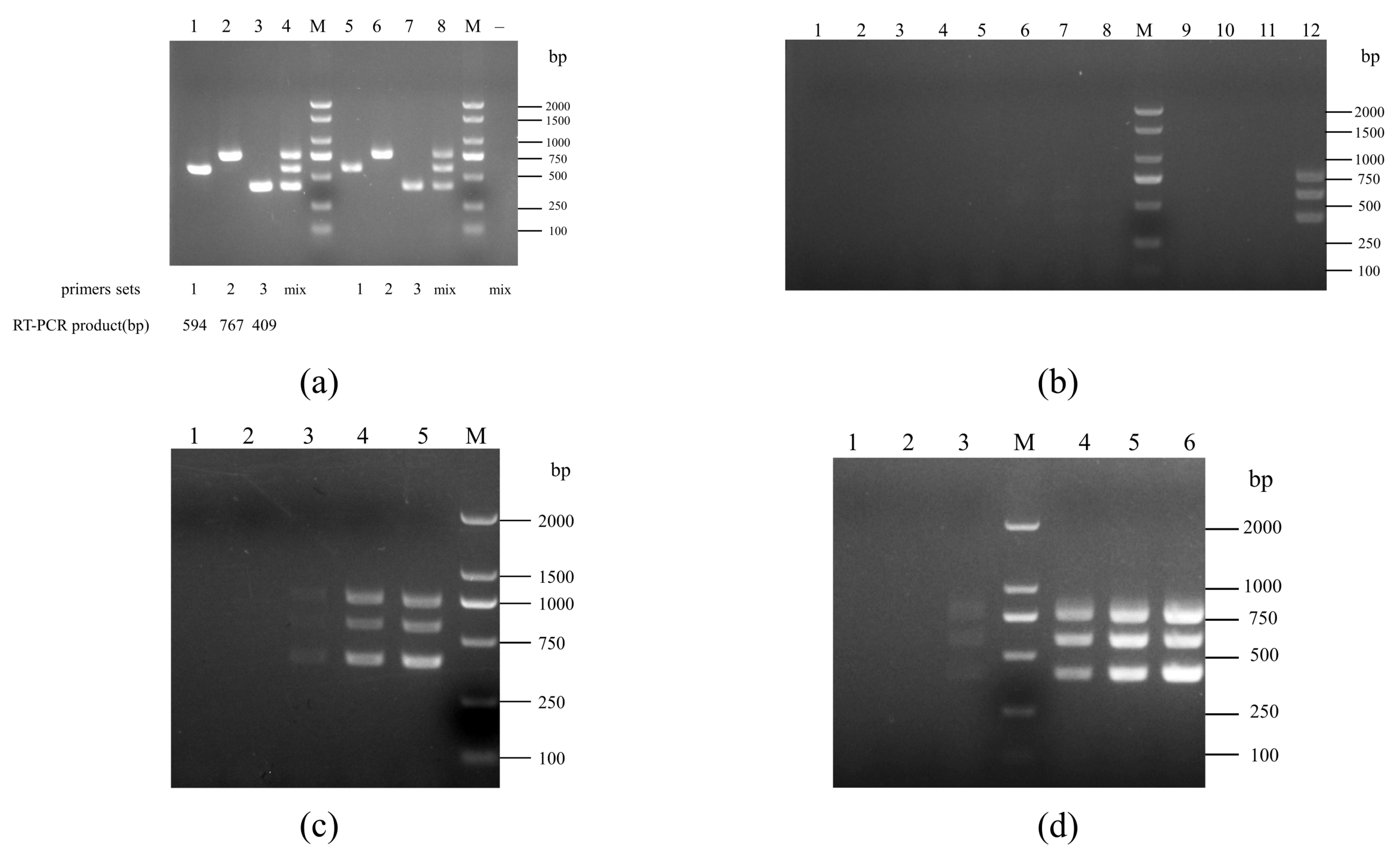
3.1. Multiplex PCR Assay for S. pasteurianus

As shown in Figure 1a, the fragments obtained by the single PCR amplification for genes E8M05_RS04035, E8M05_RS05155, and E8M05_RS06300, are 594, 767, and 409 bp, respectively, using the DNA or the culture of S. pasteurianus strain WUSP067 as templates; after optimizing reaction conditions, the multiplex PCR allowed amplification of three fragments simultaneously from the DNA or the culture of strain WUSP067. Only the culture of S. pasteurianus can amplify three fragments by this multiplex PCR, not the rest of the 11 other bacteria (Figure 1b). The sensitivity assay was performed with 10-fold dilutions of S. pasteurianus strain WUSP067 culture and DNA. The detection limit of the multiplex PCR assay was 5.46 × 10³ cfu (Figure 1c), and 17.137 pg DNA (Figure 1d). Therefore, we have developed a multiplex PCR suitable for S. pasteurianus identification.

3.2. Application of the Multiplex PCR Assay

The multiplex PCR assay was applied to detect S. pasteurianus from six fecal samples collected from cattle with diarrhea, and one sample was positive for S. pasteurianus (Table 3). In addition, 140 tonsils, 95 hilar lymph nodes, and 50 mesenteric lymph nodes from healthy pigs from different provinces were detected by this assay. The results showed that the positive rate of pig tonsils was 3.57% (5/140), the positive rate of pig hilar lymph nodes was 18.95% (18/95), and S. pasteurianus was not detected in pig mesenteric lymph nodes (Table 3). Among the 285 samples from healthy pigs, the positive rate of S. pasteurianus in the hilar lymph nodes was the highest, followed by the tonsils, and the lowest in the mesenteric lymph nodes.
To further validate this assay, sodA was amplified from these 24 S. pasteurianus positive samples. Sequence analysis showed that the sequence identity of sodA (from positions 25 to 510) obtained from 24 positive samples was all > 98.9% with that of type strain NCTC 13784 (Table S3). In addition, two S. pasteurianus strains WUSP070 and WUSP074 were isolated from these positive samples, and the information on the two strains is shown in Table 1.

3.3. The Complete Genome Sequence of Two S. pasteurianus Strains

The genome of strain WUSP070 comprises a single circular chromosome of 2,290,055 bp with G+C contents of 37.40%, 6 rRNA operons, 70 tRNA genes, and 2196 CDSs predicted in the chromosome. The genome of strain WUSP074 comprises a single circular chromosome of 2,371,672 bp with G+C contents of 37.34%, 6 rRNA operons, 70 tRNA genes, and 2309 CDSs predicted in the chromosome. No plasmid is present in the two strains.

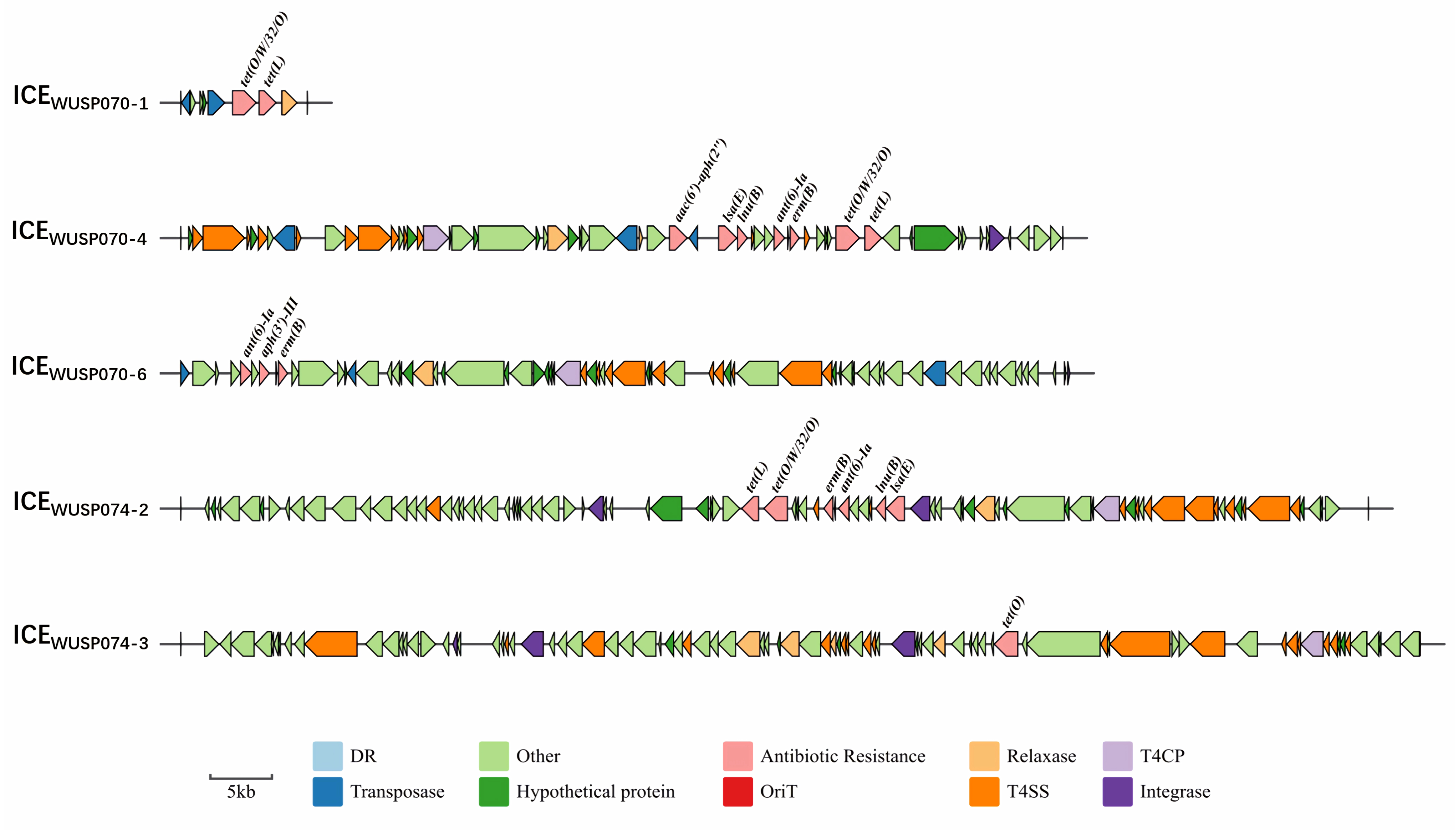
3.4. The Characteristics of Antimicrobial Resistance of Two S. pasteurianus Strains

The presence of antimicrobial resistance genes erm(B), lnu(B), tet(O/W/32/O), tet(L), aac(6′)-aph(2″), and lsa(E) in strain WUSP070 contributed to its resistance to erythromycin, lincomycin, clindamycin, doxycycline, and gentamycin (Table 4). The presence of antimicrobial resistance genes erm(B), lnu(B), lsa(E), tet(O), tet(O/W/32/O), and tet(L) in strain WUSP074 led to resistance to erythromycin, lincomycin, clindamycin, and doxycycline (Table 4). The strain resistant to three or more classes of antimicrobial agents is considered to be multidrug-resistant [32]. Thus, strains WUSP070 and WUSP074 are multidrug-resistant. To explore vehicles harboring antimicrobial resistance genes, the ICEs were predicted. Six ICEs were predicted in strain WUSP070, and three contained antimicrobial resistance genes. The ICEWUSP070-1 (from M0P24_RS00080 to M0P24_RS00115) harboring a 23-bp att sequence 5′-ggttctgttgcaaagttttaaat-3′ in the flanking region contained genes tet(O/W/32/O) and tet(L) (Figure 2). The ICEWUSP070-4 (from M0P24_RS04110 to M0P24_RS04435) harboring a 16-bp att sequence 5′-tactgttttaacaatg-3′ in the flanking region contained genes aac(6′)-aph(2″), lsa(E), lnu(B), ant(6)-Ia, erm(B), tet(O/W/32/O), and tet(L) (Figure 2). The ICEWUSP070-6 (from M0P24_RS10640 to M0P24_RS10985) contained genes ant(6)-Ia, aph(3′)-III, and ermB (Figure 2). Three ICEs were predicted in strain WUSP074, and the ICEWUSP074-2 (from M0P24_RS06290 to M0P24_RS06755) harboring a 15-bp att sequence 5′-tttttgaagttctgg-3′ in the flanking region contained genes tet(L), tet(O/W/32/O), erm(B), ant(6)-Ia, lnu(B), and lsa(E). The ICEWUSP074-3 (from M0P24_RS07560 to M0P24_RS08050) harboring a 15-bp att sequence 5′-aatatcaaaaatcag-3′ in the flanking region contained gene tet(O) (Figure 2). These ICEs were not present in a virulent strain WUSP067 isolated from a diseased pig from our previous study [22].

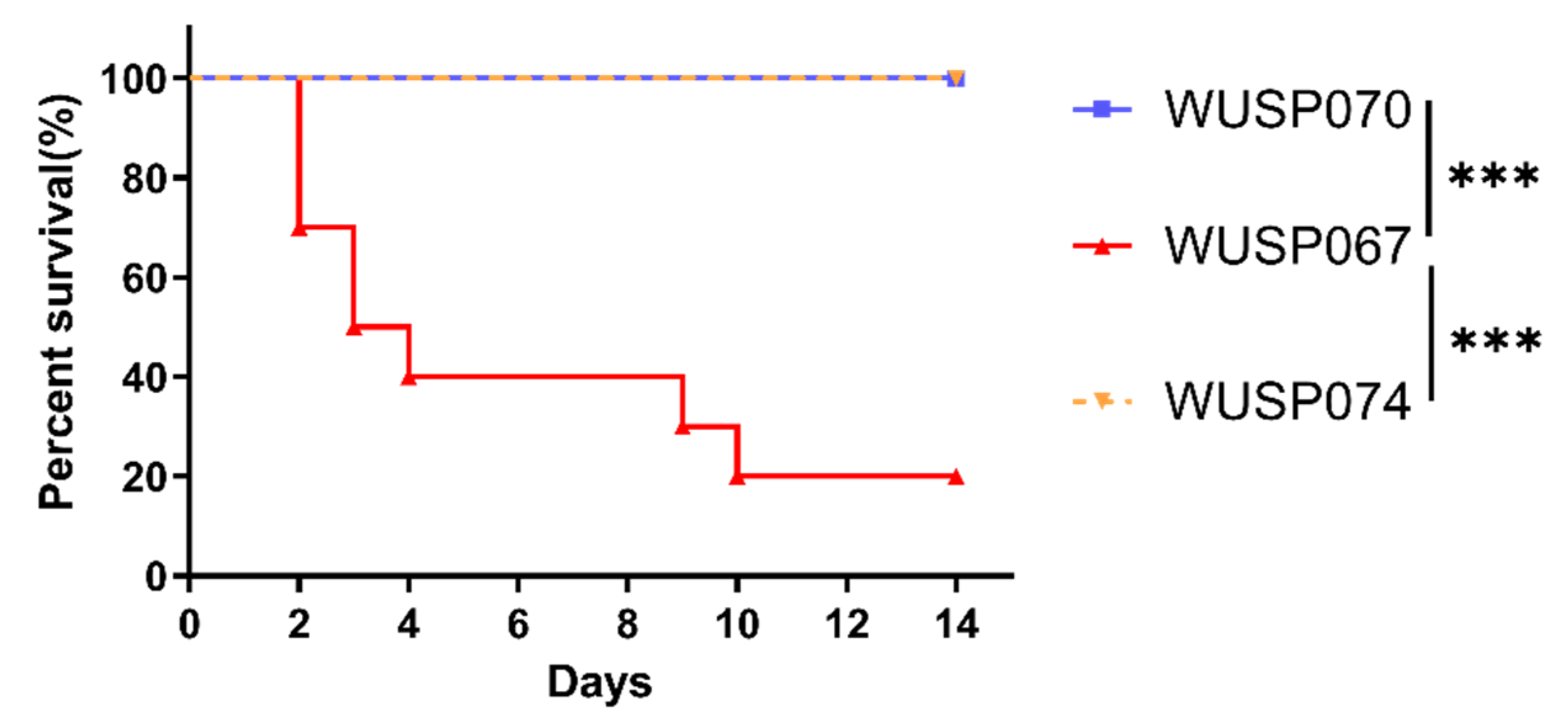
3.5. The Pathogenicity Characteristics of Two S. pasteurianus Strains

Table S4 reveals that strains WUSP070 and WUSP074 have 13 and 12 predicted virulence factors, respectively. However, on the 14th day post-infection, mice infected with strains WUSP070 and WUSP074 displayed a 100% survival rate, while mice infected with WUSP067 exhibited only a 20% survival rate (Figure 3). This observation confirms that strain WUSP067 is more pathogenic than strains WUSP070 and WUSP074.

4. Discussion

As indicated in Tables S1 and S2, there have been 44 papers reporting cases of S. pasteurianus infection in both humans and animals. Among them, 27 papers (61.36%) were published in the last decade, indicating a recent increase in attention towards S. pasteurianus. We first confirmed that this bacterium is a new pathogen of swine streptococcosis, and it can cause meningitis in pigs. In addition, S. pasteurianus was detected in pig tonsils, pulmonary hilar lymph nodes, and gut [22,33,34]. We proposed that healthy pigs’ tonsils and pulmonary hilar lymph nodes may be reservoirs of this bacterium [22]. So far, there are only three complete genome sequences of this bacterium, two human strains (ATCC 43144 and NCTC 13784) and a virulent strain WUSP067 isolated from a pig. The complete genome sequence of two non-virulent strains WUSP070 and WUSP074 provided in this study contributes to understanding the genomic characteristics of this zoonotic bacterium and identifying virulence factors by comparative genomic analysis.
So far, the identification of S. pasteurianus is mainly by biochemical testing systems, PCR assay based on SGPB0680 gene, and sodA gene sequence analysis. As mentioned above, those methods have shortcomings. In this study, we established a multiplex PCR assay to conveniently and accurately identify S. pasteurianus. The detection limit was 17.137 pg using bacterial DNA as templates. After optimization, this assay can also be used to detect bacterial cultures directly with the detection limit of 5.46 × 10³ cfu. Using this assay, 24 samples were positive for S. pasteurianus, including pig hilar lymph nodes, tonsils, and cattle feces. Furthermore, sodA sequence (from positions 25 to 510) analysis further confirmed that these 24 samples contained S. pasteurianus. However, this method should be further optimized in the future to improve the sensitivity of bacterial cultures. In our previous study, the detection rate of S. pasteurianus in pig tonsils was lower than that in pig hilar lymph nodes [22], and we wondered if it was related to tissues or regions where samples were collected. In this study, we found that the detection rate of S. pasteurianus in pig hilar lymph nodes was significantly different in different regions. For example, there was a 36% detection rate in Yunnan province, while none of the pig hilar lymph nodes was positive in Guangxi province. Thus, the detection rate of S. pasteurianus in pig tissues may be more related to regions where samples were collected, not tissues.
The virulence of bacteria such as S. suis cannot be purely defined by the source of the isolated strain [35]. Some S. suis strains isolated from healthy pigs are pathogenic and a source of infection for susceptible pigs and humans [36]. In this study, we isolated S. pasteurianus strain WUSP074 from a healthy pig tonsil, and mice infection experiments revealed that the strain was non-pathogenic. Whether, like S. suis, some tonsillar-derived S. pasteurianus strains are pathogenic and serve as a source of infection warrants further study. In addition, strain WUSP070 was isolated from cattle feces with diarrhea. Trotta et al. isolated two strains from calves with neurological hyperacute symptoms in 2019 [20]. If S. pasteurianus can cause cattle diarrhea needs further research.
For S. pasteurianus, the information on antimicrobial resistance and the spread of antibiotic resistance genes is very few. Most strains are resistant to macrolides, lincosamides, and tetracyclines [22,26,27]. In this study, two isolates WUSP070 and WUSP074 are also resistant to macrolides, lincosamides, and tetracyclines, and we first reported the presence of genes tet(O/W/32/O) and lsa(E) in S. pasteurianus that contribute to their resistance. In a duckling isolate AL101002, gene erm(B) was located at the Tn916-like element, and genes tet(M), tet(L), and erm(T) were located at the Tn916-IS1216 cluster [37]. In strain WUSP067, genes erm(B) and acc(6′)-aph(2″) were located at ICEWUSP067-1; genes tet(M) and tet(L) were located at ICEWUSP067-2 [22]. In this study, two isolates WUSP070 and WUSP074 have different vehicles, five ICEs, harboring antimicrobial resistance genes (Figure 2). Thus, ICEs may be the main vehicle for the spread of antibiotic-resistance genes in S. pasteurianus.

5. Conclusions

In this study, we established a convenient and specific multiplex PCR assay suitable for S. pasteurianus identification. This assay can be used to detect a large number of clinical samples, which provides essential technical support for the epidemiological research of S. pasteurianus. We first reported the presence of genes tet(O/W/32/O) and lsa(E) in S. pasteurianus, which leads to its resistance to lincosamides and tetracyclines. The complete genome sequence of two non-virulent strains contributes to understanding this zoonotic bacterium’s genomic characteristics and pathogenesis.

Supplementary Materials

The following supporting information can be downloaded at: https://www.mdpi.com/article/10.3390/pathogens12040615/s1, Table S1. The list of human cases infected by S. pasteurianus [38,39,40,41,42,43,44,45,46,47,48,49,50,51,52,53,54,55,56,57]; Table S2. The list of animal cases infected by S.pasteurianus; Table S3: The sodA sequence identity (from positions 25 to 510) of positive samples with that of type strain NCTC 13784; Table S4: The predicted virulence factors in strains WUSP070 and WUSP074.

Author Contributions

Z.W. and Y.B. designed the concept of the article; M.M., S.W. and Z.W. wrote the manuscript; M.M., X.L. and S.W. performed the experiments; M.M., Z.W., X.Z. and X.C. analyzed the data. All authors have read and agreed to the published version of the manuscript.

Funding

This work was supported by the Open Project Program of Engineering Research Center for the Prevention and Control of Animal Original Zoonosis, Fujian Province University under Grant number 2021ZW001 and Open Project Program of Jiangsu Key Laboratory of Zoonosis under Grant number R2103.

Institutional Review Board Statement

Mice infection experiments were carried out in the Laboratory Animal Center of Nanjing Agricultural University approved by the Laboratory Animal Monitoring Committee of Jiangsu Province (Permit number: SYXK (Su) 2021-0086).

Informed Consent Statement

Not applicable.

Data Availability Statement

The data supporting this study’s findings are available from the corresponding author upon reasonable request.

Conflicts of Interest

The authors declare no conflict of interest.

References

  1. Coykendall, A.L.; Gustafson, K.B. Deoxyribonucleic Acid Hybridizations Among Strains of Streptococcus salivarius and Streptococcus bovis. Int. J. Syst. Bacteriol. 1985, 35, 274–280. [Google Scholar] [CrossRef]
  2. Knight, R.G.; Shlaes, D.M. Physiological Characteristics and Deoxyribonucleic Acid Relatedness of Human Isolates of Streptococcus bovis and Streptococcus bovis (var.). Int. J. Syst. Bacteriol. 1985, 35, 357–361. [Google Scholar] [CrossRef]
  3. Poyart, C. Taxonomic Dissection of the Streptococcus bovis Group by Analysis of Manganese-Dependent Superoxide Dismutase Gene (SodA) Sequences: Reclassification of “Streptococcus infantarius subsp. coli” as Streptococcus lutetiensis sp. nov. and of Streptococcus bovis Biotype II.2 as Streptococcus pasteurianus sp. nov. Int. J. Syst. Evol. Microbiol. 2002, 52, 1247–1255. [Google Scholar] [CrossRef] [PubMed]
  4. Matesanz, M.; Rubal, D.; Iñiguez, I.; Rabuñal, R.; García-Garrote, F.; Coira, A.; García-País, M.J.; Pita, J.; Rodriguez-Macias, A.; López-Álvarez, M.J.; et al. Is Streptococcus Bovis a Urinary Pathogen? Eur. J. Clin. Microbiol. Infect. Dis. 2015, 34, 719–725. [Google Scholar] [CrossRef] [PubMed]
  5. Schwartz, M.C.; Dantuluri, K.; Maxey, T.; Paolillo, J. Ductal Stent Endocarditis Resulting in a Large Aortic Pseudoaneurysm. Catheter. Cardiovasc. Interv. 2021, 97, E826–E829. [Google Scholar] [CrossRef] [PubMed]
  6. Smith, A.H.; Sra, H.K.; Bawa, S.; Stevens, R. Streptococcus bovis Meningitis and Hemorrhoids. J. Clin. Microbiol. 2010, 48, 2654–2655. [Google Scholar] [CrossRef] [PubMed]
  7. Gavin, P.J.; Thomson, R.B.; Horng, S.-J.; Yogev, R. Neonatal Sepsis Caused by Streptococcus bovis Variant (Biotype II/2): Report of a Case and Review. J. Clin. Microbiol. 2003, 41, 3433–3435. [Google Scholar] [CrossRef]
  8. Lu, B.; Sui, W.; Lu, X. Intrauterine Infection and Post-Partum Bacteraemia Due to Streptococcus gallolyticus subsp. pasteurianus. J. Med. Microbiol. 2013, 62, 1617–1619. [Google Scholar] [CrossRef]
  9. Hede, S.V.; Olarte, L.; Chandramohan, L.; Kaplan, S.L.; Hulten, K.G. Streptococcus gallolyticus subsp. Pasteurianus Infection in Twin Infants. J. Clin. Microbiol. 2015, 53, 1419–1422. [Google Scholar] [CrossRef]
  10. Tarakçı, N.; Dağı, H.T.; Uğur, A.R.; Tuncer, İ.; Taştekin, A. Late-Onset Streptococcus pasteurianus Sepsis in a Preterm Baby in a Neonatal Intensive Care Unit. Turk. Pediatr. Ars. 2014, 49, 157–159. [Google Scholar] [CrossRef]
  11. Su, Y.; Miao, B.; Wang, H.; Wang, C.; Zhang, S. Splenic Abscess Caused by Streptococcus gallolyticus subsp. pasteurianus as Presentation of a Pancreatic Cancer. J. Clin. Microbiol. 2013, 51, 4249–4251. [Google Scholar] [CrossRef] [PubMed]
  12. Akahane, T.; Takahashi, K.; Matsumoto, T.; Kawakami, Y. A case of peritonitis due to Streptococcus gallolyticus subsp. pasteurianus. Kansenshogaku Zasshi 2009, 83, 56–59. [Google Scholar] [CrossRef] [PubMed]
  13. Corredoira, J.; Alonso, M.P.; García-Garrote, F.; García-Pais, M.J.; Coira, A.; Rabuñal, R.; Gonzalez-Ramirez, A.; Pita, J.; Matesanz, M.; Velasco, D.; et al. Streptococcus bovis Group and Biliary Tract Infections: An Analysis of 51 Cases. Clin. Microbiol. Infect. 2014, 20, 405–409. [Google Scholar] [CrossRef]
  14. Saegeman, V.; Cossey, V.; Loens, K.; Schuermans, A.; Glaser, P. Streptococcus gallolyticus subsp. pasteurianus Infection in a Neonatal Intensive Care Unit. Pediatr. Infect. Dis. J. 2016, 35, 1272–1275. [Google Scholar] [CrossRef]
  15. Chand, G.; Shamban, L.; Forman, A.; Sinha, P. The Association of Streptococcus gallolyticus Subspecies pasteurianus Bacteremia with the Detection of Premalignant and Malignant Colonic Lesions. Case Rep. Gastrointest. Med. 2016, 2016, 7815843. [Google Scholar] [CrossRef]
  16. Barnett, J.; Ainsworth, H.; Boon, J.D.; Twomey, D.F. Streptococcus gallolyticus subsp. pasteurianus Septicaemia in Goslings. Vet. J. 2008, 176, 251–253. [Google Scholar] [CrossRef] [PubMed]
  17. Hess, C.; Jandreski-Cvetkovic, D.; Liebhart, D.; Bilic, I.; Hess, M. Outbreaks of Streptococcus gallolyticus subsp. pasteurianus in Goslings Characterized by Central Nervous Symptoms. Avian Dis. 2021, 65, 165–170. [Google Scholar] [CrossRef] [PubMed]
  18. Li, M.; Gu, C.; Zhang, W.; Li, S.; Liu, J.; Qin, C.; Su, J.; Cheng, G.; Hu, X. Isolation and Characterization of Streptococcus gallolyticus subsp. pasteurianus Causing Meningitis in Ducklings. Vet. Microbiol. 2013, 162, 930–936. [Google Scholar] [CrossRef]
  19. Saumya, D.; Wijetunge, S.; Dunn, P.; Wallner-Pendleton, E.; Lintner, V.; Matthews, T.; Pierre, T.; Kariyawasam, S. Acute Septicemia Caused by Streptococcus gallolyticus subsp. pasteurianus in Turkey Poults. Avian Dis. 2013, 58, 318–322. [Google Scholar] [CrossRef]
  20. Trotta, A.; Sposato, A.; Marinaro, M.; Zizzo, N.; Passantino, G.; Parisi, A.; Buonavoglia, D.; Corrente, M. Neurological Symptoms and Mortality Associated with Streptococcus gallolyticus subsp. pasteurianus in Calves. Vet. Microbiol. 2019, 236, 108369. [Google Scholar] [CrossRef]
  21. Oliveira, A.R.; de Castro, M.F.; Pimentel, S.P.; de Carvalho, T.P.; Santana, C.H.; de Oliveira Santos, D.; Tinoco, H.P.; Coelho, C.M.; Pessanha, A.T.; da Paixão, T.A.; et al. Streptococcus pasteurianus-Induced Valvular Endocarditis and Sepsis in a Puerperal Emperor Tamarin (Saguinus imperator). J. Med. Primatol. 2022, 51, 388–391. [Google Scholar] [CrossRef] [PubMed]
  22. Wang, S.; Ma, M.; Liang, Z.; Zhu, X.; Yao, H.; Wang, L.; Wu, Z. Pathogenic Investigations of Streptococcus pasteurianus, an Underreported Zoonotic Pathogen, Isolated from a Diseased Piglet with Meningitis. Transbound. Emerg. Dis. 2022, 69, 2609–2620. [Google Scholar] [CrossRef]
  23. Klatte, J.M.; Clarridge, J.E.; Bratcher, D.; Selvarangan, R. A Longitudinal Case Series Description of Meningitis Due to Streptococcus gallolyticus subsp. pasteurianus in Infants. J. Clin. Microbiol. 2012, 50, 57–60. [Google Scholar] [CrossRef] [PubMed]
  24. Floret, N.; Bailly, P.; Thouverez, M.; Blanchot, C.; Alez-Martin, D.; Menget, A.; Thiriez, G.; Hoen, B.; Talon, D.; Bertrand, X. A Cluster of Bloodstream Infections Caused by Streptococcus gallolyticus Subspecies pasteurianus That Involved 5 Preterm Neonates in a University Hospital during a 2-Month Period. Infect. Control Hosp. Epidemiol. 2010, 31, 194–196. [Google Scholar] [CrossRef] [PubMed]
  25. Chang, T.-H.; Hsueh, P.-R.; Huang, Y.-T.; Chen, P.-Y.; Tang, H.-J.; Chen, J.-M. Prolonged Streptococcus gallolyticus subsp. pasteurianus Gut Colonization in Healthcare Workers and Potential Transmission Role in Neonatal Sepsis. J. Microbiol. Immunol. Infect. 2023; in press. [Google Scholar] [CrossRef]
  26. Li, Y.; Chen, X.; Zhang, Z.; Wang, L.; Wang, J.; Zeng, J.; Yang, J.; Lu, B. Microbiological and Clinical Characteristics of Streptococcus gallolyticus subsp. pasteurianus Infection in China. BMC Infect. Dis. 2019, 19, 791. [Google Scholar] [CrossRef]
  27. Hatrongjit, R.; Akeda, Y.; Hamada, S.; Gottschalk, M.; Kerdsin, A. Multiplex PCR for Identification of Six Clinically Relevant Streptococci. J. Med. Microbiol. 2017, 66, 1590–1595. [Google Scholar] [CrossRef]
  28. Wick, R.R.; Judd, L.M.; Gorrie, C.L.; Holt, K.E. Unicycler: Resolving Bacterial Genome Assemblies from Short and Long Sequencing Reads. PLoS Comput. Biol. 2017, 13, e1005595. [Google Scholar] [CrossRef]
  29. Seemann, T. Prokka: Rapid Prokaryotic Genome Annotation. Bioinformatics 2014, 30, 2068–2069. [Google Scholar] [CrossRef]
  30. Bortolaia, V.; Kaas, R.S.; Ruppe, E.; Roberts, M.C.; Schwarz, S.; Cattoir, V.; Philippon, A.; Allesoe, R.L.; Rebelo, A.R.; Florensa, A.F.; et al. ResFinder 4.0 for Predictions of Phenotypes from Genotypes. J. Antimicrob. Chemother. 2020, 75, 3491–3500. [Google Scholar] [CrossRef]
  31. Wang, M.; Goh, Y.-X.; Tai, C.; Wang, H.; Deng, Z.; Ou, H.-Y. VRprofile2: Detection of Antibiotic Resistance-Associated Mobilome in Bacterial Pathogens. Nucleic Acids Res. 2022, 50, W768–W773. [Google Scholar] [CrossRef]
  32. Schwarz, S.; Silley, P.; Simjee, S.; Woodford, N.; van Duijkeren, E.; Johnson, A.P.; Gaastra, W. Editorial: Assessing the Antimicrobial Susceptibility of Bacteria Obtained from Animals. J. Antimicrob. Chemother. 2010, 65, 601–604. [Google Scholar] [CrossRef] [PubMed]
  33. Liang, Z.; Wu, H.; Bian, C.; Chen, H.; Shen, Y.; Gao, X.; Ma, J.; Yao, H.; Wang, L.; Wu, Z. The Antimicrobial Systems of Streptococcus suis Promote Niche Competition in Pig Tonsils. Virulence 2022, 13, 781–793. [Google Scholar] [CrossRef] [PubMed]
  34. Holman, D.B.; Kommadath, A.; Tingley, J.P.; Abbott, D.W. Novel Insights into the Pig Gut Microbiome Using Metagenome-Assembled Genomes. Microbiol. Spectr. 2022, 10, e02380-22. [Google Scholar] [CrossRef] [PubMed]
  35. Segura, M.; Fittipaldi, N.; Calzas, C.; Gottschalk, M. Critical Streptococcus suis Virulence Factors: Are They All Really Critical? Trends Microbiol. 2017, 25, 585–599. [Google Scholar] [CrossRef]
  36. Liang, P.; Wang, M.; Gottschalk, M.; Vela, A.I.; Estrada, A.A.; Wang, J.; Du, P.; Luo, M.; Zheng, H.; Wu, Z. Genomic and Pathogenic Investigations of Streptococcus suis Serotype 7 Population Derived from a Human Patient and Pigs. Emerg. Microbes Infect. 2021, 10, 1960–1974. [Google Scholar] [CrossRef]
  37. Li, M.; Cai, C.; Chen, J.; Cheng, C.; Cheng, G.; Hu, X.; Liu, C. Inducible Expression of Both ErmB and ErmT Conferred High Macrolide Resistance in Streptococcus gallolyticus subsp. pasteurianus Isolates in China. Int. J. Mol. Sci. 2016, 17, 1599. [Google Scholar] [CrossRef]
  38. Cheung, M.; Pelot, M.; Nadarajah, R.; Kohl, S. Neonate with Late Onset Streptococcus bovis Meningitis: Case Report and Review of the Literature. Pediatr. Infect. Dis. J. 2000, 19, 891–893. [Google Scholar] [CrossRef]
  39. Sturt, A.S.; Yang, L.; Sandhu, K.; Pei, Z.; Cassai, N.; Blaser, M.J. Streptococcus gallolyticus Subspecies pasteurianus (Biotype II/2), a Newly Reported Cause of Adult Meningitis. J. Clin. Microbiol. 2010, 48, 2247–2249. [Google Scholar] [CrossRef]
  40. Nguyen, M.T.; Idriss, S.; Guzman, E.; De Oliveira, E.R. Neonatal Meningitis, Endocarditis, and Pneumonitis Due to Streptococcus gallolyticus subsp. pasteurianus: A Case Report. BMC Pediatr. 2019, 19, 265. [Google Scholar] [CrossRef]
  41. Orbea, M.; Desai, N.; Foster, C. Invasive Streptococcus gallolyticus Infections in Infants at Texas Children’s Hospital: A 9-Year Retrospective Review. Pediatr. Infect. Dis. J. 2022, 41, e494. [Google Scholar] [CrossRef]
  42. Vélez Balestro, L.M.; Baroni, M.R.; Ochoteco, M.C.; Zurbriggen, M.L.; Virgolini, S.M. Streptococcus gallolyticus subsp. pasteurianus isolated from cerebrospinal fluid in a pediatric patient. Rev. Argent. Microbiol. 2013, 45, 254–256. [Google Scholar] [CrossRef] [PubMed]
  43. Wardle, M.; Mu, A.; Tong, S.Y.C. Streptococcus gallolyticus subsp. pasteurianus Meningitis Complicated by Venous Sinus Thrombosis: A Case Report. Int. J. Infect. Dis. 2018, 71, 30–32. [Google Scholar] [CrossRef] [PubMed]
  44. Chen, W.-C.; Lee, P.-I.; Lin, H.-C.; Chang, L.-Y.; Lee, T.-F.; Chen, J.-M.; Hsueh, P.-R. Clustering of Streptococcus gallolyticus Subspecies pasteurianus Bacteremia and Meningitis in Neonates. J. Microbiol. Immunol. Infect. 2021, 54, 1078–1085. [Google Scholar] [CrossRef] [PubMed]
  45. Víquez-Víquez, M.; Brenes-Meléndez, M.G.; Chacón-González, C.; Ávila-Agüero, M.L. Streptococcus gallolyticus subsp. pasteurianus: A common infection caused by an unusual agent. Bol. Med. Hosp. Infant Mex. 2021, 78, 148–151. [Google Scholar] [CrossRef] [PubMed]
  46. Onoyama, S.; Ogata, R.; Wada, A.; Saito, M.; Okada, K.; Harada, T. Neonatal Bacterial Meningitis Caused by Streptococcus gallolyticus subsp. pasteurianus. J. Med. Microbiol. 2009, 58, 1252–1254. [Google Scholar] [CrossRef]
  47. Nagamatsu, M.; Takagi, T.; Ohyanagi, T.; Yamazaki, S.; Nobuoka, S.; Takemura, H.; Akita, H.; Miyai, M.; Ohkusu, K. Neonatal Meningitis Caused by Streptococcus gallolyticus subsp. pasteurianus. J. Infect. Chemother. 2012, 18, 265–268. [Google Scholar] [CrossRef]
  48. Takahashi, Y.; Ishiwada, N.; Tanaka, J.; Okusu, K.; Ichimura, S.; Hishiki, H.; Ota, S.; Kohno, Y. Streptococcus gallolyticus subsp. pasteurianus Meningitis in an Infant. Pediatr. Int. 2014, 56, 282–285. [Google Scholar] [CrossRef]
  49. Takamura, N.; Kenzaka, T.; Minami, K.; Matsumura, M. Infective Endocarditis Caused by Streptococcus gallolyticus Subspecies pasteurianus and Colon Cancer. BMJ Case Rep. 2014, 2014, bcr2013203476. [Google Scholar] [CrossRef]
  50. Yamamura, Y.; Mihara, Y.; Nakatani, K.; Nishiguchi, T.; Ikebe, T. Unexpected Ventriculitis Complication of Neonatal Meningitis Caused by Streptococcus gallolyticus subsp. pasteurianus: A Case Report. Jpn. J. Infect. Dis. 2018, 71, 68–71. [Google Scholar] [CrossRef]
  51. Kasamatsu, A.; Fukushima, K.; Horiuchi, M.; Sekiya, N. Streptococcus gallolyticus Subspecies pasteurianus Bacteremia Accompanied by Acute Pancreatitis. J. Infect. Chemother. 2022, 28, 1663–1666. [Google Scholar] [CrossRef]
  52. Shigemori, T.; Hiasa, A.; Inoue, Y.; Oka, S.; Yasuma, T.; Nishiwaki, R.; Sugimasa, N.; Hamaguchi, T.; Noji, M.; Takeuchi, K.; et al. Acute Calculous Cholecystitis Caused by Streptococcus gallolyticus Subspecies pasteurianus: A Case Report. Microorganisms 2022, 10, 1929. [Google Scholar] [CrossRef] [PubMed]
  53. Park, J.-W.; Eun, S.-H.; Kim, E.-C.; Seong, M.-W.; Kim, Y.-K. Neonatal Invasive Streptococcus gallolyticus subsp. pasteurianus Infection with Delayed Central Nervous System Complications. Korean J. Pediatr. 2015, 58, 33–36. [Google Scholar] [CrossRef] [PubMed]
  54. Van Samkar, A.; Brouwer, M.C.; Pannekoek, Y.; van der Ende, A.; van de Beek, D. Streptococcus gallolyticus Meningitis in Adults: Report of Five Cases and Review of the Literature. Clin. Microbiol. Infect. 2015, 21, 1077–1083. [Google Scholar] [CrossRef] [PubMed]
  55. Pereira, C.; Nogueira, F.; Cunha Marques, J.; Ferreira, J.P.; Almeida, J.S. Endocarditis by Streptococcus pasteurianus. Cureus 2023, 15, e34529. [Google Scholar] [CrossRef] [PubMed]
  56. Thatrimontrichai, A.; Chanvitan, P.; Janjindamai, W.; Dissaneevate, S.; Maneenil, G. Early Onset Neonatal Bacterial Meningitis Caused by Streptococcus gallolyticus subsp. pasteurianus. Southeast Asian J. Trop. Med. Public Health 2012, 43, 145–151. [Google Scholar] [PubMed]
  57. Niyas, V.K.; Arjun, R.; Sasidharan, A.; Palakunnath, G.A. Streptococcus gallolyticus Bacteremia: An Experience from a Tertiary Center in South India. Indian J. Crit. Care Med. 2020, 24, 943–945. [Google Scholar] [CrossRef]
Figure 1. Multiplex PCR Assay for S. pasteurianus. (a) Single and multiplex PCR amplification. Lanes 1–4: templates, the genome of WUSP067; primers, 1–F/R, 2–F/R, 3–F/R and a mixture of three primer pairs. Lanes 5–8: templates, culture of WUSP067; primers, 1–F/R, 2–F/R, 3–F/R and a mixture of three primer pairs. M, DNA Marker; – negative control, H2O. (b) Specificity of multiplex PCR. The culture of bacteria as templates: 1, S. equi subsp. zooepidemicus strain ATCC 35246; 2, S. agalactiae strain GD201008–001; 3, S. pluranimalium strain ML20171221B6–2; 4, B. subtilis strain 1.460; 5, A. hydrophila isolate WUQT018; 6, K. pneumoniae isolate WUQT019; 7, S. dysgalactiae isolate WUQT020; 8, E. coli isolate WUQT022; 9, Enterococcus sp isolate WUQT024; 10, S. hyovaginalis isolate WUQT033; 11, S. suis strain SC070731; 12, S. pasteurianus strain WUSP067. (c) Sensitivity of multiplex PCR. The cfu of WUSP067,1–5: 5.46 × 101 cfu, 5.46 × 10² cfu, 5.46 × 10³ cfu, 5.46 × 10⁴ cfu, and 5.46 × 10⁵ cfu. (d) Sensitivity of multiplex PCR. The DNA of WUSP067, 1–6: 171.37 fg, 1.7137 pg, 17.137 pg, 171.37 pg, 1.7137 ng, and 17.137 ng.
Pathogens 12 00615 g001
Figure 2. Vehicles for harboring antimicrobial resistance genes in S. pasteurianus. The ICEWUSP070-1 contained genes tet(O/W/32/O) and tet(L). The ICEWUSP070-4 contained genes acc(6′)-aph(2″), lsa(E), lnu(B), ant(6)-Ia, erm(B), tet(O/W/32/O), and tet(L). The ICEWUSP070-6 contained genes ant(6)-Ia, aph(3′)-III, and ermB. The ICEWUSP074-2 contained genes tet(L), tet(O/W/32/O), erm(B), ant(6)-Ia, lnu(B), and lsa(E). The ICEWUSP074-3 contained gene tet(O).
Pathogens 12 00615 g002
Figure 3. The survival curve of mice. Mice (10 per group) were injected with S. pasteurianus strains (WUSP067, WUSP070, and WUSP074) at a dose of 1.5 × 10⁸ cfu per mouse. For two weeks after infection, mortality was tracked. To analyze the results, the Log-rank (Mantel-Cox) test was used. ‘***’ indicates p < 0.001.
Pathogens 12 00615 g003
Table 1. The information of strains or isolates used in this study.
StrainSpeciesOriginNCBI Accession
WUSP067S. pasteurianusisolated from a newly weaned piglet’s brain with meningitisNZ_CP039457
WUSP070S. pasteurianusisolated from a diarrheal cattle fecal sampleNZ_CP116957.1
WUSP074S. pasteurianusisolated from a healthy porcine tonsilNZ_CP116958.1
SC070731S. suisisolated from a pig with meningitisNC_020526
ATCC 35246S. equi subsp. zooepidemicusisolated from a dead pigCP002904
GD201008-001S. agalactiaeisolated from a tilapia with meningoencephalitisNC_018646
ML20171221B6-2S. pluranimaliumisolated from a healthy pig
1.460B. subtilisChina General Microbiological Culture Collection Center
WUQT018A. hydrophilaisolated from a healthy porcine tonsil
WUQT019K. pneumoniaeisolated from a healthy porcine tonsil
WUQT020S. dysgalactiaeisolated from a healthy porcine tonsil
WUQT022E. coliisolated from a healthy porcine tonsil
WUQT024Enterococcus sp.isolated from a piglet lung
WUQT033S. hyovaginalisisolated from a piglet lung
ATCC 29213S. aureusAmerican Type Culture Collection
Table 2. The information on primers used in this study.
GenePrimer NamePrimer Sequence (5′−3′)Size (bp)
E8M05_RS040351−FGTAGATACTGATGGAGATGGT594
1−RATAATCGCCTGGTTGAGTC
E8M05_RS051552−FTTGTTCCGTTGTCAGCATA767
2−RAGCACCGATTCTATCCATAA
E8M05_RS063003−FGTTCTGGAATGGTTAGGAATC409
3−RAAGCAGCCGCAATATCAA
sodAsodA−FATGGCTATTATTTTACCAAAACTAC609
sodA−RTCACTTTGTTGCTTTTGAGTA
Table 3. The results of S. pasteurianus identification.
AreaTimeSample TypeSample NumberNumber of Positive SamplesPositive Rate (%)
Inner Mongolia2020cattle feces 6116.67
Chongqing2021pig tonsil5024.00
Yunnan2021pig hilar lymph node501836.00
Jiangsu2021pig mesenteric lymph node5000.00
Guangxi2021pig hilar lymph node4500.00
pig tonsil3000.00
Sichuan2021pig tonsil6035.00
Table 4. The MICs value and resistance mechanisms.
ClassesAntibioticsBreakpoints for Resistance (mg/L)MICs (mg/L)Resistance Mechanisms
WUSP070
MacrolidesErythromycin≥1>256erm(B)
LincosamidesLincomycin≥1>256lnu(B), erm(B), lsa(E)
Clindamycin≥1>256lnu(B), lnu(B), lsa(E)
TetracyclinesDoxycycline≥132tet(O/W/32/O), tet(L)
AminoglycosidesGentamicin≥16>256aac(6′)-aph(2″)
WUSP074
MacrolidesErythromycin≥1>256erm(B)
LincosamidesLincomycin≥1>256lsa(E), lnu(B), erm(B)
Clindamycin≥1>256lsa(E), lnu(B), erm(B)
TetracyclinesDoxycycline≥132tet(O/W/32/O), tet(L), tet(O)
Disclaimer/Publisher’s Note: The statements, opinions and data contained in all publications are solely those of the individual author(s) and contributor(s) and not of MDPI and/or the editor(s). MDPI and/or the editor(s) disclaim responsibility for any injury to people or property resulting from any ideas, methods, instructions or products referred to in the content.

