


Materials 2024, 17(3), 750; https://doi.org/10.3390/ma17030750 - 04 Feb 2024
Viewed by 850
Abstract
►
Show Figures
Metal binder jetting shows great potential for medical technology. This potential can be exploited by integrating binder jetting into existing process routes known from metal injection molding. The biggest challenge here is the flowability and packing behavior of the powders used, due to
[...] Read more.
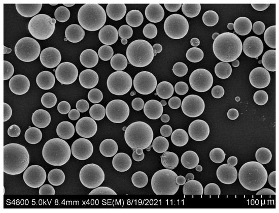
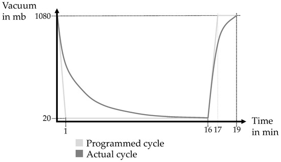
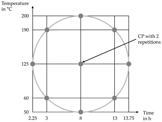
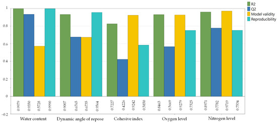
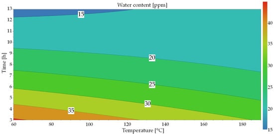
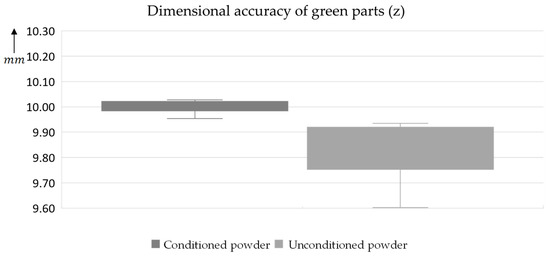
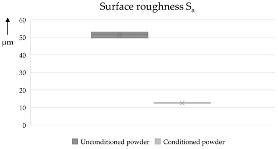
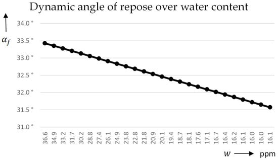
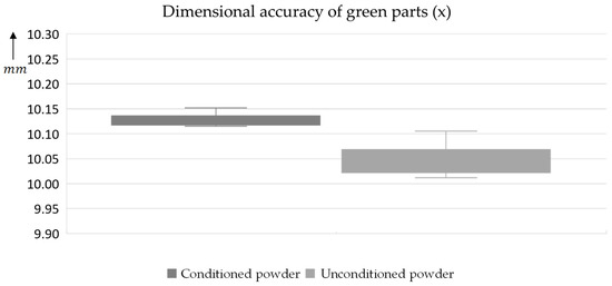
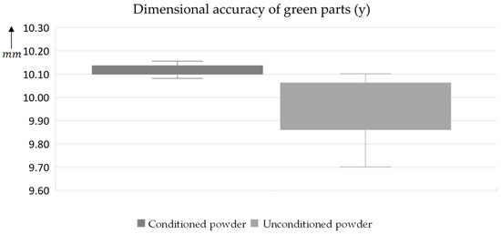
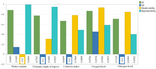
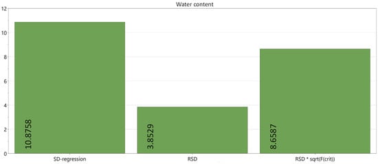
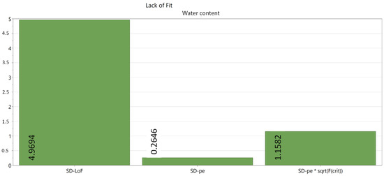
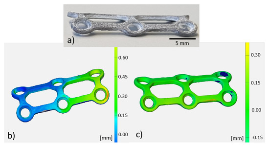
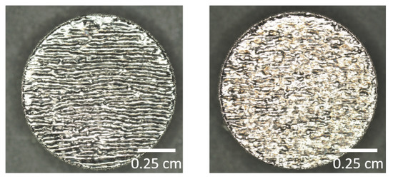
Metal binder jetting shows great potential for medical technology. This potential can be exploited by integrating binder jetting into existing process routes known from metal injection molding. The biggest challenge here is the flowability and packing behavior of the powders used, due to their low size distributions. This paper investigates different powder-drying strategies to improve flowability using a statistical experimental design. Because of its relevance for medical applications, spherical Ti-6Al-4V powder with a size distribution under 25 µm is dried under various parameters using vacuum and gas purging. The investigated parameters, time and temperature, are selected in a central-composite-circumscribed test plan with eleven tests and three center points. The target parameters—water content, flowability and impurity levels (oxygen, nitrogen)—of the powder are analyzed. For validation, practical test trials are carried out on an industrial binder jetting system with unconditioned powder and conditioning with optimized parameters, comparing the manufactured parts and the powder bed. An optimized drying cycle with a duration of 6 h at 200 °C was determined for the investigated powder. Significant improvements in the dimensional accuracy (from ±1.5 to 0.3%) of the components and the visual impression of the powder bed are demonstrated.
Full article
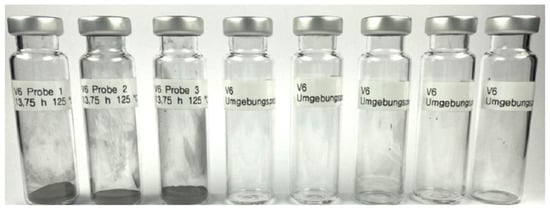
Figure 1


