Next Issue
Volume 8, April
Previous Issue
Volume 8, February


J. Fungi, Volume 8, Issue 3 (March 2022) – 109 articles

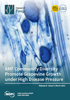
Cover Story (view full-size image): While most studies to date have researched the effect of individual AMF species on fungal pathogens associated with grapevines, this study is the first to investigate how young grapevine rootstocks inoculated with their ‘home’ and ‘away’ whole AMF communities would respond to challenges with a black foot pathogen species mixture. The outcome of this study revealed that that high disease incidence and severity did not reduce growth in vines with AMF inoculation compared to vines inoculated with the pathogen only. It also showed that the high level of disease present in rootstocks limited the effect of the AMF community with only little evidence that AMF treatments lowered disease incidence and severity in vines. View this paper
  • Issues are regarded as officially published after their release is announced to the table of contents alert mailing list.
  • You may sign up for e-mail alerts to receive table of contents of newly released issues.
  • PDF is the official format for papers published in both, html and pdf forms. To view the papers in pdf format, click on the "PDF Full-text" link, and use the free Adobe Reader to open them.
Order results
Result details
Section
Select all
Export citation of selected articles as:
9 pages, 3526 KiB  
Article
Clinical and Epidemiological Characteristics of Sporotrichosis in a Reference Center of Uruguay
by Elisa Cabeza, Annie Arrillaga, Lucía Dalcín, Mauricio Carbia, Zaida Arteta and Patricia Perera
J. Fungi 2022, 8(3), 322; https://doi.org/10.3390/jof8030322 - 21 Mar 2022
Cited by 3 | Viewed by 2508
Abstract
Background: Sporotrichosis is a fungal implantation disease of subacute/ chronic course caused by species of the dimorphic fungus Sporothrix spp. This infection usually develops after traumatic inoculation of contaminated soil, plants or organic material contaminated by Sporothrix spp. conidia into skin or mucosa. [...] Read more.
Background: Sporotrichosis is a fungal implantation disease of subacute/ chronic course caused by species of the dimorphic fungus Sporothrix spp. This infection usually develops after traumatic inoculation of contaminated soil, plants or organic material contaminated by Sporothrix spp. conidia into skin or mucosa. The objective of this work is to contribute to the knowledge of sporotrichosis in Uruguay by providing a report of a series of cases diagnosed in a reference center. Methods: We conducted a retrospective, observational, descriptive and cross-sectional study of cases of sporotrichosis diagnosed in the last 38 years. Results: In the period analyzed, 157 cases of sporotrichosis were diagnosed, 152 of those corresponded to male patients. The most frequent clinical presentation was nodular lymphatic in 120 patients. In relation to epidemiological antecedents, 128 patients had been scratched by armadillos during hunting. Conclusions: Sporotrichosis in Uruguay is a sporadic disease with a clear seasonal pattern related to particular social practices, such as hunting armadillos. Related to this practice, the affectation is greater in males and in young adults. Full article
(This article belongs to the Special Issue Fungal Infections of Implantation (Subcutaneous Mycoses))
Show Figures

Figure 1

13 pages, 1583 KiB  
Article
The Effect of Natural-Based Formulation (NBF) on the Response of RAW264.7 Macrophages to LPS as an In Vitro Model of Inflammation
by Sheelu Monga, Basem Fares, Rami Yashaev, Dov Melamed, Meygal Kahana, Fuad Fares, Abraham Weizman and Moshe Gavish
J. Fungi 2022, 8(3), 321; https://doi.org/10.3390/jof8030321 - 21 Mar 2022
Cited by 5 | Viewed by 3883
Abstract
Macrophages are some of the most important immune cells in the organism and are responsible for creating an inflammatory immune response in order to inhibit the passage of microscopic foreign bodies into the blood stream. Sometimes, their activation can be responsible for chronic [...] Read more.
Macrophages are some of the most important immune cells in the organism and are responsible for creating an inflammatory immune response in order to inhibit the passage of microscopic foreign bodies into the blood stream. Sometimes, their activation can be responsible for chronic inflammatory diseases such as asthma, tuberculosis, hepatitis, sinusitis, inflammatory bowel disease, and viral infections. Prolonged inflammation can damage the organs or may lead to death in serious conditions. In the present study, RAW264.7 macrophages were exposed to lipopolysaccharide (LPS; 20 ng/mL) and simultaneously treated with 20 µg/mL of natural-based formulation (NBF), mushroom–cannabidiol extract). Pro-inflammatory cytokines, chemokines, and other inflammatory markers were analyzed. The elevations in the presence of interleukin-6 (IL-6), cycloxygenase-2 (COX-2), C-C motif ligand-5 (CCL5), and nitrite response, following exposure to LPS, were completely inhibited by NBF administration. IL-1β and tumor necrosis factor alpha (TNF-α) release were inhibited by 3.9-fold and 1.5-fold, respectively. No toxic effect of NBF, as assessed by lactate dehydrogenase (LDH) release, was observed. Treatment of the cells with NBF significantly increased the mRNA levels of TLR2, and TLR4, but not NF-κB. Thus, it appears that the NBF possesses anti-inflammatory and immunomodulatory effects which can attenuate the release of pro-inflammatory markers. NBF may be a candidate for the treatment of acute and chronic inflammatory diseases and deserves further investigation. Full article
(This article belongs to the Topic Infectious Diseases)
Show Figures

Figure 1

16 pages, 20550 KiB  
Review
Biosynthesis of Fungal Natural Products Involving Two Separate Pathway Crosstalk
by Guangzhi Dai, Qiyao Shen, Youming Zhang and Xiaoying Bian
J. Fungi 2022, 8(3), 320; https://doi.org/10.3390/jof8030320 - 21 Mar 2022
Cited by 6 | Viewed by 3983
Abstract
Fungal natural products (NPs) usually possess complicated structures, exhibit satisfactory bioactivities, and are an outstanding source of drug leads, such as the cholesterol-lowering drug lovastatin and the immunosuppressive drug mycophenolic acid. The fungal NPs biosynthetic genes are always arranged within one single biosynthetic [...] Read more.
Fungal natural products (NPs) usually possess complicated structures, exhibit satisfactory bioactivities, and are an outstanding source of drug leads, such as the cholesterol-lowering drug lovastatin and the immunosuppressive drug mycophenolic acid. The fungal NPs biosynthetic genes are always arranged within one single biosynthetic gene cluster (BGC). However, a rare but fascinating phenomenon that a crosstalk between two separate BGCs is indispensable to some fungal dimeric NPs biosynthesis has attracted increasing attention. The hybridization of two separate BGCs not only increases the structural complexity and chemical diversity of fungal NPs, but also expands the scope of bioactivities. More importantly, the underlying mechanism for this hybridization process is poorly understood and needs further exploration, especially the determination of BGCs for each building block construction and the identification of enzyme(s) catalyzing the two biosynthetic precursors coupling processes such as Diels–Alder cycloaddition and Michael addition. In this review, we summarized the fungal NPs produced by functional crosstalk of two discrete BGCs, and highlighted their biosynthetic processes, which might shed new light on genome mining for fungal NPs with unprecedented frameworks, and provide valuable insights into the investigation of mysterious biosynthetic mechanisms of fungal dimeric NPs which are constructed by collaboration of two separate BGCs. Full article
Show Figures

Figure 1

19 pages, 3757 KiB  
Article
Diversity of Neofusicoccum parvum for the Production of the Phytotoxic Metabolites (-)-Terremutin and (R)-Mellein
by Patricia Trotel-Aziz, Guillaume Robert-Siegwald, Olivier Fernandez, Catarina Leal, Sandra Villaume, Jean-François Guise, Eliane Abou-Mansour, Marc-Henri Lebrun and Florence Fontaine
J. Fungi 2022, 8(3), 319; https://doi.org/10.3390/jof8030319 - 19 Mar 2022
Cited by 10 | Viewed by 3080
Abstract
Two Neofusicoccumparvum isolates and a UV mutant were characterized for their phytotoxin production in vitro, their pathogenicity on grapevine, and their genome sequenced. The isolate Np-Bt67 produced high level of (-)-terremutin, but almost no (R)-mellein, and it was the most [...] Read more.
Two Neofusicoccumparvum isolates and a UV mutant were characterized for their phytotoxin production in vitro, their pathogenicity on grapevine, and their genome sequenced. The isolate Np-Bt67 produced high level of (-)-terremutin, but almost no (R)-mellein, and it was the most aggressive on grapevine, triggering apoplexy. Similar symptoms were not induced by purified (-)-terremutin. The isolate Bourgogne S-116 (Np-B) produced 3-fold less (-)-terremutin and high amounts of (R)-mellein, but it was less aggressive on grapevine than Np-Bt67. The UV9 mutant obtained from Np-B (NpB-UV9) no longer produced (-)-terremutin but overproduced (R)-mellein by 2.5-fold, and it was as pathogenic as its parent. NpB-UV9 differed from its parent by simple mutations in two genes (transcription factor UCR-NP2_6692, regulatory protein UCR-NP2_9007), not located neither near (R)-mellein, nor (-)-terremutin biosynthetic genes, but likely involved in the control of (-)-terremutin biosynthesis. Grapevine immunity was disturbed upon challenge with these pathogens or purified phytotoxins, leading to an upregulation of SA-dependent defenses, while (-)-terremutin interfered with host JA/ET-dependent defenses. Our results suggest that neither (-)-terremutin nor (R)-mellein alone is essential for the pathogenicity of N. parvum on grapevine, since isolate/mutant non-producing these toxins in vitro is pathogenic. However, these phytotoxins could play a quantitative role in the infection process. Full article
Show Figures

Figure 1

12 pages, 666 KiB  
Review
Virulence Factors of Sporothrix schenckii
by Laura Cristina García-Carnero and José Ascención Martínez-Álvarez
J. Fungi 2022, 8(3), 318; https://doi.org/10.3390/jof8030318 - 19 Mar 2022
Cited by 11 | Viewed by 3546
Abstract
Sporothrix schenckii is one of the etiological agents of sporotrichosis. In this review, we discuss the virulence factors that have been proven to participate in the S. schenckii-host interaction. Among these known factors, we can find cell wall glycoproteins, adhesins, melanin, extracellular [...] Read more.
Sporothrix schenckii is one of the etiological agents of sporotrichosis. In this review, we discuss the virulence factors that have been proven to participate in the S. schenckii-host interaction. Among these known factors, we can find cell wall glycoproteins, adhesins, melanin, extracellular vesicles, and dimorphism. Furthermore, the morphological transition of S. schenckii in response to environmental conditions such as pH and temperature represents a means by which the fungus is able to establish mycosis in mammals. One of the key features in the development of sporotrichosis is the adhesion of the fungus to the host extracellular matrix. This event represents the first step to developing the mycosis, which involves adhesins such as the glycoproteins Gp70, Hsp60, and Pap1, which play a key role during the infection. The production of melanin helps the fungus to survive longer in the tissues and to neutralize or diminish many of the host’s attacks, which is why it is also considered a key factor in pathogenesis. Today, the study of human fungal pathogens’ virulence factors is a thriving area of research. Although we know some of the virulence factors in S. schenckii, much remains to be understood about the complex process of sporotrichosis development and the factors involved during the infection. Full article
(This article belongs to the Special Issue Sporothrix and Sporotrichosis 2.0)
Show Figures

Figure 1

19 pages, 5790 KiB  
Article
Mycofabrication of Mycelium-Based Leather from Brown-Rot Fungi
by Jegadeesh Raman, Da-Song Kim, Hyun-Seok Kim, Deuk-Sil Oh and Hyun-Jae Shin
J. Fungi 2022, 8(3), 317; https://doi.org/10.3390/jof8030317 - 19 Mar 2022
Cited by 27 | Viewed by 17026
Abstract
Sustainable substitutes for leather can be made from mushroom mycelium, which is an environmentally friendly alternative to animal and synthetic leather. Mycelium-based leather is derived from Polyporales, in which lignocellulosic material is used as the substrate. The plasticizing and crosslinking of mycelial mats [...] Read more.
Sustainable substitutes for leather can be made from mushroom mycelium, which is an environmentally friendly alternative to animal and synthetic leather. Mycelium-based leather is derived from Polyporales, in which lignocellulosic material is used as the substrate. The plasticizing and crosslinking of mycelial mats with various reagents might affect the leather properties and mycelial architecture. This study investigated the physicochemical and mechanical properties of mycelium-based leather (MBL) samples, including the hygroscopic nature, thermal stability, cell wall chemistry, density, micromorphology, tensile strength, elongation rate, and Young’s modulus. Micromorphological observations confirmed the mycelial networks and their binding performance, verifying their efficacy as a substitute leather. The most significant effects were observed after treatment with 20% polyethylene glycol, which resulted in an increase in Young’s modulus and tensile strength. Furthermore, the samples generally exhibited a high density (1.35, 1.46 g/cm3) and tensile strength (7.21 ± 0.93, 8.49 ± 0.90 MPa), resembling leather. The tear strength reached as low as 0.5–0.8 N/mm. However, the tensile and tear strength may be affected by leather processing and the tuning of mycelial growth. Nevertheless, high-density mycelia are shown to be suitable for the production of MBL, while mycofabrication and strain selection are sustainable for novel industrial applications of MBL. Full article
(This article belongs to the Section Fungal Cell Biology, Metabolism and Physiology)
Show Figures

Figure 1

13 pages, 722 KiB  
Article
COVID-19 Associated Pulmonary Aspergillosis (CAPA): Hospital or Home Environment as a Source of Life-Threatening Aspergillus fumigatus Infection?
by Teresa Peláez-García de la Rasilla, Irene González-Jiménez, Andrea Fernández-Arroyo, Alejandra Roldán, Jose Luis Carretero-Ares, Marta García-Clemente, Mauricio Telenti-Asensio, Emilio García-Prieto, Mar Martínez-Suarez, Fernando Vázquez-Valdés, Santiago Melón-García, Luis Caminal-Montero, Inmaculada Fernández-Simón, Emilia Mellado and María Luisa Sánchez-Núñez
J. Fungi 2022, 8(3), 316; https://doi.org/10.3390/jof8030316 - 19 Mar 2022
Cited by 13 | Viewed by 3161
Abstract
Most cases of invasive aspergillosis are caused by Aspergillus fumigatus, whose conidia are ubiquitous in the environment. Additionally, in indoor environments, such as houses or hospitals, conidia are frequently detected too. Hospital-acquired aspergillosis is usually associated with airborne fungal contamination of the [...] Read more.
Most cases of invasive aspergillosis are caused by Aspergillus fumigatus, whose conidia are ubiquitous in the environment. Additionally, in indoor environments, such as houses or hospitals, conidia are frequently detected too. Hospital-acquired aspergillosis is usually associated with airborne fungal contamination of the hospital air, especially after building construction events. A. fumigatus strain typing can fulfill many needs both in clinical settings and otherwise. The high incidence of aspergillosis in COVID patients from our hospital, made us wonder if they were hospital-acquired aspergillosis. The purpose of this study was to evaluate whether the hospital environment was the source of aspergillosis infection in CAPA patients, admitted to the Hospital Universitario Central de Asturias, during the first and second wave of the COVID-19 pandemic, or whether it was community-acquired aspergillosis before admission. During 2020, sixty-nine A. fumigatus strains were collected for this study: 59 were clinical isolates from 28 COVID-19 patients, and 10 strains were environmentally isolated from seven hospital rooms and intensive care units. A diagnosis of pulmonary aspergillosis was based on the ECCM/ISHAM criteria. Strains were genotyped by PCR amplification and sequencing of a panel of four hypervariable tandem repeats within exons of surface protein coding genes (TRESPERG). A total of seven genotypes among the 10 environmental strains and 28 genotypes among the 59 clinical strains were identified. Genotyping revealed that only one environmental A. fumigatus from UCI 5 (box 54) isolated in October (30 October 2020) and one A. fumigatus isolated from a COVID-19 patient admitted in Pneumology (Room 532-B) in November (24 November 2020) had the same genotype, but there was a significant difference in time and location. There was also no relationship in time and location between similar A. fumigatus genotypes of patients. The global A. fumigatus, environmental and clinical isolates, showed a wide diversity of genotypes. To our knowledge, this is the first study monitoring and genotyping A. fumigatus isolates obtained from hospital air and COVID-19 patients, admitted with aspergillosis, during one year. Our work shows that patients do not acquire A. fumigatus in the hospital. This proves that COVID-associated aspergillosis in our hospital is not a nosocomial infection, but supports the hypothesis of “community aspergillosis” acquisition outside the hospital, having the home environment (pandemic period at home) as the main suspected focus of infection. Full article
(This article belongs to the Special Issue Fungal Infections and COVID-19)
Show Figures

Figure 1

8 pages, 1008 KiB  
Article
Clinical Presentations, Cluster Analysis and Laboratory-Based Investigation of Aspergillus Otomycosis—A Single Center Experience
by Mila Bojanović, Aleksandra Ignjatović, Marko Stalević, Valentina Arsić-Arsenijević, Marina Ranđelović, Vladimir Gerginić, Zorica Stojanović-Radić, Ognjen Stojković, Emilija Živković-Marinkov and Suzana Otašević
J. Fungi 2022, 8(3), 315; https://doi.org/10.3390/jof8030315 - 18 Mar 2022
Cited by 6 | Viewed by 3335
Abstract
Species of Aspergillus (A.) niger complex and A. flavus complex are predominant molds that are causative agents of otomycoses. The goal of this study was to investigate the clinical presentation, diagnostic procedure, and appearance of relapse in patients with Aspergillus-otomycosis, [...] Read more.
Species of Aspergillus (A.) niger complex and A. flavus complex are predominant molds that are causative agents of otomycoses. The goal of this study was to investigate the clinical presentation, diagnostic procedure, and appearance of relapse in patients with Aspergillus-otomycosis, as well as to determine the biofilm production ability of species isolated in relapse. Thirty patients with laboratory evidenced Aspergillus-otomycosis followed by two check-ups (30 and 60 days after initiation of treatment with antimycotics for local application) were included in the study. For isolation and identification of Aspergillus spp. the standard mycological procedure was applied. Results showed very high sensitivity of microscopy, but 16.7% Aspergillus species required the optimal temperature of 27–28 °C for cultivation. Applied statistical cluster analysis showed a defined specific cluster/group of patients with A. niger complex-otomycosis. Sixty days after diagnosis and treatment initiation, six patients had a relapse, with the same species of Aspergillus genus being the cause. To establish the ability of biofilm production, the modified method described by Pierce and Kvasničková was performed, and all six species isolated in the relapse episode had the ability to produce biofilm. Official criteria and recommendations are needed due to the possibility of misdiagnosis, which leads to the prolongation and complication of the disease. Full article
(This article belongs to the Special Issue Diagnostic and Therapeutic Challenges of Human Fungal Infections)
Show Figures

Figure 1

15 pages, 1555 KiB  
Article
Chemical Constituents and Molecular Mechanism of the Yellow Phenotype of Yellow Mushroom (Floccularia luteovirens)
by Xiaolong Gan, Xuemei Bao, Baolong Liu, Yun Li, Dong Cao, Hg Zhang and Yuan Zong
J. Fungi 2022, 8(3), 314; https://doi.org/10.3390/jof8030314 - 18 Mar 2022
Cited by 6 | Viewed by 2100
Abstract
(1) Background: Yellow mushroom (Floccularia luteovirens) is a natural resource that is highly nutritional, has a high economic value, and is found in Northwest China. Despite its value, the chemical and molecular mechanisms of yellow phenotype formation are still unclear. (2) [...] Read more.
(1) Background: Yellow mushroom (Floccularia luteovirens) is a natural resource that is highly nutritional, has a high economic value, and is found in Northwest China. Despite its value, the chemical and molecular mechanisms of yellow phenotype formation are still unclear. (2) Methods: This study uses the combined analysis of transcriptome and metabolome to explain the molecular mechanism of the formation of yellow mushroom. Subcellular localization and transgene overexpression techniques were used to verify the function of the candidate gene. (3) Results: 112 compounds had a higher expression in yellow mushroom; riboflavin was the ninth most-expressed compound. HPLC showed that a key target peak at 23.128 min under visible light at 444 nm was Vb2. All proteins exhibited the closest relationship with Agaricus bisporus var. bisporus H97. One riboflavin transporter, CL911.Contig3_All (FlMCH5), was highly expressed in yellow mushrooms with a different value (log2 fold change) of −12.98, whereas it was not detected in white mushrooms. FlMCH5 was homologous to the riboflavin transporter MCH5 or MFS transporter in other strains, and the FlMCH5-GFP fusion protein was mainly located in the cell membrane. Overexpression of FlMCH5 in tobacco increased the content of riboflavin in three transgenic plants to 26 μg/g, 26.52 μg/g, and 36.94 μg/g, respectively. (4) Conclusions: In this study, it is clear that riboflavin is the main coloring compound of yellow mushrooms, and FlMCH5 is the key transport regulatory gene that produces the yellow phenotype. Full article
(This article belongs to the Section Fungal Genomics, Genetics and Molecular Biology)
Show Figures

Graphical abstract

34 pages, 9026 KiB  
Article
Colletotrichum Species Associated with Peaches in China
by Qin Tan, Guido Schnabel, Chingchai Chaisiri, Liang-Fen Yin, Wei-Xiao Yin and Chao-Xi Luo
J. Fungi 2022, 8(3), 313; https://doi.org/10.3390/jof8030313 - 18 Mar 2022
Cited by 18 | Viewed by 5948
Abstract
Colletotrichum is regarded as one of the 10 most important genera of plant pathogens in the world. It causes diseases in a wide range of economically important plants, including peaches. China is the largest producer of peaches in the world but little is [...] Read more.
Colletotrichum is regarded as one of the 10 most important genera of plant pathogens in the world. It causes diseases in a wide range of economically important plants, including peaches. China is the largest producer of peaches in the world but little is known about the Colletotrichum spp. affecting the crop. In 2017 and 2018, a total of 286 Colletotrichum isolates were isolated from symptomatic fruit and leaves in 11 peach production provinces of China. Based on multilocus phylogenetic analyses (ITS, ACT, CAL, CHS-1, GAPDH, TUB2, and HIS3) and morphological characterization, the isolates were identified to be C. nymphaeae, C. fioriniae, and C. godetiae of the C. acutatum species complex, C. fructicola and C. siamense of the C. gloeosporioides species complex, C. karsti of the C. boninense species complex, and one newly identified species, C. folicola sp. nov. This study is the first report of C. karsti and C. godetiae in peaches, and the first report of C. nymphaeae, C. fioriniae, C. fructicola, and C. siamense in peaches in China. C. nymphaeae is the most prevalent species of Colletotrichum in peaches in China, which may be the result of fungicide selection. Pathogenicity tests revealed that all species found in this study were pathogenic on both the leaves and fruit of peaches, except for C. folicola, which only infected the leaves. The present study substantially improves our understanding of the causal agents of anthracnose on peaches in China. Full article
(This article belongs to the Topic Fungal Diversity)
Show Figures

Figure 1

7 pages, 2274 KiB  
Case Report
Tinea Incognito—A Great Physician Pitfall
by Julia Nowowiejska, Anna Baran and Iwona Flisiak
J. Fungi 2022, 8(3), 312; https://doi.org/10.3390/jof8030312 - 18 Mar 2022
Cited by 6 | Viewed by 5966
Abstract
Tinea incognito is a dermatophyte infection exacerbated after inadequate administration of topical or systemic glucocorticoids. A 57-year-old man presented to the Department of Dermatology due to skin lesions persisting for one month. He reported having recently worked under hot conditions, in tight clothing, [...] Read more.
Tinea incognito is a dermatophyte infection exacerbated after inadequate administration of topical or systemic glucocorticoids. A 57-year-old man presented to the Department of Dermatology due to skin lesions persisting for one month. He reported having recently worked under hot conditions, in tight clothing, which caused sweating. Later, he noticed erythematous–exfoliative lesions in his groins and on the buttocks. He presented to the general practitioner who diagnosed him with eczema and prescribed clobetasole ointment. Since the skin lesions became more severe, he presented to the Department of Dermatology. On the physical examination, extensive erythematous–infiltrative lesions were observed in the area of medial, lateral, and posterior surface of both thighs and buttocks. Pustules were also present. Suspicion of tinea incognito was raised, and direct mycological examination and culture confirmed the presence of dermatophytes. The patient was prescribed topical terbinafine and oral itraconazole. Tinea incognito may be challenging to diagnose because the clinical presentation is relatively nonspecific and definitive culture or histopathological diagnosis such as by microscopic sample examination to identify fungal elements is not universally available. Every doctor has to keep in mind the fact that tinea may be a great mimicker of other dermatoses and to not prescribe medications without microscopic confirmation of tinea, and refer patients for dermatological consultation in case of doubt. Full article
(This article belongs to the Special Issue Fungal Infections in Dermatology)
Show Figures

Figure 1

23 pages, 5216 KiB  
Article
Phylogenomics and Comparative Genomics Highlight Specific Genetic Features in Ganoderma Species
by Yi-Fei Sun, Annie Lebreton, Jia-Hui Xing, Yu-Xuan Fang, Jing Si, Emmanuelle Morin, Shingo Miyauchi, Elodie Drula, Steven Ahrendt, Kelly Cobaugh, Anna Lipzen, Maxim Koriabine, Robert Riley, Annegret Kohler, Kerrie Barry, Bernard Henrissat, Igor V. Grigoriev, Francis M. Martin and Bao-Kai Cui
J. Fungi 2022, 8(3), 311; https://doi.org/10.3390/jof8030311 - 18 Mar 2022
Cited by 10 | Viewed by 4277
Abstract
The Ganoderma species in Polyporales are ecologically and economically relevant wood decayers used in traditional medicine, but their genomic traits are still poorly documented. In the present study, we carried out a phylogenomic and comparative genomic analyses to better understand the genetic blueprint [...] Read more.
The Ganoderma species in Polyporales are ecologically and economically relevant wood decayers used in traditional medicine, but their genomic traits are still poorly documented. In the present study, we carried out a phylogenomic and comparative genomic analyses to better understand the genetic blueprint of this fungal lineage. We investigated seven Ganoderma genomes, including three new genomes, G. australe, G. leucocontextum, and G. lingzhi. The size of the newly sequenced genomes ranged from 60.34 to 84.27 Mb and they encoded 15,007 to 20,460 genes. A total of 58 species, including 40 white-rot fungi, 11 brown-rot fungi, four ectomycorrhizal fungi, one endophyte fungus, and two pathogens in Basidiomycota, were used for phylogenomic analyses based on 143 single-copy genes. It confirmed that Ganoderma species belong to the core polyporoid clade. Comparing to the other selected species, the genomes of the Ganoderma species encoded a larger set of genes involved in terpene metabolism and coding for secreted proteins (CAZymes, lipases, proteases and SSPs). Of note, G. australe has the largest genome size with no obvious genome wide duplication, but showed transposable elements (TEs) expansion and the largest set of terpene gene clusters, suggesting a high ability to produce terpenoids for medicinal treatment. G. australe also encoded the largest set of proteins containing domains for cytochrome P450s, heterokaryon incompatibility and major facilitator families. Besides, the size of G. australe secretome is the largest, including CAZymes (AA9, GH18, A01A), proteases G01, and lipases GGGX, which may enhance the catabolism of cell wall carbohydrates, proteins, and fats during hosts colonization. The current genomic resource will be used to develop further biotechnology and medicinal applications, together with ecological studies of the Ganoderma species. Full article
(This article belongs to the Special Issue Genomics and Evolution of Macrofungi)
Show Figures

Figure 1

12 pages, 4209 KiB  
Article
Distribution of Yeast Species and Risk Factors of Oral Colonization after Oral-Care Education among the Residents of Nursing Homes
by Ming-Gene Tu, Chih-Chao Lin, Ya-Ting Chiang, Zi-Li Zhou, Li-Yun Hsieh, Kai-Ting Chen, Yin-Zhi Chen, Wen-Chi Cheng and Hsiu-Jung Lo
J. Fungi 2022, 8(3), 310; https://doi.org/10.3390/jof8030310 - 17 Mar 2022
Viewed by 2162
Abstract
Most yeasts causing infections in humans are part of commensal microflora and etiological agents of different infections when hosts become susceptible, usually due to becoming immunocompromised. The colonization of potentially pathogenic microbes in the oral cavity is increased by poor oral hygiene. This [...] Read more.
Most yeasts causing infections in humans are part of commensal microflora and etiological agents of different infections when hosts become susceptible, usually due to becoming immunocompromised. The colonization of potentially pathogenic microbes in the oral cavity is increased by poor oral hygiene. This follow-up survey was conducted approximately two months after providing information on proper oral care at 10 nursing homes in Taiwan. Among the 117 of 165 residents colonized by yeasts, 67 were colonized by more than one yeast species. A total of 231 isolates comprising eight fungal genera and 25 species were identified. Candida albicans (44.6%) was the dominant species, followed by Candida glabrata (17.7%), Candida parapsilosis (8.7%), Candida tropicalis (7.8%), and Candida pararugosa (7.3%). Residents having a yeast colony-forming unit >10 (OR, 8.897; 95% CI 2.972–26.634; p < 0.001) or using a wheelchair (OR, 4.682; 95% CI 1.599–13.705; p = 0.005) were more likely to be colonized by multiple species. By comparing before and after oral-care education, dry mouth (OR, 3.199; 95% CI 1.448–7.068; p = 0.011) and having heart disease (OR, 2.681; 95% CI 1.068–6.732; p = 0.036) emerged as two independent risk factors for increased density of colonizing yeast. Full article
(This article belongs to the Topic Infectious Diseases)
Show Figures

Figure 1

12 pages, 302 KiB  
Review
Commercial Methods for Antifungal Susceptibility Testing of Yeasts: Strengths and Limitations as Predictors of Resistance
by Ana Espinel-Ingroff
J. Fungi 2022, 8(3), 309; https://doi.org/10.3390/jof8030309 - 17 Mar 2022
Cited by 8 | Viewed by 2621
Abstract
Susceptibility testing can yield variable results because it is method (commercial or reference), agent, and species dependent. Therefore, in order for results to be clinically relevant, MICs (minimal inhibitory concentrations) or MECs (minimal effective concentrations) should help in selecting the best treatment agent [...] Read more.
Susceptibility testing can yield variable results because it is method (commercial or reference), agent, and species dependent. Therefore, in order for results to be clinically relevant, MICs (minimal inhibitory concentrations) or MECs (minimal effective concentrations) should help in selecting the best treatment agent in the clinical setting. This is accomplished by categorical endpoints, ideally, breakpoints (BPs) and/or ECVs/ECOFFs (epidemiological cutoff values). BPs and ECVs are available by the reference methods (CLSI [Clinical and Laboratory Standards Institute] and EUCAST [European Committee on Antifungal Susceptibility Testing]) for a variety of species/agent combinations. The lack of clinical data precludes establishment of BPs for susceptibility testing by the commercial methods and ECVs have only been calculated for the Etest and SYO assays. The goal of this review is to summarize the variety of commercial methods for antifungal susceptibility testing and the potential value of Etest and SYO ECVs for detecting mutants/non-wild type (NWT) Candida isolates. Therefore, the literature search focused on publications where the commercial method, meaning MICs and ECVs, were reported for specific NWT isolates; genetic mutations have also been listed. For the Etest, the best performers recognizing the NWT were anidulafungin ECVs: 92% for the common species; 97% for C. glabrata and fluconazole ECVs, mostly for C. parapsilosis (45 NWT isolates). By the SYO, posaconazole ECVs recognized 93% of the C. albicans and 96% of the C. parapsilosis NWT isolates and micafungin ECVs 94% (mostly C. albicans and C. glabrata). Smaller sets, some with clinical data, were also listed. These are promising results for the use of both commercial methods to identify antifungal resistance (NWT isolates). However, ECVs for other species and methods need to be defined, including the C. neoformans complex and emerging species. Full article
(This article belongs to the Special Issue Fungal Infections: From Diagnostics to Treatments)
15 pages, 1955 KiB  
Article
Combined Use of Presepsin and (1,3)-β-D-glucan as Biomarkers for Diagnosing Candida Sepsis and Monitoring the Effectiveness of Treatment in Critically Ill Patients
by Radim Dobiáš, Marcela Káňová, Naděžda Petejová, Štefan Kis Pisti, Robert Bocek, Eva Krejčí, Helena Stružková, Michaela Cachová, Hana Tomášková, Petr Hamal, Vladimír Havlíček and Milan Raška
J. Fungi 2022, 8(3), 308; https://doi.org/10.3390/jof8030308 - 17 Mar 2022
Cited by 3 | Viewed by 2663
Abstract
New biomarker panel was developed and validated on 165 critically ill adult patients to enable a more accurate invasive candidiasis (IC) diagnosis. Serum levels of the panfungal biomarker (1,3)-β-D-glucan (BDG) and the inflammatory biomarkers C-reactive protein, presepsin (PSEP), and procalcitonin (PCT) were correlated [...] Read more.
New biomarker panel was developed and validated on 165 critically ill adult patients to enable a more accurate invasive candidiasis (IC) diagnosis. Serum levels of the panfungal biomarker (1,3)-β-D-glucan (BDG) and the inflammatory biomarkers C-reactive protein, presepsin (PSEP), and procalcitonin (PCT) were correlated with culture-confirmed candidemia or bacteremia in 58 and 107 patients, respectively. The diagnostic utility was evaluated in sensitivity, specificity, positive predictive value (PPV), and negative predictive value (NPV). BDG was the best marker for IC, achieving 96.6% sensitivity, 97.2% specificity, 94.9% PPV, and 98.1% NPV at a cut-off of 200 pg/mL (p ≤ 0.001). PSEP exhibited 100% sensitivity and 100% NPV at a cut-off of 700 pg/mL but had a lower PPV (36.5%) and low specificity (5.6%). Combined use of PSEP and BDG, thus, seems to be the most powerful laboratory approach for diagnosing IC. Furthermore, PSEP was more accurate for 28-day mortality prediction the area under the receiver operating characteristic curve (AUC = 0.74) than PCT (AUC = 0.31; PCT cut-off = 0.5 ng/mL). Finally, serum PSEP levels decreased significantly after only 14 days of echinocandin therapy (p = 0.0012). The probability of IC is almost 100% in critically ill adults with serum BDG and PSEP concentrations > 200 pg/mL and >700 pg/mL, respectively, defining a borderline between non-invasive superficial Candida colonization and IC. Full article
(This article belongs to the Special Issue Medical Health Care and Human Fungi)
Show Figures

Figure 1

5 pages, 356 KiB  
Article
Prevalence of COVID-19 Associated Mucormycosis in a German Tertiary Care Hospital
by Ulrike Scharmann, Frank Herbstreit, Nina Kristin Steckel, Jutta Dedy, Jan Buer, Peter-Michael Rath and Hedda Luise Verhasselt
J. Fungi 2022, 8(3), 307; https://doi.org/10.3390/jof8030307 - 17 Mar 2022
Cited by 5 | Viewed by 1941
Abstract
Due to Coronavirus disease (COVID-19) a new group of patients at risk emerged with COVID-19-associated mucormycosis (CAM). Systematic studies, evaluating the prevalence of CAM are missing. To assess CAM prevalence in a tertiary care hospital in Germany, we applied direct microscopy, fungal culture [...] Read more.
Due to Coronavirus disease (COVID-19) a new group of patients at risk emerged with COVID-19-associated mucormycosis (CAM). Systematic studies, evaluating the prevalence of CAM are missing. To assess CAM prevalence in a tertiary care hospital in Germany, we applied direct microscopy, fungal culture and quantitative realtime in-house PCR targeting Mucorales-specific fragments of 18S and 28S rRNA on respiratory specimens of 100 critically ill COVID-19 patients. Overall, one Mucorales-PCR positive bronchoalevolar lavage was found whereas direct microscopy and fungal culture were negative in all cases. We conclude that a routine screening for CAM in Germany is not indicated. Full article
8 pages, 1896 KiB  
Case Report
Exclusion of Mucorales Co-Infection in a Patient with Aspergillus flavus Sinusitis by Fluorescence In Situ Hybridization (FISH)
by Johanna Kessel, Michael Hogardt, Lukas Aspacher, Thomas A. Wichelhaus, Jasmin Gerkrath, Emely Rosenow, Jan Springer and Volker Rickerts
J. Fungi 2022, 8(3), 306; https://doi.org/10.3390/jof8030306 - 16 Mar 2022
Viewed by 1944
Abstract
Invasive fungal infections are associated with increased mortality in hematological patients. Despite considerable advances in antifungal therapy, the evaluation of suspected treatment failure is a common clinical challenge requiring extensive diagnostic testing to rule out potential causes, such as mixed infections. We present [...] Read more.
Invasive fungal infections are associated with increased mortality in hematological patients. Despite considerable advances in antifungal therapy, the evaluation of suspected treatment failure is a common clinical challenge requiring extensive diagnostic testing to rule out potential causes, such as mixed infections. We present a 64-year-old patient with secondary AML, diabetes mellitus, febrile neutropenia, and sinusitis. While cultures from nasal tissue grew Aspergillus flavus, a microscopic examination of the tissue was suggestive of concomitant mucormycosis. However, fluorescence in situ hybridization (FISH) using specific probes targeting Aspergillus and Mucorales species ruled out mixed infection. This was confirmed by specific qPCR assays amplifying the DNA of Aspergillus, but not of Mucorales. These results provided a rational basis for step-down targeted therapy, i.e., the patient received posaconazole after seven days of calculated dual therapy with liposomal amphotericin B and posaconazole. Despite clinical response to the antifungal therapy, he died due to the progression of the underlying disease within two weeks after diagnosis of fungal infection. Molecular diagnostics applied to tissue blocks may reveal useful information on the etiology of invasive fungal infections, including challenging situations, such as with mixed infections. A thorough understanding of fungal etiology facilitates targeted therapy that may improve therapeutic success while limiting side effects. Full article
(This article belongs to the Special Issue Molecular Tissue Diagnosis of Fungal Infections)
Show Figures

Figure 1

14 pages, 577 KiB  
Article
Incidence of Candidemia Is Higher in COVID-19 versus Non-COVID-19 Patients, but Not Driven by Intrahospital Transmission
by Marina Machado, Agustín Estévez, Carlos Sánchez-Carrillo, Jesús Guinea, Pilar Escribano, Roberto Alonso, Maricela Valerio, Belén Padilla, Emilio Bouza and Patricia Muñoz
J. Fungi 2022, 8(3), 305; https://doi.org/10.3390/jof8030305 - 16 Mar 2022
Cited by 28 | Viewed by 3115
Abstract
There is scarce information on the actual incidence of candidemia in COVID-19 patients. In addition, comparative studies of candidemia episodes in COVID-19 and non-COVID-19 patients are heterogeneous. Here, we assessed the real incidence, epidemiology, and etiology of candidemia in COVID-19 patients, and compared [...] Read more.
There is scarce information on the actual incidence of candidemia in COVID-19 patients. In addition, comparative studies of candidemia episodes in COVID-19 and non-COVID-19 patients are heterogeneous. Here, we assessed the real incidence, epidemiology, and etiology of candidemia in COVID-19 patients, and compared them with those without COVID-19 (2020 vs. 2019 and 2020, respectively). We also genotyped all C. albicans, C. parapsilosis, and C. tropicalis isolates (n = 88), causing candidemia in both groups, providing for the first time a genotypic characterization of isolates gathered in patients with either COVID-19 or non-COVID-19. Incidence of candidemia was higher in patients with COVID-19 than non-COVID-19 (4.73 vs. 0.85 per 1000 admissions; 3.22 vs. 1.14 per 10,000 days of stay). No substantial intergroup differences were found, including mortality. Genotyping proved the presence of a low number of patients involved in clusters, allowing us to rule out rampant patient-to-patient Candida transmission. The four patients, involved in two clusters, had catheter-related candidemia diagnosed in the first COVID-19 wave, which demonstrates breaches in catheter management policies occurring in such an overwhelming situation. In conclusion, the incidence of candidemia in patients with COVID-19 is significantly higher than in those without COVID-19. However, genotyping shows that this increase is not due to uncontrolled intrahospital transmission. Full article
(This article belongs to the Special Issue Fungal Infections and COVID-19)
Show Figures

Figure 1

18 pages, 3329 KiB  
Article
Mechanism of Wheat Leaf Rust Control Using Chitosan Nanoparticles and Salicylic Acid
by Mohsen Mohamed Elsharkawy, Reda Ibrahim Omara, Yasser Sabry Mostafa, Saad Abdulrahman Alamri, Mohamed Hashem, Sulaiman A. Alrumman and Abdelmonim Ali Ahmad
J. Fungi 2022, 8(3), 304; https://doi.org/10.3390/jof8030304 - 16 Mar 2022
Cited by 18 | Viewed by 3427
Abstract
Wheat leaf rust is one of the world’s most widespread rusts. The progress of the disease was monitored using two treatments: chitosan nanoparticles and salicylic acid (SA), as well as three application methods; spraying before or after the inoculation by 24 h, and [...] Read more.
Wheat leaf rust is one of the world’s most widespread rusts. The progress of the disease was monitored using two treatments: chitosan nanoparticles and salicylic acid (SA), as well as three application methods; spraying before or after the inoculation by 24 h, and spraying both before and after the inoculation by 24 h. Urediniospore germination was significantly different between the two treatments. Wheat plants tested for latent and incubation periods, pustule size and receptivity and infection type showed significantly reduced leaf rust when compared to untreated plants. Pucciniatriticina urediniospores showed abnormalities, collapse, lysis, and shrinkage as a result of chitosan nanoparticles treatment. The enzymes, peroxidase and catalase, were increased in the activities. In both treatments, superoxide (O2) and hydrogen peroxide (H2O2), were apparent as purple and brown discolorations. Chitosan nanoparticles and SA treatments resulted in much more discoloration and quantitative measurements than untreated plants. In anatomical examinations, chitosan nanoparticles enhanced thickness of blade (µ), thickness of mesophyll tissue, thickness of the lower and upper epidermis and bundle length and width in the midrib compared to the control. In the control treatment’s top epidermis, several sori and a large number of urediniospores were found. Most anatomical characters of flag leaves in control plants were reduced by biotic stress with P. triticina. Transcription levels of PR1-PR5 and PR10 genes were activated in chitosan nanoparticles treated plants at 0, 1 and 2 days after inoculation. In light of the data, we suggest that the prospective use of chitosan nanoparticles might be an eco-friendly strategy to improve growth and control of leaf rust disease. Full article
(This article belongs to the Special Issue Fungal Nanotechnology 2.0)
Show Figures

Figure 1

18 pages, 4502 KiB  
Article
A Barcode-Based Phylogenetic Characterization of Phytophthora cactorum Identifies Two Cosmopolitan Lineages with Distinct Host Affinities and the First Report of Phytophthora pseudotsugae in California
by Tyler B. Bourret, Sebastian N. Fajardo, Cole P. Engert and David M. Rizzo
J. Fungi 2022, 8(3), 303; https://doi.org/10.3390/jof8030303 - 16 Mar 2022
Cited by 7 | Viewed by 2499
Abstract
A collection of 30 Phytophthora cactorum and 12 P. pseudotsugae (subclade 1a) strains isolated from several recent surveys across California was phylogenetically compared to a worldwide collection of 112 conspecific strains using sequences from three barcoding loci. The surveys baited P. cactorum from [...] Read more.
A collection of 30 Phytophthora cactorum and 12 P. pseudotsugae (subclade 1a) strains isolated from several recent surveys across California was phylogenetically compared to a worldwide collection of 112 conspecific strains using sequences from three barcoding loci. The surveys baited P. cactorum from soil and water across a wide variety of forested ecosystems with a geographic range of more than 1000 km. Two cosmopolitan lineages were identified within the widespread P. cactorum, one being mainly associated with strawberry production and the other more closely associated with apple orchards, oaks and ornamental trees. Two other well-sampled P. cactorum lineages, including one that dominated Californian restoration outplantings, were only found in the western United States, while a third was only found in Japan. Coastal California forest isolates of both Phytophthora species exhibited considerable diversity, suggesting both may be indigenous to the state. Many isolates with sequence accessions deposited as P. cactorum were determined to be P. hedraiandra and P. ×serendipita, with one hybrid lineage appearing relatively common across Europe and Asia. This study contains the first report of P. pseudotsugae from the state of California and one of the only reports of that species since its original description. Full article
Show Figures

Figure 1

17 pages, 2073 KiB  
Article
Sporobolomyces lactucae sp. nov. (Pucciniomycotina, Microbotryomycetes, Sporidiobolales): An Abundant Component of Romaine Lettuce Phylloplanes
by Samira Fatemi, Danny Haelewaters, Hector Urbina, Samuel Brown, Makenna L. Houston and M. Catherine Aime
J. Fungi 2022, 8(3), 302; https://doi.org/10.3390/jof8030302 - 16 Mar 2022
Viewed by 2879
Abstract
Shifts in food microbiomes may impact the establishment of human pathogens, such as virulent lineages of Escherichia coli, and thus are important to investigate. Foods that are often consumed raw, such as lettuce, are particularly susceptible to such outbreaks. We have previously [...] Read more.
Shifts in food microbiomes may impact the establishment of human pathogens, such as virulent lineages of Escherichia coli, and thus are important to investigate. Foods that are often consumed raw, such as lettuce, are particularly susceptible to such outbreaks. We have previously found that an undescribed Sporobolomyces yeast is an abundant component of the mycobiome of commercial romaine lettuce (Lactuca sativa). Here, we formally describe this species as Sporobolomyces lactucae sp. nov. (Pucciniomycotina, Microbotryomycetes, and Sporidiobolales). We isolated multiple strains of this yeast from commercial romaine lettuce purchased from supermarkets in Illinois and Indiana; additional isolates were obtained from various plant phylloplanes in California. S. lactucae is a red-pigmented species that is similar in appearance to other members of the genus Sporobolomyces. However, it can be differentiated by its ability to assimilate glucuronate and D-glucosamine. Gene genealogical concordance supports S. lactucae as a new species. The phylogenetic reconstruction of a four-locus dataset, comprising the internal transcribed spacer and large ribosomal subunit D1/D2 domain of the ribosomal RNA gene, translation elongation factor 1-α, and cytochrome B, places S. lactucae as a sister to the S. roseus clade. Sporobolomyces lactucae is one of the most common fungi in the lettuce microbiome. Full article
Show Figures

Figure 1

11 pages, 653 KiB  
Article
Incidence of Chronic Pulmonary Aspergillosis in Patients with Suspected or Confirmed NTM and TB—A Population-Based Retrospective Cohort Study
by Frederik P. Klinting, Christian B. Laursen and Ingrid L. Titlestad
J. Fungi 2022, 8(3), 301; https://doi.org/10.3390/jof8030301 - 16 Mar 2022
Cited by 1 | Viewed by 2400
Abstract
Chronic pulmonary aspergillosis (CPA) is a severe and underdiagnosed pulmonary fungal infection with a significant overlap in symptoms and imaging findings of mycobacterium tuberculosis (TB) and non-tuberculous mycobacterium (NTM). Infection with TB or NTM is a predisposing underlying condition for CPA in approximately [...] Read more.
Chronic pulmonary aspergillosis (CPA) is a severe and underdiagnosed pulmonary fungal infection with a significant overlap in symptoms and imaging findings of mycobacterium tuberculosis (TB) and non-tuberculous mycobacterium (NTM). Infection with TB or NTM is a predisposing underlying condition for CPA in approximately one-third of patients. A previously published study from Uganda showed increased incidence and complication rate of CPA with respect to pre-existing radiographic cavitation in a post-treatment TB population. The aim of this study was to investigate the incidence of CPA in a low-endemic population of confirmed or suspected TB and NTM patients. We manually reviewed 172 patients referred on suspicion or for treatment of TB or NTM at the Department of Respiratory Medicine, Odense University Hospital during the period of 1 January 2018 to 31 December 2020. We found no CPA amongst TB patients as opposed to an incidence of 8.2% (n = 4) in NTM-infected patients. We identified possible investigatory differences in Aspergillus blood sample screening protocols depending on NTM or TB, initiated at the Department of Respiratory Medicine at Odense University Hospital. A focused screening and investigatory protocol in NTM patients with persisting or developing symptoms is warranted in relation to suspected CPA. Full article
(This article belongs to the Special Issue Medical Health Care and Human Fungi)
Show Figures

Figure 1

12 pages, 5178 KiB  
Article
Insecticidal Efficacy of Metarhizium anisopliae Derived Chemical Constituents against Disease-Vector Mosquitoes
by Perumal Vivekanandhan, Kannan Swathy, Amarchand Chordia Murugan and Patcharin Krutmuang
J. Fungi 2022, 8(3), 300; https://doi.org/10.3390/jof8030300 - 15 Mar 2022
Cited by 27 | Viewed by 2946
Abstract
Insecticides can cause significant harm to both terrestrial and aquatic environments. The new insecticides derived from microbial sources are a good option with no environmental consequences. Metarhizium anisopliae (mycelia) ethyl acetate extracts were tested on larvae, pupae, and adult of Anopheles stephensi (Liston, [...] Read more.
Insecticides can cause significant harm to both terrestrial and aquatic environments. The new insecticides derived from microbial sources are a good option with no environmental consequences. Metarhizium anisopliae (mycelia) ethyl acetate extracts were tested on larvae, pupae, and adult of Anopheles stephensi (Liston, 1901), Aedes aegypti (Meigen, 1818), and Culex quinquefasciatus (Say, 1823), as well as non-target species Eudrilus eugeniae (Kinberg, 1867) and Artemia nauplii (Linnaeus, 1758) at 24 h post treatment under laboratory condition. In bioassays, Metarhizium anisopliae extracts had remarkable toxicity on all mosquito species with LC50 values, 29.631 in Ae. aegypti, 32.578 in An. stephensi and 48.003 in Cx. quinquefasciatus disease-causing mosquitoes, in A. nauplii shows (5.33–18.33 %) mortality were produced by the M. anisopliae derived crude extract. The LC50 and LC90 values were, 620.481; 6893.990 μg/mL. No behavioral changes were observed. A low lethal effect was observed in E. eugeniae treated with the fungi metabolites shows a 14.0 % mortality. The earthworm E. eugeniae mid-gut histology revealed that M. anisopliae extracts had no more harmful effects on the epidermis, circular muscle, setae, mitochondrion, and intestinal lumen tissues than chemical pesticides. By Liquid chromatography mass spectrometry (LC-MS) analysis, camphor (25.4 %), caprolactam (20.68 %), and monobutyl phthalate (19.0 %) were identified as significant components of M. anisopliae metabolites. Fourier transform infrared (FT-IR) spectral investigations revealed the presence of carboxylic acid, amides, and phenol groups, all of which could be involved in mosquito toxicity. The M. anisopliae derived chemical constituents are effective on targeted pests, pollution-free, target-specific, and are an alternative chemical insecticide. Full article
Show Figures

Graphical abstract

16 pages, 8779 KiB  
Article
Morel Production Associated with Soil Nitrogen-Fixing and Nitrifying Microorganisms
by Feng-Ming Yu, Ruvishika Shehali Jayawardena, Naritsada Thongklang, Meng-Lan Lv, Xue-Tai Zhu and Qi Zhao
J. Fungi 2022, 8(3), 299; https://doi.org/10.3390/jof8030299 - 14 Mar 2022
Cited by 23 | Viewed by 4925
Abstract
True morels (Morchella, Pezizales) cultivated in soil are subject to complex influences from soil microbial communities. To explore the characteristics of soil microbial communities on morel cultivation, and evaluate whether these microbes are related to morel production, we collected 23 [...] Read more.
True morels (Morchella, Pezizales) cultivated in soil are subject to complex influences from soil microbial communities. To explore the characteristics of soil microbial communities on morel cultivation, and evaluate whether these microbes are related to morel production, we collected 23 soil samples from four counties in Sichuan and Yunnan Provinces, China. Based on ITS and 16S rDNA amplicon sequencing, the alpha diversity analysis indicated that the biodiversity of morel cultivation soil showed a downward trend compared with the bare soil. The results also showed that there were no significant differences in soil microbial communities between OC (bare soil) and OO (after one-year suspension of sowing). This means that, after about one year of stopping sowing, the component and structure of soil that once cultivated morel would be restored. In co-occurrence networks, some noteworthy bacterial microbes involved in nitrogen fixation and nitrification have been identified in soils with high morel yields, such as Arthrobacter, Bradyhizobium, Devosia, Pseudarthrobacter, Pseudolabrys, and Nitrospira. In contrast, in soils with low or no morel yield, some pathogenic fungi accounted for a high proportion, including Gibberella, Microidium, Penicillium, Sarocladium, Streptomyces, and Trichoderma. This study provided valuable information for the isolation and culturing of some beneficial microbes for morel cultivation in further study and, potentially, to harness the power of the microbiome to improve morel production and health. Full article
(This article belongs to the Topic Mycorrhizal Fungi Mediated Sustainable Crop Production)
Show Figures

Figure 1

24 pages, 9296 KiB  
Article
Metabolomic and Physiological Changes in Fagus sylvatica Seedlings Infected with Phytophthora plurivora and the A1 and A2 Mating Types of P. ×cambivora
by Tamara Corcobado, Ivan Milenković, Iñigo Saiz-Fernández, Tomáš Kudláček, Roman Plichta, Tomáš Májek, Aneta Bačová, Henrieta Ďatková, László Benedek Dálya, Miloš Trifković, Davide Mureddu, Vladimír Račko, Monika Kardošová, Jaroslav Ďurkovič, Roman Rattunde and Thomas Jung
J. Fungi 2022, 8(3), 298; https://doi.org/10.3390/jof8030298 - 14 Mar 2022
Cited by 3 | Viewed by 3006
Abstract
Phytophthora infections are followed by histological alterations, physiological and metabolomic adjustments in the host but very few studies contemplate these changes simultaneously. Fagus sylvatica seedlings were inoculated with A1 and A2 mating types of the heterothallic P. ×cambivora and with the homothallic P. [...] Read more.
Phytophthora infections are followed by histological alterations, physiological and metabolomic adjustments in the host but very few studies contemplate these changes simultaneously. Fagus sylvatica seedlings were inoculated with A1 and A2 mating types of the heterothallic P. ×cambivora and with the homothallic P. plurivora to identify plant physiological and metabolomic changes accompanying microscope observations of the colonization process one, two and three weeks after inoculation. Phytophthora plurivora-infected plants died at a faster pace than those inoculated with P. ×cambivora and showed higher mortality than P. ×cambivora A1-infected plants. Phytophthora ×cambivora A1 and A2 caused similar progression and total rate of mortality. Most differences in the physiological parameters between inoculated and non-inoculated plants were detected two weeks after inoculation. Alterations in primary and secondary metabolites in roots and leaves were demonstrated for all the inoculated plants two and three weeks after inoculation. The results indicate that P. plurivora is more aggressive to Fagus sylvatica seedlings than both mating types of P. ×cambivora while P. ×cambivora A1 showed a slower infection mode than P. ×cambivora A2 and led to minor plant metabolomic adjustments. Full article
Show Figures

Figure 1

14 pages, 515 KiB  
Article
Prevalence of Chronic Pulmonary Aspergillosis in Patients Suspected of Chest Malignancy
by Rasmus Rønberg, Jesper Rømhild Davidsen, Helmut J. F. Salzer, Eva Van Braeckel, Flemming Schønning Rosenvinge and Christian B. Laursen
J. Fungi 2022, 8(3), 297; https://doi.org/10.3390/jof8030297 - 13 Mar 2022
Cited by 4 | Viewed by 2599
Abstract
Chronic pulmonary aspergillosis (CPA) is a potentially life-threatening fungal lung infection, and recent research suggests CPA to be more common than previously considered. Although CPA mimics other lung diseases including pulmonary cancer, awareness of this disease entity is still sparse. This study aimed [...] Read more.
Chronic pulmonary aspergillosis (CPA) is a potentially life-threatening fungal lung infection, and recent research suggests CPA to be more common than previously considered. Although CPA mimics other lung diseases including pulmonary cancer, awareness of this disease entity is still sparse. This study aimed to investigate the prevalence of CPA in a population of patients under suspicion of having lung cancer. We conducted a retrospective cohort study of 1200 patients and manually collected individual health record data from previous cancer examinations, with retrospective CPA status assessment using international criteria. Among 992 included patients, 16 (1.6%) fulfilled diagnostic criteria for CPA retrospectively, of whom 15 were undiscovered at initial lung cancer examination. The prevalence of CPA in this study population was 50 times higher than the reported prevalence of the overall European population. Our findings indicate that CPA is often missed in patients suspected of malignancy in the chest. Therefore, CPA should be kept in mind as a significant differential diagnosis. Full article
(This article belongs to the Special Issue Medical Health Care and Human Fungi)
Show Figures

Figure 1

15 pages, 7821 KiB  
Article
Selection of Polymorphic Patterns Obtained by RAPD-PCR through Qualitative and Quantitative Analyses to Differentiate Aspergillus fumigatus
by Omar E. Valencia-Ledezma, Carlos A. Castro-Fuentes, Esperanza Duarte-Escalante, María Guadalupe Frías-De-León and María del Rocío Reyes-Montes
J. Fungi 2022, 8(3), 296; https://doi.org/10.3390/jof8030296 - 13 Mar 2022
Cited by 2 | Viewed by 2221
Abstract
The objective of this work was to use the random amplification of the polymorphic DNA–polymerase chain reaction (RAPD-PCR) technique to select polymorphic patterns through qualitative and quantitative analyses to differentiate the species A. flavus, A. fumigatus, A. niger and A. tubingensis [...] Read more.
The objective of this work was to use the random amplification of the polymorphic DNA–polymerase chain reaction (RAPD-PCR) technique to select polymorphic patterns through qualitative and quantitative analyses to differentiate the species A. flavus, A. fumigatus, A. niger and A. tubingensis. Twenty-seven Aspergillus isolates from different species were typified using phenotypic (macro- and micromorphology) and genotypic (partial BenA gene sequencing) methods. Thirty-four primers were used to obtain polymorphic patterns, and with these a qualitative analysis was performed to select the primers that presented species-specific patterns to distinguish each species. For the quantitative selection, a database was built from the polymorphic patterns and used for the construction of logistic regression models; later, the model that presented the highest value of sensitivity against specificity was evaluated through ROC curves. The qualitative selection showed that the primers OPA-19, P54, 1253 and OPA-02 could differentiate the species. A quantitative analysis was carried out through logistic regression, whereby a species-specific correlation of sensitivity and specificity greater than 90% was obtained for the primers: OPC-06 with a 96.32% match to A. flavus; OPF-01 with a 100% match to A. fumigatus; OPG-13 with a 98.01% match to A. tubingensis; and OPF-07 with a 99.71% match to A. niger. The primer OPF-01 discriminated the four species as well as closely related species. The quantitative methods using the selected primers allowed discrimination between species and showed their usefulness for genotyping some of the species of medical relevance belonging to the genus Aspergillus. Full article
(This article belongs to the Special Issue Molecular Markers for Fungal Detection and Identification)
Show Figures

Figure 1

7 pages, 601 KiB  
Review
Sulfur Metabolism as a Promising Source of New Antifungal Targets
by Jorge Amich
J. Fungi 2022, 8(3), 295; https://doi.org/10.3390/jof8030295 - 13 Mar 2022
Cited by 10 | Viewed by 4105
Abstract
Fungal infections are a growing threat to human health. Despite their clinical relevance, there is a surprisingly limited availability of clinically approved antifungal agents, which is seriously aggravated by the recent appearance and fast spread of drug resistance. It is therefore clear that [...] Read more.
Fungal infections are a growing threat to human health. Despite their clinical relevance, there is a surprisingly limited availability of clinically approved antifungal agents, which is seriously aggravated by the recent appearance and fast spread of drug resistance. It is therefore clear that there is an urgent need for novel and efficient antifungals. In this context, metabolism is recognized as a promising source for new antifungal targets and, indeed, there are new drugs in development that target metabolic pathways. Fungal sulfur metabolism is particularly interesting, as many of its processes are essential for viability and/or pathogenicity and it shows substantial differences with human metabolism. This short-review will summarize our current knowledge of sulfur-related genes and routes that are important for Aspergillus fumigatus virulence, which consequently could be pursued for drug development. Full article
(This article belongs to the Special Issue Advances Against Aspergillosis and Mucormycosis 2022)
Show Figures

Figure 1

21 pages, 1876 KiB  
Review
Kinesin Motors in the Filamentous Basidiomycetes in Light of the Schizophyllum commune Genome
by Marjatta Raudaskoski
J. Fungi 2022, 8(3), 294; https://doi.org/10.3390/jof8030294 - 12 Mar 2022
Viewed by 2326
Abstract
Kinesins are essential motor molecules of the microtubule cytoskeleton. All eukaryotic organisms have several genes encoding kinesin proteins, which are necessary for various cell biological functions. During the vegetative growth of filamentous basidiomycetes, the apical cells of long leading hyphae have microtubules extending [...] Read more.
Kinesins are essential motor molecules of the microtubule cytoskeleton. All eukaryotic organisms have several genes encoding kinesin proteins, which are necessary for various cell biological functions. During the vegetative growth of filamentous basidiomycetes, the apical cells of long leading hyphae have microtubules extending toward the tip. The reciprocal exchange and migration of nuclei between haploid hyphae at mating is also dependent on cytoskeletal structures, including the microtubules and their motor molecules. In dikaryotic hyphae, resulting from a compatible mating, the nuclear location, synchronous nuclear division, and extensive nuclear separation at telophase are microtubule-dependent processes that involve unidentified molecular motors. The genome of Schizophyllum commune is analyzed as an example of a species belonging to the Basidiomycota subclass, Agaricomycetes. In this subclass, the investigation of cell biology is restricted to a few species. Instead, the whole genome sequences of several species are now available. The analyses of the mating type genes and the genes necessary for fruiting body formation or wood degrading enzymes in several genomes of Agaricomycetes have shown that they are controlled by comparable systems. This supports the idea that the genes regulating the cell biological process in a model fungus, such as the genes encoding kinesin motor molecules, are also functional in other filamentous Agaricomycetes. Full article
(This article belongs to the Special Issue Genetics and Breeding of Basidiomycetes for Biotechnology)
Show Figures

Figure 1

31 pages, 6567 KiB  
Article
Updating the Methodology of Identifying Maize Hybrids Resistant to Ear Rot Pathogens and Their Toxins—Artificial Inoculation Tests for Kernel Resistance to Fusarium graminearum, F. verticillioides, and Aspergillus flavus
by Akos Mesterhazy, Denes Szieberth, Eva Tóth Toldine, Zoltan Nagy, Balázs Szabó, Beata Herczig, Istvan Bors and Beata Tóth
J. Fungi 2022, 8(3), 293; https://doi.org/10.3390/jof8030293 - 11 Mar 2022
Cited by 8 | Viewed by 2624
Abstract
Resistance to toxigenic fungi and their toxins in maize is a highly important research topic, as mean global losses are estimated at about 10% of the yield. Resistance and toxin data of the hybrids are mostly not given, so farmers are not informed [...] Read more.
Resistance to toxigenic fungi and their toxins in maize is a highly important research topic, as mean global losses are estimated at about 10% of the yield. Resistance and toxin data of the hybrids are mostly not given, so farmers are not informed about the food safety risks of their grown hybrids. According to the findings aflatoxin regularly occurs at preharvest in Hungary and possibly other countries in the region can be jeopardized. We tested, with an improved methodology (two isolates, three pathogens, and a toxin control), 18 commercial hybrids (2017–2020) for kernel resistance (%), and for toxin contamination separately by two–two isolates of F. graminearum, F. verticillioides (mg/kg), and A. flavus (μg/kg). The preharvest toxin contamination was measured in the controls. Highly significant kernel resistance and toxin content differences were identified between hybrids to the different fungi. Extreme high toxin production was found for each toxic species. Only about 10–15% of the hybrids showed higher resistance to the fungal species tested and lower contamination level of their toxins. The lacking correlations between resistance to different fungi and toxins suggest that resistance to different fungi and response to toxin contamination inherits independently, so a toxin analysis is necessary. For safety risk estimation, separated artificial and natural kernel infection and toxin data are needed against all pathogens. Higher resistance to A. flavus and F. verticillioides stabilizes or improves feed safety in hot and dry summers, balancing the harmful effect of climate changes. Resistance and toxin tests during variety registration is an utmost necessity. The exclusion of susceptible or highly susceptible hybrids from commercial production results in reduced toxin contamination. Full article
(This article belongs to the Special Issue Plant Fungal Pathogenesis 2022)
Show Figures

Figure 1

Previous Issue
Next Issue
Back to TopTop