Next Issue
Volume 13, June
Previous Issue
Volume 13, April


Biomolecules, Volume 13, Issue 5 (May 2023) – 159 articles

Cover Story (view full-size image): 
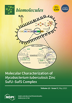
Iron-sulfur (Fe–S) clusters are essential inorganic cofactors in proteins, synthetized in vivo via complex protein machineries. Bacteria have developed several Fe–S assembly systems, such as the ISC, NIF, and SUF systems. Mycobacterium tuberculosis (Mtb), the causative agent of tuberculosis, contains only the SUF machinery. This system is essential in Mtb, and therefore constitutes an interesting therapeutic target. Two proteins of the Mtb SUF system were characterized: Rv1464(sufS) and Rv1465(sufU). SufS is a type II cysteine desulfurase enzyme and SufU is a zinc-dependent protein, endowed with sulfurtransferase activity and is able to interact with SufS. Mtb SufS-SufU is resistant to oxidative stress and the presence of zinc in SufU is likely to be responsible for this improved resistance. View this paper
  • Issues are regarded as officially published after their release is announced to the table of contents alert mailing list.
  • You may sign up for e-mail alerts to receive table of contents of newly released issues.
  • PDF is the official format for papers published in both, html and pdf forms. To view the papers in pdf format, click on the "PDF Full-text" link, and use the free Adobe Reader to open them.
Order results
Result details
Section
Select all
Export citation of selected articles as:
14 pages, 607 KiB  
Review
Organoids: Construction and Application in Gastric Cancer
by Chengdong Huo, Xiaoxia Zhang, Yanmei Gu, Daijun Wang, Shining Zhang, Tao Liu, Yumin Li and Wenting He
Biomolecules 2023, 13(5), 875; https://doi.org/10.3390/biom13050875 - 22 May 2023
Cited by 2 | Viewed by 2477
Abstract
Gastric organoids are biological models constructed in vitro using stem cell culture and 3D cell culture techniques, which are the latest research hotspots. The proliferation of stem cells in vitro is the key to gastric organoid models, making the cell subsets within the [...] Read more.
Gastric organoids are biological models constructed in vitro using stem cell culture and 3D cell culture techniques, which are the latest research hotspots. The proliferation of stem cells in vitro is the key to gastric organoid models, making the cell subsets within the models more similar to in vivo tissues. Meanwhile, the 3D culture technology also provides a more suitable microenvironment for the cells. Therefore, the gastric organoid models can largely restore the growth condition of cells in terms of morphology and function in vivo. As the most classic organoid models, patient-derived organoids use the patient’s own tissues for in vitro culture. This kind of model is responsive to the ‘disease information’ of a specific patient and has great effect on evaluating the strategies of individualized treatment. Herein, we review the current literature on the establishment of organoid cultures, and also explore organoid translational applications. Full article
Show Figures

Figure 1

18 pages, 3951 KiB  
Article
Circulating Hsp70 Levels and the Immunophenotype of Peripheral Blood Lymphocytes as Potential Biomarkers for Advanced Lung Cancer and Therapy Failure after Surgery
by Seyer Safi, Luis Messner, Merten Kliebisch, Linn Eggert, Ceyra Ceylangil, Philipp Lennartz, Benedict Jefferies, Henriette Klein, Moritz Schirren, Michael Dommasch, Dominik Lobinger and Gabriele Multhoff
Biomolecules 2023, 13(5), 874; https://doi.org/10.3390/biom13050874 - 22 May 2023
Cited by 2 | Viewed by 1526
Abstract
Lung cancer remains a devastating disease with a poor clinical outcome. A biomarker signature which could distinguish lung cancer from metastatic disease and detect therapeutic failure would significantly improve patient management and allow for individualized, risk-adjusted therapeutic decisions. In this study, circulating Hsp70 [...] Read more.
Lung cancer remains a devastating disease with a poor clinical outcome. A biomarker signature which could distinguish lung cancer from metastatic disease and detect therapeutic failure would significantly improve patient management and allow for individualized, risk-adjusted therapeutic decisions. In this study, circulating Hsp70 levels were measured using ELISA, and the immunophenotype of the peripheral blood lymphocytes were measured using multiparameter flow cytometry, to identify a predictive biomarker signature for lung cancer patients pre- and post-operatively, in patients with lung metastases and in patients with COPD as an inflammatory lung disease. The lowest Hsp70 concentrations were found in the healthy controls followed by the patients with advanced COPD. Hsp70 levels sequentially increased with an advancing tumor stage and metastatic disease. In the early-recurrence patients, Hsp70 levels started to increase within the first three months after surgery, but remained unaltered in the recurrence-free patients. An early recurrence was associated with a significant drop in B cells and an increase in Tregs, whereas the recurrence-free patients had elevated T and NK cell levels. We conclude that circulating Hsp70 concentrations might have the potential to distinguish lung cancer from metastatic disease, and might be able to predict an advanced tumor stage and early recurrence in lung cancer patients. Further studies with larger patient cohorts and longer follow-up periods are needed to validate Hsp70 and immunophenotypic profiles as predictive biomarker signatures. Full article
(This article belongs to the Special Issue HSP70: From Signaling Mechanisms to Therapeutics)
Show Figures

Figure 1

20 pages, 2231 KiB  
Review
The Potential of Edible and Medicinal Resource Polysaccharides for Prevention and Treatment of Neurodegenerative Diseases
by Qingxia Gan, Yugang Ding, Maoyao Peng, Linlin Chen, Jijing Dong, Jiaxi Hu and Yuntong Ma
Biomolecules 2023, 13(5), 873; https://doi.org/10.3390/biom13050873 - 22 May 2023
Viewed by 1858
Abstract
As natural medicines in complementary and alternative medicine, edible and medicinal resources are being gradually recognized throughout the world. According to statistics from the World Health Organization, about 80% of the worldwide population has used edible and medicinal resource products to prevent and [...] Read more.
As natural medicines in complementary and alternative medicine, edible and medicinal resources are being gradually recognized throughout the world. According to statistics from the World Health Organization, about 80% of the worldwide population has used edible and medicinal resource products to prevent and treat diseases. Polysaccharides, one of the main effective components in edible and medicinal resources, are considered ideal regulators of various biological responses due to their high effectiveness and low toxicity, and they have a wide range of possible applications for the development of functional foods for the regulation of common, frequently occurring, chronic and severe diseases. Such applications include the development of polysaccharide products for the prevention and treatment of neurodegenerative diseases that are difficult to control by a single treatment, which is of great value to the aging population. Therefore, we evaluated the potential of polysaccharides to prevent neurodegeneration by their regulation of behavioral and major pathologies, including abnormal protein aggregation and neuronal damage caused by neuronal apoptosis, autophagy, oxidative damage, neuroinflammation, unbalanced neurotransmitters, and poor synaptic plasticity. This includes multi-target and multi-pathway regulation involving the mitochondrial pathway, MAPK pathway, NF-κB pathway, Nrf2 pathway, mTOR pathway, PI3K/AKT pathway, P53/P21 pathway, and BDNF/TrkB/CREB pathway. In this paper, research into edible and medicinal resource polysaccharides for neurodegenerative diseases was reviewed in order to provide a basis for the development and application of polysaccharide health products and promote the recognition of functional products of edible and medicinal resources. Full article
Show Figures

Figure 1

19 pages, 3723 KiB  
Article
Space Environment Impacts Homeostasis: Exposure to Spaceflight Alters Mammary Gland Transportome Genes
by Osman V. Patel, Charlyn Partridge and Karen Plaut
Biomolecules 2023, 13(5), 872; https://doi.org/10.3390/biom13050872 - 22 May 2023
Viewed by 1421
Abstract
Membrane transporters and ion channels that play an indispensable role in metabolite trafficking have evolved to operate in Earth’s gravity. Dysregulation of the transportome expression profile at normogravity not only affects homeostasis along with drug uptake and distribution but also plays a key [...] Read more.
Membrane transporters and ion channels that play an indispensable role in metabolite trafficking have evolved to operate in Earth’s gravity. Dysregulation of the transportome expression profile at normogravity not only affects homeostasis along with drug uptake and distribution but also plays a key role in the pathogenesis of diverse localized to systemic diseases including cancer. The profound physiological and biochemical perturbations experienced by astronauts during space expeditions are well-documented. However, there is a paucity of information on the effect of the space environment on the transportome profile at an organ level. Thus, the goal of this study was to analyze the effect of spaceflight on ion channels and membrane substrate transporter genes in the periparturient rat mammary gland. Comparative gene expression analysis revealed an upregulation (p < 0.01) of amino acid, Ca2+, K+, Na+, Zn2+, Cl, PO43−, glucose, citrate, pyruvate, succinate, cholesterol, and water transporter genes in rats exposed to spaceflight. Genes associated with the trafficking of proton-coupled amino acids, Mg2+, Fe2+, voltage-gated K+-Na+, cation-coupled chloride, as well as Na+/Ca2+ and ATP-Mg/Pi exchangers were suppressed (p < 0.01) in these spaceflight-exposed rats. These findings suggest that an altered transportome profile contributes to the metabolic modulations observed in the rats exposed to the space environment. Full article
(This article belongs to the Collection Metabolomics and Integrated Multi-Omics in Health and Disease)
Show Figures

Figure 1

16 pages, 2719 KiB  
Review
Circulating microRNAs for Early Diagnosis of Ovarian Cancer: A Systematic Review and Meta-Analysis
by Nanna Lond Skov Frisk, Anja Elaine Sørensen, Ole Birger Vesterager Pedersen and Louise Torp Dalgaard
Biomolecules 2023, 13(5), 871; https://doi.org/10.3390/biom13050871 - 22 May 2023
Cited by 4 | Viewed by 1970
Abstract
In this study, we conducted a systematic review and meta-analysis to summarize and evaluate the global research potential of different circulating miRNAs as an early diagnostic biomarker for OC. A systematic literature search for relevant studies was conducted in June 2020 and followed [...] Read more.
In this study, we conducted a systematic review and meta-analysis to summarize and evaluate the global research potential of different circulating miRNAs as an early diagnostic biomarker for OC. A systematic literature search for relevant studies was conducted in June 2020 and followed up in November 2021. The search was conducted in English databases (PubMed, ScienceDirect). The primary search resulted in a total of 1887 articles, which were screened according to the prior established inclusion and exclusion criteria. We identified 44 relevant studies, of which 22 were eligible for the quantitative meta-analysis. Statistical analysis was performed using the Meta-package in Rstudio. Standardized mean differences (SMD) of relative levels between control subjects and OC patients were used to evaluate the differential expression. All studies were quality evaluated using a Newcastle–Ottawa Scale. Based on the meta-analysis, nine miRNAs were identified as dysregulated in OC patients compared to controls. Nine were upregulated in OC patients compared to controls (miR-21, -125, -141, -145, -205, -328, -200a, -200b, -200c). Furthermore, miR-26, -93, -106 and -200a were analyzed, but did not present an overall significant difference between OC patients and controls. These observations should be considered when performing future studies of circulating miRNAs in relation to OC: sufficient size of clinical cohorts, development of consensus guidelines for circulating miRNA measurements, and coverage of previously reported miRNAs. Full article
(This article belongs to the Special Issue Non-coding RNAs in Cancer Diagnosis and Therapy)
Show Figures

Figure 1

15 pages, 3575 KiB  
Communication
Comparison of In-Frame Deletion, Homology-Directed Repair, and Prime Editing-Based Correction of Duchenne Muscular Dystrophy Mutations
by Xiaoying Zhao, Kunli Qu, Benedetta Curci, Huanming Yang, Lars Bolund, Lin Lin and Yonglun Luo
Biomolecules 2023, 13(5), 870; https://doi.org/10.3390/biom13050870 - 22 May 2023
Cited by 1 | Viewed by 2354
Abstract
Recent progress in CRISPR gene editing tools has substantially increased the opportunities for curing devastating genetic diseases. Here we compare in-frame deletion by CRISPR-based non-homologous blunt end joining (NHBEJ), homology-directed repair (HDR), and prime editing (PE, PE2, and PE3)-based correction of two Duchenne [...] Read more.
Recent progress in CRISPR gene editing tools has substantially increased the opportunities for curing devastating genetic diseases. Here we compare in-frame deletion by CRISPR-based non-homologous blunt end joining (NHBEJ), homology-directed repair (HDR), and prime editing (PE, PE2, and PE3)-based correction of two Duchenne Muscular Dystrophy (DMD) loss-of-function mutations (c.5533G>T and c.7893delC). To enable accurate and rapid evaluation of editing efficiency, we generated a genomically integrated synthetic reporter system (VENUS) carrying the DMD mutations. The VENUS contains a modified enhanced green fluorescence protein (EGFP) gene, in which expression was restored upon the CRISPR-mediated correction of DMD loss-of-function mutations. We observed that the highest editing efficiency was achieved by NHBEJ (74–77%), followed by HDR (21–24%) and PE2 (1.5%) in HEK293T VENUS reporter cells. A similar HDR (23%) and PE2 (1.1%) correction efficiency is achieved in fibroblast VENUS cells. With PE3 (PE2 plus nicking gRNA), the c.7893delC correction efficiency was increased 3-fold. Furthermore, an approximately 31% correction efficiency of the endogenous DMD: c.7893delC is achieved in the FACS-enriched HDR-edited VENUS EGFP+ patient fibroblasts. We demonstrated that a highly efficient correction of DMD loss-of-function mutations in patient cells can be achieved by several means of CRISPR gene editing. Full article
(This article belongs to the Collection Feature Papers in Biomacromolecules: Nucleic Acids)
Show Figures

Graphical abstract

27 pages, 1949 KiB  
Review
Orchestration of Mitochondrial Function and Remodeling by Post-Translational Modifications Provide Insight into Mechanisms of Viral Infection
by Ji Woo Park, Matthew D. Tyl and Ileana M. Cristea
Biomolecules 2023, 13(5), 869; https://doi.org/10.3390/biom13050869 - 20 May 2023
Cited by 1 | Viewed by 1908
Abstract
The regulation of mitochondria structure and function is at the core of numerous viral infections. Acting in support of the host or of virus replication, mitochondria regulation facilitates control of energy metabolism, apoptosis, and immune signaling. Accumulating studies have pointed to post-translational modification [...] Read more.
The regulation of mitochondria structure and function is at the core of numerous viral infections. Acting in support of the host or of virus replication, mitochondria regulation facilitates control of energy metabolism, apoptosis, and immune signaling. Accumulating studies have pointed to post-translational modification (PTM) of mitochondrial proteins as a critical component of such regulatory mechanisms. Mitochondrial PTMs have been implicated in the pathology of several diseases and emerging evidence is starting to highlight essential roles in the context of viral infections. Here, we provide an overview of the growing arsenal of PTMs decorating mitochondrial proteins and their possible contribution to the infection-induced modulation of bioenergetics, apoptosis, and immune responses. We further consider links between PTM changes and mitochondrial structure remodeling, as well as the enzymatic and non-enzymatic mechanisms underlying mitochondrial PTM regulation. Finally, we highlight some of the methods, including mass spectrometry-based analyses, available for the identification, prioritization, and mechanistic interrogation of PTMs. Full article
(This article belongs to the Collection Metabolomics and Integrated Multi-Omics in Health and Disease)
Show Figures

Figure 1

17 pages, 5746 KiB  
Article
The IP6K Inhibitor LI-2242 Ameliorates Diet-Induced Obesity, Hyperglycemia, and Hepatic Steatosis in Mice by Improving Cell Metabolism and Insulin Signaling
by Sandip Mukherjee, Molee Chakraborty, Jake Haubner, Glen Ernst, Michael DePasquale, Danielle Carpenter, James C. Barrow and Anutosh Chakraborty
Biomolecules 2023, 13(5), 868; https://doi.org/10.3390/biom13050868 - 20 May 2023
Cited by 4 | Viewed by 1833
Abstract
Obesity and nonalcoholic fatty liver disease (NAFLD) are global health concerns, and thus, drugs for the long-term treatment of these diseases are urgently needed. We previously discovered that the inositol pyrophosphate biosynthetic enzyme IP6K1 is a target in diet-induced obesity (DIO), insulin resistance, [...] Read more.
Obesity and nonalcoholic fatty liver disease (NAFLD) are global health concerns, and thus, drugs for the long-term treatment of these diseases are urgently needed. We previously discovered that the inositol pyrophosphate biosynthetic enzyme IP6K1 is a target in diet-induced obesity (DIO), insulin resistance, and NAFLD. Moreover, high-throughput screening (HTS) assays and structure−activity relationship (SAR) studies identified LI-2242 as a potent IP6K inhibitor compound. Here, we tested the efficacy of LI-2242 in DIO WT C57/BL6J mice. LI-2242 (20 mg/kg/BW daily, i.p.) reduced body weight in DIO mice by specifically reducing the accumulation of body fat. It also improved glycemic parameters and reduced hyperinsulinemia. LI-2242-treated mice displayed reduced the weight of various adipose tissue depots and an increased expression of metabolism- and mitochondrial-energy-oxidation-inducing genes in these tissues. LI-2242 also ameliorated hepatic steatosis by reducing the expression of genes that enhance lipid uptake, lipid stabilization, and lipogenesis. Furthermore, LI-2242 enhances the mitochondrial oxygen consumption rate (OCR) and insulin signaling in adipocytes and hepatocytes in vitro. In conclusion, the pharmacologic inhibition of the inositol pyrophosphate pathway by LI-2242 has therapeutic potential in obesity and NAFLD. Full article
(This article belongs to the Special Issue Inositol Phosphates in Health and Disease)
Show Figures

Figure 1

15 pages, 612 KiB  
Review
Thermal Effect on Heat Shock Protein 70 Family to Prevent Atherosclerotic Cardiovascular Disease
by Masayo Nagai and Hidesuke Kaji
Biomolecules 2023, 13(5), 867; https://doi.org/10.3390/biom13050867 - 20 May 2023
Cited by 3 | Viewed by 1319
Abstract
Heat shock protein 70 (HSP70) is a chaperone protein induced by various stresses on cells and is involved in various disease mechanisms. In recent years, the expression of HSP70 in skeletal muscle has attracted attention for its use as a prevention of atherosclerotic [...] Read more.
Heat shock protein 70 (HSP70) is a chaperone protein induced by various stresses on cells and is involved in various disease mechanisms. In recent years, the expression of HSP70 in skeletal muscle has attracted attention for its use as a prevention of atherosclerotic cardiovascular disease (ASCVD) and as a disease marker. We have previously reported the effect of thermal stimulation targeted to skeletal muscles and skeletal muscle-derived cells. In this article, we reported review articles including our research results. HSP70 contributes to the improvement of insulin resistance as well as chronic inflammation which are underlying pathologies of type 2 diabetes, obesity, and atherosclerosis. Thus, induction of HSP70 expression by external stimulation such as heat and exercise may be useful for ASCVD prevention. It may be possible to induce HSP70 by thermal stimulus in those who have difficulty in exercise because of obesity or locomotive syndrome. It requires further investigation to determine whether monitoring serum HSP70 concentration is useful for ASCVD prevention. Full article
(This article belongs to the Special Issue HSP70: From Signaling Mechanisms to Therapeutics)
Show Figures

Figure 1

26 pages, 9848 KiB  
Review
Emerging Quantitative Biochemical, Structural, and Biophysical Methods for Studying Ribosome and Protein–RNA Complex Assembly
by Kavan Gor and Olivier Duss
Biomolecules 2023, 13(5), 866; https://doi.org/10.3390/biom13050866 - 19 May 2023
Cited by 3 | Viewed by 2467
Abstract
Ribosome assembly is one of the most fundamental processes of gene expression and has served as a playground for investigating the molecular mechanisms of how protein–RNA complexes (RNPs) assemble. A bacterial ribosome is composed of around 50 ribosomal proteins, several of which are [...] Read more.
Ribosome assembly is one of the most fundamental processes of gene expression and has served as a playground for investigating the molecular mechanisms of how protein–RNA complexes (RNPs) assemble. A bacterial ribosome is composed of around 50 ribosomal proteins, several of which are co-transcriptionally assembled on a ~4500-nucleotide-long pre-rRNA transcript that is further processed and modified during transcription, the entire process taking around 2 min in vivo and being assisted by dozens of assembly factors. How this complex molecular process works so efficiently to produce an active ribosome has been investigated over decades, resulting in the development of a plethora of novel approaches that can also be used to study the assembly of other RNPs in prokaryotes and eukaryotes. Here, we review biochemical, structural, and biophysical methods that have been developed and integrated to provide a detailed and quantitative understanding of the complex and intricate molecular process of bacterial ribosome assembly. We also discuss emerging, cutting-edge approaches that could be used in the future to study how transcription, rRNA processing, cellular factors, and the native cellular environment shape ribosome assembly and RNP assembly at large. Full article
(This article belongs to the Special Issue Ribosomal Proteins in Ribosome Assembly)
Show Figures

Graphical abstract

10 pages, 1142 KiB  
Communication
Serum and Exosomal miR-7-1-5p and miR-223-3p as Possible Biomarkers for Parkinson’s Disease
by Lorenzo Agostino Citterio, Roberta Mancuso, Simone Agostini, Mario Meloni and Mario Clerici
Biomolecules 2023, 13(5), 865; https://doi.org/10.3390/biom13050865 - 19 May 2023
Cited by 4 | Viewed by 1736
Abstract
The etiology of Parkinson’s disease (PD) is poorly understood, and is strongly suspected to include both genetic and environmental factors. In this context, it is essential to investigate possible biomarkers for both prognostic and diagnostic purposes. Several studies reported dysregulated microRNA expression in [...] Read more.
The etiology of Parkinson’s disease (PD) is poorly understood, and is strongly suspected to include both genetic and environmental factors. In this context, it is essential to investigate possible biomarkers for both prognostic and diagnostic purposes. Several studies reported dysregulated microRNA expression in neurodegenerative disorders, including PD. Using ddPCR, we investigated the concentrations of miR-7-1-5p, miR-499-3p, miR-223-3p and miR-223-5p—miRNAs involved in the α-synuclein pathway and in inflammation—in the serum and serum-isolated exosomes of 45 PD patients and 49 age- and sex-matched healthy controls (HC). While miR-499-3p and miR-223-5p showed no differences (1), serum concentration of miR-7-1-5p was significantly increased (p = 0.0007 vs. HC) and (2) miR-223-3p serum (p = 0.0006) and exosome (p = 0.0002) concentrations were significantly increased. ROC curve analysis showed that miR-223-3p and miR-7-1-5p serum concentration discriminates between PD and HC (p = 0.0001, in both cases). Notably, in PD patients, both miR-223-3p serum (p = 0.0008) and exosome (p = 0.006) concentrations correlated with levodopa equivalent daily dosage (LEDD). Finally, serum α-synuclein was increased in PD patients compared to HC (p = 0.025), and in patients correlated with serum miR-7-1-5p in (p = 0.05). Our results suggest that both miR-7-1-5p and miR-223-3p, distinguishing PD from HC, have the potential to be useful and non-invasive biomarkers in Parkinson’s disease. Full article
(This article belongs to the Special Issue Advances in Biomarkers for Neurodegenerative Diseases)
Show Figures

Figure 1

12 pages, 4316 KiB  
Article
Insight into Pathogenic Mechanism Underlying the Hereditary Cataract Caused by βB2-G149V Mutation
by Jing Wu, Silong Chen, Jingjie Xu, Wanyue Xu, Sifan Zheng, Qing Tian, Chenqi Luo, Xiangjun Chen and Xingchao Shentu
Biomolecules 2023, 13(5), 864; https://doi.org/10.3390/biom13050864 - 19 May 2023
Cited by 1 | Viewed by 1173
Abstract
Congenital cataracts account for approximately 5–20% of childhood blindness worldwide and 22–30% of childhood blindness in developing countries. Genetic disorders are the primary cause of congenital cataracts. In this work, we investigated the underlying molecular mechanism of G149V point missense mutation in βB2-crystallin, [...] Read more.
Congenital cataracts account for approximately 5–20% of childhood blindness worldwide and 22–30% of childhood blindness in developing countries. Genetic disorders are the primary cause of congenital cataracts. In this work, we investigated the underlying molecular mechanism of G149V point missense mutation in βB2-crystallin, which was first identified in a three-generation Chinese family with two affected members diagnosed with congenital cataracts. Spectroscopic experiments were performed to determine the structural differences between the wild type (WT) and the G149V mutant of βB2-crystallin. The results showed that the G149V mutation significantly changed the secondary and tertiary structure of βB2-crystallin. The polarity of the tryptophan microenvironment and the hydrophobicity of the mutant protein increased. The G149V mutation made the protein structure loose and the interaction between oligomers was reduced, which decreased the stability of the protein. Furthermore, we compared βB2-crystallin WT and the G149V mutant with their biophysical properties under environmental stress. We found that the G149V mutation makes βB2-crystallin more sensitive to environmental stresses (oxidative stress, UV irradiation, and heat shock) and more likely to aggregate and form precipitation. These features might be important to the pathogenesis of βB2-crystallin G149V mutant related to congenital cataracts. Full article
(This article belongs to the Section Biomacromolecules: Proteins)
Show Figures

Figure 1

18 pages, 977 KiB  
Review
Evidence of Metabolic Dysfunction in Amyotrophic Lateral Sclerosis (ALS) Patients and Animal Models
by Katarina Maksimovic, Mohieldin Youssef, Justin You, Hoon-Ki Sung and Jeehye Park
Biomolecules 2023, 13(5), 863; https://doi.org/10.3390/biom13050863 - 19 May 2023
Cited by 4 | Viewed by 4034
Abstract
Amyotrophic lateral sclerosis (ALS) is a neurodegenerative disease that affects motor neurons, leading to muscle weakness, paralysis, and eventual death. Research from the past few decades has appreciated that ALS is not only a disease of the motor neurons but also a disease [...] Read more.
Amyotrophic lateral sclerosis (ALS) is a neurodegenerative disease that affects motor neurons, leading to muscle weakness, paralysis, and eventual death. Research from the past few decades has appreciated that ALS is not only a disease of the motor neurons but also a disease that involves systemic metabolic dysfunction. This review will examine the foundational research of understanding metabolic dysfunction in ALS and provide an overview of past and current studies in ALS patients and animal models, spanning from full systems to various metabolic organs. While ALS-affected muscle tissue exhibits elevated energy demand and a fuel preference switch from glycolysis to fatty acid oxidation, adipose tissue in ALS undergoes increased lipolysis. Dysfunctions in the liver and pancreas contribute to impaired glucose homeostasis and insulin secretion. The central nervous system (CNS) displays abnormal glucose regulation, mitochondrial dysfunction, and increased oxidative stress. Importantly, the hypothalamus, a brain region that controls whole-body metabolism, undergoes atrophy associated with pathological aggregates of TDP-43. This review will also cover past and present treatment options that target metabolic dysfunction in ALS and provide insights into the future of metabolism research in ALS. Full article
(This article belongs to the Special Issue Stress, Aging and Metabolism)
Show Figures

Figure 1

19 pages, 3251 KiB  
Article
Enhanced L-β-Aminoisobutyric Acid Is Involved in the Pathophysiology of Effectiveness for Treatment-Resistant Schizophrenia and Adverse Reactions of Clozapine
by Kouji Fukuyama, Eishi Motomura and Motohiro Okada
Biomolecules 2023, 13(5), 862; https://doi.org/10.3390/biom13050862 - 19 May 2023
Cited by 2 | Viewed by 1588
Abstract
Clozapine is an effective antipsychotic for the treatment of antipsychotic-resistant schizophrenia; however, specific types of A/B adverse effects and clozapine-discontinuation syndromes are also well known. To date, both the critical mechanisms of clinical actions (effective for antipsychotic-resistant schizophrenia) and the adverse effects of [...] Read more.
Clozapine is an effective antipsychotic for the treatment of antipsychotic-resistant schizophrenia; however, specific types of A/B adverse effects and clozapine-discontinuation syndromes are also well known. To date, both the critical mechanisms of clinical actions (effective for antipsychotic-resistant schizophrenia) and the adverse effects of clozapine remain to be elucidated. Recently, we demonstrated that clozapine increased the synthesis of L-β-aminoisobutyric acid (L-BAIBA) in the hypothalamus. L-BAIBA is an activator of the adenosine monophosphate-activated protein kinase (AMPK), glycine receptor, GABAA receptor, and GABAB receptor (GABAB-R). These targets of L-BAIBA overlap as potential targets other than the monoamine receptors of clozapine. However, the direct binding of clozapine to these aminoacidic transmitter/modulator receptors remains to be clarified. Therefore, to explore the contribution of increased L-BAIBA on the clinical action of clozapine, this study determined the effects of clozapine and L-BAIBA on tripartite synaptic transmission, including GABAB-R and the group-III metabotropic glutamate receptor (III-mGluR) using cultured astrocytes, as well as on the thalamocortical hyper-glutamatergic transmission induced by impaired glutamate/NMDA receptors using microdialysis. Clozapine increased astroglial L-BAIBA synthesis in time/concentration-dependent manners. Increased L-BAIBA synthesis was observed until 3 days after clozapine discontinuation. Clozapine did not directly bind III-mGluR or GABAB-R, whereas L-BAIBA activated these receptors in the astrocytes. Local administration of MK801 into the reticular thalamic nucleus (RTN) increased L-glutamate release in the medial frontal cortex (mPFC) (MK801-evoked L-glutamate release). Local administration of L-BAIBA into the mPFC suppressed MK801-evoked L-glutamate release. These actions of L-BAIBA were inhibited by antagonists of III-mGluR and GABAB-R, similar to clozapine. These in vitro and in vivo analyses suggest that increased frontal L-BAIBA signaling likely plays an important role in the pharmacological actions of clozapine, such as improving the effectiveness of treating treatment-resistant schizophrenia and several clozapine discontinuation syndromes via the activation of III-mGluR and GABAB-R in the mPFC. Full article
(This article belongs to the Special Issue Glutamate and Glutamate Receptors in Health and Diseases)
Show Figures

Figure 1

14 pages, 2309 KiB  
Article
Co-Delivery of Therapeutics and Bioactive Gas Using a Novel Liposomal Platform for Enhanced Treatment of Acute Arterial Injury
by Shao-Ling Huang, Melanie R. Moody, Xing Yin, David D. McPherson and Hyunggun Kim
Biomolecules 2023, 13(5), 861; https://doi.org/10.3390/biom13050861 - 19 May 2023
Cited by 2 | Viewed by 1285
Abstract
Atherosclerosis is a complex, multi-stage disease characterized by pathological changes across the vascular wall. Endothelial dysfunction, inflammation, hypoxia, and vascular smooth muscle cell proliferation contribute to its progression. An effective strategy capable of delivering pleiotropic treatment to the vascular wall is essential to [...] Read more.
Atherosclerosis is a complex, multi-stage disease characterized by pathological changes across the vascular wall. Endothelial dysfunction, inflammation, hypoxia, and vascular smooth muscle cell proliferation contribute to its progression. An effective strategy capable of delivering pleiotropic treatment to the vascular wall is essential to limit neointimal formation. Echogenic liposomes (ELIP), which can encapsulate bioactive gases and therapeutic agents, have the potential to deliver enhanced penetration and treatment efficacy for atherosclerosis. In this study, liposomes loaded with nitric oxide (NO) and rosiglitazone, a peroxisome proliferator-activated receptor agonist, were prepared using hydration, sonication, freeze-thawing, and pressurization. The efficacy of this delivery system was evaluated in a rabbit model of acute arterial injury induced by balloon injury to the common carotid artery. Intra-arterial administration of rosiglitazone/NO co-encapsulated liposomes (R/NO-ELIP) immediately following injury resulted in reduced intimal thickening after 14 days. The anti-inflammatory and anti-proliferative effects of the co-delivery system were investigated. These liposomes were echogenic, enabling ultrasound imaging to assess their distribution and delivery. R/NO-ELIP delivery exhibited a greater attenuation (88 ± 15%) of intimal proliferation when compared to NO-ELIP (75 ± 13%) or R-ELIP (51 ± 6%) delivery alone. The study demonstrates the potential of echogenic liposomes as a promising platform for ultrasound imaging and therapeutic delivery. Full article
(This article belongs to the Section Molecular Medicine)
Show Figures

Figure 1

20 pages, 14663 KiB  
Article
Identification and Characterization of circRNAs in Non-Lactating Dairy Goat Mammary Glands Reveal Their Regulatory Role in Mammary Cell Involution and Remodeling
by Rong Xuan, Jianmin Wang, Qing Li, Yanyan Wang, Shanfeng Du, Qingling Duan, Yanfei Guo, Peipei He, Zhibin Ji and Tianle Chao
Biomolecules 2023, 13(5), 860; https://doi.org/10.3390/biom13050860 - 18 May 2023
Cited by 1 | Viewed by 1334
Abstract
This study conducted transcriptome sequencing of goat-mammary-gland tissue at the late lactation (LL), dry period (DP), and late gestation (LG) stages to reveal the expression characteristics and molecular functions of circRNAs during mammary involution. A total of 11,756 circRNAs were identified in this [...] Read more.
This study conducted transcriptome sequencing of goat-mammary-gland tissue at the late lactation (LL), dry period (DP), and late gestation (LG) stages to reveal the expression characteristics and molecular functions of circRNAs during mammary involution. A total of 11,756 circRNAs were identified in this study, of which 2528 circRNAs were expressed in all three stages. The number of exonic circRNAs was the largest, and the least identified circRNAs were antisense circRNAs. circRNA source gene analysis found that 9282 circRNAs were derived from 3889 genes, and 127 circRNAs’ source genes were unknown. Gene Ontology (GO) terms, such as histone modification, regulation of GTPase activity, and establishment or maintenance of cell polarity, were significantly enriched (FDR < 0.05), which indicates the functional diversity of circRNAs’ source genes. A total of 218 differentially expressed circRNAs were identified during the non-lactation period. The number of specifically expressed circRNAs was the highest in the DP and the lowest in LL stages. These indicated temporal specificity of circRNA expression in mammary gland tissues at different developmental stages. In addition, this study also constructed circRNA–miRNA–mRNA competitive endogenous RNA (ceRNA) regulatory networks related to mammary development, immunity, substance metabolism, and apoptosis. These findings help understand the regulatory role of circRNAs in mammary cell involution and remodeling. Full article
(This article belongs to the Special Issue Circular RNAs: Functions, Applications and Prospects)
Show Figures

Figure 1

41 pages, 2973 KiB  
Review
Dihydrocaffeic Acid—Is It the Less Known but Equally Valuable Phenolic Acid?
by Bartłomiej Zieniuk
Biomolecules 2023, 13(5), 859; https://doi.org/10.3390/biom13050859 - 18 May 2023
Cited by 4 | Viewed by 1937
Abstract
Dihydrocaffeic acid (DHCA) is a phenolic acid bearing a catechol ring and three-carbon side chain. Despite its being found in minor amounts in numerous plants and fungi of different origins, it has attracted the interest of various research groups in many fields of [...] Read more.
Dihydrocaffeic acid (DHCA) is a phenolic acid bearing a catechol ring and three-carbon side chain. Despite its being found in minor amounts in numerous plants and fungi of different origins, it has attracted the interest of various research groups in many fields of science, from food to biomedical applications. The review article presented herein aims to show a wider audience the health benefits and therapeutic, industrial, and nutritional potential of dihydrocaffeic acid, by sheddinglight on its occurrence, biosynthesis, bioavailability, and metabolism. The scientific literature describes at least 70 different derivatives of dihydrocaffeic acid, both those occurring naturally and those obtained via chemical and enzymatic methods. Among the most frequently used enzymes that were applied for the modification of the parent DHCA structure, there are lipases that allow for obtaining esters and phenolidips, tyrosinases used for the formation of the catechol ring, and laccases to functionalize this phenolic acid. In many studies, both in vitro and in vivo, the protective effect of DHCA and its derivatives on cells subjected to oxidative stress and inflammation were acknowledged. Full article
Show Figures

Figure 1

11 pages, 1501 KiB  
Article
AI-Aided Search for New HIV-1 Protease Ligands
by Roberto Arrigoni, Luigi Santacroce, Andrea Ballini and Luigi Leonardo Palese
Biomolecules 2023, 13(5), 858; https://doi.org/10.3390/biom13050858 - 18 May 2023
Cited by 2 | Viewed by 1510
Abstract
The availability of drugs capable of blocking the replication of microorganisms has been one of the greatest triumphs in the history of medicine, but the emergence of an ever-increasing number of resistant strains poses a serious problem for the treatment of infectious diseases. [...] Read more.
The availability of drugs capable of blocking the replication of microorganisms has been one of the greatest triumphs in the history of medicine, but the emergence of an ever-increasing number of resistant strains poses a serious problem for the treatment of infectious diseases. The search for new potential ligands for proteins involved in the life cycle of pathogens is, therefore, an extremely important research field today. In this work, we have considered the HIV-1 protease, one of the main targets for AIDS therapy. Several drugs are used today in clinical practice whose mechanism of action is based on the inhibition of this enzyme, but after years of use, even these molecules are beginning to be interested by resistance phenomena. We used a simple artificial intelligence system for the initial screening of a data set of potential ligands. These results were validated by docking and molecular dynamics, leading to the identification of a potential new ligand of the enzyme which does not belong to any known class of HIV-1 protease inhibitors. The computational protocol used in this work is simple and does not require large computational power. Furthermore, the availability of a large number of structural information on viral proteins and the presence of numerous experimental data on their ligands, with which it is possible to compare the results obtained with computational methods, make this research field the ideal terrain for the application of these new computational techniques. Full article
(This article belongs to the Special Issue Computer Aided Drug Discovery)
Show Figures

Figure 1

25 pages, 2710 KiB  
Review
FOXM1: Functional Roles of FOXM1 in Non-Malignant Diseases
by Zhenwang Zhang, Mengxi Li, Tian Sun, Zhengrong Zhang and Chao Liu
Biomolecules 2023, 13(5), 857; https://doi.org/10.3390/biom13050857 - 18 May 2023
Cited by 4 | Viewed by 3722
Abstract
Forkhead box (FOX) proteins are a wing-like helix family of transcription factors in the DNA-binding region. By mediating the activation and inhibition of transcription and interactions with all kinds of transcriptional co-regulators (MuvB complexes, STAT3, β-catenin, etc.), they play significant roles in carbohydrate [...] Read more.
Forkhead box (FOX) proteins are a wing-like helix family of transcription factors in the DNA-binding region. By mediating the activation and inhibition of transcription and interactions with all kinds of transcriptional co-regulators (MuvB complexes, STAT3, β-catenin, etc.), they play significant roles in carbohydrate and fat metabolism, biological aging and immune regulation, development, and diseases in mammals. Recent studies have focused on translating these essential findings into clinical applications in order to improve quality of life, investigating areas such as diabetes, inflammation, and pulmonary fibrosis, and increase human lifespan. Early studies have shown that forkhead box M1 (FOXM1) functions as a key gene in pathological processes in multiple diseases by regulating genes related to proliferation, the cell cycle, migration, and apoptosis and genes related to diagnosis, therapy, and injury repair. Although FOXM1 has long been studied in relation to human diseases, its role needs to be elaborated on. FOXM1 expression is involved in the development or repair of multiple diseases, including pulmonary fibrosis, pneumonia, diabetes, liver injury repair, adrenal lesions, vascular diseases, brain diseases, arthritis, myasthenia gravis, and psoriasis. The complex mechanisms involve multiple signaling pathways, such as WNT/β-catenin, STAT3/FOXM1/GLUT1, c-Myc/FOXM1, FOXM1/SIRT4/NF-κB, and FOXM1/SEMA3C/NRP2/Hedgehog. This paper reviews the key roles and functions of FOXM1 in kidney, vascular, lung, brain, bone, heart, skin, and blood vessel diseases to elucidate the role of FOXM1 in the development and progression of human non-malignant diseases and makes suggestions for further research. Full article
(This article belongs to the Section Molecular Medicine)
Show Figures

Figure 1

19 pages, 1589 KiB  
Review
Altered Purinergic Signaling in Neurodevelopmental Disorders: Focus on P2 Receptors
by Marta Boccazzi, Stefano Raffaele, Thomas Zanettin, Maria P. Abbracchio and Marta Fumagalli
Biomolecules 2023, 13(5), 856; https://doi.org/10.3390/biom13050856 - 18 May 2023
Cited by 3 | Viewed by 1867
Abstract
With the umbrella term ‘neurodevelopmental disorders’ (NDDs) we refer to a plethora of congenital pathological conditions generally connected with cognitive, social behavior, and sensory/motor alterations. Among the possible causes, gestational and perinatal insults have been demonstrated to interfere with the physiological processes necessary [...] Read more.
With the umbrella term ‘neurodevelopmental disorders’ (NDDs) we refer to a plethora of congenital pathological conditions generally connected with cognitive, social behavior, and sensory/motor alterations. Among the possible causes, gestational and perinatal insults have been demonstrated to interfere with the physiological processes necessary for the proper development of fetal brain cytoarchitecture and functionality. In recent years, several genetic disorders caused by mutations in key enzymes involved in purine metabolism have been associated with autism-like behavioral outcomes. Further analysis revealed dysregulated purine and pyrimidine levels in the biofluids of subjects with other NDDs. Moreover, the pharmacological blockade of specific purinergic pathways reversed the cognitive and behavioral defects caused by maternal immune activation, a validated and now extensively used rodent model for NDDs. Furthermore, Fragile X and Rett syndrome transgenic animal models as well as models of premature birth, have been successfully utilized to investigate purinergic signaling as a potential pharmacological target for these diseases. In this review, we examine results on the role of the P2 receptor signaling in the etiopathogenesis of NDDs. On this basis, we discuss how this evidence could be exploited to develop more receptor-specific ligands for future therapeutic interventions and novel prognostic markers for the early detection of these conditions. Full article
Show Figures

Figure 1

41 pages, 4159 KiB  
Review
(Patho)Physiology of Glycosylphosphatidylinositol-Anchored Proteins I: Localization at Plasma Membranes and Extracellular Compartments
by Günter A. Müller and Timo D. Müller
Biomolecules 2023, 13(5), 855; https://doi.org/10.3390/biom13050855 - 18 May 2023
Cited by 5 | Viewed by 3034
Abstract
Glycosylphosphatidylinositol (GPI)-anchored proteins (APs) are anchored at the outer leaflet of plasma membranes (PMs) of all eukaryotic organisms studied so far by covalent linkage to a highly conserved glycolipid rather than a transmembrane domain. Since their first description, experimental data have been accumulating [...] Read more.
Glycosylphosphatidylinositol (GPI)-anchored proteins (APs) are anchored at the outer leaflet of plasma membranes (PMs) of all eukaryotic organisms studied so far by covalent linkage to a highly conserved glycolipid rather than a transmembrane domain. Since their first description, experimental data have been accumulating for the capability of GPI-APs to be released from PMs into the surrounding milieu. It became evident that this release results in distinct arrangements of GPI-APs which are compatible with the aqueous milieu upon loss of their GPI anchor by (proteolytic or lipolytic) cleavage or in the course of shielding of the full-length GPI anchor by incorporation into extracellular vesicles, lipoprotein-like particles and (lyso)phospholipid- and cholesterol-harboring micelle-like complexes or by association with GPI-binding proteins or/and other full-length GPI-APs. In mammalian organisms, the (patho)physiological roles of the released GPI-APs in the extracellular environment, such as blood and tissue cells, depend on the molecular mechanisms of their release as well as the cell types and tissues involved, and are controlled by their removal from circulation. This is accomplished by endocytic uptake by liver cells and/or degradation by GPI-specific phospholipase D in order to bypass potential unwanted effects of the released GPI-APs or their transfer from the releasing donor to acceptor cells (which will be reviewed in a forthcoming manuscript). Full article
(This article belongs to the Section Biomacromolecules: Proteins)
Show Figures

Figure 1

16 pages, 1289 KiB  
Article
The Effects of Two Kinds of Dietary Interventions on Serum Metabolic Profiles in Haemodialysis Patients
by Lucyna Kozlowska, Karolina Jagiello, Krzesimir Ciura, Anita Sosnowska, Rafal Zwiech, Zbigniew Zbrog, Wojciech Wasowicz and Jolanta Gromadzinska
Biomolecules 2023, 13(5), 854; https://doi.org/10.3390/biom13050854 - 18 May 2023
Viewed by 1639
Abstract
The goal of this study was to evaluate the effects of two kinds of 24-week dietary interventions in haemodialysis patients, a traditional nutritional intervention without a meal before dialysis (HG1) and implementation of a nutritional intervention with a meal served just before dialysis [...] Read more.
The goal of this study was to evaluate the effects of two kinds of 24-week dietary interventions in haemodialysis patients, a traditional nutritional intervention without a meal before dialysis (HG1) and implementation of a nutritional intervention with a meal served just before dialysis (HG2), in terms of analysing the differences in the serum metabolic profiles and finding biomarkers of dietary efficacy. These studies were performed in two homogenous groups of patients (n = 35 in both groups). Among the metabolites with the highest statistical significance between HG1 and HG2 after the end of the study, 21 substances were putatively annotated, which had potential significance in both of the most relevant metabolic pathways and those related to diet. After the 24 weeks of the dietary intervention, the main differences between the metabolomic profiles in the HG2 vs. HG1 groups were related to the higher signal intensities from amino acid metabolites: indole-3-carboxaldehyde, 5-(hydroxymethyl-2-furoyl)glycine, homocitrulline, 4-(glutamylamino)butanoate, tryptophol, gamma-glutamylthreonine, and isovalerylglycine. These metabolites are intermediates in the metabolic pathways of the necessary amino acids (Trp, Tyr, Phe, Leu, Ile, Val, Liz, and amino acids of the urea cycle) and are also diet-related intermediates (4-guanidinobutanoic acid, indole-3-carboxyaldehyde, homocitrulline, and isovalerylglycine). Full article
(This article belongs to the Collection Metabolomics and Integrated Multi-Omics in Health and Disease)
Show Figures

Figure 1

18 pages, 3411 KiB  
Review
Ribosomal Protein uS5 and Friends: Protein–Protein Interactions Involved in Ribosome Assembly and Beyond
by Anne-Marie Landry-Voyer, Zabih Mir Hassani, Mariano Avino and François Bachand
Biomolecules 2023, 13(5), 853; https://doi.org/10.3390/biom13050853 - 18 May 2023
Cited by 4 | Viewed by 1970
Abstract
Ribosomal proteins are fundamental components of the ribosomes in all living cells. The ribosomal protein uS5 (Rps2) is a stable component of the small ribosomal subunit within all three domains of life. In addition to its interactions with proximal ribosomal proteins and rRNA [...] Read more.
Ribosomal proteins are fundamental components of the ribosomes in all living cells. The ribosomal protein uS5 (Rps2) is a stable component of the small ribosomal subunit within all three domains of life. In addition to its interactions with proximal ribosomal proteins and rRNA inside the ribosome, uS5 has a surprisingly complex network of evolutionarily conserved non-ribosome-associated proteins. In this review, we focus on a set of four conserved uS5-associated proteins: the protein arginine methyltransferase 3 (PRMT3), the programmed cell death 2 (PDCD2) and its PDCD2-like (PDCD2L) paralog, and the zinc finger protein, ZNF277. We discuss recent work that presents PDCD2 and homologs as a dedicated uS5 chaperone and PDCD2L as a potential adaptor protein for the nuclear export of pre-40S subunits. Although the functional significance of the PRMT3–uS5 and ZNF277–uS5 interactions remain elusive, we reflect on the potential roles of uS5 arginine methylation by PRMT3 and on data indicating that ZNF277 and PRMT3 compete for uS5 binding. Together, these discussions highlight the complex and conserved regulatory network responsible for monitoring the availability and the folding of uS5 for the formation of 40S ribosomal subunits and/or the role of uS5 in potential extra-ribosomal functions. Full article
(This article belongs to the Special Issue Ribosomal Proteins in Ribosome Assembly)
Show Figures

Figure 1

18 pages, 1279 KiB  
Article
Exercise-Induced Alternations of Adiponectin, Interleukin-8 and Indicators of Carbohydrate Metabolism in Males with Metabolic Syndrome
by Karol Makiel, Agnieszka Suder, Aneta Targosz, Marcin Maciejczyk and Alon Haim
Biomolecules 2023, 13(5), 852; https://doi.org/10.3390/biom13050852 - 18 May 2023
Cited by 2 | Viewed by 2105
Abstract
Adiponectin (ADIPO) and interleukin-8 (IL-8) are proteins that play a significant, albeit opposing, role in metabolic syndrome (MetS). The reported data on the effect of physical activity on the levels of these hormones in the population of people with MetS are conflicting. The [...] Read more.
Adiponectin (ADIPO) and interleukin-8 (IL-8) are proteins that play a significant, albeit opposing, role in metabolic syndrome (MetS). The reported data on the effect of physical activity on the levels of these hormones in the population of people with MetS are conflicting. The aim of the study was to evaluate the changes in hormone concentrations, insulin-resistance indices and body composition after two types of training. The study included 62 men with MetS (age 36.6 ± 6.9 years, body fat [BF] = 37.53 ± 4.5%), randomly assigned to: an experimental group EG1 (n = 21) with aerobic exercise intervention, an experimental group EG2 (n = 21) with combined aerobic and resistance exercise intervention, both for 12 weeks, and a control group CG (n = 20) without interventions. Anthropometric measurements and body composition (fat-free mass [FFM], gynoid body fat [GYNOID]), as well as a biochemical blood analysis (adiponectin [ADIPO], interleukin-8 [IL-8], homeostatic model assessment—adiponectin (HOMA-AD) and homeostatic model assessment—triglycerides (HOMA-TG) were performed at baseline, and at 6 and 12 weeks of intervention and 4 weeks after the intervention (follow-up). Intergroup (between groups) and intragroup (within each group) changes were statistically evaluated. In the experimental groups EG1 and EG2, no significant changes were observed in the ADIPO concentration, but a decrease of GYNOID and insulin-resistance indices was confirmed. The aerobic training led to favorable changes in IL-8 concentration. The use of combined resistance and aerobic training led to improved body composition, decreased waist circumference and better insulin-resistance indices in men with MetS. Full article
(This article belongs to the Topic Molecular Mechanisms of Exercise and Healthspan)
Show Figures

Figure 1

13 pages, 3081 KiB  
Article
Endocan Knockdown Down-Regulates the Expression of Angiogenesis-Associated Genes in Il-1ß Activated Chondrocytes
by Michele Scuruchi, Federica Aliquò, Angela Avenoso, Giuseppe Mandraffino, Giovanna Vermiglio, Aurelio Minuti, Salvatore Campo, Giuseppe Maurizio Campo and Angela D’Ascola
Biomolecules 2023, 13(5), 851; https://doi.org/10.3390/biom13050851 - 18 May 2023
Viewed by 1409
Abstract
Endocan is a small soluble proteoglycan (PG) known to be involved in inflammation and angiogenesis. Increased endocan expression was found in the synovia of arthritic patients and chondrocytes stimulated with IL-1ß. Considering these findings, we aimed to investigate the effects of endocan knockdown [...] Read more.
Endocan is a small soluble proteoglycan (PG) known to be involved in inflammation and angiogenesis. Increased endocan expression was found in the synovia of arthritic patients and chondrocytes stimulated with IL-1ß. Considering these findings, we aimed to investigate the effects of endocan knockdown on the modulation of pro-angiogenic molecules expression in a model of IL-1ß-induced inflammation in human articular chondrocytes. Endocan, VEGF-A, MMP-9, MMP-13, and VEGFR-2 expression was measured in both normal and endocan knockdown chondrocytes stimulated with IL-1ß. VEGFR-2 and NF-kB activation were also measured. Results have shown that endocan, VEGF-A, VEGFR-2, MMP-9, and MMP-13 were significantly up-regulated during IL-1ß-induced inflammation; interestingly, the expression of such pro-angiogenic molecules and NF-kB activation were significantly reduced by endocan knockdown. These data support the hypothesis that endocan released by activated chondrocytes may be involved in the mechanisms that stimulate cell migration and invasion, as well as angiogenesis, in the pannus of arthritic joints. Full article
(This article belongs to the Special Issue Hyaluronic Acid and Proteoglycans: Basic and Biomedical Applications)
Show Figures

Figure 1

13 pages, 643 KiB  
Review
Emerging Role and Mechanism of the FTO Gene in Cardiovascular Diseases
by Zi-Yang Xu, Xia Jing and Xing-Dong Xiong
Biomolecules 2023, 13(5), 850; https://doi.org/10.3390/biom13050850 - 17 May 2023
Cited by 4 | Viewed by 2730
Abstract
The fat mass and obesity-associated (FTO) gene was the first obesity-susceptibility gene identified through a genome-wide association study (GWAS). A growing number of studies have suggested that genetic variants of FTO are strongly associated with the risk of cardiovascular diseases, including [...] Read more.
The fat mass and obesity-associated (FTO) gene was the first obesity-susceptibility gene identified through a genome-wide association study (GWAS). A growing number of studies have suggested that genetic variants of FTO are strongly associated with the risk of cardiovascular diseases, including hypertension and acute coronary syndrome. In addition, FTO was also the first N6-methyladenosine (m6A) demethylase, suggesting the reversible nature of m6A modification. m6A is dynamically deposited, removed, and recognized by m6A methylases, demethylases, and m6A binding proteins, respectively. By catalyzing m6A demethylation on mRNA, FTO may participate in various biological processes by modulating RNA function. Recent studies demonstrated that FTO plays a pivotal role in the initiation and progression of cardiovascular diseases such as myocardial fibrosis, heart failure, and atherosclerosis and may hold promise as a potential therapeutic target for treating or preventing a variety of cardiovascular diseases. Here, we review the association between FTO genetic variants and cardiovascular disease risk, summarize the role of FTO as an m6A demethylase in cardiovascular disorders, and discuss future research directions and possible clinical implications. Full article
Show Figures

Figure 1

13 pages, 1571 KiB  
Article
Association of Long Noncoding RNA Expression Signatures with Stress-Induced Myocardial Perfusion Defects
by Yu-Chieh Chang, Jun-Ting Liou, Yu-Min Peng, Guan-Jun Chen, Chien-Yu Lin and Chin-An Yang
Biomolecules 2023, 13(5), 849; https://doi.org/10.3390/biom13050849 - 17 May 2023
Viewed by 1113
Abstract
Stress-induced myocardial perfusion defects found in dipyridamole–thallium-201 single-photon emission computed tomography imaging may indicate vascular perfusion abnormalities and risk of obstructive or nonobstructive coronary heart disease. Besides nuclear imaging and subsequent coronary angiography (CAG), no blood test can indicate whether dysregulated homeostasis is [...] Read more.
Stress-induced myocardial perfusion defects found in dipyridamole–thallium-201 single-photon emission computed tomography imaging may indicate vascular perfusion abnormalities and risk of obstructive or nonobstructive coronary heart disease. Besides nuclear imaging and subsequent coronary angiography (CAG), no blood test can indicate whether dysregulated homeostasis is associated with stress-induced myocardial perfusion defects. This study investigated the expression signature of long noncoding RNAs (lncRNAs) and genes involved in vascular inflammation and stress response in the blood of patients with stress-induced myocardial perfusion abnormalities (n = 27). The results revealed an expression signature consisting of the upregulation of RMRP (p < 0.01) and downregulations of THRIL (p < 0.01) and HIF1A (p < 0.01) among patients with a positive thallium stress test and no significant coronary artery stenosis within 6 months after baseline treatment. We developed a scoring system based on the expression signatures of RMRP, MIAT, NTT, MALAT1, HSPA1A, and NLRP3 to predict the need for further CAG among patients with moderate-to-significant stress-induced myocardial perfusion defects (area under the receiver operating characteristic curve = 0.963). Therefore, we identified a dysregulated expression profile of lncRNA-based genes in the blood that could be valuable for the early detection of vascular homeostasis imbalance and personalized therapy. Full article
(This article belongs to the Special Issue Classical and Novel Biomarkers for Cardiovascular Disease)
Show Figures

Figure 1

17 pages, 28499 KiB  
Article
An Overview of Two Old Friends Associated with Platelet Redox Signaling, the Protein Disulfide Isomerase and NADPH Oxidase
by Andrés Trostchansky and Marcelo Alarcon
Biomolecules 2023, 13(5), 848; https://doi.org/10.3390/biom13050848 - 17 May 2023
Cited by 1 | Viewed by 1391
Abstract
Oxidative stress participates at the baseline of different non-communicable pathologies such as cardiovascular diseases. Excessive formation of reactive oxygen species (ROS), above the signaling levels necessary for the correct function of organelles and cells, may contribute to the non-desired effects of oxidative stress. [...] Read more.
Oxidative stress participates at the baseline of different non-communicable pathologies such as cardiovascular diseases. Excessive formation of reactive oxygen species (ROS), above the signaling levels necessary for the correct function of organelles and cells, may contribute to the non-desired effects of oxidative stress. Platelets play a relevant role in arterial thrombosis, by aggregation triggered by different agonists, where excessive ROS formation induces mitochondrial dysfunction and stimulate platelet activation and aggregation. Platelet is both a source and a target of ROS, thus we aim to analyze both the platelet enzymes responsible for ROS generation and their involvement in intracellular signal transduction pathways. Among the proteins involved in these processes are Protein Disulphide Isomerase (PDI) and NADPH oxidase (NOX) isoforms. By using bioinformatic tools and information from available databases, a complete bioinformatic analysis of the role and interactions of PDI and NOX in platelets, as well as the signal transduction pathways involved in their effects was performed. We focused the study on analyzing whether these proteins collaborate to control platelet function. The data presented in the current manuscript support the role that PDI and NOX play on activation pathways necessary for platelet activation and aggregation, as well as on the platelet signaling imbalance produced by ROS production. Our data could be used to design specific enzyme inhibitors or a dual inhibition for these enzymes with an antiplatelet effect to design promising treatments for diseases involving platelet dysfunction. Full article
Show Figures

Figure 1

8 pages, 1573 KiB  
Communication
The Impact of Maternal Probiotics on Intestinal Vitamin D Receptor Expression in Early Life
by Anita Sharma, Yueyue Yu, Jing Lu, Lei Lu, Yong-Guo Zhang, Yinglin Xia, Jun Sun and Erika C. Claud
Biomolecules 2023, 13(5), 847; https://doi.org/10.3390/biom13050847 - 16 May 2023
Cited by 1 | Viewed by 1345
Abstract
Vitamin D signaling via the Vitamin D Receptor (VDR) has been shown to protect against intestinal inflammation. Previous studies have also reported the mutual interactions of intestinal VDR and the microbiome, indicating a potential role of probiotics in modulating VDR expression. In preterm [...] Read more.
Vitamin D signaling via the Vitamin D Receptor (VDR) has been shown to protect against intestinal inflammation. Previous studies have also reported the mutual interactions of intestinal VDR and the microbiome, indicating a potential role of probiotics in modulating VDR expression. In preterm infants, although probiotics have been shown to reduce the incidence of necrotizing enterocolitis (NEC), they are not currently recommended by the FDA due to potential risks in this population. No previous studies have delved into the effect of maternally administered probiotics on intestinal VDR expression in early life. Using an infancy mouse model, we found that young mice exposed to maternally administered probiotics (SPF/LB) maintained higher colonic VDR expression than our unexposed mice (SPF) in the face of a systemic inflammatory stimulus. These findings indicate a potential role for microbiome-modulating therapies in preventing diseases such as NEC through the enhancement of VDR signaling. Full article
Show Figures

Figure 1

11 pages, 1160 KiB  
Article
Non-Psychoactive Cannabinoid Modulation of Nociception and Inflammation Associated with a Rat Model of Pulpitis
by Elana Y. Laks, Hongbo Li and Sara Jane Ward
Biomolecules 2023, 13(5), 846; https://doi.org/10.3390/biom13050846 - 16 May 2023
Cited by 1 | Viewed by 2123
Abstract
Despite advancements in dental pain management, one of the most common reasons for emergency dental care is orofacial pain. Our study aimed to determine the effects of non-psychoactive Cannabis constituents in the treatment of dental pain and related inflammation. We tested the therapeutic [...] Read more.
Despite advancements in dental pain management, one of the most common reasons for emergency dental care is orofacial pain. Our study aimed to determine the effects of non-psychoactive Cannabis constituents in the treatment of dental pain and related inflammation. We tested the therapeutic potential of two non-psychoactive Cannabis constituents, cannabidiol (CBD) and β-caryophyllene (β-CP), in a rodent model of orofacial pain associated with pulp exposure. Sham or left mandibular molar pulp exposures were performed on Sprague Dawley rats treated with either vehicle, the phytocannabinoid CBD (5 mg/kg i.p.) or the sesquiterpene β-CP (30 mg/kg i.p.) administered 1 h pre-exposure and on days 1, 3, 7, and 10 post-exposure. Orofacial mechanical allodynia was evaluated at baseline and post-pulp exposure. Trigeminal ganglia were harvested for histological evaluation at day 15. Pulp exposure was associated with significant orofacial sensitivity and neuroinflammation in the ipsilateral orofacial region and trigeminal ganglion. β-CP but not CBD produced a significant reduction in orofacial sensitivity. β-CP also significantly reduced the expression of the inflammatory markers AIF and CCL2, while CBD only decreased AIF expression. These data represent the first preclinical evidence that non-psychoactive cannabinoid-based pharmacotherapy may provide a therapeutic benefit for the treatment of orofacial pain associated with pulp exposure. Full article
(This article belongs to the Special Issue New Advances of Cannabinoid Receptors in Health and Disease)
Show Figures

Figure 1

Previous Issue
Next Issue
Back to TopTop