Next Issue
Volume 12, August
Previous Issue
Volume 12, June


Biosensors, Volume 12, Issue 7 (July 2022) – 118 articles

Cover Story (view full-size image): 
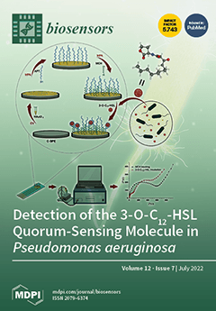
In this study, the development of the first electrochemical aptasensor for the detection of 3-O-C12-HSL is reported. A carbon-based screen-printed electrode modified with gold nanoparticles proved to be the best platform for the aptasensor. Each step in the fabrication of the aptasensor (i.e., the deposition of gold nanoparticles, aptamer immobilization, and incubation with the analyte) was optimized and characterized using cyclic voltammetry, differential pulse voltammetry, and electrochemical impedance spectroscopy. Different redox probes were evaluated in the solution, with the best results being obtained in the presence of [Fe(CN)6]4−/[Fe(CN)6]3−. The binding affinity of 106.7 nM for the immobilized thiol-terminated aptamer was determined using surface plasmon resonance. View this paper
  • Issues are regarded as officially published after their release is announced to the table of contents alert mailing list.
  • You may sign up for e-mail alerts to receive table of contents of newly released issues.
  • PDF is the official format for papers published in both, html and pdf forms. To view the papers in pdf format, click on the "PDF Full-text" link, and use the free Adobe Reader to open them.
Order results
Result details
Section
Select all
Export citation of selected articles as:
31 pages, 4132 KiB  
Review
Recent Advancements in Electrochemical Biosensors for Monitoring the Water Quality
by Yun Hui, Zhaoling Huang, Md Eshrat E. Alahi, Anindya Nag, Shilun Feng and Subhas Chandra Mukhopadhyay
Biosensors 2022, 12(7), 551; https://doi.org/10.3390/bios12070551 - 21 Jul 2022
Cited by 19 | Viewed by 4325
Abstract
The release of chemicals and microorganisms from various sources, such as industry, agriculture, animal farming, wastewater treatment plants, and flooding, into water systems have caused water pollution in several parts of our world, endangering aquatic ecosystems and individual health. World Health Organization (WHO) [...] Read more.
The release of chemicals and microorganisms from various sources, such as industry, agriculture, animal farming, wastewater treatment plants, and flooding, into water systems have caused water pollution in several parts of our world, endangering aquatic ecosystems and individual health. World Health Organization (WHO) has introduced strict standards for the maximum concentration limits for nutrients and chemicals in drinking water, surface water, and groundwater. It is crucial to have rapid, sensitive, and reliable analytical detection systems to monitor the pollution level regularly and meet the standard limit. Electrochemical biosensors are advantageous analytical devices or tools that convert a bio-signal by biorecognition elements into a significant electrical response. Thanks to the micro/nano fabrication techniques, electrochemical biosensors for sensitive, continuous, and real-time detection have attracted increasing attention among researchers and users worldwide. These devices take advantage of easy operation, portability, and rapid response. They can also be miniaturized, have a long-life span and a quick response time, and possess high sensitivity and selectivity and can be considered as portable biosensing assays. They are of special importance due to their great advantages such as affordability, simplicity, portability, and ability to detect at on-site. This review paper is concerned with the basic concepts of electrochemical biosensors and their applications in various water quality monitoring, such as inorganic chemicals, nutrients, microorganisms’ pollution, and organic pollutants, especially for developing real-time/online detection systems. The basic concepts of electrochemical biosensors, different surface modification techniques, bio-recognition elements (BRE), detection methods, and specific real-time water quality monitoring applications are reviewed thoroughly in this article. Full article
(This article belongs to the Special Issue Nano-Biosensors for Detection and Monitoring)
Show Figures

Figure 1

29 pages, 9373 KiB  
Review
Pyrene-Based AIE Active Materials for Bioimaging and Theranostics Applications
by Muthaiah Shellaiah and Kien-Wen Sun
Biosensors 2022, 12(7), 550; https://doi.org/10.3390/bios12070550 - 21 Jul 2022
Cited by 38 | Viewed by 3787
Abstract
Aggregation-induced emission (AIE) is a unique research topic and property that can lead to a wide range of applications, including cellular imaging, theranostics, analyte quantitation and the specific detection of biologically important species. Towards the development of the AIE-active materials, many aromatic moieties [...] Read more.
Aggregation-induced emission (AIE) is a unique research topic and property that can lead to a wide range of applications, including cellular imaging, theranostics, analyte quantitation and the specific detection of biologically important species. Towards the development of the AIE-active materials, many aromatic moieties composed of tetraphenylethylene, anthracene, pyrene, etc., have been developed. Among these aromatic moieties, pyrene is an aromatic hydrocarbon with a polycyclic flat structure containing four fused benzene rings to provide an unusual electron delocalization feature that is important in the AIE property. Numerous pyrene-based AIE-active materials have been reported with the AIE property towards sensing, imaging and theranostics applications. Most importantly, these AIE-active pyrene moieties exist as small molecules, Schiff bases, polymers, supramolecules, metal-organic frameworks, etc. This comprehensive review outlines utilizations of AIE-active pyrene-based materials on the imaging and theranostics studies. Moreover, the design and synthesis of these pyrene-based molecules are delivered with discussions on their future scopes. Full article
Show Figures

Figure 1

23 pages, 5642 KiB  
Article
Exploring Orientation Invariant Heuristic Features with Variant Window Length of 1D-CNN-LSTM in Human Activity Recognition
by Arnab Barua, Daniel Fuller, Sumayyah Musa and Xianta Jiang
Biosensors 2022, 12(7), 549; https://doi.org/10.3390/bios12070549 - 21 Jul 2022
Cited by 5 | Viewed by 2021
Abstract
Many studies have explored divergent deep neural networks in human activity recognition (HAR) using a single accelerometer sensor. Multiple types of deep neural networks, such as convolutional neural networks (CNN), long short-term memory (LSTM), or their hybridization (CNN-LSTM), have been implemented. However, the [...] Read more.
Many studies have explored divergent deep neural networks in human activity recognition (HAR) using a single accelerometer sensor. Multiple types of deep neural networks, such as convolutional neural networks (CNN), long short-term memory (LSTM), or their hybridization (CNN-LSTM), have been implemented. However, the sensor orientation problem poses challenges in HAR, and the length of windows as inputs for the deep neural networks has mostly been adopted arbitrarily. This paper explores the effect of window lengths with orientation invariant heuristic features on the performance of 1D-CNN-LSTM in recognizing six human activities; sitting, lying, walking and running at three different speeds using data from an accelerometer sensor encapsulated into a smartphone. Forty-two participants performed the six mentioned activities by keeping smartphones in their pants pockets with arbitrary orientation. We conducted an inter-participant evaluation using 1D-CNN-LSTM architecture. We found that the average accuracy of the classifier was saturated to 80 ± 8.07% for window lengths greater than 65 using only four selected simple orientation invariant heuristic features. In addition, precision, recall and F1-measure in recognizing stationary activities such as sitting and lying decreased with increment of window length, whereas we encountered an increment in recognizing the non-stationary activities. Full article
(This article belongs to the Special Issue Wearable Sensing for Health Monitoring)
Show Figures

Figure 1

14 pages, 1911 KiB  
Review
Plasmonic Approaches for the Detection of SARS-CoV-2 Viral Particles
by Sabine Szunerits, Hiba Saada, Quentin Pagneux and Rabah Boukherroub
Biosensors 2022, 12(7), 548; https://doi.org/10.3390/bios12070548 - 21 Jul 2022
Cited by 7 | Viewed by 2316
Abstract
The ongoing highly contagious Coronavirus disease 2019 (COVID-19) pandemic, caused by severe acute respiratory syndrome coronavirus 2 (SARS-CoV-2), underlines the fundamental position of diagnostic testing in outbreak control by allowing a distinction of the infected from the non-infected people. Diagnosis of COVID-19 remains [...] Read more.
The ongoing highly contagious Coronavirus disease 2019 (COVID-19) pandemic, caused by severe acute respiratory syndrome coronavirus 2 (SARS-CoV-2), underlines the fundamental position of diagnostic testing in outbreak control by allowing a distinction of the infected from the non-infected people. Diagnosis of COVID-19 remains largely based on reverse transcription PCR (RT-PCR), identifying the genetic material of the virus. Molecular testing approaches have been largely proposed in addition to infectivity testing of patients via sensing the presence of viral particles of SARS-CoV-2 specific structural proteins, such as the spike glycoproteins (S1, S2) and the nucleocapsid (N) protein. While the S1 protein remains the main target for neutralizing antibody treatment upon infection and the focus of vaccine and therapeutic design, it has also become a major target for the development of point-of care testing (POCT) devices. This review will focus on the possibility of surface plasmon resonance (SPR)-based sensing platforms to convert the receptor-binding event of SARS-CoV-2 viral particles into measurable signals. The state-of-the-art SPR-based SARS-CoV-2 sensing devices will be provided, and highlights about the applicability of plasmonic sensors as POCT for virus particle as well as viral protein sensing will be discussed. Full article
(This article belongs to the Special Issue Fundamentals of SARS-CoV-2 Biosensors)
Show Figures

Figure 1

14 pages, 3372 KiB  
Article
Red Blood Cell Sedimentation Index Using Shear Stress of Blood Flow in Microfluidic Channel
by Yang Jun Kang
Biosensors 2022, 12(7), 547; https://doi.org/10.3390/bios12070547 - 21 Jul 2022
Cited by 3 | Viewed by 2063
Abstract
Red blood cell sedimentation has been used as a promising indicator of hematological diseases and disorders. However, to address several issues (i.e., syringe installation direction, blood on-off flow control, image-based quantification, and hemodilution) raised by the previous methods, it is necessary to devise [...] Read more.
Red blood cell sedimentation has been used as a promising indicator of hematological diseases and disorders. However, to address several issues (i.e., syringe installation direction, blood on-off flow control, image-based quantification, and hemodilution) raised by the previous methods, it is necessary to devise a new method for the effective quantification of red blood cell sedimentation under a constant blood flow. In this study, the shear stress of a blood flow is estimated by analyzing an interface in a co-flowing channel to quantify the red blood cell sedimentation in blood syringes filled with blood (hematocrit = 50%). A red blood cell sedimentation index is newly suggested by analyzing the temporal variations in the shear stress. According to the experimental investigation, the sedimentation index tends to decrease at a higher flow rate. A higher level of hematocrit has a negative influence on the sedimentation index. As a performance demonstration of the present method, the red blood cell sedimentation processes of various test bloods were quantitatively compared in terms of the shear stress, image intensity, and sedimentation velocity. It was found that the proposed index provided a more than 10-fold increase in sensitivity over the previous method (i.e., image intensity). Additionally, it provided more consistent results than another conventional sedimentation method (sedimentation velocity). In conclusion, the present index can be effectively adopted to monitor the red blood cell sedimentation in a 10-min blood delivery. Full article
(This article belongs to the Special Issue Microfluidics for Biomedical Applications)
Show Figures

Figure 1

16 pages, 6440 KiB  
Article
A Neural Sensor with a Nanocomposite Interface for the Study of Spike Characteristics of Hippocampal Neurons under Learning Training
by Shihong Xu, Yu Deng, Jinping Luo, Yaoyao Liu, Enhui He, Yan Yang, Kui Zhang, Longze Sha, Yuchun Dai, Tao Ming, Yilin Song, Luyi Jing, Chengyu Zhuang, Qi Xu and Xinxia Cai
Biosensors 2022, 12(7), 546; https://doi.org/10.3390/bios12070546 - 21 Jul 2022
Cited by 4 | Viewed by 2373
Abstract
Both the cellular- and population-level properties of involved neurons are essential for unveiling the learning and memory functions of the brain. To give equal attention to these two aspects, neural sensors based on microelectrode arrays (MEAs) have been in the limelight due to [...] Read more.
Both the cellular- and population-level properties of involved neurons are essential for unveiling the learning and memory functions of the brain. To give equal attention to these two aspects, neural sensors based on microelectrode arrays (MEAs) have been in the limelight due to their noninvasive detection and regulation capabilities. Here, we fabricated a neural sensor using carboxylated graphene/3,4-ethylenedioxythiophene:polystyrenesulfonate (cGO/PEDOT:PSS), which is effective in sensing and monitoring neuronal electrophysiological activity in vitro for a long time. The cGO/PEDOT:PSS-modified microelectrodes exhibited a lower electrochemical impedance (7.26 ± 0.29 kΩ), higher charge storage capacity (7.53 ± 0.34 mC/cm2), and improved charge injection (3.11 ± 0.25 mC/cm2). In addition, their performance was maintained after 2 to 4 weeks of long-term cell culture and 50,000 stimulation pulses. During neural network training, the sensors were able to induce learning function in hippocampal neurons through precise electrical stimulation and simultaneously detect changes in neural activity at multiple levels. At the cellular level, not only were three kinds of transient responses to electrical stimulation sensed, but electrical stimulation was also found to affect inhibitory neurons more than excitatory neurons. As for the population level, changes in connectivity and firing synchrony were identified. The cGO/PEDOT:PSS-based neural sensor offers an excellent tool in brain function development and neurological disease treatment. Full article
(This article belongs to the Special Issue Recent Progress in Nanomaterial-Enhanced Biosensing)
Show Figures

Figure 1

11 pages, 3504 KiB  
Article
A Polypyrrole/Nanoclay Hybrid Film for Ultra-Sensitive Cardiac Troponin T Electrochemical Immunosensor
by Vicente P. A. Landim, Marcos V. Foguel, Cecília M. Prado, Maria P. T. Sotomayor, Iolanda C. Vieira, Bárbara V. M. Silva and Rosa F. Dutra
Biosensors 2022, 12(7), 545; https://doi.org/10.3390/bios12070545 - 21 Jul 2022
Cited by 6 | Viewed by 1888
Abstract
An electrochemical immunosensor based on a nanohybrid film of carboxylated polypyrrole and amine nanoclay was developed for label-free detection of the human cardiac troponin T (cTnT). The nanohybrid film was formed in situ on the surface of the glassy carbon electrode, followed by [...] Read more.
An electrochemical immunosensor based on a nanohybrid film of carboxylated polypyrrole and amine nanoclay was developed for label-free detection of the human cardiac troponin T (cTnT). The nanohybrid film was formed in situ on the surface of the glassy carbon electrode, followed by the covalent immobilization of anti-troponin T antibodies by glutaraldehyde. Morphological and chemical characterizations of the nanohybrid film were performed by scanning electron microscopy and Fourier-transform infrared spectroscopy. Under the optimized conditions, a calibration curve for cTnT in spiked serum was obtained by square wave voltammetry, and a low limit of detection and quantification was achieved (0.35 and 1.05 pg mL−1, respectively). This was the first time that this type of nanohybrid film was used in the development of an immunosensor for cTnT that proved to be a simple and efficient strategy for the manufacture of a label-free electrochemical device that could be applied in the diagnosis of acute myocardial infarction. Full article
(This article belongs to the Special Issue Smart Materials for Chemical and Biosensing)
Show Figures

Graphical abstract

12 pages, 3736 KiB  
Article
RhB@MOF-5 Composite Film as a Fluorescence Sensor for Detection of Chilled Pork Freshness
by Jingyi Li, Ning Zhang, Xin Yang, Xinting Yang, Zengli Wang and Huan Liu
Biosensors 2022, 12(7), 544; https://doi.org/10.3390/bios12070544 - 20 Jul 2022
Cited by 5 | Viewed by 2081
Abstract
This study presents a novel composite thin film based on rhodamine B encapsulated into MOF-5 (Metal Organic Frameworks) as a fluorescence sensor for the real-time detection of the freshness of chilled pork. The composite film can adsorb and respond to the volatile amines [...] Read more.
This study presents a novel composite thin film based on rhodamine B encapsulated into MOF-5 (Metal Organic Frameworks) as a fluorescence sensor for the real-time detection of the freshness of chilled pork. The composite film can adsorb and respond to the volatile amines produced by the quality deterioration of pork during storage at 4 °C, with the fluorescence intensity of RhB decreasing over time. The quantitative model used for predicting the freshness indicator (total volatile base nitrogen) of pork was built using the fluorescence spectra (excited at 340 nm) of the RhB@MOF-5 composite film combined with the partial least squares (PLS) algorithm, providing Rc2 and Rp2 values of 0.908 and 0.821 and RMSEC (root mean square error of calibration) and RMSEP (root mean square error of prediction) values of 3.435 mg/100 g and 3.647 mg/100 g, respectively. The qualitative model established by the partial least squares discriminant analysis (PLS-DA) algorithm was able to accurately classify pork samples as fresh, acceptable or spoiled, and the accuracy was 86.67%. Full article
Show Figures

Figure 1

25 pages, 8161 KiB  
Review
Biosensors and Microfluidic Biosensors: From Fabrication to Application
by Madhusudan B. Kulkarni, Narasimha H. Ayachit and Tejraj M. Aminabhavi
Biosensors 2022, 12(7), 543; https://doi.org/10.3390/bios12070543 - 20 Jul 2022
Cited by 47 | Viewed by 10845
Abstract
Biosensors are ubiquitous in a variety of disciplines, such as biochemical, electrochemical, agricultural, and biomedical areas. They can integrate various point-of-care applications, such as in the food, healthcare, environmental monitoring, water quality, forensics, drug development, and biological domains. Multiple strategies have been employed [...] Read more.
Biosensors are ubiquitous in a variety of disciplines, such as biochemical, electrochemical, agricultural, and biomedical areas. They can integrate various point-of-care applications, such as in the food, healthcare, environmental monitoring, water quality, forensics, drug development, and biological domains. Multiple strategies have been employed to develop and fabricate miniaturized biosensors, including design, optimization, characterization, and testing. In view of their interactions with high-affinity biomolecules, they find application in the sensitive detection of analytes, even in small sample volumes. Among the many developed techniques, microfluidics have been widely explored; these use fluid mechanics to operate miniaturized biosensors. The currently used commercial devices are bulky, slow in operation, expensive, and require human intervention; thus, it is difficult to automate, integrate, and miniaturize the existing conventional devices for multi-faceted applications. Microfluidic biosensors have the advantages of mobility, operational transparency, controllability, and stability with a small reaction volume for sensing. This review addresses biosensor technologies, including the design, classification, advances, and challenges in microfluidic-based biosensors. The value chain for developing miniaturized microfluidic-based biosensor devices is critically discussed, including fabrication and other associated protocols for application in various point-of-care testing applications. Full article
(This article belongs to the Special Issue Biosensors for Bioanalytical and Healthcare Applications)
Show Figures

Figure 1

15 pages, 7263 KiB  
Article
HCTNet: A Hybrid ConvNet-Transformer Network for Retinal Optical Coherence Tomography Image Classification
by Zongqing Ma, Qiaoxue Xie, Pinxue Xie, Fan Fan, Xinxiao Gao and Jiang Zhu
Biosensors 2022, 12(7), 542; https://doi.org/10.3390/bios12070542 - 20 Jul 2022
Cited by 10 | Viewed by 1989
Abstract
Automatic and accurate optical coherence tomography (OCT) image classification is of great significance to computer-assisted diagnosis of retinal disease. In this study, we propose a hybrid ConvNet-Transformer network (HCTNet) and verify the feasibility of a Transformer-based method for retinal OCT image classification. The [...] Read more.
Automatic and accurate optical coherence tomography (OCT) image classification is of great significance to computer-assisted diagnosis of retinal disease. In this study, we propose a hybrid ConvNet-Transformer network (HCTNet) and verify the feasibility of a Transformer-based method for retinal OCT image classification. The HCTNet first utilizes a low-level feature extraction module based on the residual dense block to generate low-level features for facilitating the network training. Then, two parallel branches of the Transformer and the ConvNet are designed to exploit the global and local context of the OCT images. Finally, a feature fusion module based on an adaptive re-weighting mechanism is employed to combine the extracted global and local features for predicting the category of OCT images in the testing datasets. The HCTNet combines the advantage of the convolutional neural network in extracting local features and the advantage of the vision Transformer in establishing long-range dependencies. A verification on two public retinal OCT datasets shows that our HCTNet method achieves an overall accuracy of 91.56% and 86.18%, respectively, outperforming the pure ViT and several ConvNet-based classification methods. Full article
(This article belongs to the Special Issue Advanced Surface Plasmon Resonance Sensor and Its Application)
Show Figures

Figure 1

12 pages, 3451 KiB  
Article
Millimeter Wave-Based Non-Destructive Biosensor System for Live Fish Monitoring
by Meng Wang, Yunyue Yang, Boyu Mu, Marina A. Nikitina and Xinqing Xiao
Biosensors 2022, 12(7), 541; https://doi.org/10.3390/bios12070541 - 20 Jul 2022
Cited by 6 | Viewed by 1696
Abstract
Waterless transportation for live grouper is a novel mode of transport that not only saves money, but also lowers wastewater pollution. Technical obstacles remain, however, in achieving intelligent monitoring and a greater survival rate. During live grouper waterless transportation, the stress response is [...] Read more.
Waterless transportation for live grouper is a novel mode of transport that not only saves money, but also lowers wastewater pollution. Technical obstacles remain, however, in achieving intelligent monitoring and a greater survival rate. During live grouper waterless transportation, the stress response is a key indicator that affects the survival life-span of the grouper. Studies based on breathing rate analysis have demonstrated that among many stress response parameters, breathing rate is the most direct parameter to reflect the intensity. Conventional measurement methods, which set up sensors on the gills of groupers, interfere with the normal breathing of living aquatic products and are complex in system design. We designed a new breathing monitoring system based on a completely non-destructive approach. The system allows the real-time monitoring of living aquatic products’ breathing rate by simply placing the millimeter wave radar on the inner wall of the incubator and facing the gills. The system we developed can detect more parameters in the future, and can replace the existing system to simplify the study of stress responses. Full article
Show Figures

Figure 1

18 pages, 2475 KiB  
Article
Flexible Glassy Carbon Multielectrode Array for In Vivo Multisite Detection of Tonic and Phasic Dopamine Concentrations
by Elisa Castagnola, Elaine M. Robbins, Bingchen Wu, May Yoon Pwint, Raghav Garg, Tzahi Cohen-Karni and Xinyan Tracy Cui
Biosensors 2022, 12(7), 540; https://doi.org/10.3390/bios12070540 - 20 Jul 2022
Cited by 10 | Viewed by 3469
Abstract
Dopamine (DA) plays a central role in the modulation of various physiological brain functions, including learning, motivation, reward, and movement control. The DA dynamic occurs over multiple timescales, including fast phasic release, as a result of neuronal firing and slow tonic release, which [...] Read more.
Dopamine (DA) plays a central role in the modulation of various physiological brain functions, including learning, motivation, reward, and movement control. The DA dynamic occurs over multiple timescales, including fast phasic release, as a result of neuronal firing and slow tonic release, which regulates the phasic firing. Real-time measurements of tonic and phasic DA concentrations in the living brain can shed light on the mechanism of DA dynamics underlying behavioral and psychiatric disorders and on the action of pharmacological treatments targeting DA. Current state-of-the-art in vivo DA detection technologies are limited in either spatial or temporal resolution, channel count, longitudinal stability, and ability to measure both phasic and tonic dynamics. We present here an implantable glassy carbon (GC) multielectrode array on a SU-8 flexible substrate for integrated multichannel phasic and tonic measurements of DA concentrations. The GC MEA demonstrated in vivo multichannel fast-scan cyclic voltammetry (FSCV) detection of electrically stimulated phasic DA release simultaneously at different locations of the mouse dorsal striatum. Tonic DA measurement was enabled by coating GC electrodes with poly(3,4-ethylenedioxythiophene)/carbon nanotube (PEDOT/CNT) and using optimized square-wave voltammetry (SWV). Implanted PEDOT/CNT-coated MEAs achieved stable detection of tonic DA concentrations for up to 3 weeks in the mouse dorsal striatum. This is the first demonstration of implantable flexible MEA capable of multisite electrochemical sensing of both tonic and phasic DA dynamics in vivo with chronic stability. Full article
(This article belongs to the Special Issue Biosensors in 2022)
Show Figures

Figure 1

12 pages, 4811 KiB  
Article
Ultralow Laser Power Three-Dimensional Superresolution Microscopy Based on Digitally Enhanced STED
by Xiaochun Shen, Luwei Wang, Wei Li, He Wang, Hanqiu Zhou, Yinru Zhu, Wei Yan and Junle Qu
Biosensors 2022, 12(7), 539; https://doi.org/10.3390/bios12070539 - 20 Jul 2022
Cited by 1 | Viewed by 1633
Abstract
The resolution of optical microscopes is limited by the optical diffraction limit; in particular, the axial resolution is much lower than the lateral resolution, which hinders the clear distinction of the three-dimensional (3D) structure of cells. Although stimulated emission depletion (STED) superresolution microscopy [...] Read more.
The resolution of optical microscopes is limited by the optical diffraction limit; in particular, the axial resolution is much lower than the lateral resolution, which hinders the clear distinction of the three-dimensional (3D) structure of cells. Although stimulated emission depletion (STED) superresolution microscopy can break through the optical diffraction limit to achieve 3D superresolution imaging, traditional 3D STED requires high depletion laser power to acquire high-resolution images, which can cause irreversible light damage to biological samples and probes. Therefore, we developed an ultralow laser power 3D STED superresolution imaging method. On the basis of this method, we obtained lateral and axial resolutions of 71 nm and 144 nm, respectively, in fixed cells with 0.65 mW depletion laser power. This method will have broad application prospects in 3D superresolution imaging of living cells. Full article
(This article belongs to the Special Issue Optical Imaging and Biophotonic Sensors (OIBS))
Show Figures

Figure 1

24 pages, 5552 KiB  
Article
Development of Nanomaterial-Modified Impedimetric Aptasensor—A Single-Step Strategy for 3,4-Methylenedioxymethylamphetamine Detection
by Shringika Soni, Utkarsh Jain, Donald H. Burke and Nidhi Chauhan
Biosensors 2022, 12(7), 538; https://doi.org/10.3390/bios12070538 - 20 Jul 2022
Cited by 4 | Viewed by 2206
Abstract
Developing rapid, sensitive detection methods for 3,4-Methylenedioxymethylamphetamine (MDMA) is crucial to reduce its current misuse in the world population. With that aim, we developed an aptamer-modified tin nanoparticle (SnNP)-based nanoarchitecture as an electrochemical sensor in this study. This platform exhibited a high electron [...] Read more.
Developing rapid, sensitive detection methods for 3,4-Methylenedioxymethylamphetamine (MDMA) is crucial to reduce its current misuse in the world population. With that aim, we developed an aptamer-modified tin nanoparticle (SnNP)-based nanoarchitecture as an electrochemical sensor in this study. This platform exhibited a high electron transfer rate with enhanced conductivity arising from its large surface area in comparison to the bare electrode. This observation was explained by the 40-fold higher electroactive surface area of SnNPs@Au, which provided a large space for 1.0 μM AptMDMA (0.68 ± 0.36 × 10¹² molecule/cm2) immobilization and yielded a significant electrochemical response in the presence of MDMA. Furthermore, the AptMDMA-modified SnNPs@Au sensing platform proved to be a simple yet ultrasensitive analytical device for MDMA detection in spiked biological and water samples. This novel electrochemical aptasensor showed good linearity in the range of 0.01–1.0 nM for MDMA (R2 = 0.97) with a limit of detection of 0.33 nM and a sensitivity of 0.54 ohm/nM. In addition, the device showed high accuracy and stability along with signal recoveries in the range of 92–96.7% (Relative Standard Deviation, RSD, 1.1–2.18%). In conclusion, the proposed aptasensor developed here is the first to combine SnNPs and aptamers for illicit compound detection, and it offers a reliable platform for recreational drug detection. Full article
(This article belongs to the Section Nano- and Micro-Technologies in Biosensors)
Show Figures

Figure 1

15 pages, 4296 KiB  
Article
A Point-of-Care Device for Fully Automated, Fast and Sensitive Protein Quantification via qPCR
by Francesca Romana Cavallo, Khalid Baig Mirza, Sara de Mateo, Luca Miglietta, Jesus Rodriguez-Manzano , Konstantin Nikolic and Christofer Toumazou
Biosensors 2022, 12(7), 537; https://doi.org/10.3390/bios12070537 - 19 Jul 2022
Viewed by 2211
Abstract
This paper presents a fully automated point-of-care device for protein quantification using short-DNA aptamers, where no manual sample preparation is needed. The device is based on our novel aptamer-based methodology combined with real-time polymerase chain reaction (qPCR), which we employ for very sensitive [...] Read more.
This paper presents a fully automated point-of-care device for protein quantification using short-DNA aptamers, where no manual sample preparation is needed. The device is based on our novel aptamer-based methodology combined with real-time polymerase chain reaction (qPCR), which we employ for very sensitive protein quantification. DNA amplification through qPCR, sensing and real-time data processing are seamlessly integrated into a point-of-care device equipped with a disposable cartridge for automated sample preparation. The system’s modular nature allows for easy assembly, adjustment and expansion towards a variety of biomarkers for applications in disease diagnostics and personalised medicine. Alongside the device description, we also present a new algorithm, which we named PeakFluo, to perform automated and real-time quantification of proteins. PeakFluo achieves better linearity than proprietary software from a commercially available qPCR machine, and it allows for early detection of the amplification signal. Additionally, we propose an alternative way to use the proposed device beyond the quantitative reading, which can provide clinically relevant advice. We demonstrate how a convolutional neural network algorithm trained on qPCR images can classify samples into high/low concentration classes. This method can help classify obese patients from their leptin values to optimise weight loss therapies in clinical settings. Full article
(This article belongs to the Section Biosensors and Healthcare)
Show Figures

Figure 1

11 pages, 2193 KiB  
Article
Highly Sensitive Love Mode Acoustic Wave Platform with SiO2 Wave-Guiding Layer and Gold Nanoparticles for Detection of Carcinoembryonic Antigens
by Chong Li, Jikai Zhang, Haiyu Xie, Jingting Luo, Chen Fu, Ran Tao, Honglang Li and Yongqing Fu
Biosensors 2022, 12(7), 536; https://doi.org/10.3390/bios12070536 - 18 Jul 2022
Cited by 5 | Viewed by 1695
Abstract
A highly sensitive and precise Love wave mode surface acoustic wave (SAW) immunosensor based on an ST-cut 90°X quartz substrate and an SiO2 wave-guiding layer was developed to detect cancer-related biomarkers of carcinoembryonic antigens (CEAs). A delay line structure of the SAW [...] Read more.
A highly sensitive and precise Love wave mode surface acoustic wave (SAW) immunosensor based on an ST-cut 90°X quartz substrate and an SiO2 wave-guiding layer was developed to detect cancer-related biomarkers of carcinoembryonic antigens (CEAs). A delay line structure of the SAW device with a resonant frequency of 196 MHz was designed/fabricated, and its surface was functionalized through CEA antibody immobilization. The CEA antibodies were bound with gold nanoparticles and CEA antibodies to form a sandwich structure, which significantly amplified the mass loading effect and enhanced the maximum responses by 30 times. The center frequency of the Love wave immunosensor showed a linear response as a function of the CEA concentration in the range of 0.2–5 ng/mL. It showed a limit of detection of 0.2 ng/mL, and its coefficient of determination was 0.983. The sensor also showed minimal interference from nonspecific adsorptions, thus demonstrating its promise for point-of-care applications for cancer biomarkers. Full article
(This article belongs to the Section Biosensor and Bioelectronic Devices)
Show Figures

Figure 1

13 pages, 1597 KiB  
Article
Optical Detection of Fat Concentration in Milk Using MXene-Based Surface Plasmon Resonance Structure
by Abdulkarem H. M. Almawgani, Malek G. Daher, Sofyan A. Taya, Mohammad Mashagbeh and Ilhami Colak
Biosensors 2022, 12(7), 535; https://doi.org/10.3390/bios12070535 - 18 Jul 2022
Cited by 29 | Viewed by 1826
Abstract
MXene (Ti3C2Tx) has emerged very recently as an interacting material for surface plasmon resonance (SPR) configuration. It was discovered that Ti3C2Tx can facilitate the adsorption of biomolecules due to its higher binding [...] Read more.
MXene (Ti3C2Tx) has emerged very recently as an interacting material for surface plasmon resonance (SPR) configuration. It was discovered that Ti3C2Tx can facilitate the adsorption of biomolecules due to its higher binding energies, stronger interaction between matter and light, and larger surface area. In this work, a two-dimensional Ti3C2Tx and silicon layer-based SPR refractometric sensor is proposed for the sensitive and fast detection of milk fat concentration due to the high significance of this issue to people all over the world. The proposed SPR structure employs BK7 (BK7 is a designation for the most common Borosilicate Crown glass used for a variety of applications in the visible range) as a coupling prism and silver as a metal layer. The layer thicknesses and the number of Ti3C2Tx sheets are optimized for the highest performance. The highest reached sensitivity is 350 deg./RIU with 50 nm silver and 4 nm silicon with a monolayer of Ti3C2Tx, which is ultra-high sensitivity compared to the latest work that utilizes SPR configuration. The proposed SPR-based sensor’s ultra-high sensitivity makes it more attractive for usage in a variety of biosensing applications. Full article
(This article belongs to the Special Issue Highly Sensitive Biosensors Based on Two-Dimensional Materials)
Show Figures

Figure 1

15 pages, 6137 KiB  
Article
Design and Realization of MEMS Heart Sound Sensor with Concave, Racket-Shaped Cilium
by Yuhua Yang, Bo Wang, Jiangong Cui, Guojun Zhang, Renxin Wang, Wendong Zhang, Changde He, Yirui Li, Pengcheng Shi and Shuotong Wang
Biosensors 2022, 12(7), 534; https://doi.org/10.3390/bios12070534 - 18 Jul 2022
Viewed by 2229
Abstract
The biomedical acoustic signal plays an important role in clinical non-invasive diagnosis. In view of the deficiencies in early diagnosis of cardiovascular diseases, acoustic properties of S1 and S2 heart sounds are utilized. In this paper, we propose an integrated concave [...] Read more.
The biomedical acoustic signal plays an important role in clinical non-invasive diagnosis. In view of the deficiencies in early diagnosis of cardiovascular diseases, acoustic properties of S1 and S2 heart sounds are utilized. In this paper, we propose an integrated concave cilium MEMS heart sound sensor. The concave structure enlarges the area for receiving sound waves to improve the low-frequency sensitivity, and realizes the low-frequency and high-sensitivity characteristics of an MEMS heart sound sensor by adopting a reasonable acoustic package design, reducing the loss of heart sound distortion and faint heart murmurs, and improving the auscultation effect. Finally, experimental results show that the integrated concave ciliated MEMS heart sound sensor’s sensitivity reaches −180.6 dB@500 Hz, as compared with the traditional bionic ciliated MEMS heart sound sensor; the sensitivity is 8.9 dB higher. The sensor has a signal-to-noise ratio of 27.05 dB, and has good heart sound detection ability, improving the accuracy of clinical detection methods. Full article
(This article belongs to the Section Nano- and Micro-Technologies in Biosensors)
Show Figures

Figure 1

14 pages, 2833 KiB  
Article
Ratiometric Fluorescence Detection of Colorectal Cancer-Associated Exosomal miR-92a-3p with DSN-Assisted Signal Amplification by a MWCNTs@Au NCs Nanoplatform
by Zhiwei Sun, Juan Li, Yao Tong, Li Zhao, Xiaoyu Zhou, Hui Li, Chuanxin Wang, Lutao Du and Yanyan Jiang
Biosensors 2022, 12(7), 533; https://doi.org/10.3390/bios12070533 - 17 Jul 2022
Cited by 7 | Viewed by 2388
Abstract
The detection of miRNA shows great promise in disease diagnosis. In this work, a ratiometric fluorescent biosensor based on multi-walled carbon nanotubes@gold nanoclusters (MWCNTs@Au NCs) and duplex-specific nuclease (DSN)-assisted signal amplification was fabricated for miRNA detection. Colorectal cancer (CRC)-associated miR-92a-3p extracted from exosomes [...] Read more.
The detection of miRNA shows great promise in disease diagnosis. In this work, a ratiometric fluorescent biosensor based on multi-walled carbon nanotubes@gold nanoclusters (MWCNTs@Au NCs) and duplex-specific nuclease (DSN)-assisted signal amplification was fabricated for miRNA detection. Colorectal cancer (CRC)-associated miR-92a-3p extracted from exosomes was selected as the target. MWCNTs@Au NCs performs the dual functions of fluorescence quencher and internal fluorescence reference. In the absence of miR-92a-3p, an Atto-425-modified single-stranded DNA probe is adsorbed on MWCNTs@Au NCs, resulting in the quenching of Atto-425. In the presence of miR-92a-3p, the duplex is formed by hybridization of the probe and miR-92a-3p and leaves the MWCNTs@Au NCs, resulting in the fluorescence recovery of Atto-425. DSN can cleave the probe and result in the release of miR-92a-3p. The released miR-92a-3p can hybridize with other probes to form a signal amplification cycle. The fluorescence of MWCNTs@Au NCs remains stable and constitutes a ratiometric fluorescence system with that of Atto-425. A detection concentration interval of 0.1–10 pM and a limit of detection of 31 fM was obtained under optimized measurement conditions. In addition, the accuracy of the biosensor was validated by detecting the concentration of miR-92a-3p extracted from clinical exosome samples. Full article
(This article belongs to the Special Issue Electrochemistry and Spectroscopy-Based Biosensors)
Show Figures

Figure 1

29 pages, 2608 KiB  
Review
Colorimetric Systems for the Detection of Bacterial Contamination: Strategy and Applications
by Dong-Min Kim and Seung-Min Yoo
Biosensors 2022, 12(7), 532; https://doi.org/10.3390/bios12070532 - 16 Jul 2022
Cited by 12 | Viewed by 3770
Abstract
Bacterial contamination is a public health concern worldwide causing enormous social and economic losses. For early diagnosis and adequate management to prevent or treat pathogen-related illnesses, extensive effort has been put into the development of pathogenic bacterial detection systems. Colorimetric sensing systems have [...] Read more.
Bacterial contamination is a public health concern worldwide causing enormous social and economic losses. For early diagnosis and adequate management to prevent or treat pathogen-related illnesses, extensive effort has been put into the development of pathogenic bacterial detection systems. Colorimetric sensing systems have attracted increasing attention due to their simple and single-site operation, rapid signal readout with the naked eye, ability to operate without external instruments, portability, compact design, and low cost. In this article, recent trends and advances in colorimetric systems for the detection and monitoring of bacterial contamination are reviewed. This article focuses on pathogen detection strategies and technologies based on reaction factors that affect the color change for visual readout. Reactions used in each strategy are introduced by dividing them into the following five categories: external pH change-induced pH indicator reactions, intracellular enzyme-catalyzed chromogenic reactions, enzyme-like nanoparticle (NP)-catalyzed substrate reactions, NP aggregation-based reactions, and NP accumulation-based reactions. Some recently developed colorimetric systems are introduced, and their challenges and strategies to improve the sensing performance are discussed. Full article
(This article belongs to the Special Issue Smart Materials for Chemical and Biosensing)
Show Figures

Figure 1

15 pages, 6508 KiB  
Article
Gold Nanoparticles Prepared with Cyclodextrin Applied to Rapid Vertical Flow Technology for the Detection of Brucellosis
by Ashe Fang, Danni Feng, Xiushuang Luo and Feng Shi
Biosensors 2022, 12(7), 531; https://doi.org/10.3390/bios12070531 - 15 Jul 2022
Cited by 4 | Viewed by 1641
Abstract
Currently, brucellosis seriously threatens the health of humans and animals and hinders the development of animal husbandry. However, the diagnostic methods for brucellosis have some disadvantages, such as low sensitivity, long detection time, professional operation, and high cost. This study aims to establish [...] Read more.
Currently, brucellosis seriously threatens the health of humans and animals and hinders the development of animal husbandry. However, the diagnostic methods for brucellosis have some disadvantages, such as low sensitivity, long detection time, professional operation, and high cost. This study aims to establish a convenient, fast, effective, and inexpensive detection method for brucellosis. Gold nanoparticles with β-cyclodextrin as a reducing agent were prepared and optimized, applied to rapid vertical flow technology (RVFT), and used to establish a kit for the detection of brucellosis. In this study, gold nanoparticles prepared from β-cyclodextrin were applied to RVFT for the first time, and on this basis, silver staining amplification technology was introduced, which further improved the sensitivity and reduced the detection limit of this method. Standard Brucella-Positive Serum (containing Brucella antibody at 4000 IU/mL) could be detected in this system even for a dilution factor of 1 × 10⁻³. The detection limit was 4 IU/mL. RVFT is simple to operate, has a short reaction time, and is 5–6 min visible to the naked eye, without any equipment. Full article
(This article belongs to the Section Biosensor and Bioelectronic Devices)
Show Figures

Graphical abstract

14 pages, 3291 KiB  
Article
Probing Denaturation of Protein A via Surface-Enhanced Infrared Absorption Spectroscopy
by Valentina Di Meo, Massimo Moccia, Gennaro Sanità, Alessio Crescitelli, Annalisa Lamberti, Vincenzo Galdi, Ivo Rendina and Emanuela Esposito
Biosensors 2022, 12(7), 530; https://doi.org/10.3390/bios12070530 - 15 Jul 2022
Cited by 6 | Viewed by 2127
Abstract
We apply surface-enhanced infrared absorption (SEIRA) spectroscopy to monitor the denaturation process of a surface-bound protein A monolayer. Our proposed platform relies on a plasmonic metasurface comprising different spatial subregions (“pixels”) that are engineered to exhibit different resonances covering the infrared region of [...] Read more.
We apply surface-enhanced infrared absorption (SEIRA) spectroscopy to monitor the denaturation process of a surface-bound protein A monolayer. Our proposed platform relies on a plasmonic metasurface comprising different spatial subregions (“pixels”) that are engineered to exhibit different resonances covering the infrared region of the electromagnetic spectrum that is matched to the vibrational modes of the Amide groups. Specifically, we are able to determine changes in the Amide I and Amide II vibration coupled modes, by comparing the SEIRA reflectance spectra pertaining to the native state and a denatured state induced by a pH variation. In particular, we observe some evident red-shifts in the principal Amide I mode and the Amide II vibration coupled modes (attributable to the breaking of hydrogen bonds), which result in insurmountable barriers for refolding. Thanks to the strong field localization, and consequent enhancement of the light-matter interactions, our proposed sensing platform can operate with extremely small amounts of an analyte, with an estimated detection limit of about 3 femtomoles of molecules. Full article
Show Figures

Figure 1

19 pages, 4309 KiB  
Article
Integrated Polarization-Splitting Grating Coupler for Chip-Scale Atomic Magnetometer
by Jinsheng Hu, Jixi Lu, Zihua Liang, Lu Liu, Weiyi Wang, Peng Zhou and Mao Ye
Biosensors 2022, 12(7), 529; https://doi.org/10.3390/bios12070529 - 15 Jul 2022
Cited by 11 | Viewed by 2493
Abstract
Atomic magnetometers (AMs) are widely acknowledged as one of the most sensitive kind of instruments for bio-magnetic field measurement. Recently, there has been growing interest in developing chip-scale AMs through nanophotonics and current CMOS-compatible nanofabrication technology, in pursuit of substantial reduction in volume [...] Read more.
Atomic magnetometers (AMs) are widely acknowledged as one of the most sensitive kind of instruments for bio-magnetic field measurement. Recently, there has been growing interest in developing chip-scale AMs through nanophotonics and current CMOS-compatible nanofabrication technology, in pursuit of substantial reduction in volume and cost. In this study, an integrated polarization-splitting grating coupler is demonstrated to achieve both efficient coupling and polarization splitting at the D1 transition wavelength of rubidium (795 nm). With this device, linearly polarized probe light that experienced optical rotation due to magnetically induced circular birefringence (of alkali medium) can be coupled and split into individual output ports. This is especially advantageous for emerging chip-scale AMs in that differential detection of ultra-weak magnetic field can be achieved through compact planar optical components. In addition, the device is designed with silicon nitride material on silicon dioxide that is deposited on a silicon substrate, being compatible with the current CMOS nanofabrication industry. Our study paves the way for the development of on-chip AMs that are the foundation for future multi-channel high-spatial resolution bio-magnetic imaging instruments. Full article
(This article belongs to the Special Issue Micro/Nano and Electromagnetic Sensors)
Show Figures

Figure 1

9 pages, 2517 KiB  
Article
Label-Free and Homogeneous Electrochemical Biosensor for Flap Endonuclease 1 Based on the Target-Triggered Difference in Electrostatic Interaction between Molecular Indicators and Electrode Surface
by Jianping Zheng, Xiaolin Xu, Hanning Zhu, Zhipeng Pan, Xianghui Li, Fang Luo and Zhenyu Lin
Biosensors 2022, 12(7), 528; https://doi.org/10.3390/bios12070528 - 15 Jul 2022
Cited by 3 | Viewed by 1595
Abstract
Target-induced differences in the electrostatic interactions between methylene blue (MB) and indium tin oxide (ITO) electrode surface was firstly employed to develop a homogeneous electrochemical biosensor for flap endonuclease 1 (FEN1) detection. In the absence of FEN1, the positively charged methylene blue (MB) [...] Read more.
Target-induced differences in the electrostatic interactions between methylene blue (MB) and indium tin oxide (ITO) electrode surface was firstly employed to develop a homogeneous electrochemical biosensor for flap endonuclease 1 (FEN1) detection. In the absence of FEN1, the positively charged methylene blue (MB) is free in the solution and can diffuse onto the negatively charged ITO electrode surface easily, resulting in an obvious electrochemical signal. Conversely, with the presence of FEN1, a 5′-flap is cleaved from the well-designed flapped dumbbell DNA probe (FDP). The remained DNA fragment forms a closed dumbbell DNA probe to trigger hyperbranched rolling circle amplification (HRCA) reaction, generating plentiful dsDNA sequences. A large amount of MB could be inserted into the produced dsDNA sequences to form MB-dsDNA complexes, which contain a large number of negative charges. Due to the strong electrostatic repulsion between MB-dsDNA complexes and the ITO electrode surface, a significant signal drop occurs. The signal change (ΔCurrent) shows a linear relationship with the logarithm of FEN1 concentration from 0.04 to 80.0 U/L with a low detection limit of 0.003 U/L (S/N = 3). This study provides a label-free and homogeneous electrochemical platform for evaluating FEN1 activity. Full article
(This article belongs to the Special Issue State-of-the-Art Biosensors in China)
Show Figures

Figure 1

19 pages, 8014 KiB  
Article
Copper–Ruthenium Composite as Perspective Material for Bioelectrodes: Laser-Assisted Synthesis, Biocompatibility Study, and an Impedance-Based Cellular Biosensor as Proof of Concept
by Daniil D. Stupin, Anna A. Abelit, Andrey S. Mereshchenko, Maxim S. Panov and Mikhail N. Ryazantsev
Biosensors 2022, 12(7), 527; https://doi.org/10.3390/bios12070527 - 14 Jul 2022
Cited by 4 | Viewed by 1795
Abstract
Copper is an inexpensive material that has found wide application in electronics due to its remarkable electric properties. However, the high toxicity of both copper and copper oxide imposes restrictions on the application of this metal as a material for bioelectronics. One way [...] Read more.
Copper is an inexpensive material that has found wide application in electronics due to its remarkable electric properties. However, the high toxicity of both copper and copper oxide imposes restrictions on the application of this metal as a material for bioelectronics. One way to increase the biocompatibility of pure copper while keeping its remarkable properties is to use copper-based composites. In the present study, we explored a new copper–ruthenium composite as a potential biocompatible material for bioelectrodes. Sample electrodes were obtained by subsequent laser deposition of copper and ruthenium on glass plates from a solution containing salts of these metals. The fabricated Cu–Ru electrodes exhibit high effective area and their impedance properties can be described by simple R-CPE equivalent circuits that make them perspective for sensing applications. Finally, we designed a simple impedance cell-based biosensor using this material that allows us to distinguish between dead and alive HeLa cells. Full article
(This article belongs to the Section Biosensor and Bioelectronic Devices)
Show Figures

Figure 1

18 pages, 4410 KiB  
Article
Large Area Microfluidic Bioreactor for Production of Recombinant Protein
by Natalia Bourguignon, Paola Karp, Carolina Attallah, Daniel A. Chamorro, Marcos Oggero, Ross Booth, Sol Ferrero, Shekhar Bhansali, Maximiliano S. Pérez, Betiana Lerner and Gustavo Helguera
Biosensors 2022, 12(7), 526; https://doi.org/10.3390/bios12070526 - 14 Jul 2022
Cited by 2 | Viewed by 2326
Abstract
To produce innovative biopharmaceuticals, highly flexible, adaptable, robust, and affordable bioprocess platforms for bioreactors are essential. In this article, we describe the development of a large-area microfluidic bioreactor (LM bioreactor) for mammalian cell culture that works at laminar flow and perfusion conditions. The [...] Read more.
To produce innovative biopharmaceuticals, highly flexible, adaptable, robust, and affordable bioprocess platforms for bioreactors are essential. In this article, we describe the development of a large-area microfluidic bioreactor (LM bioreactor) for mammalian cell culture that works at laminar flow and perfusion conditions. The 184 cm2 32 cisterns LM bioreactor is the largest polydimethylsiloxane (PDMS) microfluidic device fabricated by photopolymer flexographic master mold methodology, reaching a final volume of 2.8 mL. The LM bioreactor was connected to a syringe pump system for culture media perfusion, and the cells’ culture was monitored by photomicrograph imaging. CHO-ahIFN-α2b adherent cell line expressing the anti-hIFN-a2b recombinant scFv-Fc monoclonal antibody (mAb) for the treatment of systemic lupus erythematosus were cultured on the LM bioreactor. Cell culture and mAb production in the LM bioreactor could be sustained for 18 days. Moreover, the anti-hIFN-a2b produced in the LM bioreactor showed higher affinity and neutralizing antiproliferative activity compared to those mAbs produced in the control condition. We demonstrate for the first-time, a large area microfluidic bioreactor for mammalian cell culture that enables a controlled microenvironment suitable for the development of high-quality biologics with potential for therapeutic use. Full article
(This article belongs to the Collection Microsystems for Cell Cultures)
Show Figures

Figure 1

15 pages, 3286 KiB  
Article
Ultrasensitive Diamond Microelectrode Application in the Detection of Ca2+ Transport by AnnexinA5-Containing Nanostructured Liposomes
by Alberto Pasquarelli, Luiz Henrique Silva Andrilli, Maytê Bolean, Claudio Reis Ferreira, Marcos Antônio Eufrásio Cruz, Flavia Amadeu de Oliveira, Ana Paula Ramos, José Luis Millán, Massimo Bottini and Pietro Ciancaglini
Biosensors 2022, 12(7), 525; https://doi.org/10.3390/bios12070525 - 14 Jul 2022
Cited by 7 | Viewed by 1649
Abstract
This report describes the innovative application of high sensitivity Boron-doped nanocrystalline diamond microelectrodes for tracking small changes in Ca2+ concentration due to binding to Annexin-A5 inserted into the lipid bilayer of liposomes (proteoliposomes), which could not be assessed using common Ca2+ [...] Read more.
This report describes the innovative application of high sensitivity Boron-doped nanocrystalline diamond microelectrodes for tracking small changes in Ca2+ concentration due to binding to Annexin-A5 inserted into the lipid bilayer of liposomes (proteoliposomes), which could not be assessed using common Ca2+ selective electrodes. Dispensing proteoliposomes to an electrolyte containing 1 mM Ca2+ resulted in a potential jump that decreased with time, reaching the baseline level after ~300 s, suggesting that Ca2+ ions were incorporated into the vesicle compartment and were no longer detected by the microelectrode. This behavior was not observed when liposomes (vesicles without AnxA5) were dispensed in the presence of Ca2+. The ion transport appears Ca2+-selective, since dispensing proteoliposomes in the presence of Mg2+ did not result in potential drop. The experimental conditions were adjusted to ensure an excess of Ca2+, thus confirming that the potential reduction was not only due to the binding of Ca2+ to AnxA5 but to the transfer of ions to the lumen of the proteoliposomes. Ca2+ uptake stopped immediately after the addition of EDTA. Therefore, our data provide evidence of selective Ca2+ transport into the proteoliposomes and support the possible function of AnxA5 as a hydrophilic pore once incorporated into lipid membrane, mediating the mineralization initiation process occurring in matrix vesicles. Full article
(This article belongs to the Special Issue Diamond Technology for Biosensing and Quantum Sensing)
Show Figures

Graphical abstract

18 pages, 2998 KiB  
Article
New ECG Compression Method for Portable ECG Monitoring System Merged with Binary Convolutional Auto-Encoder and Residual Error Compensation
by Jiguang Shi, Fei Wang, Moran Qin, Aiyun Chen, Wenhan Liu, Jin He, Hao Wang, Sheng Chang and Qijun Huang
Biosensors 2022, 12(7), 524; https://doi.org/10.3390/bios12070524 - 14 Jul 2022
Cited by 6 | Viewed by 1917
Abstract
In the past few years, deep learning-based electrocardiogram (ECG) compression methods have achieved high-ratio compression by reducing hidden nodes. However, this reduction can result in severe information loss, which will lead to poor quality of the reconstructed signal. To overcome this problem, a [...] Read more.
In the past few years, deep learning-based electrocardiogram (ECG) compression methods have achieved high-ratio compression by reducing hidden nodes. However, this reduction can result in severe information loss, which will lead to poor quality of the reconstructed signal. To overcome this problem, a novel quality-guaranteed ECG compression method based on a binary convolutional auto-encoder (BCAE) equipped with residual error compensation (REC) was proposed. In traditional compression methods, ECG signals are compressed into floating-point numbers. BCAE directly compresses the ECG signal into binary codes rather than floating-point numbers, whereas binary codes take up fewer bits than floating-point numbers. Compared with the traditional floating-point number compression method, the hidden nodes of the BCAE network can be artificially increased without reducing the compression ratio, and as many hidden nodes as possible can ensure the quality of the reconstructed signal. Furthermore, a novel optimization method named REC was developed. It was used to compensate for the residual between the ECG signal output by BCAE and the original signal. Complemented with the residual error, the restoration of the compression signal was improved, so the reconstructed signal was closer to the original signal. Control experiments were conducted to verify the effectiveness of this novel method. Validated by the MIT-BIH database, the compression ratio was 117.33 and the root mean square difference (PRD) was 7.76%. Furthermore, a portable compression device was designed based on the proposed algorithm using Raspberry Pi. It indicated that this method has attractive prospects in telemedicine and portable ECG monitoring systems. Full article
(This article belongs to the Special Issue Wearable Sensing for Health Monitoring)
Show Figures

Figure 1

14 pages, 5994 KiB  
Article
SARS-CoV-2 Surveillance in Indoor Air Using Electrochemical Sensor for Continuous Monitoring and Real-Time Alerts
by Fei Lu, Ozhan Gecgel, Ashwin Ramanujam and Gerardine G. Botte
Biosensors 2022, 12(7), 523; https://doi.org/10.3390/bios12070523 - 13 Jul 2022
Cited by 2 | Viewed by 3257
Abstract
The severe acute respiratory syndrome related coronavirus 2 (SARS-CoV-2) has spread globally and there is still a lack of rapid detection techniques for SARS-CoV-2 surveillance in indoor air. In this work, two test rigs were developed that enable continuous air monitoring for the [...] Read more.
The severe acute respiratory syndrome related coronavirus 2 (SARS-CoV-2) has spread globally and there is still a lack of rapid detection techniques for SARS-CoV-2 surveillance in indoor air. In this work, two test rigs were developed that enable continuous air monitoring for the detection of SARS-CoV-2 by sample collection and testing. The collected samples from simulated SARS-CoV-2 contaminated air were analyzed using an ultra-fast COVID-19 diagnostic sensor (UFC-19). The test rigs utilized two air sampling methods: cyclone-based collection and internal impaction. The former achieved a limit of detection (LoD) of 0.004 cp/L in the air (which translates to 0.5 cp/mL when tested in aqueous solution), lower than the latter with a limit of 0.029 cp/L in the air. The LoD of 0.5 cp/mL using the UFC-19 sensor in aqueous solution is significantly lower than the best-in-class assays (100 cp/mL) and FDA EUA RT-PCR test (6250 cp/mL). In addition, the developed test rig provides an ultra-fast method to detect airborne SARS-CoV-2. The required time to test 250 L air is less than 5 min. While most of the time is consumed by the air collection process, the sensing is completed in less than 2 s using the UFC-19 sensor. This method is much faster than both the rapid antigen (<20 min) and RT-PCR test (<90 min). Full article
(This article belongs to the Section Biosensors and Healthcare)
Show Figures

Figure 1

22 pages, 8641 KiB  
Article
Interrogation on the Cellular Nano-Interface and Biosafety of Repeated Nano-Electroporation by Nanostraw System
by Aihua Zhang, Jiaru Fang, Ji Wang, Xi Xie, Hui-Jiuan Chen and Gen He
Biosensors 2022, 12(7), 522; https://doi.org/10.3390/bios12070522 - 13 Jul 2022
Cited by 2 | Viewed by 1786
Abstract
Cell perforation is a critical step for intracellular drug delivery and real-time biosensing of intracellular signals. In recent years, the nanostraws system has been developed to achieve intracellular drug delivery with minimal invasiveness to the cells. Repeated cell perforation via nano-system could allow [...] Read more.
Cell perforation is a critical step for intracellular drug delivery and real-time biosensing of intracellular signals. In recent years, the nanostraws system has been developed to achieve intracellular drug delivery with minimal invasiveness to the cells. Repeated cell perforation via nano-system could allow delivery of multiple drugs into cells for cell editing, but the biosafety is rarely explored. In this work, a nanostraw-mediated nano-electroporation system was developed, which allowed repeated perforation of the same set of cells in a minimally invasive manner, while the biosafety aspect of this system was investigated. Highly controllable fabrication of Al2O3 nanostraw arrays based on a porous polyethylene terephthalate (PET) membrane was integrated with a microfluidic device to construct the nanostraw-electroporation system. The pulse conditions and intervals of nano-electroporation were systematically optimized to achieve efficient cells perforation and maintain the viability of the cells. The cells proliferation, the early apoptosis activities after nanostraw-electroporation and the changes of gene functions and gene pathways of cells after repeated nano-electroporation were comprehensively analyzed. These results revealed that the repeated nanostraw-electroporation did not induce obvious negative effects on the cells. This work demonstrates the feasibility of repeated nano-electroporation on cells and provides a promising strategy for future biomedical applications. Full article
(This article belongs to the Special Issue Lab on a Chip Technology for Pathogen Detection and Disease Diagnosis)
Show Figures

Figure 1

Previous Issue
Next Issue
Back to TopTop