


Biosensors 2024, 14(4), 199; https://doi.org/10.3390/bios14040199 (registering DOI) - 18 Apr 2024
Abstract
Rapid surface charge mapping of a solid surface remains a challenge. In this study, we present a novel microchip based on liquid crystals for assessing the surface charge distribution of a planar or soft surface. This chip enables rapid measurements of the local
[...] Read more.
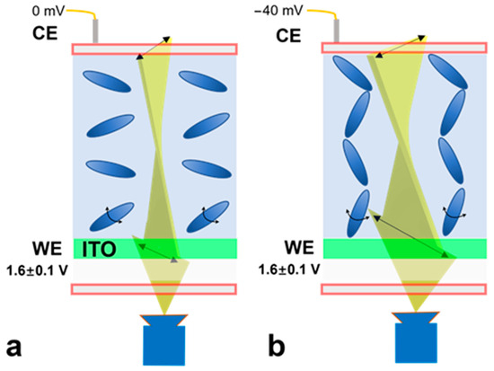
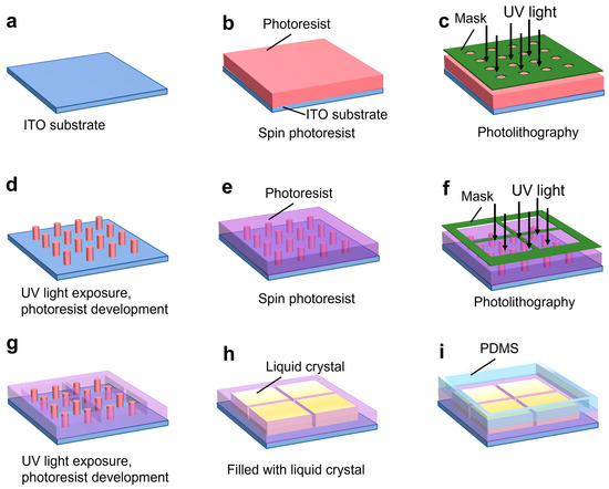
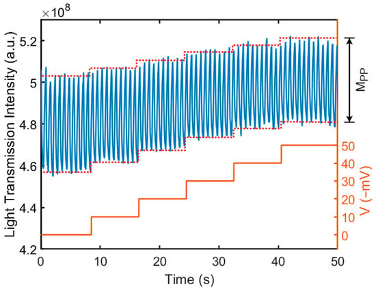
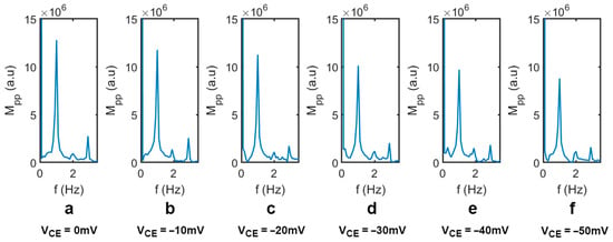
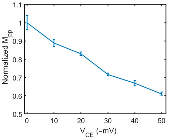
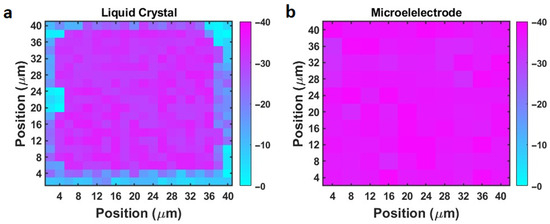
Rapid surface charge mapping of a solid surface remains a challenge. In this study, we present a novel microchip based on liquid crystals for assessing the surface charge distribution of a planar or soft surface. This chip enables rapid measurements of the local surface charge distribution of a charged surface. The chip consists of a micropillar array fabricated on a transparent indium tin oxide substrate, while the liquid crystal is used to fill in the gaps between the micropillar structures. When an object is placed on top of the chip, the local surface charge (or zeta potential) influences the orientation of the liquid crystal molecules, resulting in changes in the magnitude of transmitted light. By measuring the intensity of the transmitted light, the distribution of the surface charge can be accurately quantified. We calibrated the chip in a three-electrode configuration and demonstrated the validity of the chip for rapid surface charge mapping using a borosilicate glass slide. This chip offers noninvasive, rapid mapping of surface charges on charged surfaces, with no need for physical or chemical modifications, and has broad potential applications in biomedical research and advanced material design.
Full article
(This article belongs to the Special Issue Advancing Biomedical Biosensing with Microelectrode Arrays)
►
Show Figures
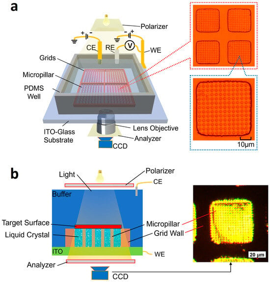
Figure 1


