Next Issue
Volume 12, October-2
Previous Issue
Volume 12, September-2


Cells, Volume 12, Issue 19 (October-1 2023) – 91 articles

Cover Story (view full-size image): 
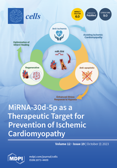
“MicroRNA-30d-5p as a Therapeutic Target for Cardiac Remodeling Post-Myocardial Infarction” describes the potential of miR-30d-5p as a therapeutic intervention for alleviating adverse cardiac remodeling after myocardial infarction (MI). The research reveals a significant downregulation of miR-30d-5p in ischemic myocardium. Administering a miR-30d-5p mimic in an experimental setting leads to a notable reduction in the left ventricle's infarct area size following MI. In vitro experiments employing HUVECs demonstrate accelerated cell migration in response to miR-30d-5p mimic treatment. The study highlights the potential anti-apoptotic effect of miR-30d-5p, implying a decrease in the apoptosis rate in human cardiomyocytes. The findings underscore miR-30d-5p’s cardioprotective potential and its role in mitigating the risk of ischemic cardiomyopathy development following MI. View this paper
  • Issues are regarded as officially published after their release is announced to the table of contents alert mailing list.
  • You may sign up for e-mail alerts to receive table of contents of newly released issues.
  • PDF is the official format for papers published in both, html and pdf forms. To view the papers in pdf format, click on the "PDF Full-text" link, and use the free Adobe Reader to open them.
Order results
Result details
Section
Select all
Export citation of selected articles as:
18 pages, 866 KiB  
Review
Current Nutritional and Pharmacological Approaches for Attenuating Sarcopenia
by Kunihiro Sakuma, Kento Hamada, Akihiko Yamaguchi and Wataru Aoi
Cells 2023, 12(19), 2422; https://doi.org/10.3390/cells12192422 - 09 Oct 2023
Cited by 2 | Viewed by 2552
Abstract
Sarcopenia is characterized by a gradual slowing of movement due to loss of muscle mass and quality, decreased power and strength, increased risk of injury from falls, and often weakness. This review will focus on recent research trends in nutritional and pharmacological approaches [...] Read more.
Sarcopenia is characterized by a gradual slowing of movement due to loss of muscle mass and quality, decreased power and strength, increased risk of injury from falls, and often weakness. This review will focus on recent research trends in nutritional and pharmacological approaches to controlling sarcopenia. Because nutritional studies in humans are fairly limited, this paper includes many results from nutritional studies in mammals. The combination of resistance training with supplements containing amino acids is the gold standard for preventing sarcopenia. Amino acid (HMB) supplementation alone has no significant effect on muscle strength or muscle mass in sarcopenia, but the combination of HMB and exercise (whole body vibration stimulation) is likely to be effective. Tea catechins, soy isoflavones, and ursolic acid are interesting candidates for reducing sarcopenia, but both more detailed basic research on this treatment and clinical studies in humans are needed. Vitamin D supplementation has been shown not to improve sarcopenia in elderly individuals who are not vitamin D-deficient. Myostatin inhibitory drugs have been tried in many neuromuscular diseases, but increases in muscle mass and strength are less likely to be expected. Validation of myostatin inhibitory antibodies in patients with sarcopenia has been positive, but excessive expectations are not warranted. Full article
(This article belongs to the Special Issue Cellular Plasticity of the Neuromuscular System)
Show Figures

Figure 1

24 pages, 4014 KiB  
Review
Exosomal microRNA/miRNA Dysregulation in Respiratory Diseases: From Mycoplasma-Induced Respiratory Disease to COVID-19 and Beyond
by Yingjie Wang, Mengyun Zou, Yabo Zhao, Md. Ahsanul Kabir and Xiuli Peng
Cells 2023, 12(19), 2421; https://doi.org/10.3390/cells12192421 - 09 Oct 2023
Cited by 2 | Viewed by 1561
Abstract
Respiratory diseases represent a significant economic and health burden worldwide, affecting millions of individuals each year in both human and animal populations. MicroRNAs (miRNAs) play crucial roles in gene expression regulation and are involved in various physiological and pathological processes. Exosomal miRNAs and [...] Read more.
Respiratory diseases represent a significant economic and health burden worldwide, affecting millions of individuals each year in both human and animal populations. MicroRNAs (miRNAs) play crucial roles in gene expression regulation and are involved in various physiological and pathological processes. Exosomal miRNAs and cellular miRNAs have been identified as key regulators of several immune respiratory diseases, such as chronic respiratory diseases (CRD) caused by Mycoplasma gallisepticum (MG), Mycoplasma pneumoniae pneumonia (MMP) caused by the bacterium Mycoplasma pneumoniae, coronavirus disease 2019 (COVID-19), chronic obstructive pulmonary disease (COPD), asthma, and acute lung injury/acute respiratory distress syndrome (ALI/ARDS). Consequently, miRNAs seem to have the potential to serve as diagnostic biomarkers and therapeutic targets in respiratory diseases. In this review, we summarize the current understanding of the functional roles of miRNAs in the above several respiratory diseases and discuss the potential use of miRNAs as stable diagnostic biomarkers and therapeutic targets for several immune respiratory diseases, focusing on the identification of differentially expressed miRNAs and their targeting of various signaling pathways implicated in disease pathogenesis. Despite the progress made, unanswered questions and future research directions are discussed to facilitate personalized and targeted therapies for patients with these debilitating conditions. Full article
Show Figures

Figure 1

15 pages, 6364 KiB  
Article
Role of Nucleobindin-2 in the Clinical Pathogenesis and Treatment Resistance of Glioblastoma
by I-Cheng Lin, Chih-Hui Chang, Yoon Bin Chong, Shih-Hsun Kuo, Yu-Wen Cheng, Ann-Shung Lieu, Tzu-Ting Tseng, Chien-Ju Lin, Hung-Pei Tsai and Aij-Lie Kwan
Cells 2023, 12(19), 2420; https://doi.org/10.3390/cells12192420 - 09 Oct 2023
Cited by 1 | Viewed by 874
Abstract
Glioblastoma (GBM) stands as the most prevalent primary malignant brain tumor, typically resulting in a median survival period of approximately thirteen to fifteen months after undergoing surgery, chemotherapy, and radiotherapy. Nucleobindin-2 (NUCB2) is a protein involved in appetite regulation and energy homeostasis. In [...] Read more.
Glioblastoma (GBM) stands as the most prevalent primary malignant brain tumor, typically resulting in a median survival period of approximately thirteen to fifteen months after undergoing surgery, chemotherapy, and radiotherapy. Nucleobindin-2 (NUCB2) is a protein involved in appetite regulation and energy homeostasis. In this study, we assessed the impact of NUCB2 expression on tumor progression and prognosis of GBM. We further evaluated the relationship between NUCB2 expression and the sensitivity to chemotherapy and radiotherapy in GBM cells. Additionally, we compared the survival of mice intracranially implanted with GBM cells. High NUCB2 expression was associated with poor prognosis in patients with GBM. Knockdown of NUCB2 reduced cell viability, migration ability, and invasion ability of GBM cells. Overexpression of NUCB2 resulted in reduced apoptosis following temozolomide treatment and increased levels of DNA damage repair proteins after radiotherapy. Furthermore, mice intracranially implanted with NUCB2 knockdown GBM cells exhibited longer survival compared to the control group. NUCB2 may serve as a prognostic biomarker for poor outcomes in patients with GBM. Additionally, NUCB2 not only contributes to tumor progression but also influences the sensitivity of GBM cells to chemotherapy and radiotherapy. Therefore, targeting NUCB2 protein expression may represent a novel therapeutic approach for the treatment of GBM. Full article
Show Figures

Figure 1

14 pages, 3881 KiB  
Article
Age-Dependent Surface Receptor Expression Patterns in Immature Versus Mature Platelets in Mouse Models of Regenerative Thrombocytopenia
by Anita Pirabe, Sabine Frühwirth, Laura Brunnthaler, Hubert Hackl, Anna Schmuckenschlager, Waltraud C. Schrottmaier and Alice Assinger
Cells 2023, 12(19), 2419; https://doi.org/10.3390/cells12192419 - 08 Oct 2023
Viewed by 1087
Abstract
Aging is a multifaceted process that unfolds at both the individual and cellular levels, resulting in changes in platelet count and platelet reactivity. These alterations are influenced by shifts in platelet production, as well as by various environmental factors that affect circulating platelets. [...] Read more.
Aging is a multifaceted process that unfolds at both the individual and cellular levels, resulting in changes in platelet count and platelet reactivity. These alterations are influenced by shifts in platelet production, as well as by various environmental factors that affect circulating platelets. Aging also triggers functional changes in platelets, including a reduction in RNA content and protein production capacity. Older individuals and RNA-rich immature platelets often exhibit hyperactivity, contributing significantly to pathologic conditions such as cardiovascular diseases, sepsis, and thrombosis. However, the impact of aging on surface receptor expression of circulating platelets, particularly whether these effects vary between immature and mature platelets, remains largely unexplored. Thus, we investigated the expression of certain surface and activation receptors on platelets from young and old mice as well as on immature and mature platelets from mouse models of regenerative thrombocytopenia by flow cytometry. Our findings indicate that aged mice show an upregulated expression of the platelet endothelial cell adhesion molecule-1 (CD31), tetraspanin-29 (CD9), and Toll-like receptor 2 (TLR2) compared to their younger counterparts. Interestingly, when comparing immature and mature platelets in both young and old mice, no differences were observed in mature platelets. However, immature platelets from young mice displayed higher surface expression compared to immature platelets from old mice. Additionally, in mouse models of regenerative thrombocytopenia, the majority of receptors were upregulated in immature platelets. These results suggest that distinct surface receptor expressions are increased on platelets from old mice and immature platelets, which may partially explain their heightened activity and contribute to an increased thrombotic risk. Full article
(This article belongs to the Special Issue Platelet Function and Platelet Proteomics in Ageing-Related Diseases)
Show Figures

Graphical abstract

15 pages, 5873 KiB  
Article
Mapping Cell-in-Cell Structures in Oral Squamous Cell Carcinoma
by Leonardo de Oliveira Siquara da Rocha, Bruno Solano de Freitas Souza, Ricardo Della Coletta, Daniel W. Lambert and Clarissa A. Gurgel Rocha
Cells 2023, 12(19), 2418; https://doi.org/10.3390/cells12192418 - 08 Oct 2023
Viewed by 1414
Abstract
Cell-in-cell (CIC) structures contribute to tumor aggressiveness and poor prognosis in oral squamous cell carcinoma (OSCC). In vitro 3D models may contribute to the understanding of the underlying molecular mechanisms of these events. We employed a spheroid model to study the CIC structures [...] Read more.
Cell-in-cell (CIC) structures contribute to tumor aggressiveness and poor prognosis in oral squamous cell carcinoma (OSCC). In vitro 3D models may contribute to the understanding of the underlying molecular mechanisms of these events. We employed a spheroid model to study the CIC structures in OSCC. Spheroids were obtained from OSCC (HSC3) and cancer-associated fibroblast (CAF) lines using the Nanoshuttle-PLTM bioprinting system (Greiner Bio-One). Spheroid form, size, and reproducibility were evaluated over time (EvosTM XL; ImageJ version 1.8). Slides were assembled, stained (hematoxylin and eosin), and scanned (Axio Imager Z2/VSLIDE) using the OlyVIA System (Olympus Life Science) and ImageJ software (NIH) for cellular morphology and tumor zone formation (hypoxia and/or proliferative zones) analysis. CIC occurrence, complexity, and morphology were assessed considering the spheroid regions. Well-formed spheroids were observed within 6 h of incubation, showing the morphological aspects of the tumor microenvironment, such as hypoxic (core) and proliferative zone (periphery) formation. CIC structures were found in both homotypic and heterotypic groups, predominantly in the proliferative zone of the mixed HSC3/CAF spheroids. “Complex cannibalism” events were also noted. These results showcase the potential of this model in further studies on CIC morphology, formation, and relationship with tumor prognosis. Full article
(This article belongs to the Collection Feature Papers in ‘Cellular Pathology’)
Show Figures

Figure 1

48 pages, 1090 KiB  
Review
Anti-Inflammatory and Immunomodulatory Effect of High-Dose Immunoglobulins in Children: From Approved Indications to Off-Label Use
by Francesca Conti, Mattia Moratti, Lucia Leonardi, Arianna Catelli, Elisa Bortolamedi, Emanuele Filice, Anna Fetta, Marianna Fabi, Elena Facchini, Maria Elena Cantarini, Angela Miniaci, Duccio Maria Cordelli, Marcello Lanari, Andrea Pession and Daniele Zama
Cells 2023, 12(19), 2417; https://doi.org/10.3390/cells12192417 - 07 Oct 2023
Cited by 1 | Viewed by 1578
Abstract
Background: The large-scale utilization of immunoglobulins in patients with inborn errors of immunity (IEIs) since 1952 prompted the discovery of their key role at high doses as immunomodulatory and anti-inflammatory therapy, in the treatment of IEI-related immune dysregulation disorders, according to labelled and [...] Read more.
Background: The large-scale utilization of immunoglobulins in patients with inborn errors of immunity (IEIs) since 1952 prompted the discovery of their key role at high doses as immunomodulatory and anti-inflammatory therapy, in the treatment of IEI-related immune dysregulation disorders, according to labelled and off-label indications. Recent years have been dominated by a progressive imbalance between the gradual but constant increase in the use of immunoglobulins and their availability, exacerbated by the SARS-CoV-2 pandemic. Objectives: To provide pragmatic indications for a need-based application of high-dose immunoglobulins in the pediatric context. Sources: A literature search was performed using PubMed, from inception until 1st August 2023, including the following keywords: anti-inflammatory; children; high dose gammaglobulin; high dose immunoglobulin; immune dysregulation; immunomodulation; immunomodulatory; inflammation; intravenous gammaglobulin; intravenous immunoglobulin; off-label; pediatric; subcutaneous gammaglobulin; subcutaneous immunoglobulin. All article types were considered. Implications: In the light of the current imbalance between gammaglobulins’ demand and availability, this review advocates the urgency of a more conscious utilization of this medical product, giving indications about benefits, risks, cost-effectiveness, and administration routes of high-dose immunoglobulins in children with hematologic, neurologic, and inflammatory immune dysregulation disorders, prompting further research towards a responsible employment of gammaglobulins and improving the therapeutical decisional process. Full article
(This article belongs to the Special Issue Immunopharmacology: Current Applications and Future Perspectives)
Show Figures

Figure 1

17 pages, 7863 KiB  
Article
Calcein-Modified CeO2 for Intracellular ROS Detection: Mechanisms of Action and Cytotoxicity Analysis In Vitro
by Nikita N. Chukavin, Vladimir K. Ivanov and Anton L. Popov
Cells 2023, 12(19), 2416; https://doi.org/10.3390/cells12192416 - 07 Oct 2023
Cited by 2 | Viewed by 1219
Abstract
Cerium oxide nanoparticles (CeO2 NPs) are metal-oxide-based nanozymes with unique reactive oxygen species (ROS) scavenging abilities. Here, we studied new CeO2 NPs modified with calcein (CeO2-calcein) as an intracellular ROS inactivation/visualization theranostic agent. The molecular mechanisms of the CeO [...] Read more.
Cerium oxide nanoparticles (CeO2 NPs) are metal-oxide-based nanozymes with unique reactive oxygen species (ROS) scavenging abilities. Here, we studied new CeO2 NPs modified with calcein (CeO2-calcein) as an intracellular ROS inactivation/visualization theranostic agent. The molecular mechanisms of the CeO2-calcein intracellular activity, allowing for the direct monitoring of ROS inactivation in living cells, were studied. CeO2-calcein was taken up by both normal (human mesenchymal stem cells, hMSc) and cancer (human osteosarcoma, MNNG/Hos cell line) cells, and was easily decomposed via endogenous or exogenous ROS, releasing brightly fluorescent calcein, which could be quantitatively detected using fluorescence microscopy. It was shown that the CeO2-calcein has selective cytotoxicity, inducing the death of human osteosarcoma cells and modulating the expression of key genes responsible for cell redox status as well as proliferative and migration activity. Such cerium-based theranostic agents can be used in various biomedical applications. Full article
(This article belongs to the Special Issue Nanobiophotonics: From Cell Imaging to Clinical Applications)
Show Figures

Figure 1

16 pages, 5088 KiB  
Article
The Study of the Caudal Vertebrae of Thick-Toed Geckos after a Prolonged Space Flight by X-ray Phase-Contrast Micro-CT
by Inna Bukreeva, Victoria I. Gulimova, Yuri S. Krivonosov, Alexey V. Buzmakov, Olga Junemann, Alessia Cedola, Michela Fratini, Laura Maugeri, Ginevra Begani Provinciali, Francesca Palermo, Alessia Sanna, Nicola Pieroni, Victor E. Asadchikov and Sergey V. Saveliev
Cells 2023, 12(19), 2415; https://doi.org/10.3390/cells12192415 - 07 Oct 2023
Viewed by 1111
Abstract
The proximal caudal vertebrae and notochord in thick-toed geckos (TG) (Chondrodactylus turneri, Gray, 1864) were investigated after a 30-day space flight onboard the biosatellite Bion-M1. This region has not been explored in previous studies. Our research focused on finding sites most [...] Read more.
The proximal caudal vertebrae and notochord in thick-toed geckos (TG) (Chondrodactylus turneri, Gray, 1864) were investigated after a 30-day space flight onboard the biosatellite Bion-M1. This region has not been explored in previous studies. Our research focused on finding sites most affected by demineralization caused by microgravity (G0). We used X-ray phase-contrast tomography to study TG samples without invasive prior preparation to clarify our previous findings on the resistance of TG’s bones to demineralization in G0. The results of the present study confirmed that geckos are capable of preserving bone mass after flight, as neither cortical nor trabecular bone volume fraction showed statistically significant changes after flight. On the other hand, we observed a clear decrease in the mineralization of the notochordal septum and a substantial rise in intercentrum volume following the flight. To monitor TG’s mineral metabolism in G0, we propose to measure the volume of mineralized tissue in the notochordal septum. This technique holds promise as a sensitive approach to track the demineralization process in G0, given that the volume of calcification within the septum is limited, making it easy to detect even slight changes in mineral content. Full article
(This article belongs to the Special Issue New Insights into Microgravity and Space Biology)
Show Figures

Figure 1

19 pages, 1924 KiB  
Article
Mitochondrial F-ATP Synthase Co-Migrating Proteins and Ca2+-Dependent Formation of Large Channels
by Anna B. Nikiforova, Yulia L. Baburina, Marina P. Borisova, Alexey K. Surin, Ekaterina S. Kharechkina, Olga V. Krestinina, Maria Y. Suvorina, Svetlana A. Kruglova and Alexey G. Kruglov
Cells 2023, 12(19), 2414; https://doi.org/10.3390/cells12192414 - 07 Oct 2023
Cited by 1 | Viewed by 1031
Abstract
Monomers, dimers, and individual FOF1-ATP synthase subunits are, presumably, involved in the formation of the mitochondrial permeability transition pore (PTP), whose molecular structure, however, is still unknown. We hypothesized that, during the Ca2+-dependent assembly of a PTP [...] Read more.
Monomers, dimers, and individual FOF1-ATP synthase subunits are, presumably, involved in the formation of the mitochondrial permeability transition pore (PTP), whose molecular structure, however, is still unknown. We hypothesized that, during the Ca2+-dependent assembly of a PTP complex, the F-ATP synthase (subunits) recruits mitochondrial proteins that do not interact or weakly interact with the F-ATP synthase under normal conditions. Therefore, we examined whether the PTP opening in mitochondria before the separation of supercomplexes via BN-PAGE will increase the channel stability and channel-forming capacity of isolated F-ATP synthase dimers and monomers in planar lipid membranes. Additionally, we studied the specific activity and the protein composition of F-ATP synthase dimers and monomers from rat liver and heart mitochondria before and after PTP opening. Against our expectations, preliminary PTP opening dramatically suppressed the high-conductance channel activity of F-ATP synthase dimers and monomers and decreased their specific “in-gel” activity. The decline in the channel-forming activity correlated with the reduced levels of as few as two proteins in the bands: methylmalonate–semialdehyde dehydrogenase and prohibitin 2. These results indicate that proteins co-migrating with the F-ATP synthase may be important players in PTP formation and stabilization. Full article
(This article belongs to the Special Issue Mitochondria at the Crossroad of Health and Disease)
Show Figures

Figure 1

17 pages, 2270 KiB  
Review
Potential Therapeutic Use of Stem Cells for Prion Diseases
by Mohammed Zayed, Sung-Ho Kook and Byung-Hoon Jeong
Cells 2023, 12(19), 2413; https://doi.org/10.3390/cells12192413 - 07 Oct 2023
Viewed by 1586
Abstract
Prion diseases are neurodegenerative disorders that are progressive, incurable, and deadly. The prion consists of PrPSc, the misfolded pathogenic isoform of the cellular prion protein (PrPC). PrPC is involved in a variety of physiological functions, including cellular proliferation, [...] Read more.
Prion diseases are neurodegenerative disorders that are progressive, incurable, and deadly. The prion consists of PrPSc, the misfolded pathogenic isoform of the cellular prion protein (PrPC). PrPC is involved in a variety of physiological functions, including cellular proliferation, adhesion, differentiation, and neural development. Prion protein is expressed on the membrane surface of a variety of stem cells (SCs), where it plays an important role in the pluripotency and self-renewal matrix, as well as in SC differentiation. SCs have been found to multiply the pathogenic form of the prion protein, implying their potential as an in vitro model for prion diseases. Furthermore, due to their capability to self-renew, differentiate, immunomodulate, and regenerate tissue, SCs are prospective cell treatments in many neurodegenerative conditions, including prion diseases. Regenerative medicine has become a new revolution in disease treatment in recent years, particularly with the introduction of SC therapy. Here, we review the data demonstrating prion diseases’ biology and molecular mechanism. SC biology, therapeutic potential, and its role in understanding prion disease mechanisms are highlighted. Moreover, we summarize preclinical studies that use SCs in prion diseases. Full article
(This article belongs to the Special Issue Mesenchymal Stem Cells and Their Role in Neurodegenerative Diseases)
Show Figures

Figure 1

18 pages, 5177 KiB  
Article
Unveiling Neuroprotection and Regeneration Mechanisms in Optic Nerve Injury: Insight from Neural Progenitor Cell Therapy with Focus on Vps35 and Syntaxin12
by Hyun-Ah Shin, Mira Park, Hey Jin Lee, Van-An Duong, Hyun-Mun Kim, Dong-Youn Hwang, Hookeun Lee and Helen Lew
Cells 2023, 12(19), 2412; https://doi.org/10.3390/cells12192412 - 06 Oct 2023
Viewed by 1638
Abstract
Axonal degeneration resulting from optic nerve damage can lead to the progressive death of retinal ganglion cells (RGCs), culminating in irreversible vision loss. We contrasted two methods for inducing optic nerve damage: optic nerve compression (ONCo) and optic nerve crush (ONCr). These were [...] Read more.
Axonal degeneration resulting from optic nerve damage can lead to the progressive death of retinal ganglion cells (RGCs), culminating in irreversible vision loss. We contrasted two methods for inducing optic nerve damage: optic nerve compression (ONCo) and optic nerve crush (ONCr). These were assessed for their respective merits in simulating traumatic optic neuropathies and neurodegeneration. We also administered neural progenitor cells (NPCs) into the subtenon space to validate their potential in mitigating optic nerve damage. Our findings indicate that both ONCo and ONCr successfully induced optic nerve damage, as shown by increases in ischemia and expression of genes linked to neuronal regeneration. Post NPC injection, recovery in the expression of neuronal regeneration-related genes was more pronounced in the ONCo model than in the ONCr model, while inflammation-related gene expression saw a better recovery in ONCr. In addition, the proteomic analysis of R28 cells in hypoxic conditions identified Vps35 and Syntaxin12 genes. Vps35 preserved the mitochondrial function in ONCo, while Syntaxin12 appeared to restrain inflammation via the Wnt/β-catenin signaling pathway in ONCr. NPCs managed to restore damaged RGCs by elevating neuroprotection factors and controlling inflammation through mitochondrial homeostasis and Wnt/β-catenin signaling in hypoxia-injured R28 cells and in both animal models. Our results suggest that ischemic injury and crush injury cause optic nerve damage via different mechanisms, which can be effectively simulated using ONCo and ONCr, respectively. Moreover, cell-based therapies such as NPCs may offer promising avenues for treating various optic neuropathies, including ischemic and crush injuries. Full article
(This article belongs to the Section Cell and Gene Therapy)
Show Figures

Graphical abstract

16 pages, 5819 KiB  
Article
Multiphoton In Vivo Microscopy of Embryonic Thrombopoiesis Reveals the Generation of Platelets through Budding
by Huan Liu, Hellen Ishikawa-Ankerhold, Julia Winterhalter, Michael Lorenz, Mykhailo Vladymyrov, Steffen Massberg, Christian Schulz and Mathias Orban
Cells 2023, 12(19), 2411; https://doi.org/10.3390/cells12192411 - 06 Oct 2023
Viewed by 1135
Abstract
Platelets are generated by specialized cells called megakaryocytes (MKs). However, MK’s origin and platelet release mode have remained incompletely understood. Here, we established direct visualization of embryonic thrombopoiesis in vivo by combining multiphoton intravital microscopy (MP-IVM) with a fluorescence switch reporter mouse model [...] Read more.
Platelets are generated by specialized cells called megakaryocytes (MKs). However, MK’s origin and platelet release mode have remained incompletely understood. Here, we established direct visualization of embryonic thrombopoiesis in vivo by combining multiphoton intravital microscopy (MP-IVM) with a fluorescence switch reporter mouse model under control of the platelet factor 4 promoter (Pf4CreRosa26mTmG). Using this microscopy tool, we discovered that fetal liver MKs provide higher thrombopoietic activity than yolk sac MKs. Mechanistically, fetal platelets were released from MKs either by membrane buds or the formation of proplatelets, with the former constituting the key process. In E14.5 c-Myb-deficient embryos that lack definitive hematopoiesis, MK and platelet numbers were similar to wild-type embryos, indicating the independence of embryonic thrombopoiesis from definitive hematopoiesis at this stage of development. In summary, our novel MP-IVM protocol allows the characterization of thrombopoiesis with high spatio-temporal resolution in the mouse embryo and has identified membrane budding as the main mechanism of fetal platelet production. Full article
(This article belongs to the Special Issue Recent Advances in Intravital and Live Cell Imaging)
Show Figures

Figure 1

10 pages, 2647 KiB  
Article
A Combinatorial Code for CPEB-Mediated c-myc Repression
by Koichi Ogami, Keima Ogawa, Shoko Sanpei, Fumito Ichikawa, Tsuyoshi Udagawa and Shin-ichi Hoshino
Cells 2023, 12(19), 2410; https://doi.org/10.3390/cells12192410 - 06 Oct 2023
Viewed by 907
Abstract
During early embryonic development, the RNA-binding protein CPEB mediates cytoplasmic polyadenylation and translational activation through a combinatorial code defined by the cy-toplasmic polyadenylation element (CPE) present in maternal mRNAs. However, in non-neuronal somatic cells, CPEB accelerates deadenylation to repress translation of the target, [...] Read more.
During early embryonic development, the RNA-binding protein CPEB mediates cytoplasmic polyadenylation and translational activation through a combinatorial code defined by the cy-toplasmic polyadenylation element (CPE) present in maternal mRNAs. However, in non-neuronal somatic cells, CPEB accelerates deadenylation to repress translation of the target, including c-myc mRNA, through an ill-defined cis-regulatory mechanism. Using RNA mutagenesis and electrophoretic mobility shift assays, we demonstrated that a combination of tandemly arranged consensus (cCPE) and non-consensus (ncCPE) cytoplasmic polyadenylation elements (CPEs) constituted a combinatorial code for CPEB-mediated c-myc mRNA decay. CPEB binds to cCPEs with high affinity (Kd = ~250 nM), whereas it binds to ncCPEs with low affinity (Kd > ~900 nM). CPEB binding to a cCPE enhances CPEB binding to the proximal ncCPE. In contrast, while a cCPE did not activate mRNA degradation, an ncCPE was essential for the induction of degradation, and a combination of a cCPE and ncCPEs further promoted degradation. Based on these findings, we propose a model in which the high-affinity binding of CPEB to the cCPE accelerates the binding of the second CPEB to the ncCPEs, resulting in the recruitment of deadenylases, acceleration of deadenylation, and repression of c-myc mRNAs. Full article
Show Figures

Graphical abstract

24 pages, 4071 KiB  
Article
A Thia-Analogous Indirubin N-Glycoside Disrupts Mitochondrial Function and Causes the Death of Human Melanoma and Cutaneous Squamous Cell Carcinoma Cells
by Franziska Wendt, Felix Wittig, Anne Rupprecht, Robert Ramer, Peter Langer, Steffen Emmert, Marcus Frank and Burkhard Hinz
Cells 2023, 12(19), 2409; https://doi.org/10.3390/cells12192409 - 05 Oct 2023
Viewed by 1138
Abstract
Skin cancer is the most common malignant disease worldwide and, therefore, also poses a challenge from a pharmacotherapeutic perspective. Derivatives of indirubin are an interesting option in this context. In the present study, the effects of 3-[3′-oxo-benzo[b]thiophen-2′-(Z)-ylidene]-1-(β-d-glucopyranosyl)-oxindole [...] Read more.
Skin cancer is the most common malignant disease worldwide and, therefore, also poses a challenge from a pharmacotherapeutic perspective. Derivatives of indirubin are an interesting option in this context. In the present study, the effects of 3-[3′-oxo-benzo[b]thiophen-2′-(Z)-ylidene]-1-(β-d-glucopyranosyl)-oxindole (KD87), a thia-analogous indirubin N-glycoside, on the viability and mitochondrial properties of melanoma (A375) and squamous cell carcinoma cells (A431) of the skin were investigated. In both cell lines, KD87 caused decreased viability, the activation of caspases-3 and -7, and the inhibition of colony formation. At the mitochondrial level, a concentration-dependent decrease in both the basal and ATP-linked oxygen consumption rate and in the reserve capacity of oxidative respiration were registered in the presence of KD87. These changes were accompanied by morphological alterations in the mitochondria, a release of mitochondrial cytochrome c into the cytosol and significant reductions in succinate dehydrogenase complex subunit B (SDHB, subunit of complex II) in A375 and A431 cells and NADH:ubiquinone oxidoreductase subunit B8 (NDUFB8, subunit of complex I) in A375 cells. The effect of KD87 was accompanied by a significant upregulation of the enzyme heme oxygenase-1, whose inhibition led to a partial but significant reduction in the metabolic-activity-reducing effect of KD87. In summary, our data show a mitochondria-targeting effect of KD87 as part of the cytotoxic effect of this compound on skin cancer cells, which should be considered in future studies with this class of compounds. Full article
(This article belongs to the Special Issue Skin Research: Cellular Mechanism and Therapeutic Potentials)
Show Figures

Figure 1

19 pages, 4276 KiB  
Article
Impact of the Physical Cellular Microenvironment on the Structure and Function of a Model Hepatocyte Cell Line for Drug Toxicity Applications
by Benjamin Allcock, Wenbin Wei, Kirsty Goncalves, Henry Hoyle, Alisha Robert, Rebecca Quelch-Cliffe, Adam Hayward, Jim Cooper and Stefan Przyborski
Cells 2023, 12(19), 2408; https://doi.org/10.3390/cells12192408 - 05 Oct 2023
Viewed by 1019
Abstract
It is widely recognised that cells respond to their microenvironment, which has implications for cell culture practices. Growth cues provided by 2D cell culture substrates are far removed from native 3D tissue structure in vivo. Geometry is one of many factors that differs [...] Read more.
It is widely recognised that cells respond to their microenvironment, which has implications for cell culture practices. Growth cues provided by 2D cell culture substrates are far removed from native 3D tissue structure in vivo. Geometry is one of many factors that differs between in vitro culture and in vivo cellular environments. Cultured cells are far removed from their native counterparts and lose some of their predictive capability and reliability. In this study, we examine the cellular processes that occur when a cell is cultured on 2D or 3D surfaces for a short period of 8 days prior to its use in functional assays, which we term: “priming”. We follow the process of mechanotransduction from cytoskeletal alterations, to changes to nuclear structure, leading to alterations in gene expression, protein expression and improved functional capabilities. In this study, we utilise HepG2 cells as a hepatocyte model cell line, due to their robustness for drug toxicity screening. Here, we demonstrate enhanced functionality and improved drug toxicity profiles that better reflect the in vivo clinical response. However, findings more broadly reflect in vitro cell culture practises across many areas of cell biology, demonstrating the fundamental impact of mechanotransduction in bioengineering and cell biology. Full article
(This article belongs to the Collection Advances in Cell Culture and Tissue Engineering)
Show Figures

Graphical abstract

11 pages, 613 KiB  
Review
An Overview of the Role of Furin in Type 2 Diabetes
by Sulaiman K. Marafie and Fahd Al-Mulla
Cells 2023, 12(19), 2407; https://doi.org/10.3390/cells12192407 - 05 Oct 2023
Cited by 1 | Viewed by 1434
Abstract
Post-translational modifications (PTMs) play important roles in regulating several human diseases, like cancer, neurodegenerative disorders, and metabolic disorders. Investigating PTMs’ contribution to protein functions is critical for modern biology and medicine. Proprotein convertases (PCs) are irreversible post-translational modifiers that have been extensively studied [...] Read more.
Post-translational modifications (PTMs) play important roles in regulating several human diseases, like cancer, neurodegenerative disorders, and metabolic disorders. Investigating PTMs’ contribution to protein functions is critical for modern biology and medicine. Proprotein convertases (PCs) are irreversible post-translational modifiers that have been extensively studied and are considered as key targets for novel therapeutics. They cleave proteins at specific sites causing conformational changes affecting their functions. Furin is considered as a PC model in regulating growth factors and is involved in regulating many pro-proteins. The mammalian target of the rapamycin (mTOR) signaling pathway is another key player in regulating cellular processes and its dysregulation is linked to several diseases including type 2 diabetes (T2D). The role of furin in the context of diabetes has been rarely explored and is currently lacking. Moreover, furin variants have altered activity that could have implications on overall health. In this review, we aim to highlight the role of furin in T2D in relation to mTOR signaling. We will also address furin genetic variants and their potential effect on T2D and β-cell functions. Understanding the role of furin in prediabetes and dissecting it from other confounding factors like obesity is crucial for future therapeutic interventions in metabolic disorders. Full article
Show Figures

Figure 1

35 pages, 11910 KiB  
Article
PolyQ-Expansion Causes Mitochondria Fragmentation Independent of Huntingtin and Is Distinct from Traumatic Brain Injury (TBI)/Mechanical Stress-Mediated Fragmentation Which Results from Cell Death
by Kelsey Swinter, Dania Salah, Rasika Rathnayake and Shermali Gunawardena
Cells 2023, 12(19), 2406; https://doi.org/10.3390/cells12192406 - 05 Oct 2023
Viewed by 1232
Abstract
Mitochondrial dysfunction has been reported in many Huntington’s disease (HD) models; however, it is unclear how these defects occur. Here, we test the hypothesis that excess pathogenic huntingtin (HTT) impairs mitochondrial homeostasis, using Drosophila genetics and pharmacological inhibitors in HD and polyQ-expansion disease [...] Read more.
Mitochondrial dysfunction has been reported in many Huntington’s disease (HD) models; however, it is unclear how these defects occur. Here, we test the hypothesis that excess pathogenic huntingtin (HTT) impairs mitochondrial homeostasis, using Drosophila genetics and pharmacological inhibitors in HD and polyQ-expansion disease models and in a mechanical stress-induced traumatic brain injury (TBI) model. Expression of pathogenic HTT caused fragmented mitochondria compared to normal HTT, but HTT did not co-localize with mitochondria under normal or pathogenic conditions. Expression of pathogenic polyQ (127Q) alone or in the context of Machado Joseph Disease (MJD) caused fragmented mitochondria. While mitochondrial fragmentation was not dependent on the cellular location of polyQ accumulations, the expression of a chaperone protein, excess of mitofusin (MFN), or depletion of dynamin-related protein 1 (DRP1) rescued fragmentation. Intriguingly, a higher concentration of nitric oxide (NO) was observed in polyQ-expressing larval brains and inhibiting NO production rescued polyQ-mediated fragmented mitochondria, postulating that DRP1 nitrosylation could contribute to excess fission. Furthermore, while excess PI3K, which suppresses polyQ-induced cell death, did not rescue polyQ-mediated fragmentation, it did rescue fragmentation caused by mechanical stress/TBI. Together, our observations suggest that pathogenic polyQ alone is sufficient to cause DRP1-dependent mitochondrial fragmentation upstream of cell death, uncovering distinct physiological mechanisms for mitochondrial dysfunction in polyQ disease and mechanical stress. Full article
(This article belongs to the Section Mitochondria)
Show Figures

Figure 1

13 pages, 2524 KiB  
Article
UCHL1 Regulates Radiation Lung Injury via Sphingosine Kinase-1
by Yulia Epshtein, Biji Mathew, Weiguo Chen and Jeffrey R. Jacobson
Cells 2023, 12(19), 2405; https://doi.org/10.3390/cells12192405 - 05 Oct 2023
Viewed by 925
Abstract
GADD45a is a gene we previously reported as a mediator of responses to acute lung injury. GADD45a−/− mice express decreased Akt and increased Akt ubiquitination due to the reduced expression of UCHL1 (ubiquitin c-terminal hydrolase L1), a deubiquitinating enzyme, while GADD45a−/− mice have [...] Read more.
GADD45a is a gene we previously reported as a mediator of responses to acute lung injury. GADD45a−/− mice express decreased Akt and increased Akt ubiquitination due to the reduced expression of UCHL1 (ubiquitin c-terminal hydrolase L1), a deubiquitinating enzyme, while GADD45a−/− mice have increased their susceptibility to radiation-induced lung injury (RILI). Separately, we have reported a role for sphingolipids in RILI, evidenced by the increased RILI susceptibility of SphK1−/− (sphingosine kinase 1) mice. A mechanistic link between UCHL1 and sphingolipid signaling in RILI is suggested by the known polyubiquitination of SphK1. Thus, we hypothesized that the regulation of SphK1 ubiquitination by UCHL1 mediates RILI. Initially, human lung endothelial cells (EC) subjected to radiation demonstrated a significant upregulation of UCHL1 and SphK1. The ubiquitination of EC SphK1 after radiation was confirmed via the immunoprecipitation of SphK1 and Western blotting for ubiquitin. Further, EC transfected with siRNA specifically for UCHL1 or pretreated with LDN-5744, as a UCHL1 inhibitor, prior to radiation were noted to have decreased ubiquitinated SphK1 in both conditions. Further, the inhibition of UCHL1 attenuated sphingolipid-mediated EC barrier enhancement was measured by transendothelial electrical resistance. Finally, LDN pretreatment significantly augmented murine RILI severity. Our data support the fact that the regulation of SphK1 expression after radiation is mediated by UCHL1. The modulation of UCHL1 affecting sphingolipid signaling may represent a novel RILI therapeutic strategy. Full article
Show Figures

Figure 1

29 pages, 3132 KiB  
Review
Dendritic Cell Vaccination in Non-Small Cell Lung Cancer: Remodeling the Tumor Immune Microenvironment
by Jensen Abascal, Michael S. Oh, Elvira L. Liclican, Steven M. Dubinett, Ramin Salehi-Rad and Bin Liu
Cells 2023, 12(19), 2404; https://doi.org/10.3390/cells12192404 - 04 Oct 2023
Cited by 1 | Viewed by 2038
Abstract
Non-small-cell lung cancer (NSCLC) remains one of the leading causes of death worldwide. While NSCLCs possess antigens that can potentially elicit T cell responses, defective tumor antigen presentation and T cell activation hinder host anti-tumor immune responses. The NSCLC tumor microenvironment (TME) is [...] Read more.
Non-small-cell lung cancer (NSCLC) remains one of the leading causes of death worldwide. While NSCLCs possess antigens that can potentially elicit T cell responses, defective tumor antigen presentation and T cell activation hinder host anti-tumor immune responses. The NSCLC tumor microenvironment (TME) is composed of cellular and soluble mediators that can promote or combat tumor growth. The composition of the TME plays a critical role in promoting tumorigenesis and dictating anti-tumor immune responses to immunotherapy. Dendritic cells (DCs) are critical immune cells that activate anti-tumor T cell responses and sustain effector responses. DC vaccination is a promising cellular immunotherapy that has the potential to facilitate anti-tumor immune responses and transform the composition of the NSCLC TME via tumor antigen presentation and cell–cell communication. Here, we will review the features of the NSCLC TME with an emphasis on the immune cell phenotypes that directly interact with DCs. Additionally, we will summarize the major preclinical and clinical approaches for DC vaccine generation and examine how effective DC vaccination can transform the NSCLC TME toward a state of sustained anti-tumor immune signaling. Full article
(This article belongs to the Special Issue Tumor Immune Microenvironment in Lung Cancer)
Show Figures

Figure 1

17 pages, 4251 KiB  
Article
Axon-Autonomous Effects of the Amyloid Precursor Protein Intracellular Domain (AICD) on Kinase Signaling and Fast Axonal Transport
by Svenja König, Nadine Schmidt, Karin Bechberger, Sarah Morris, Mercedes Priego, Hannah Zaky, Yuyu Song, Jan Pielage, Silke Brunholz, Scott T. Brady, Stefan Kins and Gerardo Morfini
Cells 2023, 12(19), 2403; https://doi.org/10.3390/cells12192403 - 04 Oct 2023
Viewed by 1703
Abstract
The amyloid precursor protein (APP) is a key molecular component of Alzheimer’s disease (AD) pathogenesis. Proteolytic APP processing generates various cleavage products, including extracellular amyloid beta (Aβ) and the cytoplasmic APP intracellular domain (AICD). Although the role of AICD in the activation of [...] Read more.
The amyloid precursor protein (APP) is a key molecular component of Alzheimer’s disease (AD) pathogenesis. Proteolytic APP processing generates various cleavage products, including extracellular amyloid beta (Aβ) and the cytoplasmic APP intracellular domain (AICD). Although the role of AICD in the activation of kinase signaling pathways is well established in the context of full-length APP, little is known about intracellular effects of the AICD fragment, particularly within discrete neuronal compartments. Deficits in fast axonal transport (FAT) and axonopathy documented in AD-affected neurons prompted us to evaluate potential axon-autonomous effects of the AICD fragment for the first time. Vesicle motility assays using the isolated squid axoplasm preparation revealed inhibition of FAT by AICD. Biochemical experiments linked this effect to aberrant activation of selected axonal kinases and heightened phosphorylation of the anterograde motor protein conventional kinesin, consistent with precedents showing phosphorylation-dependent regulation of motors proteins powering FAT. Pharmacological inhibitors of these kinases alleviated the AICD inhibitory effect on FAT. Deletion experiments indicated this effect requires a sequence encompassing the NPTY motif in AICD and interacting axonal proteins containing a phosphotyrosine-binding domain. Collectively, these results provide a proof of principle for axon-specific effects of AICD, further suggesting a potential mechanistic framework linking alterations in APP processing, FAT deficits, and axonal pathology in AD. Full article
(This article belongs to the Special Issue Molecular Insights into Neurodegenerative Diseases)
Show Figures

Figure 1

20 pages, 7270 KiB  
Article
Disheveled-1 Interacts with Claudin-5 and Contributes to Norrin-Induced Endothelial Barrier Restoration
by Mónica Díaz-Coránguez, Laura González-González, Amy Wang, Xuwen Liu and David A. Antonetti
Cells 2023, 12(19), 2402; https://doi.org/10.3390/cells12192402 - 04 Oct 2023
Cited by 2 | Viewed by 1424
Abstract
Previous studies have revealed that norrin can reverse vascular endothelial-growth-factor (VEGF)-induced permeability in a β-catenin-dependent pathway. Here, we have explored the contribution of disheveled-1 (DVL1) in norrin-induced blood-retinal barrier (BRB) restoration. We provide evidence that in addition to canonical signaling, DVL1 promotes tight [...] Read more.
Previous studies have revealed that norrin can reverse vascular endothelial-growth-factor (VEGF)-induced permeability in a β-catenin-dependent pathway. Here, we have explored the contribution of disheveled-1 (DVL1) in norrin-induced blood-retinal barrier (BRB) restoration. We provide evidence that in addition to canonical signaling, DVL1 promotes tight junction (TJ) stabilization through a novel, non-canonical signaling pathway involving direct claudin-5 (CLDN5) binding. Immunofluorescence staining of rat retinal cross-sections showed enriched expression of DVL1 and 3 at endothelial capillaries and co-localization with CLDN5 and ZO-1 at the TJ complex in primary bovine retinal endothelial cells (BRECs). Barrier properties of BRECs were determined via measurements of trans-endothelial electrical resistance (TEER) or permeability to 70 kDa RITC-dextran. These studies demonstrated that norrin restoration of barrier properties after VEGF treatment required DVL1 as an siRNA knockdown of Dvl1 but not Dvl2 or Dvl3, reduced basal barrier properties and ablated norrin-induced barrier restoration. However, loss of Dvl1 did not decrease β-catenin signaling activity as measured by Axin2 mRNA expression, suggesting the contribution of a non-canonical pathway. DVL and TJ protein interactions were analyzed via co-immunoprecipitation of endogenous protein in BRECs, which demonstrated that DVL1 interacts with both CLDN5 and ZO-1, while DVL3 interacts only with ZO-1. These interactions were most abundant after inducing BRB restoration by treating BRECs with VEGF and norrin. DVL has previously been shown to form intramolecular bindings between the C-terminal PDZ-binding motif (PDZ-BM) with an internal PDZ domain. Co-transfection of HEK293 cells with DVL1 and CLDN5 or relevant mutants revealed that DVL1 interacts with CLDN5 through the DVL PDZ domain binding, CLDN5 PDZ-BM, in competition with DVL1 PDZ-BM, since DVL/CLDN5 interaction increases with deletion of the DVL1 PDZ-BM and decreases by co-expressing the C-terminal fragment of DVL1 containing the PDZ-BM or through deletion of CLDN5 PDZ-BM. In BREC cells, transfection of the C-terminal fragment of DVL1 downregulates the expression of CLDN5 but does not affect the expression of other proteins of the TJs, including ZO-1, occludin, CLDN1 or VE-cadherin. Blocking DVL1/CLDN5 interaction increased basal permeability and prevented norrin induction of barrier properties after VEGF. Combined with previous data, these results demonstrate that norrin signals through both a canonical β-catenin pathway and a non-canonical signaling pathway by which DVL1 directly binds to CLDN5 to promote barrier properties. Full article
(This article belongs to the Special Issue Retinal Cell Biology in Health and Disease)
Show Figures

Graphical abstract

15 pages, 3413 KiB  
Article
Measuring Melanoma Nanomechanical Properties in Relation to Metastatic Ability and Anti-Cancer Drug Treatment Using Scanning Ion Conductance Microscopy
by Emily Woodcock, Peter V. Gorelkin, Philip S. Goff, Christopher R. W. Edwards, Yanjun Zhang, Yuri Korchev and Elena V. Sviderskaya
Cells 2023, 12(19), 2401; https://doi.org/10.3390/cells12192401 - 04 Oct 2023
Cited by 1 | Viewed by 1360
Abstract
A cell’s mechanical properties have been linked to cancer development, motility and metastasis and are therefore an attractive target as a universal, reliable cancer marker. For example, it has been widely published that cancer cells show a lower Young’s modulus than their non-cancerous [...] Read more.
A cell’s mechanical properties have been linked to cancer development, motility and metastasis and are therefore an attractive target as a universal, reliable cancer marker. For example, it has been widely published that cancer cells show a lower Young’s modulus than their non-cancerous counterparts. Furthermore, the effect of anti-cancer drugs on cellular mechanics may offer a new insight into secondary mechanisms of action and drug efficiency. Scanning ion conductance microscopy (SICM) offers a nanoscale resolution, non-contact method of nanomechanical data acquisition. In this study, we used SICM to measure the nanomechanical properties of melanoma cell lines from different stages with increasing metastatic ability. Young’s modulus changes following treatment with the anti-cancer drugs paclitaxel, cisplatin and dacarbazine were also measured, offering a novel perspective through the use of continuous scan mode SICM. We found that Young’s modulus was inversely correlated to metastatic ability in melanoma cell lines from radial growth, vertical growth and metastatic phases. However, Young’s modulus was found to be highly variable between cells and cell lines. For example, the highly metastatic cell line A375M was found to have a significantly higher Young’s modulus, and this was attributed to a higher level of F-actin. Furthermore, our data following nanomechanical changes after 24 hour anti-cancer drug treatment showed that paclitaxel and cisplatin treatment significantly increased Young’s modulus, attributed to an increase in microtubules. Treatment with dacarbazine saw a decrease in Young’s modulus with a significantly lower F-actin corrected total cell fluorescence. Our data offer a new perspective on nanomechanical changes following drug treatment, which may be an overlooked effect. This work also highlights variations in cell nanomechanical properties between previous studies, cancer cell lines and cancer types and questions the usefulness of using nanomechanics as a diagnostic or prognostic tool. Full article
(This article belongs to the Special Issue Advances in Scanning Probe Microscopy in Cell Biology)
Show Figures

Figure 1

17 pages, 6134 KiB  
Article
c-Cbl Regulates Murine Subventricular Zone-Derived Neural Progenitor Cells in Dependence of the Epidermal Growth Factor Receptor
by Maximilian Vogt, Madhukrishna Kolothara Unnikrishnan, Nora Heinig, Ulrike Schumann, Mirko H. H. Schmidt and Kathrin Barth
Cells 2023, 12(19), 2400; https://doi.org/10.3390/cells12192400 - 03 Oct 2023
Viewed by 979
Abstract
The localization, expression, and physiological role of regulatory proteins in the neurogenic niches of the brain is fundamental to our understanding of adult neurogenesis. This study explores the expression and role of the E3-ubiquitin ligase, c-Cbl, in neurogenesis within the subventricular zone (SVZ) [...] Read more.
The localization, expression, and physiological role of regulatory proteins in the neurogenic niches of the brain is fundamental to our understanding of adult neurogenesis. This study explores the expression and role of the E3-ubiquitin ligase, c-Cbl, in neurogenesis within the subventricular zone (SVZ) of mice. In vitro neurosphere assays and in vivo analyses were performed in specific c-Cbl knock-out lines to unravel c-Cbl’s role in receptor tyrosine kinase signaling, including the epidermal growth factor receptor (EGFR) pathway. Our findings suggest that c-Cbl is significantly expressed within EGFR-expressing cells, playing a pivotal role in neural stem cell proliferation and differentiation. However, c-Cbl’s function extends beyond EGFR signaling, as its loss upon knock-out stimulated progenitor cell proliferation in neurosphere cultures. Yet, this effect was not detected in hippocampal progenitor cells, reflecting the lack of the EGFR in the hippocampus. In vivo, c-Cbl exerted only a minor proneurogenic influence with no measurable impact on the formation of adult-born neurons. In conclusion, c-Cbl regulates neural stem cells in the subventricular zone via the EGFR pathway but, likely, its loss is compensated by other signaling modules in vivo. Full article
(This article belongs to the Section Cells of the Nervous System)
Show Figures

Figure 1

43 pages, 4907 KiB  
Article
In Cerebellar Atrophy of 12-Month-Old ATM-Null Mice, Transcriptome Upregulations Concern Most Neurotransmission and Neuropeptide Pathways, While Downregulations Affect Prominently Itpr1, Usp2 and Non-Coding RNA
by Marina Reichlmeir, Júlia Canet-Pons, Gabriele Koepf, Wasifa Nurieva, Ruth Pia Duecker, Claudia Doering, Kathryn Abell, Jana Key, Matthew P. Stokes, Stefan Zielen, Ralf Schubert, Zoltán Ivics and Georg Auburger
Cells 2023, 12(19), 2399; https://doi.org/10.3390/cells12192399 - 03 Oct 2023
Cited by 1 | Viewed by 1369
Abstract
The autosomal recessive disorder Ataxia-Telangiectasia is caused by a dysfunction of the stress response protein, ATM. In the nucleus of proliferating cells, ATM senses DNA double-strand breaks and coordinates their repair. This role explains T-cell dysfunction and tumour risk. However, it remains unclear [...] Read more.
The autosomal recessive disorder Ataxia-Telangiectasia is caused by a dysfunction of the stress response protein, ATM. In the nucleus of proliferating cells, ATM senses DNA double-strand breaks and coordinates their repair. This role explains T-cell dysfunction and tumour risk. However, it remains unclear whether this function is relevant for postmitotic neurons and underlies cerebellar atrophy, since ATM is cytoplasmic in postmitotic neurons. Here, we used ATM-null mice that survived early immune deficits via bone-marrow transplantation, and that reached initial neurodegeneration stages at 12 months of age. Global cerebellar transcriptomics demonstrated that ATM depletion triggered upregulations in most neurotransmission and neuropeptide systems. Downregulated transcripts were found for the ATM interactome component Usp2, many non-coding RNAs, ataxia genes Itpr1, Grid2, immediate early genes and immunity factors. Allelic splice changes affected prominently the neuropeptide machinery, e.g., Oprm1. Validation experiments with stressors were performed in human neuroblastoma cells, where ATM was localised only to cytoplasm, similar to the brain. Effect confirmation in SH-SY5Y cells occurred after ATM depletion and osmotic stress better than nutrient/oxidative stress, but not after ATM kinase inhibition or DNA stressor bleomycin. Overall, we provide pioneer observations from a faithful A-T mouse model, which suggest general changes in synaptic and dense-core vesicle stress adaptation. Full article
Show Figures

Figure 1

23 pages, 922 KiB  
Review
Noncoding RNAs and Human Induced Pluripotent Stem Cell-Derived Cardiomyocytes in Cardiac Arrhythmic Brugada Syndrome
by Benjamin Theisen, Austin Holtz and Viswanathan Rajagopalan
Cells 2023, 12(19), 2398; https://doi.org/10.3390/cells12192398 - 03 Oct 2023
Cited by 1 | Viewed by 1338
Abstract
Hundreds of thousands of people die each year as a result of sudden cardiac death, and many are due to heart rhythm disorders. One of the major causes of these arrhythmic events is Brugada syndrome, a cardiac channelopathy that results in abnormal cardiac [...] Read more.
Hundreds of thousands of people die each year as a result of sudden cardiac death, and many are due to heart rhythm disorders. One of the major causes of these arrhythmic events is Brugada syndrome, a cardiac channelopathy that results in abnormal cardiac conduction, severe life-threatening arrhythmias, and, on many occasions, death. This disorder has been associated with mutations and dysfunction of about two dozen genes; however, the majority of the patients do not have a definite cause for the diagnosis of Brugada Syndrome. The protein-coding genes represent only a very small fraction of the mammalian genome, and the majority of the noncoding regions of the genome are actively transcribed. Studies have shown that most of the loci associated with electrophysiological traits are located in noncoding regulatory regions and are expected to affect gene expression dosage and cardiac ion channel function. Noncoding RNAs serve an expanding number of regulatory and other functional roles within the cells, including but not limited to transcriptional, post-transcriptional, and epigenetic regulation. The major noncoding RNAs found in Brugada Syndrome include microRNAs; however, others such as long noncoding RNAs are also identified. They contribute to pathogenesis by interacting with ion channels and/or are detectable as clinical biomarkers. Stem cells have received significant attention in the recent past, and can be differentiated into many different cell types including those in the heart. In addition to contractile and relaxational properties, BrS-relevant electrophysiological phenotypes are also demonstrated in cardiomyocytes differentiated from stem cells induced from adult human cells. In this review, we discuss the current understanding of noncoding regions of the genome and their RNA biology in Brugada Syndrome. We also delve into the role of stem cells, especially human induced pluripotent stem cell-derived cardiac differentiated cells, in the investigation of Brugada syndrome in preclinical and clinical studies. Full article
Show Figures

Figure 1

18 pages, 2091 KiB  
Article
Mitochondrial Dysfunction and Decreased Cytochrome c in Cell and Animal Models of Machado–Joseph Disease
by Filipa Almeida, Ildete L. Ferreira, Luana Naia, Daniela Marinho, Ana Catarina Vilaça-Ferreira, Marta D. Costa, Sara Duarte-Silva, Patrícia Maciel and A. Cristina Rego
Cells 2023, 12(19), 2397; https://doi.org/10.3390/cells12192397 - 03 Oct 2023
Viewed by 1193
Abstract
Mitochondrial dysfunction has been described in many neurodegenerative disorders; however, there is less information regarding mitochondrial deficits in Machado–Joseph disease (MJD), a polyglutamine (polyQ) disorder caused by CAG repeat expansion in the ATXN3 gene. In the present study, we characterized the changes in [...] Read more.
Mitochondrial dysfunction has been described in many neurodegenerative disorders; however, there is less information regarding mitochondrial deficits in Machado–Joseph disease (MJD), a polyglutamine (polyQ) disorder caused by CAG repeat expansion in the ATXN3 gene. In the present study, we characterized the changes in mitochondrial function and biogenesis markers in two MJD models, CMVMJD135 (MJD135) transgenic mice at a fully established phenotype stage and tetracycline-regulated PC6-3 Q108 cell line expressing mutant ataxin-3 (mATXN3). We detected mATXN3 in the mitochondrial fractions of PC6-3 Q108 cells, suggesting the interaction of expanded ATXN3 with the organelle. Interestingly, in both the cerebella of the MJD135 mouse model and in PC6-3 Q108 cells, we found decreased mitochondrial respiration, ATP production and mitochondrial membrane potential, strongly suggesting mitochondrial dysfunction in MJD. Also, in PC6-3 Q108 cells, an additional enhanced glycolytic flux was observed. Supporting the functional deficits observed in MJD mitochondria, MJD135 mouse cerebellum and PC6-3 Q108 cells showed reduced cytochrome c mRNA and protein levels. Overall, our findings show compromised mitochondrial function associated with decreased cytochrome c levels in both cell and animal models of MJD. Full article
Show Figures

Figure 1

22 pages, 4876 KiB  
Article
NAD+ Acts as a Protective Factor in Cellular Stress Response to DNA Alkylating Agents
by Joanna Ruszkiewicz, Ylea Papatheodorou, Nathalie Jäck, Jasmin Melzig, Franziska Eble, Annika Pirker, Marius Thomann, Andreas Haberer, Simone Rothmiller, Alexander Bürkle and Aswin Mangerich
Cells 2023, 12(19), 2396; https://doi.org/10.3390/cells12192396 - 02 Oct 2023
Cited by 1 | Viewed by 1297
Abstract
Sulfur mustard (SM) and its derivatives are potent genotoxic agents, which have been shown to trigger the activation of poly (ADP-ribose) polymerases (PARPs) and the depletion of their substrate, nicotinamide adenine dinucleotide (NAD+). NAD+ is an essential molecule involved in [...] Read more.
Sulfur mustard (SM) and its derivatives are potent genotoxic agents, which have been shown to trigger the activation of poly (ADP-ribose) polymerases (PARPs) and the depletion of their substrate, nicotinamide adenine dinucleotide (NAD+). NAD+ is an essential molecule involved in numerous cellular pathways, including genome integrity and DNA repair, and thus, NAD+ supplementation might be beneficial for mitigating mustard-induced (geno)toxicity. In this study, the role of NAD+ depletion and elevation in the genotoxic stress response to SM derivatives, i.e., the monofunctional agent 2-chloroethyl-ethyl sulfide (CEES) and the crosslinking agent mechlorethamine (HN2), was investigated with the use of NAD+ booster nicotinamide riboside (NR) and NAD+ synthesis inhibitor FK866. The effects were analyzed in immortalized human keratinocytes (HaCaT) or monocyte-like cell line THP-1. In HaCaT cells, NR supplementation, increased NAD+ levels, and elevated PAR response, however, did not affect ATP levels or DNA damage repair, nor did it attenuate long- and short-term cytotoxicities. On the other hand, the depletion of cellular NAD+ via FK866 sensitized HaCaT cells to genotoxic stress, particularly CEES exposure, whereas NR supplementation, by increasing cellular NAD+ levels, rescued the sensitizing FK866 effect. Intriguingly, in THP-1 cells, the NR-induced elevation of cellular NAD+ levels did attenuate toxicity of the mustard compounds, especially upon CEES exposure. Together, our results reveal that NAD+ is an important molecule in the pathomechanism of SM derivatives, exhibiting compound-specificity. Moreover, the cell line-dependent protective effects of NR are indicative of system-specificity of the application of this NAD+ booster. Full article
Show Figures

Figure 1

24 pages, 2253 KiB  
Review
Enhancing Antisense Oligonucleotide-Based Therapeutic Delivery with DG9, a Versatile Cell-Penetrating Peptide
by Umme Sabrina Haque and Toshifumi Yokota
Cells 2023, 12(19), 2395; https://doi.org/10.3390/cells12192395 - 02 Oct 2023
Cited by 1 | Viewed by 4026
Abstract
Antisense oligonucleotide-based (ASO) therapeutics have emerged as a promising strategy for the treatment of human disorders. Charge-neutral PMOs have promising biological and pharmacological properties for antisense applications. Despite their great potential, the efficient delivery of these therapeutic agents to target cells remains a [...] Read more.
Antisense oligonucleotide-based (ASO) therapeutics have emerged as a promising strategy for the treatment of human disorders. Charge-neutral PMOs have promising biological and pharmacological properties for antisense applications. Despite their great potential, the efficient delivery of these therapeutic agents to target cells remains a major obstacle to their widespread use. Cellular uptake of naked PMO is poor. Cell-penetrating peptides (CPPs) appear as a possibility to increase the cellular uptake and intracellular delivery of oligonucleotide-based drugs. Among these, the DG9 peptide has been identified as a versatile CPP with remarkable potential for enhancing the delivery of ASO-based therapeutics due to its unique structural features. Notably, in the context of phosphorodiamidate morpholino oligomers (PMOs), DG9 has shown promise in enhancing delivery while maintaining a favorable toxicity profile. A few studies have highlighted the potential of DG9-conjugated PMOs in DMD (Duchenne Muscular Dystrophy) and SMA (Spinal Muscular Atrophy), displaying significant exon skipping/inclusion and functional improvements in animal models. The article provides an overview of a detailed understanding of the challenges that ASOs face prior to reaching their targets and continued advances in methods to improve their delivery to target sites and cellular uptake, focusing on DG9, which aims to harness ASOs’ full potential in precision medicine. Full article
(This article belongs to the Section Cell and Gene Therapy)
Show Figures

Figure 1

13 pages, 843 KiB  
Review
Pulmonary Hypertension in Interstitial Lung Disease: Updates in Disease, Diagnosis, and Therapeutics
by Zachary A. Haynes, Abhimanyu Chandel and Christopher S. King
Cells 2023, 12(19), 2394; https://doi.org/10.3390/cells12192394 - 01 Oct 2023
Cited by 1 | Viewed by 2155
Abstract
Pulmonary hypertension is a debilitating condition that frequently develops in the setting of interstitial lung disease, likely related to chronic alveolar hypoxemia and pulmonary vascular remodeling. This disease process is likely to be identified more frequently by providers given recent advancements in definitions [...] Read more.
Pulmonary hypertension is a debilitating condition that frequently develops in the setting of interstitial lung disease, likely related to chronic alveolar hypoxemia and pulmonary vascular remodeling. This disease process is likely to be identified more frequently by providers given recent advancements in definitions and diagnostic modalities, and provides practitioners with emerging opportunities to improve patient outcomes and quality of life. Despite years of data suggesting against the efficacy of pulmonary vasodilator therapy in patients with pulmonary hypertension due to interstitial lung disease, new data have emerged identifying promising advancements in therapeutics. The authors present to you a comprehensive review of pulmonary hypertension in interstitial lung disease, reviewing our current understanding of pathophysiology, updates in diagnostic approaches, and highlights of recent clinical trials which provide an effective approach for medical management. Full article
(This article belongs to the Special Issue Recent Advances in Interstitial Lung Diseases)
Show Figures

Figure 1

14 pages, 3722 KiB  
Article
A CD276-Targeted Antibody-Drug Conjugate to Treat Non-Small Lung Cancer (NSCLC)
by Jiashuai Zhang, Zhuoxin (Zora) Zhou, Kai Chen, Seulhee Kim, Irene Soohyun Cho, Tanvi Varadkar, Hailey Baker, Ju Hwan Cho, Lufang Zhou and Xiaoguang (Margaret) Liu
Cells 2023, 12(19), 2393; https://doi.org/10.3390/cells12192393 - 30 Sep 2023
Viewed by 1645
Abstract
Non-small cell lung cancer (NSCLC) patients, accounting for approximately 85% of lung cancer cases, are usually diagnosed in advanced stages. Traditional surgical resection and radiotherapy have very limited clinical benefits. The objective of this study was to develop and evaluate a targeted therapy, [...] Read more.
Non-small cell lung cancer (NSCLC) patients, accounting for approximately 85% of lung cancer cases, are usually diagnosed in advanced stages. Traditional surgical resection and radiotherapy have very limited clinical benefits. The objective of this study was to develop and evaluate a targeted therapy, antibody-drug conjugate (ADC), for NSCLC treatment. Specifically, the CD276 receptor was evaluated and confirmed as an ideal surface target of NSCLC in the immunohistochemistry (IHC) staining of seventy-three patient tumor microarrays and western blotting analysis of eight cell lines. Our anti-CD276 monoclonal antibody (mAb) with cross-activity to both human and mouse receptors showed high surface binding, effective drug delivery and tumor-specific targeting in flow cytometry, confocal microscopy, and in vivo imaging system analysis. The ADC constructed with our CD276 mAb and payload monomethyl auristatin F (MMAF) showed high anti-NSCLC cytotoxicity to multiple lines and effective anti-tumor efficacy in both immunocompromised and immunocompetent NSCLC xenograft mouse models. The brief mechanism study revealed the integration of cell proliferation inhibition and immune cell reactivation in tumor microenvironments. The toxicity study did not detect off-target immune toxicity or peripheral toxicity. Altogether, this study suggested that anti-CD276 ADC could be a promising candidate for NSCLC treatment. Full article
Show Figures

Figure 1

Previous Issue
Back to TopTop