Next Issue
Volume 14, October
Previous Issue
Volume 14, August


Viruses, Volume 14, Issue 9 (September 2022) – 241 articles

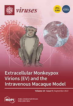
Cover Story (view full-size image): The 2022 monkeypox pandemic has reinforced the need to expand our understanding of virological mechanisms that govern disease and transmission. The intravenous macaque model is an important tool to study these mechanisms and to evaluate countermeasures against monkeypox and smallpox. We tested whether enriching our inoculums for the rarely utilized morphogenic form of poxviruses, extracellular virus (EV), could invoke disease more efficiently in macaques. Under our experimental conditions, we discovered that the integrity of the extracellular form was compromised by inherent factors in nonhuman primate serum. We also determined that the EV that is detectable in the oropharynx of infected macaques could play a role in respiratory transmission. View this paper
  • Issues are regarded as officially published after their release is announced to the table of contents alert mailing list.
  • You may sign up for e-mail alerts to receive table of contents of newly released issues.
  • PDF is the official format for papers published in both, html and pdf forms. To view the papers in pdf format, click on the "PDF Full-text" link, and use the free Adobe Reader to open them.
Order results
Result details
Section
Select all
Export citation of selected articles as:
9 pages, 892 KiB  
Review
Romania, a Harbour of HIV-1 Subtype F1: Where Are We after 33 Years of HIV-1 Infection?
by Mădălina Preda and Loredana Cornelia Sabina Manolescu
Viruses 2022, 14(9), 2081; https://doi.org/10.3390/v14092081 - 19 Sep 2022
Cited by 4 | Viewed by 1782
Abstract
Infection with the human immunodeficiency virus (HIV) has been a major public health concern worldwide for more than 30 years, including in Romania. The F1 HIV-1 subtype was exported from Angola to Romania most probably because of the two countries’ close political connections. [...] Read more.
Infection with the human immunodeficiency virus (HIV) has been a major public health concern worldwide for more than 30 years, including in Romania. The F1 HIV-1 subtype was exported from Angola to Romania most probably because of the two countries’ close political connections. Patients infected with HIV-1 via re-used and improperly sterilized injection equipment and through transfusions of unscreened blood, also known as the “Romanian cohort”, were the most common type of HIV-1 infection in Romania in the early 1990s, when the virus’s presence was recognized. Recently, subtype B started to increase in our country, mostly diagnosed in people using intravenous drugs or in men having sex with men. The evolution of the HIV-1 infection in Romania has been unique, with a dominance of the subtype F1, making it different from other countries in Europe. Full article
Show Figures

Figure 1

12 pages, 1201 KiB  
Article
Antiviral Activity of Crude Polysaccharide Derived from Seaweed against IHNV and IPNV In Vitro
by Guangming Ren, Liming Xu, Jingzhuang Zhao, Yizhi Shao, Yujie Lin, Linfang Li, Qi Liu, Tongyan Lu and Qiya Zhang
Viruses 2022, 14(9), 2080; https://doi.org/10.3390/v14092080 - 19 Sep 2022
Cited by 4 | Viewed by 1808
Abstract
Both infectious hematopoietic necrosis virus (IHNV) and infectious pancreatic necrosis virus (IPNV) are the causative agents of acute and highly contagious diseases of juvenile salmonids, resulting in severe economic losses to these cold-water fish globally. There is an urgent need to explore antiviral [...] Read more.
Both infectious hematopoietic necrosis virus (IHNV) and infectious pancreatic necrosis virus (IPNV) are the causative agents of acute and highly contagious diseases of juvenile salmonids, resulting in severe economic losses to these cold-water fish globally. There is an urgent need to explore antiviral agents against IHNV and IPNV due to the lack of commercially available vaccines and antiviral drugs. More importantly, the co-infection of IHNV and IPNV is prevalent in nature, which not only aggravates extensive damage to the salmonids but also poses challenges to its prevention and control. The antiviral effects of a crude polysaccharide derived from seaweed (CSP) on IHNV and IPNV were evaluated in this study separately. Furthermore, the underlying antiviral mechanisms of CSP to IHNV and IPNV were analyzed, respectively. The results showed that CSP possessed excellent safety and good ability to inhibit IHNV, IPNV, and their co-infection. CSP preferred to act at the early stage of viral infection. The antiviral mechanism of CSP on IHNV is possibly involved in preventing viral attachment and release, while in IPNV, it is involved in suppressing viral attachment, entry, and release. Taken together, the results of this study shed new light on developing novel agents against viral infection in salmonid fish. Full article
Show Figures

Figure 1

17 pages, 1920 KiB  
Review
Pathogenicity of Avian Polyomaviruses and Prospect of Vaccine Development
by Chen-Wei Wang, Yung-Liang Chen, Simon J. T. Mao, Tzu-Chieh Lin, Ching-Wen Wu, Duangsuda Thongchan, Chi-Young Wang and Hung-Yi Wu
Viruses 2022, 14(9), 2079; https://doi.org/10.3390/v14092079 - 19 Sep 2022
Cited by 3 | Viewed by 2349
Abstract
Polyomaviruses are nonenveloped icosahedral viruses with a double-stranded circular DNA containing approximately 5000 bp and 5–6 open reading frames. In contrast to mammalian polyomaviruses (MPVs), avian polyomaviruses (APVs) exhibit high lethality and multipathogenicity, causing severe infections in birds without oncogenicity. APVs are classified [...] Read more.
Polyomaviruses are nonenveloped icosahedral viruses with a double-stranded circular DNA containing approximately 5000 bp and 5–6 open reading frames. In contrast to mammalian polyomaviruses (MPVs), avian polyomaviruses (APVs) exhibit high lethality and multipathogenicity, causing severe infections in birds without oncogenicity. APVs are classified into 10 major species: Adélie penguin polyomavirus, budgerigar fledgling disease virus, butcherbird polyomavirus, canary polyomavirus, cormorant polyomavirus, crow polyomavirus, Erythrura gouldiae polyomavirus, finch polyomavirus, goose hemorrhagic polyomavirus, and Hungarian finch polyomavirus under the genus Gammapolyomavirus. This paper briefly reviews the genomic structure and pathogenicity of the 10 species of APV and some of their differences in terms of virulence from MPVs. Each gene’s genomic size, number of amino acid residues encoding each gene, and key biologic functions are discussed. The rationale for APV classification from the Polyomavirdae family and phylogenetic analyses among the 10 APVs are also discussed. The clinical symptoms in birds caused by APV infection are summarized. Finally, the strategies for developing an effective vaccine containing essential epitopes for preventing virus infection in birds are discussed. We hope that more effective and safe vaccines with diverse protection will be developed in the future to solve or alleviate the problems of viral infection. Full article
(This article belongs to the Special Issue State-of-the-Art Avian Viruses Research in Asia)
Show Figures

Figure 1

2 pages, 163 KiB  
Reply
Reply to Alberts, P. Comment on “Hietanen et al. Cytolytic Properties and Genome Analysis of Rigvir® Oncolytic Virotherapy Virus and Other Echovirus 7 Isolates. Viruses 2022, 14, 525”
by Eero Hietanen and Petri Susi
Viruses 2022, 14(9), 2078; https://doi.org/10.3390/v14092078 - 19 Sep 2022
Viewed by 978
Abstract
Our work focused on comparing the cellular effects of Rigvir to other echovirus 7 isolates, because originally Rigvir is also an echovirus 7 isolate [...] Full article
(This article belongs to the Section Viral Immunology, Vaccines, and Antivirals)
15 pages, 2192 KiB  
Article
Quantification of APOBEC3 Mutation Rates Affecting the VP1 Gene of BK Polyomavirus In Vivo
by Dorian McIlroy, Cécile Peltier, My-Linh Nguyen, Louise Manceau, Lenha Mobuchon, Nicolas Le Baut, Ngoc-Khanh Nguyen, Minh-Chau Tran, The-Cuong Nguyen and Céline Bressollette-Bodin
Viruses 2022, 14(9), 2077; https://doi.org/10.3390/v14092077 - 19 Sep 2022
Cited by 2 | Viewed by 1804
Abstract
Mutations in the BK polyomavirus (BKPyV) capsid accumulate in kidney transplant (KTx) recipients with persistent virus replication. They are associated with neutralization escape and appear to arise as a result of cytosine deamination by host cell APOBEC3A/B enzymes. To study the mutagenic processes [...] Read more.
Mutations in the BK polyomavirus (BKPyV) capsid accumulate in kidney transplant (KTx) recipients with persistent virus replication. They are associated with neutralization escape and appear to arise as a result of cytosine deamination by host cell APOBEC3A/B enzymes. To study the mutagenic processes occurring in patients, we amplified the typing region of the VP1 gene, sequenced the amplicons to a depth of 5000–10,000×, and identified rare mutations, which were fitted to COSMIC mutational signatures. Background mutations were identified in amplicons from plasmids carrying the BKPyV genome and compared to mutations observed in 148 samples from 23 KTx recipients in France and in Vietnam. Three mutational signatures were consistently observed in urine, serum, and kidney biopsy samples, two of which, SBS2 and SBS13, corresponded to APOBEC3A/B activity. In addition, a third signature with no known etiology, SBS89, was detected both in patient samples, and in cells infected in vitro with BKPyV. Quantitatively, APOBEC3A/B mutation rates in urine samples were strongly correlated with urine viral load, and also appeared to vary between individuals. These results confirm that APOBEC3A/B is a major, but not the only, source of BKPyV genome mutations in patients. Full article
(This article belongs to the Special Issue Human Polyomaviruses (HPyVs) in Human Diseases and Cancer Development)
Show Figures

Figure 1

3 pages, 197 KiB  
Comment
Comment on Hietanen et al. Cytolytic Properties and Genome Analysis of Rigvir® Oncolytic Virotherapy Virus and Other Echovirus 7 Isolates. Viruses 2022, 14, 525
by Pēteris Alberts
Viruses 2022, 14(9), 2076; https://doi.org/10.3390/v14092076 - 19 Sep 2022
Cited by 1 | Viewed by 1240
Abstract
In a recent article published in Viruses by Hietanen et al. [...] Full article
(This article belongs to the Section Viral Immunology, Vaccines, and Antivirals)
11 pages, 2899 KiB  
Article
Structural Basis for the Inhibition of Coronaviral Main Proteases by a Benzothiazole-Based Inhibitor
by Xiaohui Hu, Cheng Lin, Qin Xu, Xuelan Zhou, Pei Zeng, Peter J. McCormick, Haihai Jiang, Jian Li and Jin Zhang
Viruses 2022, 14(9), 2075; https://doi.org/10.3390/v14092075 - 18 Sep 2022
Cited by 9 | Viewed by 2001
Abstract
The ongoing spread of severe acute respiratory syndrome coronavirus-2 (SARS-CoV-2) has caused hundreds of millions of cases and millions of victims worldwide with serious consequences to global health and economies. Although many vaccines protecting against SARS-CoV-2 are currently available, constantly emerging new variants [...] Read more.
The ongoing spread of severe acute respiratory syndrome coronavirus-2 (SARS-CoV-2) has caused hundreds of millions of cases and millions of victims worldwide with serious consequences to global health and economies. Although many vaccines protecting against SARS-CoV-2 are currently available, constantly emerging new variants necessitate the development of alternative strategies for prevention and treatment of COVID-19. Inhibitors that target the main protease (Mpro) of SARS-CoV-2, an essential enzyme that promotes viral maturation, represent a key class of antivirals. Here, we showed that a peptidomimetic compound with benzothiazolyl ketone as warhead, YH-53, is an effective inhibitor of SARS-CoV-2, SARS-CoV, and MERS-CoV Mpros. Crystal structures of Mpros from SARS-CoV-2, SARS-CoV, and MERS-CoV bound to the inhibitor YH-53 revealed a unique ligand-binding site, which provides new insights into the mechanism of inhibition of viral replication. A detailed analysis of these crystal structures defined the key molecular determinants required for inhibition and illustrate the binding mode of Mpros from other coronaviruses. In consideration of the important role of Mpro in developing antivirals against coronaviruses, insights derived from this study should add to the design of pan-coronaviral Mpro inhibitors that are safer and more effective. Full article
(This article belongs to the Special Issue Viral Enzyme Inhibitors: Structure and Dynamics)
Show Figures

Figure 1

22 pages, 6518 KiB  
Article
Genetic Evolution of Avian Influenza A (H9N2) Viruses Isolated from Domestic Poultry in Uganda Reveals Evidence of Mammalian Host Adaptation, Increased Virulence and Reduced Sensitivity to Baloxavir
by Gladys Atim, Titus Tugume, Qouilazoni A. Ukuli, Bernard Erima, Andrew Mubiru, Hannah Kibuuka, Edison Mworozi, Pamela McKenzie, Jasmine C. M. Turner, David Walker, Trushar Jeevan, Robert G. Webster, Jeremy Jones, Richard J. Webby, Mariette F. Ducatez, Fred Wabwire-Mangen and Denis K. Byarugaba
Viruses 2022, 14(9), 2074; https://doi.org/10.3390/v14092074 - 18 Sep 2022
Cited by 2 | Viewed by 2828
Abstract
A (H9N2) avian influenza A viruses were first detected in Uganda in 2017 and have since established themselves in live bird markets. The aim of this study was to establish the subsequent genetic evolution of H9N2 viruses in Uganda. Cloacal samples collected from [...] Read more.
A (H9N2) avian influenza A viruses were first detected in Uganda in 2017 and have since established themselves in live bird markets. The aim of this study was to establish the subsequent genetic evolution of H9N2 viruses in Uganda. Cloacal samples collected from live bird market stalls in Kampala from 2017 to 2019 were screened by RT-PCR for influenza A virus and H9N2 viruses were isolated in embryonated eggs. One hundred and fifty H9N2 isolates were subjected to whole genome sequencing on the Illumina MiSeq platform. The sequence data analysis and comparison with contemporary isolates revealed that the virus was first introduced into Uganda in 2014 from ancestors in the Middle East. There has since been an increase in nucleotide substitutions and reassortments among the viruses within and between live bird markets, leading to variations in phylogeny of the different segments, although overall diversity remained low. The isolates had several mutations such as HA-Q226L and NS-I106M that enable mammalian host adaptation, NP-M105V, PB1-D3V, and M1-T215A known for increased virulence/pathogenicity and replication, and PA-E199D, NS-P42S, and M2-S31N that promote drug resistance. The PA-E199D substitution in particular confers resistance to the endonuclease inhibitor Baloxavir acid, which is one of the new anti-influenza drugs. Higher EC50 was observed in isolates with a double F105L+E199D substitution that may suggest a possible synergistic effect. These H9N2 viruses have established an endemic situation in live bird markets in Uganda because of poor biosecurity practices and therefore pose a zoonotic threat. Regular surveillance is necessary to further generate the needed evidence for effective control strategies and to minimize the threats. Full article
(This article belongs to the Section Animal Viruses)
Show Figures

Figure 1

17 pages, 1901 KiB  
Article
Development and Characterization of a Genetically Stable Infectious Clone for a Genotype I Isolate of Dengue Virus Serotype 1
by Mingyue Hu, Tiantian Wu, Yang Yang, Tongling Chen, Jiawei Hao, Youchuan Wei, Tingrong Luo, De Wu and Yi-Ping Li
Viruses 2022, 14(9), 2073; https://doi.org/10.3390/v14092073 - 18 Sep 2022
Cited by 2 | Viewed by 2529
Abstract
Dengue virus (DENV) is primarily transmitted by the bite of an infected mosquito of Aedes aegypti and Aedes albopictus, and symptoms caused may range from mild dengue fever to severe dengue hemorrhagic fever and dengue shock syndrome. Reverse genetic system represents a [...] Read more.
Dengue virus (DENV) is primarily transmitted by the bite of an infected mosquito of Aedes aegypti and Aedes albopictus, and symptoms caused may range from mild dengue fever to severe dengue hemorrhagic fever and dengue shock syndrome. Reverse genetic system represents a valuable tool for the study of DENV virology, infection, pathogenesis, etc. Here, we generated and characterized an eukaryotic-activated full-length infectious cDNA clone for a DENV serotype 1 (DENV-1) isolate, D19044, collected in 2019. Initially, nearly the full genome was determined by sequencing overlapping RT-PCR products, and was classified to be genotype I DENV-1. D19044 wild-type cDNA clone (D19044_WT) was assembled by four subgenomic fragments, in a specific order, into a low-copy vector downstream the CMV promoter. D19044_WT released the infectious virus at a low level (1.26 × 10³ focus forming units per milliliter [FFU/mL]) following plasmid transfection of BHK-21 cells. Further adaptation by consecutive virus passages up to passage 37, and seven amino acid substitutions (7M) were identified from passage-recovered viruses. The addition of 7M (D19044_7M) greatly improved viral titer (7.5 × 10⁴ FFU/mL) in transfected BHK-21 culture, and virus infections in 293T, Huh7.5.1, and C6/36 cells were also efficient. D19044_7M plasmid was genetically stable in transformant bacteria after five transformation-purification cycles, which did not change the capacity of producing infectious virus. Moreover, the D19044_7M virus was inhibited by mycophenolic acid in a dose-dependent manner. In conclusion, we have developed a DNA-launched full-length infectious clone for a genotype I isolate of DENV-1, with genetic stability in transformant bacteria, thus providing a useful tool for the study of DENV-1. Full article
(This article belongs to the Special Issue Molecular Biology of RNA Viruses)
Show Figures

Figure 1

19 pages, 3385 KiB  
Article
Lysyl Oxidase-like Protein Recognizes Viral Envelope Proteins and Bacterial Polysaccharides against Pathogen Infection via Induction of Expression of Antimicrobial Peptides
by Peng-Yuan Lu, Guo-Juan Niu, Pan-Pan Hong and Jin-Xing Wang
Viruses 2022, 14(9), 2072; https://doi.org/10.3390/v14092072 - 18 Sep 2022
Cited by 1 | Viewed by 1904
Abstract
Lysyl oxidases (LOXs) are copper-dependent monoamine oxidases, and they play critical roles in extracellular matrix (ECM) remodeling. The LOX and LOX-like (LOXL) proteins also have a variety of biological functions, such as development and growth regulation, tumor suppression, and cellular senescence. However, the [...] Read more.
Lysyl oxidases (LOXs) are copper-dependent monoamine oxidases, and they play critical roles in extracellular matrix (ECM) remodeling. The LOX and LOX-like (LOXL) proteins also have a variety of biological functions, such as development and growth regulation, tumor suppression, and cellular senescence. However, the functions of LOXLs containing repeated scavenger receptor cysteine-rich (SRCR) domains in immunity are rarely reported. In this study, we characterized the antiviral and antibacterial functions of a lysyl oxidase-like (LOXL) protein containing tandem SRCR domains in Marsupenaeus japonicus. The mRNA level of LoxL was significantly upregulated in the hemocytes and intestines of shrimp challenged using white spot syndrome virus (WSSV) or bacteria. After the knockdown of LoxL via RNA interference, WSSV replication and bacterial loads were apparently increased, and the survival rate of the shrimp decreased significantly, suggesting that LOXL functions against pathogen infection in shrimp. Mechanistically, LOXL interacted with the envelope proteins of WSSV or with lipopolysaccharide and peptidoglycan from bacteria in shrimp challenged using WSSV or bacteria, and it promoted the expression of a battery of antimicrobial peptides (AMPs) via the induction of Dorsal nuclear translocation against viral and bacterial infection. Moreover, LOXL expression was also positively regulated by Dorsal in the shrimp challenged by pathogens. These results indicate that, by acting as a pattern recognition receptor, LOXL plays vital roles in antiviral and antibacterial innate immunity by enhancing the expression of AMPs in shrimp. Full article
(This article belongs to the Special Issue State-of-the-Art Aquatic Viruses Research in China)
Show Figures

Figure 1

8 pages, 649 KiB  
Brief Report
Vaccination Decreases the Infectious Viral Load of Delta Variant SARS-CoV-2 in Asymptomatic Patients
by Jessica A. Plante, Rafael R. G. Machado, Brooke M. Mitchell, Divya P. Shinde, Jordyn Walker, Dionna Scharton, Allan McConnell, Nehad Saada, Jianying Liu, Bilal Khan, Rafael K. Campos, Bryan A. Johnson, Vineet D. Menachery, Corri B. Levine, Ping Ren, Susan L. F. McLellan, Kenneth S. Plante and Scott C. Weaver
Viruses 2022, 14(9), 2071; https://doi.org/10.3390/v14092071 - 18 Sep 2022
Cited by 6 | Viewed by 1965
Abstract
The Delta variant of SARS-CoV-2 has caused many breakthrough infections in fully vaccinated individuals. While vaccine status did not generally impact the number of viral RNA genome copies in nasopharyngeal swabs of breakthrough patients, as measured by Ct values, it has been previously [...] Read more.
The Delta variant of SARS-CoV-2 has caused many breakthrough infections in fully vaccinated individuals. While vaccine status did not generally impact the number of viral RNA genome copies in nasopharyngeal swabs of breakthrough patients, as measured by Ct values, it has been previously found to decrease the infectious viral load in symptomatic patients. We quantified the viral RNA, infectious virus, and anti-spike IgA in nasopharyngeal swabs collected from individuals asymptomatically infected with the Delta variant of SARS-CoV-2. Vaccination decreased the infectious viral load, but not the amount of viral RNA. Furthermore, vaccinees with asymptomatic infections had significantly higher levels of anti-spike IgA in their nasal secretions compared to unvaccinated individuals with asymptomatic infections. Thus, vaccination may decrease the transmission risk of Delta, and perhaps other variants, despite not affecting the amount of viral RNA measured in nasopharyngeal swabs. Full article
(This article belongs to the Section SARS-CoV-2 and COVID-19)
Show Figures

Figure 1

16 pages, 2088 KiB  
Article
COS-7 and SVGp12 Cellular Models to Study JCPyV Replication and MicroRNA Expression after Infection with Archetypal and Rearranged-NCCR Viral Strains
by Carla Prezioso, Sara Passerini, Dolores Limongi, Anna Teresa Palamara, Ugo Moens and Valeria Pietropaolo
Viruses 2022, 14(9), 2070; https://doi.org/10.3390/v14092070 - 17 Sep 2022
Cited by 5 | Viewed by 1802
Abstract
Since the non-coding control region (NCCR) and microRNA (miRNA) could represent two different and independent modalities of regulating JC polyomavirus (JCPyV) replication at the transcriptional and post-transcriptional levels, the interplay between JC viral load based on NCCR architecture and miRNA levels, following JCPyV [...] Read more.
Since the non-coding control region (NCCR) and microRNA (miRNA) could represent two different and independent modalities of regulating JC polyomavirus (JCPyV) replication at the transcriptional and post-transcriptional levels, the interplay between JC viral load based on NCCR architecture and miRNA levels, following JCPyV infection with archetypal and rearranged (rr)-NCCR JCPyV variants, was explored in COS-7 and SVGp12 cells infected by different JCPyV strains. Specifically, the involvement of JCPyV miRNA in regulating viral replication was investigated for the archetypal CY strain—which is the transmissible form—and for the rearranged MAD-1 strain, which is the first isolated variant from patients with progressive multifocal leukoencephalopathy. The JCPyV DNA viral load was low in cells infected with CY compared with that in MAD-1-infected cells. Productive viral replication was observed in both cell lines. The expression of JCPyV miRNAs was observed from 3 days after viral infection in both cell types, and miR-J1-5p expression was inversely correlated with the JCPyV replication trend. The JCPyV miRNAs in the exosomes present in the supernatants produced by the infected cells could be carried into uninfected cells. Additional investigations of the expression of JCPyV miRNAs and their presence in exosomes are necessary to shed light on their regulatory role during viral reactivation. Full article
(This article belongs to the Special Issue Host Cell–Virus Interaction 2.0)
Show Figures

Figure 1

12 pages, 525 KiB  
Article
Autopsy Study Defines Composition and Dynamics of the HIV-1 Reservoir after Allogeneic Hematopoietic Stem Cell Transplantation with CCR5Δ32/Δ32 Donor Cells
by Laura E. P. Huyveneers, Anke Bruns, Arjen Stam, Pauline Ellerbroek, Dorien de Jong, Noémi A. Nagy, Stephanie B. H. Gumbs, Kiki Tesselaar, Kobus Bosman, Maria Salgado, Gero Hütter, Lodewijk A. A. Brosens, Mi Kwon, Jose Diez Martin, Jan T. M. van der Meer, Theun M. de Kort, Asier Sáez-Cirión, Julian Schulze zur Wiesch, Jaap Jan Boelens, Javier Martinez-Picado, Jürgen H. E. Kuball, Annemarie M. J. Wensing and Monique Nijhuisadd Show full author list remove Hide full author list
Viruses 2022, 14(9), 2069; https://doi.org/10.3390/v14092069 - 17 Sep 2022
Cited by 5 | Viewed by 3081
Abstract
Allo-HSCT with CCR5Δ32/Δ32 donor cells is the only curative HIV-1 intervention. We investigated the impact of allo-HSCT on the viral reservoir in PBMCs and post-mortem tissue in two patients. IciS-05 and IciS-11 both received a CCR5Δ32/Δ32 allo-HSCT. Before allo-HSCT, ultrasensitive HIV-1 RNA quantification; [...] Read more.
Allo-HSCT with CCR5Δ32/Δ32 donor cells is the only curative HIV-1 intervention. We investigated the impact of allo-HSCT on the viral reservoir in PBMCs and post-mortem tissue in two patients. IciS-05 and IciS-11 both received a CCR5Δ32/Δ32 allo-HSCT. Before allo-HSCT, ultrasensitive HIV-1 RNA quantification; HIV-1-DNA quantification; co-receptor tropism analysis; deep-sequencing and viral characterization in PBMCs and bone marrow; and post-allo-HSCT, ultrasensitive RNA and HIV-1-DNA quantification were performed. Proviral quantification, deep sequencing, and viral characterization were done in post-mortem tissue samples. Both patients harbored subtype B CCR5-tropic HIV-1 as determined genotypically and functionally by virus culture. Pre-allo-HSCT, HIV-1-DNA could be detected in both patients in bone marrow, PBMCs, and T-cell subsets. Chimerism correlated with detectable HIV-1-DNA LTR copies in cells and tissues. Post-mortem analysis of IciS-05 revealed proviral DNA in all tissue biopsies, but not in PBMCs. In patient IciS-11, who was transplanted twice, no HIV-1-DNA could be detected in PBMCs at the time of death, whereas HIV-1-DNA was detectable in the lymph node. In conclusion, shortly after CCR5Δ32/Δ32, allo-HSCT HIV-1-DNA became undetectable in PBMCs. However, HIV-1-DNA variants identical to those present before transplantation persisted in post-mortem-obtained tissues, indicating that these tissues play an important role as viral reservoirs. Full article
Show Figures

Figure 1

15 pages, 3025 KiB  
Article
Complete Genome and Molecular Characterization of a New Cyprinid Herpesvirus 2 (CyHV-2) SH-01 Strain Isolated from Cultured Crucian Carp
by Jia Yang, Jinxuan Wen, Simin Xiao, Chang Wei, Fei Yu, Patarida Roengjit, Liqun Lu and Hao Wang
Viruses 2022, 14(9), 2068; https://doi.org/10.3390/v14092068 - 17 Sep 2022
Cited by 3 | Viewed by 2039
Abstract
Cyprinid herpesvirus 2 (CyHV-2) is a causative factor of herpesviral hematopoietic necrosis (HVHN) in farmed crucian carp (Carassius carassius) and goldfish (Carassius auratus). In this study, we analyzed the genomic characteristics of a new strain, CyHV-2 SH-01, isolated during [...] Read more.
Cyprinid herpesvirus 2 (CyHV-2) is a causative factor of herpesviral hematopoietic necrosis (HVHN) in farmed crucian carp (Carassius carassius) and goldfish (Carassius auratus). In this study, we analyzed the genomic characteristics of a new strain, CyHV-2 SH-01, isolated during outbreaks in crucian carp at a local fish farm near Shanghai, China. CyHV-2 SH-01 exhibited a high sensitivity to goldfish and crucian carp in our previous research. The complete genome of SH-01 is 290,428 bp with 154 potential open reading frames (ORFs) and terminal repeat (TR) regions at both ends. Compared to the sequenced genomes of other CyHVs, Carassius auratus herpesvirus (CaHV) and Anguillid herpesvirus 1 (AngHV-1), several variations were found in SH-01, including nucleotide mutations, deletions, and insertions, as well as gene duplications, rearrangements, and horizontal transfers. Overall, the genome of SH-01 shares 99.60% of its identity with that of ST-J1. Genomic collinearity analysis showed that SH-01 has a high degree of collinearity with another three CyHV-2 isolates, and it is generally closely related to CaHV, CyHV-1, and CyHV-3, although it contains many differences in locally collinear blocks (LCBs). The lowest degree of collinearity was found with AngHV-1, despite some homologous LCBs, indicating that they are evolutionarily the most distantly related. The results provide new clues to better understand the CyHV-2 genome through sequencing and sequence mining. Full article
Show Figures

Figure 1

20 pages, 4923 KiB  
Article
Diet-Induced Obesity and NASH Impair Disease Recovery in SARS-CoV-2-Infected Golden Hamsters
by François Briand, Valentin Sencio, Cyril Robil, Séverine Heumel, Lucie Deruyter, Arnaud Machelart, Johanna Barthelemy, Gemma Bogard, Eik Hoffmann, Fabrice Infanti, Oliver Domenig, Audrey Chabrat, Virgile Richard, Vincent Prévot, Ruben Nogueiras, Isabelle Wolowczuk, Florence Pinet, Thierry Sulpice and François Trottein
Viruses 2022, 14(9), 2067; https://doi.org/10.3390/v14092067 - 17 Sep 2022
Cited by 4 | Viewed by 2665
Abstract
Obese patients with non-alcoholic steatohepatitis (NASH) are prone to severe forms of COVID-19. There is an urgent need for new treatments that lower the severity of COVID-19 in this vulnerable population. To better replicate the human context, we set up a diet-induced model [...] Read more.
Obese patients with non-alcoholic steatohepatitis (NASH) are prone to severe forms of COVID-19. There is an urgent need for new treatments that lower the severity of COVID-19 in this vulnerable population. To better replicate the human context, we set up a diet-induced model of obesity associated with dyslipidemia and NASH in the golden hamster (known to be a relevant preclinical model of COVID-19). A 20-week, free-choice diet induces obesity, dyslipidemia, and NASH (liver inflammation and fibrosis) in golden hamsters. Obese NASH hamsters have higher blood and pulmonary levels of inflammatory cytokines. In the early stages of a SARS-CoV-2 infection, the lung viral load and inflammation levels were similar in lean hamsters and obese NASH hamsters. However, obese NASH hamsters showed worse recovery (i.e., less resolution of lung inflammation 10 days post-infection (dpi) and lower body weight recovery on dpi 25). Obese NASH hamsters also exhibited higher levels of pulmonary fibrosis on dpi 25. Unlike lean animals, obese NASH hamsters infected with SARS-CoV-2 presented long-lasting dyslipidemia and systemic inflammation. Relative to lean controls, obese NASH hamsters had lower serum levels of angiotensin-converting enzyme 2 activity and higher serum levels of angiotensin II—a component known to favor inflammation and fibrosis. Even though the SARS-CoV-2 infection resulted in early weight loss and incomplete body weight recovery, obese NASH hamsters showed sustained liver steatosis, inflammation, hepatocyte ballooning, and marked liver fibrosis on dpi 25. We conclude that diet-induced obesity and NASH impair disease recovery in SARS-CoV-2-infected hamsters. This model might be of value for characterizing the pathophysiologic mechanisms of COVID-19 and evaluating the efficacy of treatments for the severe forms of COVID-19 observed in obese patients with NASH. Full article
(This article belongs to the Special Issue Aging and Comorbidities of COVID-19)
Show Figures

Figure 1

13 pages, 2585 KiB  
Article
Human Virome in Cervix Controlled by the Domination of Human Papillomavirus
by Thanayod Sasivimolrattana, Wasun Chantratita, Insee Sensorn, Arkom Chaiwongkot, Shina Oranratanaphan and Parvapan Bhattarakosol
Viruses 2022, 14(9), 2066; https://doi.org/10.3390/v14092066 - 17 Sep 2022
Cited by 3 | Viewed by 2026
Abstract
Although other co-viral infections could also be considered influencing factors, cervical human papillomavirus (HPV) infection is the main cause of cervical cancer. Metagenomics have been employed in the NGS era to study the microbial community in each habitat. Thus, in this investigation, virome [...] Read more.
Although other co-viral infections could also be considered influencing factors, cervical human papillomavirus (HPV) infection is the main cause of cervical cancer. Metagenomics have been employed in the NGS era to study the microbial community in each habitat. Thus, in this investigation, virome capture sequencing was used to examine the virome composition in the HPV-infected cervix. Based on the amount of HPV present in each sample, the results revealed that the cervical virome of HPV-infected individuals could be split into two categories: HPV-dominated (HD; ≥60%) and non-HPV-dominated (NHD; <60%). Cervical samples contained traces of several human viral species, including the molluscum contagiosum virus (MCV), human herpesvirus 4 (HHV4), torque teno virus (TTV), and influenza A virus. When compared to the HD group, the NHD group had a higher abundance of several viruses. Human viral diversity appears to be influenced by HPV dominance. This is the first proof that the diversity of human viruses in the cervix is impacted by HPV abundance. However, more research is required to determine whether human viral variety and the emergence of cancer are related. Full article
(This article belongs to the Special Issue New Frontiers in Small DNA Virus Research)
Show Figures

Figure 1

16 pages, 1661 KiB  
Article
Predicting Egg Passage Adaptations to Design Better Vaccines for the H3N2 Influenza Virus
by Yunsong Liu, Hui Chen, Wenyuan Duan, Xinyi Zhang, Xionglei He, Rasmus Nielsen, Liang Ma and Weiwei Zhai
Viruses 2022, 14(9), 2065; https://doi.org/10.3390/v14092065 - 17 Sep 2022
Viewed by 2168
Abstract
Seasonal H3N2 influenza evolves rapidly, leading to an extremely poor vaccine efficacy. Substitutions employed during vaccine production using embryonated eggs (i.e., egg passage adaptation) contribute to the poor vaccine efficacy (VE), but the evolutionary mechanism remains elusive. Using an unprecedented number of hemagglutinin [...] Read more.
Seasonal H3N2 influenza evolves rapidly, leading to an extremely poor vaccine efficacy. Substitutions employed during vaccine production using embryonated eggs (i.e., egg passage adaptation) contribute to the poor vaccine efficacy (VE), but the evolutionary mechanism remains elusive. Using an unprecedented number of hemagglutinin sequences (n = 89,853), we found that the fitness landscape of passage adaptation is dominated by pervasive epistasis between two leading residues (186 and 194) and multiple other positions. Convergent evolutionary paths driven by strong epistasis explain most of the variation in VE, which has resulted in extremely poor vaccines for the past decade. Leveraging the unique fitness landscape, we developed a novel machine learning model that can predict egg passage substitutions for any candidate vaccine strain before the passage experiment, providing a unique opportunity for the selection of optimal vaccine viruses. Our study presents one of the most comprehensive characterizations of the fitness landscape of a virus and demonstrates that evolutionary trajectories can be harnessed for improved influenza vaccines. Full article
(This article belongs to the Special Issue Molecular Biology of Influenza Viruses)
Show Figures

Figure 1

18 pages, 2506 KiB  
Article
RIOK3 and Its Alternatively Spliced Isoform Have Disparate Roles in the Innate Immune Response to Rift Valley Fever Virus (MP12) Infection
by Thomas C. Bisom, Luke A. White, Jean-Marc Lanchy and J. Stephen Lodmell
Viruses 2022, 14(9), 2064; https://doi.org/10.3390/v14092064 - 17 Sep 2022
Cited by 8 | Viewed by 1920
Abstract
Rift Valley fever virus (RVFV) is a pathogenic human and livestock RNA virus that poses a significant threat to public health and biosecurity. During RVFV infection, the atypical kinase RIOK3 plays important roles in the innate immune response. Although its exact functions in [...] Read more.
Rift Valley fever virus (RVFV) is a pathogenic human and livestock RNA virus that poses a significant threat to public health and biosecurity. During RVFV infection, the atypical kinase RIOK3 plays important roles in the innate immune response. Although its exact functions in innate immunity are not completely understood, RIOK3 has been shown to be necessary for mounting an antiviral interferon (IFN) response to RVFV in epithelial cells. Furthermore, after immune stimulation, the splicing pattern for RIOK3 mRNA changes markedly, and RIOK3′s dominant alternatively spliced isoform, RIOK3 X2, exhibits an opposite effect on the IFN response by dampening it. Here, we further investigate the roles of RIOK3 and its spliced isoform in other innate immune responses to RVFV, namely the NFκB-mediated inflammatory response. We find that while RIOK3 is important for negatively regulating this inflammatory pathway, its alternatively spliced isoform, RIOK3 X2, stimulates it. Overall, these data demonstrate that both RIOK3 and its X2 isoform have unique roles in separate innate immune pathways that respond to RVFV infection. Full article
(This article belongs to the Special Issue Bunyavirus, Volume II)
Show Figures

Figure 1

13 pages, 1761 KiB  
Article
Transmission Patterns of Seasonal Influenza in China between 2010 and 2018
by Hao Lei, Lei Yang, Gang Wang, Chi Zhang, Yuting Xin, Qianru Sun, Bing Zhang, Tao Chen, Jing Yang, Weijuan Huang, Modi Xu, Yu Xie, Yinghan Wang, Pei Xu, Litao Sun, Deyin Guo, Xiangjun Du, Dayan Wang and Yuelong Shu
Viruses 2022, 14(9), 2063; https://doi.org/10.3390/v14092063 - 17 Sep 2022
Cited by 7 | Viewed by 2609
Abstract
Background Understanding the transmission source, pattern, and mechanism of infectious diseases is essential for targeted prevention and control. Though it has been studied for many years, the detailed transmission patterns and drivers for the seasonal influenza epidemics in China remain elusive. Methods In [...] Read more.
Background Understanding the transmission source, pattern, and mechanism of infectious diseases is essential for targeted prevention and control. Though it has been studied for many years, the detailed transmission patterns and drivers for the seasonal influenza epidemics in China remain elusive. Methods In this study, utilizing a suite of epidemiological and genetic approaches, we analyzed the updated province-level weekly influenza surveillance, sequence, climate, and demographic data between 1 April 2010 and 31 March 2018 from continental China, to characterize detailed transmission patterns and explore the potential initiating region and drivers of the seasonal influenza epidemics in China. Results An annual cycle for influenza A(H1N1)pdm09 and B and a semi-annual cycle for influenza A(H3N2) were confirmed. Overall, the seasonal influenza A(H3N2) virus caused more infection in China and dominated the summer season in the south. The summer season epidemics in southern China were likely initiated in the “Lingnan” region, which includes the three most southern provinces of Hainan, Guangxi, and Guangdong. Additionally, the regions in the south play more important seeding roles in maintaining the circulation of seasonal influenza in China. Though intense human mobility plays a role in the province-level transmission of influenza epidemics on a temporal scale, climate factors drive the spread of influenza epidemics on both the spatial and temporal scales. Conclusion The surveillance of seasonal influenza in the south, especially the “Lingnan” region in the summer, should be strengthened. More broadly, both the socioeconomic and climate factors contribute to the transmission of seasonal influenza in China. The patterns and mechanisms revealed in this study shed light on the precise forecasting, prevention, and control of seasonal influenza in China and worldwide. Full article
(This article belongs to the Special Issue State-of-the-Art Influenza Virus Research in China)
Show Figures

Figure 1

12 pages, 1779 KiB  
Article
Use of LoopDeelab during the COVID-19 Pandemic: An Innovative Device for Field Diagnosis
by Nefert Candace Dossou, Isidore Gaubert, Elodie Maille, Remy Morello, Renaud Cassier, Cécile Schanen, Jean-Jacques Dutheil, Louis-Marie Rocque, Astrid Vabret and Meriadeg Ar Gouilh
Viruses 2022, 14(9), 2062; https://doi.org/10.3390/v14092062 - 17 Sep 2022
Viewed by 1644
Abstract
Rapid and accurate diagnosis of SARS-CoV-2 infection is essential for the management of the COVID-19 outbreak. RT-LAMP LoopDeetect COVID-19 (LoopDeescience, France) is a rapid molecular diagnostic tool which operates with the LoopDeelab (LoopDeescience, France) device. RAPID COVID is a prospective double-blind research protocol [...] Read more.
Rapid and accurate diagnosis of SARS-CoV-2 infection is essential for the management of the COVID-19 outbreak. RT-LAMP LoopDeetect COVID-19 (LoopDeescience, France) is a rapid molecular diagnostic tool which operates with the LoopDeelab (LoopDeescience, France) device. RAPID COVID is a prospective double-blind research protocol which was conducted to evaluate the concordance between Loopdeetect COVID-19 and RT-PCR Allplex 2019 n-Cov (Seegene, Korea). Between 11 May 2020 and 14 June 2021, a total of 1122 nasopharyngeal swab specimens were collected, of which 741 were finally analysed. There were 32 “positive” and “indeterminate” RT-PCR results. The intrinsic performances of Loopdeetect COVID-19 are equivalent to other commercial RT-LAMP PCR COVID-19 kits, with a sensitivity and specificity of 69.23% [CI 95%: 48.21–85.67] and 100% [CI 95%: 99.58–100.00], respectively. To the best of our knowledge, LoopDeelab is the only LAMP PCR diagnostic device allowing such a fast and reliable analysis with low-cost equipment; this makes it a new and innovative technology, designed for field use. This device being portable, the development of other detection kits will be useful for the management of epidemics with a high attack rate and would facilitate the rapid application of health measures. Full article
(This article belongs to the Section SARS-CoV-2 and COVID-19)
Show Figures

Figure 1

11 pages, 1080 KiB  
Article
Receptor-Binding Domain (RBD) Antibodies Contribute More to SARS-CoV-2 Neutralization When Target Cells Express High Levels of ACE2
by Ariana Ghez Farrell, Bernadeta Dadonaite, Allison J. Greaney, Rachel Eguia, Andrea N. Loes, Nicholas M. Franko, Jennifer Logue, Juan Manuel Carreño, Anass Abbad, Helen Y. Chu, Kenneth A. Matreyek and Jesse D. Bloom
Viruses 2022, 14(9), 2061; https://doi.org/10.3390/v14092061 - 16 Sep 2022
Cited by 12 | Viewed by 2707
Abstract
Neutralization assays are experimental surrogates for the effectiveness of infection- or vaccine-elicited polyclonal antibodies and therapeutic monoclonal antibodies targeting SARS-CoV-2. However, the measured neutralization can depend on the details of the experimental assay. Here, we systematically assess how ACE2 expression in target cells [...] Read more.
Neutralization assays are experimental surrogates for the effectiveness of infection- or vaccine-elicited polyclonal antibodies and therapeutic monoclonal antibodies targeting SARS-CoV-2. However, the measured neutralization can depend on the details of the experimental assay. Here, we systematically assess how ACE2 expression in target cells affects neutralization by antibodies to different spike epitopes in lentivirus pseudovirus neutralization assays. For high ACE2-expressing target cells, receptor-binding domain (RBD) antibodies account for nearly all neutralizing activity in polyclonal human sera. However, for lower ACE2-expressing target cells, antibodies targeting regions outside the RBD make a larger (although still modest) contribution to serum neutralization. These serum-level results are mirrored for monoclonal antibodies: N-terminal domain (NTD) antibodies and RBD antibodies that do not compete for ACE2 binding incompletely neutralize on high ACE2-expressing target cells, but completely neutralize on cells with lower ACE2 expression. Our results show that the ACE2 expression level in the target cells is an important experimental variable, and that high ACE2 expression emphasizes the role of a subset of RBD-directed antibodies. Full article
(This article belongs to the Section Coronaviruses)
Show Figures

Figure 1

8 pages, 859 KiB  
Communication
Foot-and-Mouth Disease Virus 3Cpro Cleaves BP180 to Induce Blister Formation
by Pathum Ekanayaka, Asela Weerawardhana, Kiramage Chathuranga, Jong-Hyeon Park and Jong-Soo Lee
Viruses 2022, 14(9), 2060; https://doi.org/10.3390/v14092060 - 16 Sep 2022
Cited by 1 | Viewed by 1923
Abstract
Foot-and-mouth disease (FMD) is mainly characterized by blister formation (vesicles) in animals infected with foot-and-mouth disease virus (FMDV). However, the molecular basis of the blister formation in FMD is still unknown. BP180 is one of the main anchoring proteins connecting the dermal and [...] Read more.
Foot-and-mouth disease (FMD) is mainly characterized by blister formation (vesicles) in animals infected with foot-and-mouth disease virus (FMDV). However, the molecular basis of the blister formation in FMD is still unknown. BP180 is one of the main anchoring proteins connecting the dermal and epidermal layers of the skin. Previous studies have shown that the cleavage of BP180 by proteases produced by the inflammatory cells and the resulting skin loosening are major causes of the blister formation in bullous pemphigoid (BP) disease. Similar to BP, here we have demonstrated that, among the FMDV-encoded proteases, only FMDV 3Cpro contributes to the cleavage of BP180 at multiple sites, consequently inducing the degradation of BP180, leading to skin loosening. Additionally, we confirmed that FMDV 3Cpro interacts directly with BP180 and the FMDV 3Cpro C142T mutant, known to have reduced protease activity, is less effective for BP180 degradation than wild-type FMDV 3Cpro. In conclusion, for the first time, our results demonstrate the function of FMDV 3Cpro on the connective-tissue protein BP180 associated with blister formation. Full article
Show Figures

Figure 1

9 pages, 1602 KiB  
Communication
Zika Virus Exploits Lipid Rafts to Infect Host Cells
by Daniela Peruzzu, Antonello Amendola, Giulietta Venturi, Valeria de Turris, Giulia Marsili, Claudia Fortuna, Katia Fecchi and Maria Cristina Gagliardi
Viruses 2022, 14(9), 2059; https://doi.org/10.3390/v14092059 - 16 Sep 2022
Cited by 7 | Viewed by 2000
Abstract
Several flaviviruses such as Hepatitis C virus, West Nile virus, Dengue virus and Japanese Encephalitis virus exploit the raft platform to enter host cells whereas the involvement of lipid rafts in Zika virus–host cell interaction has not yet been demonstrated. Zika virus disease [...] Read more.
Several flaviviruses such as Hepatitis C virus, West Nile virus, Dengue virus and Japanese Encephalitis virus exploit the raft platform to enter host cells whereas the involvement of lipid rafts in Zika virus–host cell interaction has not yet been demonstrated. Zika virus disease is caused by a flavivirus transmitted by Aedes spp. Mosquitoes, although other mechanisms such as blood transfusion, sexual and maternal–fetal transmission have been demonstrated. Symptoms are generally mild, such as fever, rash, joint pain and conjunctivitis, but neurological complications, including Guillain-Barré syndrome, have been associated to this viral infection. During pregnancy, it can cause microcephaly and other congenital abnormalities in the fetus, as well as pregnancy complications, representing a serious health threat. In this study, we show for the first time that Zika virus employs cell membrane lipid rafts as a portal of entry into Vero cells. We previously demonstrated that the antifungal drug Amphotericin B (AmphB) hampers a microbe–host cell interaction through the disruption of lipid raft architecture. Here, we found that Amphotericin B by the same mechanism of action inhibits both Zika virus cell entry and replication. These data encourage further studies on the off-label use of Amphotericin B in Zika virus infections as a new and alternate antiviral therapy. Full article
(This article belongs to the Special Issue Emerging Zoonotic Viral Diseases)
Show Figures

Figure 1

19 pages, 2994 KiB  
Article
FDA-Approved Inhibitors of RTK/Raf Signaling Potently Impair Multiple Steps of In Vitro and Ex Vivo Influenza A Virus Infections
by Robert Meineke, Sonja Stelz, Maximilian Busch, Christopher Werlein, Mark Kühnel, Danny Jonigk, Guus F. Rimmelzwaan and Husni Elbahesh
Viruses 2022, 14(9), 2058; https://doi.org/10.3390/v14092058 - 16 Sep 2022
Cited by 7 | Viewed by 2321
Abstract
Influenza virus (IV) infections pose a burden on global public health with significant morbidity and mortality. The limited range of currently licensed IV antiviral drugs is susceptible to the rapid rise of resistant viruses. In contrast, FDA-approved kinase inhibitors can be repurposed as [...] Read more.
Influenza virus (IV) infections pose a burden on global public health with significant morbidity and mortality. The limited range of currently licensed IV antiviral drugs is susceptible to the rapid rise of resistant viruses. In contrast, FDA-approved kinase inhibitors can be repurposed as fast-tracked host-targeted antivirals with a higher barrier of resistance. Extending our recent studies, we screened 21 FDA-approved small-molecule kinase inhibitors (SMKIs) and identified seven candidates as potent inhibitors of pandemic and seasonal IV infections. These SMKIs were further validated in a biologically and clinically relevant ex vivo model of human precision-cut lung slices. We identified steps of the virus infection cycle affected by these inhibitors (entry, replication, egress) and found that most SMKIs affected both entry and egress. Based on defined and overlapping targets of these inhibitors, the candidate SMKIs target receptor tyrosine kinase (RTK)-mediated activation of Raf/MEK/ERK pathways to limit influenza A virus infection. Our data and the established safety profiles of these SMKIs support further clinical investigations and repurposing of these SMKIs as host-targeted influenza therapeutics. Full article
Show Figures

Figure 1

10 pages, 1230 KiB  
Article
Aerosol Survival, Disinfection and Formalin Inactivation of Nipah Virus
by Sophie J. Smither, Lin S. Eastaugh, Lyn M. O’Brien, Amanda L. Phelps and Mark S. Lever
Viruses 2022, 14(9), 2057; https://doi.org/10.3390/v14092057 - 16 Sep 2022
Cited by 2 | Viewed by 1823
Abstract
Nipah virus is a relatively newly discovered emerging virus on the WHO list of priority pathogens which has the potential to cause outbreaks with high fatality rates. Whilst progress is being made in the development of animal models for evaluating vaccines and therapies, [...] Read more.
Nipah virus is a relatively newly discovered emerging virus on the WHO list of priority pathogens which has the potential to cause outbreaks with high fatality rates. Whilst progress is being made in the development of animal models for evaluating vaccines and therapies, some of the more fundamental data on Nipah virus are lacking. We performed studies to generate novel information on the aerosol survival of Nipah virus and to look at the efficacy of two common disinfectants. We also performed studies to evaluate the inactivation of Nipah virus by using neutral buffered formalin. Nipah virus was relatively stable in a small particle (1–5 µm) aerosol in the dark, with it having a decay rate of 1.46%min−1. Sodium hypochlorite (at 10%) and ethanol (at 80%) reduced the titre of Nipah virus to undetectable levels. Nipah virus that was in tissue culture medium was also inactivated after 24 h in the presence of 10% formalin. Full article
(This article belongs to the Special Issue Aerosol Transmission of Viral Disease)
Show Figures

Figure 1

14 pages, 2314 KiB  
Article
CD163-Expressing Porcine Macrophages Support NADC30-like and NADC34-like PRRSV Infections
by Yulin Xu, Mengxue Ye, Shaohua Sun, Qi Cao, Jia Luo, Yuening Wang, Wanglong Zheng, François Meurens, Nanhua Chen and Jianzhong Zhu
Viruses 2022, 14(9), 2056; https://doi.org/10.3390/v14092056 - 16 Sep 2022
Cited by 8 | Viewed by 2334
Abstract
Porcine reproductive and respiratory syndrome virus (PRRSV) has a strict cell tropism. In addition to the primary alveolar macrophages, PRRSV is strictly cytotropic to African green monkey kidney cells, such as MARC-145 cells; however, MARC-145 cells are not infected by most NADC30-like and [...] Read more.
Porcine reproductive and respiratory syndrome virus (PRRSV) has a strict cell tropism. In addition to the primary alveolar macrophages, PRRSV is strictly cytotropic to African green monkey kidney cells, such as MARC-145 cells; however, MARC-145 cells are not infected by most NADC30-like and NADC34-like PRRSV strains. The essential scavenger receptor CD163 has been proved to mediate productive infection of PRRSV in various non-permissive cell lines. In this study, we systematically tested the porcine CD163 stably expressing 3D4/21 cells for infections with various PRRSV strains. The results showed that the porcine CD163-expressing macrophages support the infections of PRRSV2 of lineages 1, 5, and 8, as evidenced by Western blotting, immunofluorescence assay, quantitative PCR, and virus titration assay. Considering the current prevalence of NADC30-like and NADC34-like PRRSV2 of lineage 1 in China, the CD163-expressing macrophages are very useful for PRRSV research and disease management. Full article
(This article belongs to the Special Issue Enteric and Respiratory Viruses in Animals 3.0)
Show Figures

Figure 1

16 pages, 4027 KiB  
Article
Chemically Modified Bovine β-Lactoglobulin as a Broad-Spectrum Influenza Virus Entry Inhibitor with the Potential to Combat Influenza Outbreaks
by Yuhong Fu, Peiyu Li, Wei Xu, Zezhong Liu, Cong Wang, Qian Wang, Jiayi Tang, Weihua Li, Lu Lu and Shibo Jiang
Viruses 2022, 14(9), 2055; https://doi.org/10.3390/v14092055 - 16 Sep 2022
Cited by 2 | Viewed by 2104
Abstract
Frequent outbreaks of the highly pathogenic influenza A virus (AIV) infection, together with the lack of broad-spectrum influenza vaccines, call for the development of broad-spectrum prophylactic agents. Previously, 3-hydroxyphthalic anhydride-modified bovine β-lactoglobulin (3HP-β-LG) was proven to be effective against human immunodeficiency virus (HIV) [...] Read more.
Frequent outbreaks of the highly pathogenic influenza A virus (AIV) infection, together with the lack of broad-spectrum influenza vaccines, call for the development of broad-spectrum prophylactic agents. Previously, 3-hydroxyphthalic anhydride-modified bovine β-lactoglobulin (3HP-β-LG) was proven to be effective against human immunodeficiency virus (HIV) and severe acute respiratory syndrome coronavirus 2 (SARS-CoV-2) and it has also been used in the clinical control of cervical human papillomavirus (HPV) infections. Here, we show its efficacy in potently inhibiting infection by divergent influenza A and B viruses. Mechanistic studies suggest that 3HP-β-LG binds, possibly through its negatively charged residues, to the receptor-binding domain in the hemagglutinin 1 (HA1) subunit in the HA of the influenza virus, thus inhibiting the attachment of the HA to sialic acid on host cells. The intranasal administration of 3HP-β-LG led to the protection of mice against challenges by influenza A(H1N1)/PR8, A(H3N2), and A(H7N9) viruses. Furthermore, 3HP-β-LG is highly stable when stored at 50 °C for 30 days and it shows excellent safety in vitro and in vivo. Collectively, our findings suggest that 3HP-β-LG could be successfully repurposed as an intranasal prophylactic agent to prevent influenza virus infections during influenza outbreaks. Full article
(This article belongs to the Special Issue Viral Entry Inhibitors 2022)
Show Figures

Figure 1

16 pages, 2886 KiB  
Article
The First Human Vulvar Intraepithelial Neoplasia Cell Line with Naturally Infected Episomal HPV18 Genome
by Ming Wu, Xiu Zhang, Yiyi Kang, Yaqi Zhu, Zhaoyu Su, Jun Liu, Wei Zhang, Hong Chen and Hui Li
Viruses 2022, 14(9), 2054; https://doi.org/10.3390/v14092054 - 16 Sep 2022
Cited by 5 | Viewed by 1831
Abstract
Persistent infection with high-risk HPV leads to cervical cancers and other anogenital cancers and head and neck carcinomas in both men and women. There is no effective drug fortreating HPV infection and HPV-associated carcinomas, largely due to a lack of models of natural [...] Read more.
Persistent infection with high-risk HPV leads to cervical cancers and other anogenital cancers and head and neck carcinomas in both men and women. There is no effective drug fortreating HPV infection and HPV-associated carcinomas, largely due to a lack of models of natural HPV infection and the complexity of the HPV life cycle. There are no available cell lines from vulvar, anal, or penile lesions and cancers in the field. In this study, we established the first human cell line from vulvar intraepithelial neoplasia (VIN) with naturally infected HPV18 by conditional reprogramming (CR) method. Our data demonstrated that VIN cells possessed different biological characteristics and diploid karyotypes from HPV18-positive cancer cells (HeLa). Then, we determined that VIN cells contained episomal HPV18 using approaches including the ratio of HPV E2copy/E7copy, rolling cycle amplification, and sequencing. The VIN cells expressed squamous epithelium-specific markers that are different from HeLa cells, a cervical adenocarcinoma cell line. When cultured under 3D air–liquid interface (ALI) system, we observed the expression of both early and late differentiation markers involucrin and filaggrin. Most importantly, we were able to detect the expression of viral late gene L1 in the cornified layer of ALI 3D culture derived from VIN cells, suggesting quite different HPV genomic status from cancer cells. We also observed progeny viral particles under transmission electron microscopy (TEM) in ALI 3D cultures, confirming the episomal HPV18 genome and active viral life cycle in the new cell line. To our knowledge, this is the first human VIN cell line with naturally infected HPV18 genome and provides a valuable model for HPV biology studies, HPV-associated cancer initiation and progression, and drug-screening platforms. Full article
(This article belongs to the Special Issue Conditional Cell Reprogramming: Applications in Virology)
Show Figures

Figure 1

16 pages, 1583 KiB  
Article
Norovirus Infection in Young Nicaraguan Children Induces Durable and Genotype-Specific Antibody Immunity
by Paul D. Brewer-Jensen, Yaoska Reyes, Sylvia Becker-Dreps, Fredman González, Michael L. Mallory, Lester Gutiérrez, Omar Zepeda, Edwing Centeno, Nadja Vielot, Marta Diez-Valcarce, Jan Vinjé, Ralph Baric, Lisa C. Lindesmith and Filemon Bucardo
Viruses 2022, 14(9), 2053; https://doi.org/10.3390/v14092053 - 16 Sep 2022
Cited by 3 | Viewed by 2064
Abstract
There are significant challenges to the development of a pediatric norovirus vaccine, mainly due to the antigenic diversity among strains infecting young children. Characterizing human norovirus serotypes and understanding norovirus immunity in naïve children would provide key information for designing rational vaccine platforms. [...] Read more.
There are significant challenges to the development of a pediatric norovirus vaccine, mainly due to the antigenic diversity among strains infecting young children. Characterizing human norovirus serotypes and understanding norovirus immunity in naïve children would provide key information for designing rational vaccine platforms. In this study, 26 Nicaraguan children experiencing their first norovirus acute gastroenteritis (AGE) episode during the first 18 months of life were investigated. We used a surrogate neutralization assay that measured antibodies blocking the binding of 13 different norovirus virus-like particles (VLPs) to histo-blood group antigens (HBGAs) in pre- and post-infection sera. To assess for asymptomatic norovirus infections, stools from asymptomatic children were collected monthly, screened for norovirus by RT-qPCR and genotyped by sequencing. Seroconversion of an HBGA-blocking antibody matched the infecting genotype in 25 (96%) of the 26 children. A subset of 13 (50%) and 4 (15%) of the 26 children experienced monotypic GII and GI seroconversion, respectively, strongly suggesting a type-specific response in naïve children, and 9 (35%) showed multitypic seroconversion. The most frequent pairing in multitypic seroconversion (8/12) were GII.4 Sydney and GII.12 noroviruses, both co-circulating at the time. Blocking antibody titers to these two genotypes did not correlate with each other, suggesting multiple exposure rather than cross-reactivity between genotypes. In addition, GII titers remained consistent for at least 19 months post-infection, demonstrating durable immunity. In conclusion, the first natural norovirus gastroenteritis episodes in these young children were dominated by a limited number of genotypes and induced responses of antibodies blocking binding of norovirus VLPs in a genotype-specific manner, suggesting that an effective pediatric norovirus vaccine likely needs to be multivalent and include globally dominant genotypes. The duration of protection from natural infections provides optimism for pediatric norovirus vaccines administered early in life. Full article
(This article belongs to the Special Issue Human Norovirus)
Show Figures

Figure 1

19 pages, 1808 KiB  
Review
Oligonucleotide-Based Therapies for Chronic HBV Infection: A Primer on Biochemistry, Mechanisms and Antiviral Effects
by Andrew Vaillant
Viruses 2022, 14(9), 2052; https://doi.org/10.3390/v14092052 - 16 Sep 2022
Cited by 9 | Viewed by 2815
Abstract
Three types of oligonucleotide-based medicines are under clinical development for the treatment of chronic HBV infection. Antisense oligonucleotides (ASOs) and synthetic interfering RNA (siRNA) are designed to degrade HBV mRNA, and nucleic acid polymers (NAPs) stop the assembly and secretion of HBV subviral [...] Read more.
Three types of oligonucleotide-based medicines are under clinical development for the treatment of chronic HBV infection. Antisense oligonucleotides (ASOs) and synthetic interfering RNA (siRNA) are designed to degrade HBV mRNA, and nucleic acid polymers (NAPs) stop the assembly and secretion of HBV subviral particles. Extensive clinical development of ASOs and siRNA for a variety of liver diseases has established a solid understanding of their pharmacodynamics, accumulation in different tissue types in the liver, pharmacological effects, off-target effects and how chemical modifications and delivery approaches affect these parameters. These effects are highly conserved for all ASO and siRNA used in human studies to date. The clinical assessment of several ASO and siRNA compounds in chronic HBV infection in recent years is complicated by the different delivery approaches used. Moreover, these assessments have not considered the large clinical database of ASO/siRNA function in other liver diseases and known off target effects in other viral infections. The goal of this review is to summarize the current understanding of ASO/siRNA/NAP pharmacology and integrate these concepts into current clinical results for these compounds in the treatment of chronic HBV infection. Full article
(This article belongs to the Special Issue Hepatitis B Virus: New Breakthroughs to Conquer an Ancient Disease)
Show Figures

Graphical abstract

Previous Issue
Next Issue
Back to TopTop