


J. Funct. Biomater. 2024, 15(5), 115; https://doi.org/10.3390/jfb15050115 - 25 Apr 2024
Abstract
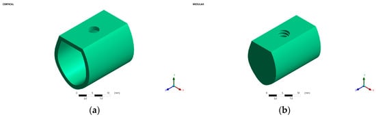
This study aimed to analyze the effect of the height of the proximal axial wall of the prepared tooth and the distance between the adjacent tooth and the prepared tooth on the scan accuracy of intraoral scanners. Ten working casts with maxillary first
[...] Read more.
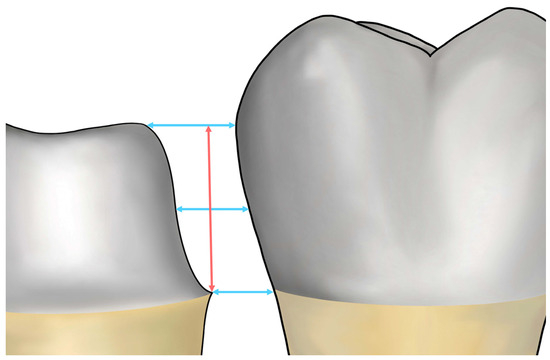
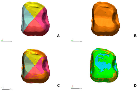
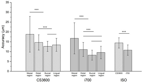
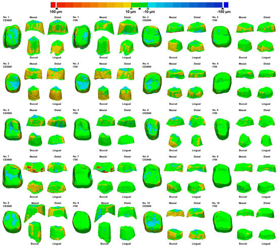
This study aimed to analyze the effect of the height of the proximal axial wall of the prepared tooth and the distance between the adjacent tooth and the prepared tooth on the scan accuracy of intraoral scanners. Ten working casts with maxillary first molars prepared to receive zirconia crowns were randomly obtained from a dental clinic. Each of the 10 casts was scanned using two intraoral scanners (i700; MEDIT and CS3600; Carestream; computer-aided design [CAD] test model, CTM; N = 15 per working cast) 15 times per scanner. Individual dies of the prepared teeth were fabricated, and high-precision scan data were acquired using a laboratory scanner (CAD reference model, CRM; N = 1). CTMs were aligned relative to the prepared tooth of CRMs by using three-dimensional inspection software (Ver 2018.1.0; Control X; 3D Systems). Data were statistically analyzed using an independent t-test and one-way analysis of variance for between-group comparisons (α = 0.05). The inaccuracy in the proximal regions (mesial or distal) of the prepared tooth was higher than that in the buccal and lingual regions (p < 0.05). The scan accuracy was not correlated with the variables when the distance between the adjacent tooth and the prepared tooth was ≥2.0 mm and the height of the proximal axial wall of the prepared tooth was <3.0 mm (p > 0.05). Therefore, an excellent scan accuracy can be obtained using an intraoral scanner when the distance between the adjacent tooth and the prepared tooth is ≥2.0 mm and the proximal axial wall height of the prepared tooth is <3.0 mm.
Full article
(This article belongs to the Special Issue Functional Biomaterials and Digital Technologies in Dentistry: From Bench to Bedside—Volume II)
►
Show Figures

Figure 1


