Next Issue
Volume 12, September
Previous Issue
Volume 12, July


Diagnostics, Volume 12, Issue 8 (August 2022) – 257 articles

Cover Story (view full-size image): 
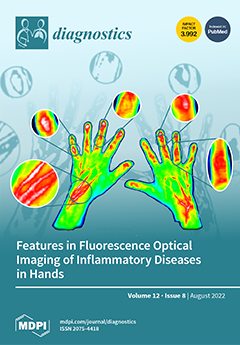
Increasing evidence shows the benefit of near-infrared fluorescence optical imaging (FOI, Xiralite™) for the diagnosis of rheumatoid arthritis, psoriatic arthritis, and osteoarthritis, among others. FOI visualizes impaired microcirculation of the hands, which in most cases leads to an increased accumulation of the contrast agent ICG in the affected regions, indicative of inflammation. This work summarizes the important image features, i.e., location, shape, and timing, found in patients with various rheumatic diseases. Enhancement patterns of patients’ FOI examinations are characterized and assigned to inflammatory anatomical structures, known to be typical for the respective diseases, in their clinical diagnoses. View this paper
  • Issues are regarded as officially published after their release is announced to the table of contents alert mailing list.
  • You may sign up for e-mail alerts to receive table of contents of newly released issues.
  • PDF is the official format for papers published in both, html and pdf forms. To view the papers in pdf format, click on the "PDF Full-text" link, and use the free Adobe Reader to open them.
Order results
Result details
Section
Select all
Export citation of selected articles as:
23 pages, 7799 KiB  
Article
Multi-Scale Hybrid Network for Polyp Detection in Wireless Capsule Endoscopy and Colonoscopy Images
by Meryem Souaidi and Mohamed El Ansari
Diagnostics 2022, 12(8), 2030; https://doi.org/10.3390/diagnostics12082030 - 22 Aug 2022
Cited by 13 | Viewed by 2062
Abstract
The trade-off between speed and precision is a key step in the detection of small polyps in wireless capsule endoscopy (WCE) images. In this paper, we propose a hybrid network of an inception v4 architecture-based single-shot multibox detector (Hyb-SSDNet) to detect small polyp [...] Read more.
The trade-off between speed and precision is a key step in the detection of small polyps in wireless capsule endoscopy (WCE) images. In this paper, we propose a hybrid network of an inception v4 architecture-based single-shot multibox detector (Hyb-SSDNet) to detect small polyp regions in both WCE and colonoscopy frames. Medical privacy concerns are considered the main barriers to WCE image acquisition. To satisfy the object detection requirements, we enlarged the training datasets and investigated deep transfer learning techniques. The Hyb-SSDNet framework adopts inception blocks to alleviate the inherent limitations of the convolution operation to incorporate contextual features and semantic information into deep networks. It consists of four main components: (a) multi-scale encoding of small polyp regions, (b) using the inception v4 backbone to enhance more contextual features in shallow and middle layers, and (c) concatenating weighted features of mid-level feature maps, giving them more importance to highly extract semantic information. Then, the feature map fusion is delivered to the next layer, followed by some downsampling blocks to generate new pyramidal layers. Finally, the feature maps are fed to multibox detectors, consistent with the SSD process-based VGG16 network. The Hyb-SSDNet achieved a 93.29% mean average precision (mAP) and a testing speed of 44.5 FPS on the WCE dataset. This work proves that deep learning has the potential to develop future research in polyp detection and classification tasks. Full article
Show Figures

Figure 1

12 pages, 2067 KiB  
Article
Papillary and Trabecular Muscles Have Substantial Impact on Quantification of Left Ventricle in Patients with Hypertrophic Obstructive Cardiomyopathy
by Chengzhi Yang, Haobo Xu, Shubin Qiao, Ruofei Jia, Zening Jin and Jiansong Yuan
Diagnostics 2022, 12(8), 2029; https://doi.org/10.3390/diagnostics12082029 - 22 Aug 2022
Cited by 4 | Viewed by 1328
Abstract
Patients with obstructive hypertrophic cardiomyopathy (HOCM) have large papillary and trabecular muscles (PTMs), which are myocardial tissue. PTMs are usually excluded from the myocardium and included in the left ventricular (LV) cavity when determining LV mass (LVM) and volumes using cardiac magnetic resonance [...] Read more.
Patients with obstructive hypertrophic cardiomyopathy (HOCM) have large papillary and trabecular muscles (PTMs), which are myocardial tissue. PTMs are usually excluded from the myocardium and included in the left ventricular (LV) cavity when determining LV mass (LVM) and volumes using cardiac magnetic resonance (CMR). This conventional method may result in large distortion of LVM and other indices. We investigated 74 patients with HOCM undergoing CMR imaging. LV short-axis cine images were obtained. LV contours were drawn using two different methods: (1) the conventional method, where PTMs were included in the LV cavity; and (2) the mask method, which includes the TPMs in the LV myocardium. The LV end-diastolic volume (LV-EDV), LV end-systolic volume (LV-ESV), LV ejection fraction (LVEF), and the LVM were then calculated. Fasting NT-proBNP and CK-MB levels were measured with ELISA. In patients with HOCM, mass of PTMs (MOPTM) was 47.9 ± 18.7 g, which represented 26.9% of total LVM. Inclusion of PTMs with the mask method resulted in significantly greater LVM and LVM index (both p < 0.0001) in comparison with those measured with the conventional method. In addition, the mask method produced a significant decrease in LV-EDV and LV-ESV. LVEF was significantly increased with the mask method (64.3 ± 7.9% vs. 77.2 ± 7.1%, p < 0.0001). MOPTM was positively correlated with BMI, septal wall thickness, LVM, LV-EDV, and LV-ESV. LVEF was inversely correlated with MOPTM. In addition, MOPTM correlated positively with NT-proBNP (r = 0.265, p = 0.039) and CK-MB (r = 0.356, p = 0.002). In conclusion, inclusion of PTMs in the myocardium has a substantial impact on quantification of the LVM, LV-EDV, LV-ESV, and LVEF in patients with HOCM. The effects of the PTMs in women was greater than that in men. Furthermore, the MOPTM was positively associated with NT-proBNP and CK-MB. The PTMs might be included in the myocardium when measuring the LV volumes and mass of patients with HOCM. At present, the clinical and prognostic meaning and relevance of the PTMs is not clear and should be further studied. Full article
(This article belongs to the Section Medical Imaging and Theranostics)
Show Figures

Figure 1

16 pages, 7441 KiB  
Article
Interstitial Lung Fibrosis Following COVID-19 Pneumonia
by Mihai Lazar, Ecaterina Constanta Barbu, Cristina Emilia Chitu, Catalin Tiliscan, Laurentiu Stratan, Sorin Stefan Arama, Victoria Arama and Daniela Adriana Ion
Diagnostics 2022, 12(8), 2028; https://doi.org/10.3390/diagnostics12082028 - 22 Aug 2022
Cited by 11 | Viewed by 2800
Abstract
Background and Objectives: Pulmonary fibrosis represents a stage of normal physiologic response to inflammatory aggression, mostly self-limiting and reversible; however, numerous patients treated for SARS-CoV-2 pneumonia present after release from hospital residual lung fibrosis. In this article, we aim to present an optimization [...] Read more.
Background and Objectives: Pulmonary fibrosis represents a stage of normal physiologic response to inflammatory aggression, mostly self-limiting and reversible; however, numerous patients treated for SARS-CoV-2 pneumonia present after release from hospital residual lung fibrosis. In this article, we aim to present an optimization method for evaluating pulmonary fibrosis by quantitative analysis, to identify the risk factors/predictors for pulmonary fibrosis in patients with SARS-CoV-2 infection, and to characterize the impact of pulmonary fibrosis on the symptomatology of patients after release from the hospital. Materials and Methods: We performed a prospective observational study on 100 patients with severe forms of pneumonia, with a control group of 61 non-COVID normal patients. Results: We found persistent interstitial changes consistent with fibrotic changes in 69% of patients. The risk of fibrosis was proportional to the values of erythrocyte sedimentation rate (ESR), C reactive protein (CRP), and lactate dehydrogenase (LDH), and to the duration of hospitalization. The imaging parameters correlated with increased risk for interstitial fibrosis were the number of affected pulmonary lobes and the percent of interstitial pulmonary fibrosis. Conclusions: The main risk factors for pulmonary fibrosis post-COVID-19 identified in our study are increased ESR, CRP, LDH, duration of hospitalization and the severity of pneumonia. Full article
Show Figures

Figure 1

11 pages, 1224 KiB  
Review
Recent Advances in the Diagnosis of Enamel Cracks: A Narrative Review
by Bassam Zidane
Diagnostics 2022, 12(8), 2027; https://doi.org/10.3390/diagnostics12082027 - 22 Aug 2022
Cited by 5 | Viewed by 6770
Abstract
Cracked teeth can pose a diagnostic dilemma for a clinician as they can mimic several other conditions. The constant physiological stress along with any pathological strain like trauma or iatrogenic causes can lead to the development of microcracks in the teeth. Constant exposure [...] Read more.
Cracked teeth can pose a diagnostic dilemma for a clinician as they can mimic several other conditions. The constant physiological stress along with any pathological strain like trauma or iatrogenic causes can lead to the development of microcracks in the teeth. Constant exposure to immense stress can cause the progression of these often-undiagnosed tooth cracks to cause tooth fractures. This review aims to outline the etiology of tooth cracks, their classification, and recent advances in the diagnosis of enamel cracks. Diagnosing a cracked tooth can be an arduous task as symptoms differ according to the location and extension of the incomplete fracture. Early detection is critical because restorative treatment can prevent fracture propagation, microleakage, pulpal or periodontal tissue involvement, and catastrophic cusp failure. Older methods of crack detection are not sensitive or specific. They include clinical examination, visual inspection, exploratory excavation, and percussion test. The dye test used blue or gentian violet stains to highlight fracture lines. Modern methods include transillumination, optical coherence tomography Swept-Source Optical Coherence Tomography (SSOCT), near-infrared imaging, ultrasonic system, infrared thermography, and near-infrared laser. These methods appear to be more efficacious than traditional clinical dental imaging techniques in detecting longitudinal tooth cracks. Clinically distinguishing between the various types of cracks can be difficult with patient-reported signs and symptoms varying according to the location and extension of the incomplete fracture. Cracks are more common in restored teeth. Technological advances such as transillumination allow for early detection and enhanced prognosis. Full article
(This article belongs to the Special Issue Current Concepts and Prospects of Diagnostics in Oral Diseases)
Show Figures

Figure 1

11 pages, 2085 KiB  
Article
Detection of Autoantibodies in Saliva as New Avenue for the Diagnosis and Management of Autoimmune Patients
by Savino Sciascia, Chelsea Bentow, Massimo Radin, Alice Barinotti, Irene Cecchi, Silvia Foddai, Dario Roccatello and Michael Mahler
Diagnostics 2022, 12(8), 2026; https://doi.org/10.3390/diagnostics12082026 - 22 Aug 2022
Cited by 3 | Viewed by 1422
Abstract
(1) Background: Autoimmune diseases are characterized by autoantibodies directed to a large number of antigenic targets and are measured using serum as sample matrix. Although serum is a very common specimen type, it comes with certain drawbacks. Most importantly, it depends on venous [...] Read more.
(1) Background: Autoimmune diseases are characterized by autoantibodies directed to a large number of antigenic targets and are measured using serum as sample matrix. Although serum is a very common specimen type, it comes with certain drawbacks. Most importantly, it depends on venous puncture and requires medical personnel for sampling. This is of particular importance in light of the limited healthcare access of patients with autoimmune diseases during the COVID-19 pandemic. Consequently, alternative sample matrices are being explored for the measurement of autoantibodies. Our study aimed to establish the feasibility of measuring autoantibodies in saliva samples using a novel and highly sensitive method for the detection of autoantibodies. (2) Methods: A total of 48 serum/saliva pairs were collected and tested using a novel particle-based multi-analyte technology (PMAT) system for the presence of a wide range of autoantibodies. (3) Results: A high level of correlation was observed between the results obtained with serum and saliva (Spearman’s rho = 0.725). Study participants clearly preferred saliva over serum sampling as part of the usability assessment. (4) Conclusions: Saliva represents a promising alternative sample matrix for the detection of autoantibodies. The usability study showed a clear preference of saliva over serum as a sample matrix. Full article
(This article belongs to the Section Pathology and Molecular Diagnostics)
Show Figures

Figure 1

13 pages, 336 KiB  
Review
The Management of Pregnancy Complicated with the Previable Preterm and Preterm Premature Rupture of the Membranes: What about a Limit of Neonatal Viability?—A Review
by Stepan Feduniw, Zuzanna Gaca, Olga Malinowska, Weronika Brunets, Magdalena Zgliczyńska, Marta Włodarczyk, Anna Wójcikiewicz and Michał Ciebiera
Diagnostics 2022, 12(8), 2025; https://doi.org/10.3390/diagnostics12082025 - 22 Aug 2022
Cited by 4 | Viewed by 2903
Abstract
Preterm premature rupture of the membranes (PPROM) at the limit of viability is associated with low neonatal survival rates and a high rate of neonatal complications in survivors. It carries a major risk of maternal morbidity and mortality. The limit of viability can [...] Read more.
Preterm premature rupture of the membranes (PPROM) at the limit of viability is associated with low neonatal survival rates and a high rate of neonatal complications in survivors. It carries a major risk of maternal morbidity and mortality. The limit of viability can be defined as the earliest stage of fetal maturity when a fetus has a reasonable chance, although not a high likelihood, for extra-uterine survival. The study reviews available data on preventing preterm delivery caused by the previable PPROM, pregnancy latency, therapeutic options including the use of antibiotics and steroids, neonatal outcomes, and future directions and opportunities. Full article
(This article belongs to the Special Issue Advances in Prenatal Diagnostics)
13 pages, 1929 KiB  
Article
Postmortem Bacteriology in Forensic Autopsies—A Single Center Retrospective Study in Romania
by Iuliana Diac, Arthur-Atilla Keresztesi, Anda-Mihaela Cerghizan, Mihai Negrea and Cătălin Dogăroiu
Diagnostics 2022, 12(8), 2024; https://doi.org/10.3390/diagnostics12082024 - 21 Aug 2022
Cited by 3 | Viewed by 1964
Abstract
Postmortem bacteriology examinations have been a controversial topic over the years, though the value of postmortem bacteriology cultures remains promising. The aim of this study was to review the postmortem bacteriological sampling procedures and results in a single centre in Bucharest over a [...] Read more.
Postmortem bacteriology examinations have been a controversial topic over the years, though the value of postmortem bacteriology cultures remains promising. The aim of this study was to review the postmortem bacteriological sampling procedures and results in a single centre in Bucharest over a period of 10 years. Material and methods: The present study was a retrospective, single-center study, performed at the Mina Minovici National Institute of Legal Medicine in Bucharest, Romania, from 2011–2020. Results: Postmortem bacteriology was requested 630 forensic autopsies, 245 female (38.9%) and 385 male (61.1%), age range 0 and 94 years, median age of 52 years. Deaths occurred in hospital for 594 cases (94.3%) and out-of-hospital for 36 cases (5.7%—field case). Blood cultures were requested in the majority of cases, followed by tracheal swabs and lung tissue. In-hospital and out of hospital deaths did not differ significantly regarding the number of microorganisms identified in a positive blood culture. Postmortem bacteriology cultures of the respiratory tract showed a statistically significant association to microscopically confirmed lung infections. Conclusions Postmortem sampling for bacteriology testing in our center in Bucharest is heterogeneous with a high variation of patterns. A positive blood culture result for Staphylococcus species without the identification of a specific microorganism is more likely due to postmortem contamination. Full article
(This article belongs to the Section Diagnostic Microbiology and Infectious Disease)
Show Figures

Figure 1

15 pages, 3971 KiB  
Article
Advancing Brain Metastases Detection in T1-Weighted Contrast-Enhanced 3D MRI Using Noisy Student-Based Training
by Engin Dikici, Xuan V. Nguyen, Matthew Bigelow, John L. Ryu and Luciano M. Prevedello
Diagnostics 2022, 12(8), 2023; https://doi.org/10.3390/diagnostics12082023 - 21 Aug 2022
Cited by 2 | Viewed by 1602
Abstract
The detection of brain metastases (BM) in their early stages could have a positive impact on the outcome of cancer patients. The authors previously developed a framework for detecting small BM (with diameters of <15 mm) in T1-weighted contrast-enhanced 3D magnetic resonance images [...] Read more.
The detection of brain metastases (BM) in their early stages could have a positive impact on the outcome of cancer patients. The authors previously developed a framework for detecting small BM (with diameters of <15 mm) in T1-weighted contrast-enhanced 3D magnetic resonance images (T1c). This study aimed to advance the framework with a noisy-student-based self-training strategy to use a large corpus of unlabeled T1c data. Accordingly, a sensitivity-based noisy-student learning approach was formulated to provide high BM detection sensitivity with a reduced count of false positives. This paper (1) proposes student/teacher convolutional neural network architectures, (2) presents data and model noising mechanisms, and (3) introduces a novel pseudo-labeling strategy factoring in the sensitivity constraint. The evaluation was performed using 217 labeled and 1247 unlabeled exams via two-fold cross-validation. The framework utilizing only the labeled exams produced 9.23 false positives for 90% BM detection sensitivity, whereas the one using the introduced learning strategy led to ~9% reduction in false detections (i.e., 8.44). Significant reductions in false positives (>10%) were also observed in reduced labeled data scenarios (using 50% and 75% of labeled data). The results suggest that the introduced strategy could be utilized in existing medical detection applications with access to unlabeled datasets to elevate their performances. Full article
(This article belongs to the Special Issue Artificial Intelligence in Radiology 2.0)
Show Figures

Figure 1

14 pages, 2357 KiB  
Article
Assessment of Pulpal Status in Primary Teeth Following Direct Pulp Capping in an Experimental Canine Model
by Andreea Igna, Cornel Igna, Mariana Ioana Miron, Larisa Schuszler, Roxana Dascălu, Mihaela Moldovan, Adrian Aristide Voicu, Carmen Darinca Todea, Marius Boariu, Maria-Alexandra Mârțu and Ștefan-Ioan Stratul
Diagnostics 2022, 12(8), 2022; https://doi.org/10.3390/diagnostics12082022 - 21 Aug 2022
Cited by 4 | Viewed by 2216
Abstract
(1) Background: This study aimed to assess the pulpal response of primary teeth by pulse-oximetry (PO) in a canine model, following direct pulp capping (DPC). (2) Methods: Forty-eight primary teeth from eight canine subjects were divided into three treatment groups, based on the [...] Read more.
(1) Background: This study aimed to assess the pulpal response of primary teeth by pulse-oximetry (PO) in a canine model, following direct pulp capping (DPC). (2) Methods: Forty-eight primary teeth from eight canine subjects were divided into three treatment groups, based on the DPC material—calcium hydroxide (CH), MTA, BiodentineTM)—and three corresponding control groups. Data from PO pulp testing were correlated with laser Doppler flowmetry (LDF) testing, computer tomographic (CT) densitometry and histological analysis; the experiment lasted 14 days. (3) Results: SpO₂ recordings revealed statistically significant differences (p = 0.002, <0.05) between the treatment and control groups, and no significant differences (p = 0.257, >0.05) were observed between treatment groups. LDF recordings showed significant differences (p = 0.002, <0.05) between the treatment and control groups and identified significant differences between materials (p = 0.001, <0.05). CT densitometry indicated vital pulps in all teeth, with pulpal inflammation detected in 6/8 CH-capped teeth and 2/8 MTA-capped teeth. Histologic evaluation confirmed vital pulp in all specimens, with different degrees of inflammation. (4) Conclusions: Within its limitations, the present study confirms the diagnostic value of PO evaluation of pulpal status in primary teeth with histologic means after pulp-capping procedures in a canine model. However, various degrees of pulpal inflammation elicited by different pulp-capping materials seem not to correlate with the obtained PO values. Full article
(This article belongs to the Special Issue Diagnosis and Treatment of Periodontal Diseases in 2022)
Show Figures

Figure 1

9 pages, 399 KiB  
Article
Cell Toxicity Study of Antiseptic Solutions Containing Povidone–Iodine and Hydrogen Peroxide
by Veronica Romano, Donato Di Gennaro, Anna Maria Sacco, Enrico Festa, Emanuela Roscetto, Morena Anna Basso, Tiziana Ascione and Giovanni Balato
Diagnostics 2022, 12(8), 2021; https://doi.org/10.3390/diagnostics12082021 - 21 Aug 2022
Cited by 9 | Viewed by 1928
Abstract
The increasing incidence of periprosthetic joint infections (PJIs) has led to a growing interest in developing strategies to prevent and treat this severe complication. The surgical site’s application of antiseptic solutions to eliminate contaminating bacteria and eradicate the bacterial biofilm has been increasing [...] Read more.
The increasing incidence of periprosthetic joint infections (PJIs) has led to a growing interest in developing strategies to prevent and treat this severe complication. The surgical site’s application of antiseptic solutions to eliminate contaminating bacteria and eradicate the bacterial biofilm has been increasing over time. Even though it has been proven that combining antimicrobials could enhance their activities and help overcome acquired microbial resistance related to the topical use of antibiotics, the toxicity of integrated solutions is not well described. This study aimed to evaluate the cytotoxicity of solutions containing povidone–iodine (PI) and hydrogen peroxide (H2O2), alone or in combination, after 1.3 and 5 min of exposure. Chondrocytes, tenocytes, and fibroblast-like synoviocytes were used for cytotoxicity analysis. Trypan blue stain (0.4% in PBS) was applied to evaluate the dead cells. All solutions tested showed a progressive increase in toxicity as exposure time increased except for PI at 0.3%, which exhibited the lowest toxicity. The combined solutions reported a reduced cellular killing at 3 and 5 min than H2O2 at equal concentrations, similar results to PI solutions. Full article
(This article belongs to the Special Issue Diagnosis and Treatment of Periprosthetic Joint Infections)
Show Figures

Figure 1

15 pages, 2536 KiB  
Article
Electrocardiographic Patterns of Depolarization Abnormalities Help to Identify Reduced Left Ventricular Ejection Fraction
by Maria Gordeeva, Irina Serdiukova, Alexander Krasichkov and Elena Parmon
Diagnostics 2022, 12(8), 2020; https://doi.org/10.3390/diagnostics12082020 - 21 Aug 2022
Viewed by 1384
Abstract
The aim of the study was to investigate the relationship between a decrease in the left ventricular ejection fraction (EF) and traditional ECG signs associated with structural changes of the myocardium (pathological Q wave, ventricular arrhythmias) and relatively new and poorly understood (fragmented [...] Read more.
The aim of the study was to investigate the relationship between a decrease in the left ventricular ejection fraction (EF) and traditional ECG signs associated with structural changes of the myocardium (pathological Q wave, ventricular arrhythmias) and relatively new and poorly understood (fragmented QRS complex (fQRS), early repolarization pattern (ERP)) and evaluate their significance for identifying patients with mildly reduced EF (mrEF). The study included 148 patients who were treated and examined at the Almazov Medical Research Center. FQRS, ERP, pathological Q wave, and premature ventricular contractions (PVC) were described in the analysis of the ECG, and the results of echocardiography and statistical data were analyzed: Fisher’s test and chi-square, correlation analysis, and ROC analysis. According to the level of EF, patients were divided into three groups: group 1—patients with low EF (lEF) (less than 40%), group 2—patients with mildly reduced EF (mrEF) (40–49%); group 3—patients with preserved EF (pEF) (more than 50%). In the first group (EF), fQRS was registered in 16 (51.6%) patients, in the mrEF in 16 (18.2%). Pathological Q wave was detected in lEF in 20 (65%), in mrEF in 10 (35%), 15 (18%), in pEF in 15 (18%). The fQRS has been found to be more important in identifying patients with mrEF. In lEF in 2 (6.5%) patients, in mrEF in 2 (6.9%), in pEF in 11 (12.5%). There was no relationship between ERP, the amount of PVC, and the presence of ventricular tachycardia with EF. FQRS is significantly more common occurred with a decrease in EF and may be a marker of a mrEF. Thus, fQRS is associated with mrEF and pay close attention in routine clinical practice to identify patients at high risk of developing systolic dysfunction. Full article
(This article belongs to the Special Issue Diagnostic Advances in Cardiovascular Prevention)
Show Figures

Figure 1

3 pages, 590 KiB  
Interesting Images
Unconventional Obturator Artery Nutrient Branch: Image of an Anatomical Variation
by Benjamin L. Bosse, Victoria J. Palacios, Dustin W. Dutcher, Emily J. Etter, Peter C. Lim, Caroline A. Cobine and Gillian L. Moritz
Diagnostics 2022, 12(8), 2019; https://doi.org/10.3390/diagnostics12082019 - 21 Aug 2022
Cited by 3 | Viewed by 1443
Abstract
Variations in vascular anatomy are of great concern to surgeons, as proper identification of aberrant arteries can reduce the risk of iatrogenic injury and improve patient outcomes. Several studies have highlighted the irregular branching pattern of pelvic arteries, with a recent focus on [...] Read more.
Variations in vascular anatomy are of great concern to surgeons, as proper identification of aberrant arteries can reduce the risk of iatrogenic injury and improve patient outcomes. Several studies have highlighted the irregular branching pattern of pelvic arteries, with a recent focus on the obturator artery (OA). The OA has an inconstant origin from the internal iliac artery, external iliac artery, or inferior epigastric artery. Within the pelvis, the OA can give off muscular branches and nutrient vessels to the ilium and pubis. Though occasionally described in text, few resources employ images of human donors that depict branches arising from the OAs. Out of the 34 hemisected pelves studied, we identified 1 individual with a substantial nutrient vessel branching unilaterally from the OA. Herein, we present the first image of this unconventional nutrient artery. This vessel should be highlighted given that its size and course make it particularly vulnerable during intrapelvic surgeries such as pelvic lymph node dissection or in procedures requiring arterial embolization of the OA. Full article
(This article belongs to the Section Medical Imaging and Theranostics)
Show Figures

Figure 1

10 pages, 744 KiB  
Article
A Fused Deep Learning Architecture for the Detection of the Relationship between the Mandibular Third Molar and the Mandibular Canal
by Cansu Buyuk, Nurullah Akkaya, Belde Arsan, Gurkan Unsal, Secil Aksoy and Kaan Orhan
Diagnostics 2022, 12(8), 2018; https://doi.org/10.3390/diagnostics12082018 - 20 Aug 2022
Cited by 4 | Viewed by 2220
Abstract
The study aimed to generate a fused deep learning algorithm that detects and classifies the relationship between the mandibular third molar and mandibular canal on orthopantomographs. Radiographs (n = 1880) were randomly selected from the hospital archive. Two dentomaxillofacial radiologists annotated the [...] Read more.
The study aimed to generate a fused deep learning algorithm that detects and classifies the relationship between the mandibular third molar and mandibular canal on orthopantomographs. Radiographs (n = 1880) were randomly selected from the hospital archive. Two dentomaxillofacial radiologists annotated the data via MATLAB and classified them into four groups according to the overlap of the root of the mandibular third molar and mandibular canal. Each radiograph was segmented using a U-Net-like architecture. The segmented images were classified by AlexNet. Accuracy, the weighted intersection over union score, the dice coefficient, specificity, sensitivity, and area under curve metrics were used to quantify the performance of the models. Also, three dental practitioners were asked to classify the same test data, their success rate was assessed using the Intraclass Correlation Coefficient. The segmentation network achieved a global accuracy of 0.99 and a weighted intersection over union score of 0.98, average dice score overall images was 0.91. The classification network achieved an accuracy of 0.80, per class sensitivity of 0.74, 0.83, 0.86, 0.67, per class specificity of 0.92, 0.95, 0.88, 0.96 and AUC score of 0.85. The most successful dental practitioner achieved a success rate of 0.79. The fused segmentation and classification networks produced encouraging results. The final model achieved almost the same classification performance as dental practitioners. Better diagnostic accuracy of the combined artificial intelligence tools may help to improve the prediction of the risk factors, especially for recognizing such anatomical variations. Full article
(This article belongs to the Section Machine Learning and Artificial Intelligence in Diagnostics)
Show Figures

Figure 1

4 pages, 197 KiB  
Editorial
Special Issue: Next-Generation Sequencing in Tumor Diagnosis and Treatment II
by Umberto Malapelle, Matteo Fassan and Dario de Biase
Diagnostics 2022, 12(8), 2017; https://doi.org/10.3390/diagnostics12082017 - 20 Aug 2022
Viewed by 1249
Abstract
Next-generation sequencing (NGS) allows for the sequencing of multiple genes at a very high depth of coverage [...] Full article
(This article belongs to the Special Issue Next-Generation Sequencing in Tumor Diagnosis and Treatment II)
14 pages, 6996 KiB  
Article
Retinal and Choroidal Thinning—A Predictor of Coronary Artery Occlusion?
by Indrė Matulevičiūtė, Agnė Sidaraitė, Vacis Tatarūnas, Audronė Veikutienė, Olivija Dobilienė and Dalia Žaliūnienė
Diagnostics 2022, 12(8), 2016; https://doi.org/10.3390/diagnostics12082016 - 20 Aug 2022
Cited by 13 | Viewed by 1528
Abstract
Introduction. Optical coherence tomography (OCT) and optical coherence tomography angiography (OCTA) allowed visualization of retina and choroid to nearly the capillary level; however, the relationship between systemic macrovascular status and retinal microvascular changes is not yet known well. Aim. Our purpose [...] Read more.
Introduction. Optical coherence tomography (OCT) and optical coherence tomography angiography (OCTA) allowed visualization of retina and choroid to nearly the capillary level; however, the relationship between systemic macrovascular status and retinal microvascular changes is not yet known well. Aim. Our purpose was to assess the impact of retinal optical coherence tomography (OCT) and optical coherence tomography angiography (OCTA) parameters on prediction of coronary heart disease (CHD) in acute myocardial infarction (MI) and chronic three vessel disease (3VD) groups. Methods. This observational study included 184 patients—26 in 3VD, 76 in MI and 82 in healthy participants groups. Radial scans of the macula and OCTA scans of the central macula (superficial (SCP) and deep (DCP) capillary plexuses) were performed on all participants. All participants underwent coronary angiography. Results. Patients in MI groups showed decreased parafoveal total retinal thickness as well as GCL+ retinal thickness. Outer circle total retinal thickness and GCL+ retinal thickness were lowest in the 3VD group. The MI group had thinner, while 3VD the thinnest, choroid. A decrease in choroidal thickness and vascular density could predict 3VD. Conclusions. A decrease in retinal and choroidal thickness as well as decreased vascular density in the central retinal region may predict coronary artery disease. OCT and OCTA could be a significant, safe, and noninvasive tool for the prediction of coronary artery disease. Full article
(This article belongs to the Section Optical Diagnostics)
Show Figures

Figure 1

11 pages, 2544 KiB  
Article
Plasmodium malariae Detected by Microscopy in the International Bordering Area of Mizoram, a Northeastern State of India
by Kuldeep Singh, Praveen Kumar Bharti, Naorem Chaoba Devi, Naseem Ahmed and Amit Sharma
Diagnostics 2022, 12(8), 2015; https://doi.org/10.3390/diagnostics12082015 - 20 Aug 2022
Cited by 1 | Viewed by 2853
Abstract
Northeastern states of India share international borders with Myanmar, China, Bangladesh, and Bhutan, contributing 7.45% of the overall malaria cases in the country. Mizoram accounts for the highest malaria burden in the northeastern states, with perennial transmission in the hilly and deep-forested areas. [...] Read more.
Northeastern states of India share international borders with Myanmar, China, Bangladesh, and Bhutan, contributing 7.45% of the overall malaria cases in the country. Mizoram accounts for the highest malaria burden in the northeastern states, with perennial transmission in the hilly and deep-forested areas. Plasmodium falciparum (93%) is the most prevalent human Plasmodium species, followed by P. vivax; however, information on P. ovale and P. malariae is negligible. Rapid diagnostic tests (RDTs) are the most preferred malaria diagnostic tool followed by microscopy in this high malaria-endemic region. The present epidemiological study was carried out in July and August 2019 to assess the malaria burden in and around the Chawngte primary health center, Lawngtlai District of Mizoram, using RDTs and microscopy as diagnostic tools. World Health Organization-certified level I microscopists examined the blood smears. Diagnosis using RDTs resulted in 151 malaria cases (P. falciparum: 136; P. vivax: 15) out of 948 screened fever cases. However, blood smear examination detected 179 cases (P. falciparum: 154; P. vivax: 17; mixed P. falciparum + P. vivax infection: 3; P. malariae: 5). Analysis revealed that the risk of malaria infection was higher in the ≥5-year-old subjects than in the under-5 age group. The mean parasite density of P. malariae (1455.00/μL blood) was the lowest; cf. with P. falciparum: 12,275.08/μL blood. Surveillance at the point-of-care level using microscopy was able to detect all the four human Plasmodium species and their mixed infections, including P. malariae, which were missed with RDTs. Thus, the quality of microscopy along with trained manpower should be strengthened to diagnose all human malaria parasite species (particularly P. malariae and P. ovale) until the molecular tools are deployed at the field level to achieve malaria elimination by 2030. Full article
(This article belongs to the Special Issue Diagnosis and Management of Malaria)
Show Figures

Figure 1

14 pages, 1486 KiB  
Article
Expression of CD44 in Leukocyte Subpopulations in Patients with Inflammatory Bowel Diseases
by Ivana Franić, Nikolina Režić-Mužinić, Anita Markotić, Piero Marin Živković, Marino Vilović, Doris Rušić and Joško Božić
Diagnostics 2022, 12(8), 2014; https://doi.org/10.3390/diagnostics12082014 - 20 Aug 2022
Cited by 1 | Viewed by 1537
Abstract
CD44 expressed in monocytes and lymphocytes seems to play a crucial role in gastrointestinal inflammation, such as the one occurring in the context of inflammatory bowel diseases. Differentially methylated genes are distinctly expressed across monocyte subpopulations related to the state of Crohn’s disease. [...] Read more.
CD44 expressed in monocytes and lymphocytes seems to play a crucial role in gastrointestinal inflammation, such as the one occurring in the context of inflammatory bowel diseases. Differentially methylated genes are distinctly expressed across monocyte subpopulations related to the state of Crohn’s disease. Hence, the aim of this study was to detect CD44 expression in leukocyte subpopulations in relation to the type of IBD, therapy, and disease duration. Monocyte subpopulations CD14++CD16, CD14++CD16++, and CD14+CD16+ as well as other leukocytes were analyzed for their CD44 expression using flow cytometry in 46 patients with IBD and 48 healthy controls. Patients with Crohn’s disease treated with non-biological therapy (NBT) exhibited a lower percentage of anti-inflammatory CD14+CD16++ monocytes, whereas NBT-treated patients with ulcerative colitis had lower expression of CD44 on CD14+CD44+ lymphocytes in comparison to controls, respectively. Conversely, patients with Crohn’s disease treated with biological therapy had a higher percentage of CD44+ granulocytes but lower expression of CD44 on anti-inflammatory monocytes compared to controls. Median fluorescence intensity (MFI) of CD44 on CD44+CD14+ lymphocytes was higher in ulcerative colitis patients treated with biological therapy compared to NBT. The percentage of classical CD14++CD16 monocytes was lower in the <9 years of IBD duration subgroup compared with the longer disease duration subgroup. The present study addresses the putative role of differentiation and regulation of leukocytes in tailoring IBD therapeutic regimes. Full article
(This article belongs to the Section Pathology and Molecular Diagnostics)
Show Figures

Figure 1

18 pages, 1049 KiB  
Article
Custom 3D fMRI Registration Template Construction Method Based on Time-Series Fusion
by Zhongyang Wang, Junchang Xin, Huixian Shen, Qi Chen, Zhiqiong Wang and Xinlei Wang
Diagnostics 2022, 12(8), 2013; https://doi.org/10.3390/diagnostics12082013 - 20 Aug 2022
Viewed by 1215
Abstract
As the brain standard template for medical image registration has only been constructed with an MRI template, there is no three-dimensional fMRI standard template for use, and when the subject’s brain structure is quite different from the standard brain structure, the registration to [...] Read more.
As the brain standard template for medical image registration has only been constructed with an MRI template, there is no three-dimensional fMRI standard template for use, and when the subject’s brain structure is quite different from the standard brain structure, the registration to the standard space will lead to large errors. Registration to an individual space can avoid this problem. However, in the current fMRI registration algorithm based on individual space, the reference image is often selected by researchers or randomly selected fMRI images at a certain time point. This makes the quality of the reference image very dependent on the experience and ability of the researchers and has great contingency. Whether the reference image is appropriate and reasonable affects the rationality and accuracy of the registration results to a great extent. Therefore, a method for constructing a 3D custom fMRI template is proposed. First, the data are preprocessed; second, by taking a group of two-dimensional slices corresponding to the same layer of the brain in three-dimensional fMRI images at multiple time points as image sequences, each group of slice sequences are registered and fused; and finally, a group of fused slices corresponding to different layers of the brain are obtained. In the process of registration, in order to make full use of the correlation information between the sequence data, the feature points of each two slices of adjacent time points in the sequence are matched, and then according to the transformation relationship between the adjacent images, they are recursively forwarded and mapped to the same space. Then, the fused slices are stacked in order to form a three-dimensional customized fMRI template with individual pertinence. Finally, in the classic registration algorithm, the difference in the registration accuracy between using a custom fMRI template and different standard spaces is compared, which proves that using a custom template can improve the registration effect to a certain extent. Full article
(This article belongs to the Special Issue Advancements in Neuroimaging)
Show Figures

Figure 1

11 pages, 378 KiB  
Article
Dose Descriptors and Assessment of Risk of Exposure-Induced Death in Patients Undergoing COVID-19 Related Chest Computed Tomography
by Lejla M. Čiva, Adnan Beganović, Mustafa Busuladžić, Merim Jusufbegović, Ta’a Awad-Dedić and Sandra Vegar-Zubović
Diagnostics 2022, 12(8), 2012; https://doi.org/10.3390/diagnostics12082012 - 19 Aug 2022
Cited by 4 | Viewed by 2507
Abstract
For more than two years, coronavirus disease 19 (COVID-19) has represented a threat to global health and lifestyles. Computed tomography (CT) imaging provides useful information in patients with COVID-19 pneumonia. However, this diagnostic modality is based on exposure to ionizing radiation, which is [...] Read more.
For more than two years, coronavirus disease 19 (COVID-19) has represented a threat to global health and lifestyles. Computed tomography (CT) imaging provides useful information in patients with COVID-19 pneumonia. However, this diagnostic modality is based on exposure to ionizing radiation, which is associated with an increased risk of radiation-induced cancer. In this study, we evaluated the common dose descriptors, CTDIvol and DLP, for 1180 adult patients. This data was used to estimate the effective dose, and risk of exposure-induced death (REID). Awareness of the extensive use of CT as a diagnostic tool in the management of COVID-19 during the pandemic is vital for the evaluation of radiation exposure parameters, dose reduction methods development and radiation protection. Full article
(This article belongs to the Special Issue The Role of CT in 2019 Novel Coronavirus Pneumonia (COVID-19))
Show Figures

Figure 1

31 pages, 9803 KiB  
Review
Imaging More than Skin-Deep: Radiologic and Dermatologic Presentations of Systemic Disorders
by Mehrzad Shafiei, Firoozeh Shomal Zadeh, Bahar Mansoori, Hunter Pyle, Nnenna Agim, Jorge Hinojosa, Arturo Dominguez, Cristina Thomas and Majid Chalian
Diagnostics 2022, 12(8), 2011; https://doi.org/10.3390/diagnostics12082011 - 19 Aug 2022
Cited by 1 | Viewed by 2322
Abstract
Background: Cutaneous manifestations of systemic diseases are diverse and sometimes precede more serious diseases and symptomatology. Similarly, radiologic imaging plays a key role in early diagnosis and determination of the extent of systemic involvement. Simultaneous awareness of skin and imaging manifestations can help [...] Read more.
Background: Cutaneous manifestations of systemic diseases are diverse and sometimes precede more serious diseases and symptomatology. Similarly, radiologic imaging plays a key role in early diagnosis and determination of the extent of systemic involvement. Simultaneous awareness of skin and imaging manifestations can help the radiologist to narrow down differential diagnosis even if imaging findings are nonspecific. Aims: To improve diagnostic accuracy and patient care, it is important that clinicians and radiologists be familiar with both cutaneous and radiologic features of various systemic disorders. This article reviews cutaneous manifestations and imaging findings of commonly encountered systemic diseases. Conclusions: Familiarity with the most disease-specific skin lesions help the radiologist pinpoint a specific diagnosis and consequently, in preventing unnecessary invasive workups and contributing to improved patient care. Full article
(This article belongs to the Special Issue Skeletal Radiology)
Show Figures

Figure 1

12 pages, 1979 KiB  
Article
The Low Expression of Fc-Gamma Receptor III (CD16) and High Expression of Fc-Gamma Receptor I (CD64) on Neutrophil Granulocytes Mark Severe COVID-19 Pneumonia
by Joerg Hoffmann, Rojin Etati, Cornelia Brendel, Andreas Neubauer and Elisabeth Mack
Diagnostics 2022, 12(8), 2010; https://doi.org/10.3390/diagnostics12082010 - 19 Aug 2022
Cited by 1 | Viewed by 1364
Abstract
Hyperinflammation through neutrophil granulocytes contributes to disease severity in COVID-19 pneumonia and promotes acute lung failure. Understanding the mechanisms of the dysregulations within the myeloid cell compartment may help to improve therapies for severe COVID-19 infection. Here, we investigated the immunopathological characteristics of [...] Read more.
Hyperinflammation through neutrophil granulocytes contributes to disease severity in COVID-19 pneumonia and promotes acute lung failure. Understanding the mechanisms of the dysregulations within the myeloid cell compartment may help to improve therapies for severe COVID-19 infection. Here, we investigated the immunopathological characteristics of circulating neutrophil granulocytes and monocytes in 16 patients with COVID-19 pneumonia by multiparameter flow cytometry in comparison to 9 patients with pulmonary infiltrates but without COVID-19. We correlated the immunophenotypes with the scores of the severity-of-disease classification system, APACHE-II. We found that the mean fluorescence intensity (MFI) of CD15, which is important for the transendothelial migration, was significantly reduced in the patients with COVID-19 (difference ± SD; 295.70 ± 117.50 MFI; p = 0.02). In addition, the granularity was significantly lower in the neutrophil granulocytes of patients with COVID-19 (difference ± SD; 1.11 ± 0.43 side-scatter ratio; p = 0.02). Moreover, the Fc-gamma receptor III (CD16) and Fc-gamma receptor I (CD64) on the neutrophil granulocytes were expressed discordantly with COVID-19 severity. CD16 correlated as inversely proportional (ρ = (−)0.72; 95% CI (−)0.92–(−)0.23; p = 0.01) and CD64 as proportional (ρ = 0.76; 95% CI 0.31–0.93; p = 0.01) with the APACHE-II scores of the patients. We conclude that the deviant expression of the Fc-gamma receptors might play role in a dysregulated antibody-mediated phagocytosis in severe cases of COVID-19 pneumonia. Full article
(This article belongs to the Section Diagnostic Microbiology and Infectious Disease)
Show Figures

Figure 1

8 pages, 1445 KiB  
Article
Changes in Cystoscopic Findings after Intravesical Hyaluronic Acid Instillation Therapy in Patients with Interstitial Cystitis
by Chia-Ju Lin, Chih-Ku Liu, Hsiao-Yun Hsieh, Ming-Jer Chen and Ching-Pei Tsai
Diagnostics 2022, 12(8), 2009; https://doi.org/10.3390/diagnostics12082009 - 19 Aug 2022
Cited by 4 | Viewed by 3125
Abstract
(1) Background: Limited data showed changes in glomerulation in the bladder mucosa of patients with interstitial cystitis (IC) after intravesical hyaluronic acid (HA) bladder infusion. We aimed to investigate the above changes. (2) Methods: Medical records of IC patients were reviewed retrospectively, from [...] Read more.
(1) Background: Limited data showed changes in glomerulation in the bladder mucosa of patients with interstitial cystitis (IC) after intravesical hyaluronic acid (HA) bladder infusion. We aimed to investigate the above changes. (2) Methods: Medical records of IC patients were reviewed retrospectively, from January 2010 to October 2019. Patients who had received repeated cystoscopy after intravesical HA treatment were enrolled. The associations of multiple parameters, including the ages, symptoms, initial glomerulation stage, HA doses, and the interval period of repeated cystoscopy between the glomerulation change in the repeated cystoscopy were analyzed. (3) Results: Among the 35 patients, 9 cases (25.7%) showed better glomerulation grades in the repeated cystoscope (Group 1), 20 cases (57.1%) showed the same grades (Group 2), and 6 cases showed worse grades (Group 3). No difference was seen in the initial grades or treatment course among the three groups. The interval periods from the initial to the repeated cystoscopy of Group 1 were longer than Group 2 and Group 3 (p = 0.031). Group 3 presents an elder age trend than the other two groups. (4) Conclusion: Intravesical HA repaired bladder glomerulation in a small group of patients with IC. Prolonged treatment has potential benefits, while older age is possibly a negative factor. However, no strong correlation was found between the initial glomerulation grades or changes in glomerulation grades with clinical symptoms. Full article
(This article belongs to the Special Issue Imaging of Gynecological Disease)
Show Figures

Figure 1

16 pages, 2369 KiB  
Systematic Review
Comparison of Diagnostic Performance of Ultrasonography and Magnetic Resonance Enterography in the Assessment of Active Bowel Lesions in Patients with Crohn’s Disease: A Systematic Review and Meta-Analysis
by Da In Lee, Myung-Won You, So Hyun Park, Mirinae Seo and Seong Jin Park
Diagnostics 2022, 12(8), 2008; https://doi.org/10.3390/diagnostics12082008 - 19 Aug 2022
Cited by 3 | Viewed by 1530
Abstract
We aimed to evaluate and compare the diagnostic performances of ultrasonography (US) and magnetic resonance enterography (MRE) in assessing active bowel lesions in patients with Crohn’s disease (CD). Materials and Methods: We searched PubMed and EMBASE for studies in which US and MRE [...] Read more.
We aimed to evaluate and compare the diagnostic performances of ultrasonography (US) and magnetic resonance enterography (MRE) in assessing active bowel lesions in patients with Crohn’s disease (CD). Materials and Methods: We searched PubMed and EMBASE for studies in which US and MRE were used to assess active bowel lesions in CD patients. Bivariate random effect meta-analytic methods were used to estimate pooled sensitivity, specificity, and hierarchical summary receiver operating characteristic (HSROC) curves. We performed a meta-regression analysis to explore the source of study heterogeneity. Results: Eleven studies involving 752 patients were included. US exhibited a pooled sensitivity of 86% (95% confidence interval (CI) 72–94), pooled specificity of 88% (95% CI 78–94), and HSROC of 0.93 in 10 studies. MRE exhibited a pooled sensitivity of 88% (95% CI 76–95), pooled specificity of 87% (95% CI 73–95), and an HSROC of 0.94 in eight studies. In seven studies comparing the diagnostic performances of US and MRE, the summary sensitivity of US and MRE were 86% (95% CI 65–96, I2 = 92.1) and 86% (95% CI 72–93, I2 = 88.1) (p = 0.841), respectively. The summary specificity of US and MRE were 87% (95% CI 78–93, I2 = 79.8%) and 84% (72–90, I2 = 72.5%) (p = 0.431), respectively, which showed no statistical differences. On meta-regression analysis, studies from Europe (p = 0.002), those that used linear US probes (p = 0.012), those on small bowel lesions (p = 0.01), and those with outcomes as combined features (active inflammation) reported higher US sensitivity than those from other regions, those that used both linear and convex US probes, those on small and large bowels, and those with outcome as one feature (bowel wall thickening or ulcer). Studies with pediatric patients (p = 0.001), those with reference standards including US (p = 0.001), and outcomes as combined features (p = 0.01) reported higher MRE specificity than those with adult populations, reference standards other than the US, and outcomes as one feature. Conclusions: In spite of considerable heterogeneity in the included studies, both US and MRE can diagnose active bowel lesions with comparable diagnostic accuracy in patients with CD. The study region, type of US probe, lesion location, investigated outcome for US sensitivity and study population, reference standards, and investigated outcomes for MRE specificity were potential sources of heterogeneity. Full article
(This article belongs to the Special Issue Advances in the Diagnosis of Gastrointestinal Diseases)
Show Figures

Figure 1

17 pages, 2997 KiB  
Article
Reproducibility and Repeatability of Coronary Computed Tomography Angiography (CCTA) Image Segmentation in Detecting Atherosclerosis: A Radiomics Study
by Mardhiyati Mohd Yunus, Akmal Sabarudin, Muhammad Khalis Abdul Karim, Puteri N. E. Nohuddin, Isa Azzaki Zainal, Mohd Shahril Mohd Shamsul and Ahmad Khairuddin Mohamed Yusof
Diagnostics 2022, 12(8), 2007; https://doi.org/10.3390/diagnostics12082007 - 19 Aug 2022
Cited by 4 | Viewed by 2125
Abstract
Atherosclerosis is known as the leading factor in heart disease with the highest mortality rate among the Malaysian population. Usually, the gold standard for diagnosing atherosclerosis is by using the coronary computed tomography angiography (CCTA) technique to look for plaque within the coronary [...] Read more.
Atherosclerosis is known as the leading factor in heart disease with the highest mortality rate among the Malaysian population. Usually, the gold standard for diagnosing atherosclerosis is by using the coronary computed tomography angiography (CCTA) technique to look for plaque within the coronary artery. However, qualitative diagnosis for noncalcified atherosclerosis is vulnerable to false-positive diagnoses, as well as inconsistent reporting between observers. In this study, we assess the reproducibility and repeatability of segmenting atherosclerotic lesions manually and semiautomatically in CCTA images to identify the most appropriate CCTA image segmentation method for radiomics analysis to quantitatively extract the atherosclerotic lesion. Thirty (30) CCTA images were taken retrospectively from the radiology image database of Hospital Canselor Tuanku Muhriz (HCTM), Kuala Lumpur, Malaysia. We extract 11,700 radiomics features which include the first-order, second-order and shape features from 180 times of image segmentation. The interest vessels were segmentized manually and semiautomatically using LIFEx (Version 7.0.15, Institut Curie, Orsay, France) software by two independent radiology experts, focusing on three main coronary blood vessels. As a result, manual segmentation with a soft-tissuewindowing setting yielded higher repeatability as compared to semiautomatic segmentation with a significant intraclass correlation coefficient (intra-CC) 0.961 for thefirst-order and shape features; intra-CC of 0.924 for thesecond-order features with p < 0.001. Meanwhile, the semiautomatic segmentation has higher reproducibility as compared to manual segmentation with significant interclass correlation coefficient (inter-CC) of 0.920 (first-order features) and a good interclass correlation coefficient of 0.839 for the second-order features with p < 0.001. The first-order, shape order and second-order features for both manual and semiautomatic segmentation have an excellent percentage of reproducibility and repeatability (intra-CC > 0.9). In conclusion, semi-automated segmentation is recommended for inter-observer study while manual segmentation with soft tissue-windowing can be used for single observer study. Full article
(This article belongs to the Special Issue Advances in Diagnostic Medical Imaging)
Show Figures

Figure 1

14 pages, 16835 KiB  
Case Report
Dentigerous Cysts with Diverse Radiological Presentation Highlighting Diagnostic Challenges
by Alexandre Perez, Vincent Lenoir and Tommaso Lombardi
Diagnostics 2022, 12(8), 2006; https://doi.org/10.3390/diagnostics12082006 - 19 Aug 2022
Cited by 3 | Viewed by 10704
Abstract
Dentigerous cyst is an odontogenic developmental cyst arising from the pericoronal tissue of an impacted tooth, and that may exhibit various radiological aspects. The aim of this article is to present four cases of histologically confirmed mandibular dentigerous cysts to highlight diverse radiological [...] Read more.
Dentigerous cyst is an odontogenic developmental cyst arising from the pericoronal tissue of an impacted tooth, and that may exhibit various radiological aspects. The aim of this article is to present four cases of histologically confirmed mandibular dentigerous cysts to highlight diverse radiological presentations: one of classical appearance (well-limited unilocular radiolucent lesion surrounding the crown) and three which have shown radiological peculiarities (one cyst displacing the adjacent tooth, with bone but no root resorption, one cyst presenting hallmarks of infection and one multilocular cyst with thin septa). Such radiologic diversity may, on occasion, suggest a clinical aggressive lesion such as an odontogenic keratocyst or ameloblastoma. The diagnosis of dentigerous cyst requires a thorough evaluation of the clinical presentation and accurate radiological studies. Full article
(This article belongs to the Topic Diagnostic Imaging in Oral and Maxillofacial Diseases)
Show Figures

Figure 1

13 pages, 2665 KiB  
Systematic Review
The Role of Fluorescence In Situ Hybridization in the Surveillance of Non-Muscle Invasive Bladder Cancer: An Updated Systematic Review and Meta-Analysis
by Weitao Zheng, Tianhai Lin, Zeyu Chen, Dehong Cao, Yige Bao, Peng Zhang, Lu Yang and Qiang Wei
Diagnostics 2022, 12(8), 2005; https://doi.org/10.3390/diagnostics12082005 - 19 Aug 2022
Viewed by 1337
Abstract
Background: Fluorescence in situ hybridization (FISH) has become a popular biomarker for subsequent monitoring the recurrence of non-muscle invasive bladder cancer (NMIBC), several studies have investigated the ability of FISH to detect recurrence in the surveillance of NMIBC. However, the results were inconsistent. [...] Read more.
Background: Fluorescence in situ hybridization (FISH) has become a popular biomarker for subsequent monitoring the recurrence of non-muscle invasive bladder cancer (NMIBC), several studies have investigated the ability of FISH to detect recurrence in the surveillance of NMIBC. However, the results were inconsistent. Methods: We conducted a systematic literature search extensively on authenticated databases including PubMed/Medline, Embase, Web of Science, Ovid, and Cochrane Library. Meta-analysis was performed to find out the sensitivity and specificity of FISH in predicting recurrence of NMIBC. Results: 15 studies were ultimately included in this meta-analysis, a total of 2941 FISH evaluations from 2385 NMIBC patients were available. The pooled sensitivity of FISH was 68% (95% CI: 0.58–0.76), and the pooled specificity was 64% (95% CI: 0.53–0.74). Subgroup analyses were performed in 7 studies without Bacillus Calmette–Guerin (BCG) treatment, the pooled sensitivity was 82% (95% CI: 0.68–0.90), and the pooled specificity was 63% (95% CI: 0.37–0.82). And in 9 studies using “UroVysion standard” to define positive FISH results showed a pooled sensitivity of 60% (95% CI: 0.50–0.70) and specificity of 70% (95% CI: 0.61–0.78). Conclusions: The findings of this study indicate that FISH has a satisfactory sensitivity (68%) and specificity (64%) and could be a potential biomarker in the surveillance of NMIBC. Moreover, BCG treatment and different FISH methods may have an impact on the sensitivity and specificity, these factors should be taken into account when making clinical strategy. Full article
(This article belongs to the Special Issue State of the Art of Bladder Cancer: From Diagnosis to Therapy)
Show Figures

Figure 1

12 pages, 421 KiB  
Systematic Review
The Expression Levels and Concentrations of PD-1 and PD-L1 Proteins in Septic Patients: A Systematic Review
by Mutiara Indah Sari and Syafruddin Ilyas
Diagnostics 2022, 12(8), 2004; https://doi.org/10.3390/diagnostics12082004 - 19 Aug 2022
Cited by 10 | Viewed by 1745 | Correction
Abstract
Sepsis is a series of life-threatening organ dysfunction caused by an impaired host response to infection. A large number of molecular studies of sepsis have revealed complex interactions between infectious agents and hosts that result in heterogeneous manifestations of sepsis. Sepsis can cause [...] Read more.
Sepsis is a series of life-threatening organ dysfunction caused by an impaired host response to infection. A large number of molecular studies of sepsis have revealed complex interactions between infectious agents and hosts that result in heterogeneous manifestations of sepsis. Sepsis can cause immunosuppression and increase the expression of checkpoint inhibitor molecules, including programmed death protein (PD-1) and programmed death ligand 1 (PD-L1), and thus PD-1 and PD-L1 are thought to be useful as diagnostic and prognostic tools for sepsis. PD-1 is an inhibitor of both adaptive and innate immune responses, and is expressed on activated T lymphocytes, natural killer (NK) cells, B lymphocytes, macrophages, dendritic cells (DCs), and monocytes, whereas PD-L1 is expressed on macrophages, some activated T and B cells, and mesenchymal stem cells as well as various non-hematopoietic cells. This systematic review aims to assess the PD-1 and PD-L1 protein expression levels and concentrations in septic and other infectious patients. Full article
(This article belongs to the Special Issue Biomarkers of Sepsis)
Show Figures

Figure 1

24 pages, 2674 KiB  
Review
Imperative Role of Machine Learning Algorithm for Detection of Parkinson’s Disease: Review, Challenges and Recommendations
by Arti Rana, Ankur Dumka, Rajesh Singh, Manoj Kumar Panda, Neeraj Priyadarshi and Bhekisipho Twala
Diagnostics 2022, 12(8), 2003; https://doi.org/10.3390/diagnostics12082003 - 19 Aug 2022
Cited by 39 | Viewed by 3983
Abstract
Parkinson’s disease (PD) is a neurodegenerative disease that affects the neural, behavioral, and physiological systems of the brain. This disease is also known as tremor. The common symptoms of this disease are a slowness of movement known as ‘bradykinesia’, loss of automatic movements, [...] Read more.
Parkinson’s disease (PD) is a neurodegenerative disease that affects the neural, behavioral, and physiological systems of the brain. This disease is also known as tremor. The common symptoms of this disease are a slowness of movement known as ‘bradykinesia’, loss of automatic movements, speech/writing changes, and difficulty with walking at early stages. To solve these issues and to enhance the diagnostic process of PD, machine learning (ML) algorithms have been implemented for the categorization of subjective disease and healthy controls (HC) with comparable medical appearances. To provide a far-reaching outline of data modalities and artificial intelligence techniques that have been utilized in the analysis and diagnosis of PD, we conducted a literature analysis of research papers published up until 2022. A total of 112 research papers were included in this study, with an examination of their targets, data sources and different types of datasets, ML algorithms, and associated outcomes. The results showed that ML approaches and new biomarkers have a lot of promise for being used in clinical decision-making, resulting in a more systematic and informed diagnosis of PD. In this study, some major challenges were addressed along with a future recommendation. Full article
Show Figures

Figure 1

19 pages, 9315 KiB  
Article
GamaComet: A Deep Learning-Based Tool for the Detection and Classification of DNA Damage from Buccal Mucosa Comet Assay Images
by Afiahayati, Edgar Anarossi, Ryna Dwi Yanuaryska and Sri Mulyana
Diagnostics 2022, 12(8), 2002; https://doi.org/10.3390/diagnostics12082002 - 18 Aug 2022
Cited by 3 | Viewed by 1755
Abstract
Comet assay is a simple and precise method to analyze DNA damage. Nowadays, many research studies have demonstrated the effectiveness of buccal mucosa cells usage in comet assays. However, several software tools do not perform well for detecting and classifying comets from a [...] Read more.
Comet assay is a simple and precise method to analyze DNA damage. Nowadays, many research studies have demonstrated the effectiveness of buccal mucosa cells usage in comet assays. However, several software tools do not perform well for detecting and classifying comets from a comet assay image of buccal mucosa cells because the cell has a lot more noise. Therefore, a specific software tool is required for fully automated comet detection and classification from buccal mucosa cell swabs. This research proposes a deep learning-based fully automated framework using Faster R-CNN to detect and classify comets in a comet assay image taken from buccal mucosa swab. To train the Faster R-CNN model, buccal mucosa samples were collected from 24 patients in Indonesia. We acquired 275 comet assay images containing 519 comets. Furthermore, two strategies were used to overcome the lack of dataset problems during the model training, namely transfer learning and data augmentation. We implemented the proposed Faster R-CNN model as a web-based tool, GamaComet, that can be accessed freely for academic purposes. To test the GamaComet, buccal mucosa samples were collected from seven patients in Indonesia. We acquired 43 comet assay images containing 73 comets. GamaComet can give an accuracy of 81.34% for the detection task and an accuracy of 66.67% for the classification task. Furthermore, we also compared the performance of GamaComet with an existing free software tool for comet detection, OpenComet. The experiment results showed that GamaComet performed significantly better than OpenComet that could only give an accuracy of 11.5% for the comet detection task. Downstream analysis can be well conducted based on the detection and classification results from GamaComet. The analysis showed that patients owning comet assay images containing comets with class 3 and class 4 had a smoking habit, meaning they had more cells with a high level of DNA damage. Although GamaComet had a good performance, the performance for the classification task could still be improved. Therefore, it will be one of the future works for the research development of GamaComet. Full article
(This article belongs to the Special Issue Artificial Intelligence in Dental Medicine)
Show Figures

Figure 1

21 pages, 1950 KiB  
Review
Pathological and Molecular Features of Nodal Peripheral T-Cell Lymphomas
by Akira Satou, Taishi Takahara and Toyonori Tsuzuki
Diagnostics 2022, 12(8), 2001; https://doi.org/10.3390/diagnostics12082001 - 18 Aug 2022
Cited by 4 | Viewed by 2453
Abstract
Peripheral T-cell lymphomas (PTCLs) are uncommon neoplasms derived from mature T cells or NK cells. PTCLs comprise numerous disease entities, with over 30 distinct entities listed in the latest WHO classification. They predominantly affect adults and elderly people and usually exhibit an aggressive [...] Read more.
Peripheral T-cell lymphomas (PTCLs) are uncommon neoplasms derived from mature T cells or NK cells. PTCLs comprise numerous disease entities, with over 30 distinct entities listed in the latest WHO classification. They predominantly affect adults and elderly people and usually exhibit an aggressive clinical course with poor prognosis. According to their presentation, PTCLs can be divided into nodal, extranodal or cutaneous, and leukemic types. The most frequent primary sites of PTCLs are lymph nodes, with over half of cases showing nodal presentation. Nodal PTCLs include ALK-positive and ALK-negative anaplastic large cell lymphoma; nodal T-cell lymphoma with T follicular helper cell origin; and PTCL, not otherwise specified. Adult T-cell leukemia/lymphoma also frequently affects lymph nodes. Recent pathological and molecular findings in nodal PTCLs have profoundly advanced the identification of tumor signatures and the refinement of the classification. Therefore, the therapies and pathological diagnosis of nodal PTCLs are continually evolving. This paper aims to provide a summary and update of the pathological and molecular features of nodal PTCLs, which will be helpful for diagnostic practice. Full article
(This article belongs to the Special Issue Diagnostic Pathology of Lymphomas and Lymphoproliferative Disorders)
Show Figures

Figure 1

Previous Issue
Back to TopTop