Next Issue
Volume 12, August
Previous Issue
Volume 12, June


Diagnostics, Volume 12, Issue 7 (July 2022) – 264 articles

Cover Story (view full-size image): 
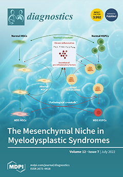
Myelodysplastic syndromes (MDSs) are clonal disorders characterized by ineffective hematopoiesis, resulting in cytopenias and a risk of developing acute myeloid leukemia. In addition to mutations affecting hematopoietic stem cells (HSCs), numerous studies have highlighted the role of the bone marrow microenvironment (BMME) in the development of MDSs. The mesenchymal niche represents a key component of the BMME. In this review, we discuss the role of the mesenchymal niche in the pathophysiology of MDS and provide an overview of currently available in vitro and in vivo models that can be used to study the effects of the mesenchymal niche on HSCs. View this paper
  • Issues are regarded as officially published after their release is announced to the table of contents alert mailing list.
  • You may sign up for e-mail alerts to receive table of contents of newly released issues.
  • PDF is the official format for papers published in both, html and pdf forms. To view the papers in pdf format, click on the "PDF Full-text" link, and use the free Adobe Reader to open them.
Order results
Result details
Section
Select all
Export citation of selected articles as:
14 pages, 965 KiB  
Article
Anti-Citrullinated Protein Antibody Titers Are Independently Modulated by Both Disease Activity and Conventional or Biologic Anti-Rheumatic Drugs
by Miren Uriarte Ecenarro, Daniel Useros, Aranzazu Alfranca, Reyes Tejedor, Isidoro González-Alvaro and Rosario García-Vicuña
Diagnostics 2022, 12(7), 1773; https://doi.org/10.3390/diagnostics12071773 - 21 Jul 2022
Viewed by 1210
Abstract
This study aimed to analyze the factors that influence anti-citrullinated protein antibody (ACPA) titers in a seropositive early arthritis (EA) population under non-protocolized treatment with disease-modifying anti-rheumatic drugs (DMARDs). A total of 130 ACPA-positive patients from the PEARL (Princesa Early Arthritis Longitudinal) study [...] Read more.
This study aimed to analyze the factors that influence anti-citrullinated protein antibody (ACPA) titers in a seropositive early arthritis (EA) population under non-protocolized treatment with disease-modifying anti-rheumatic drugs (DMARDs). A total of 130 ACPA-positive patients from the PEARL (Princesa Early Arthritis Longitudinal) study were studied along a 5-year follow-up. Sociodemographic, clinical, and therapeutic variables, along with serum samples, were collected at five visits by protocol. Anti-cyclic citrullinated peptide 2 (CCP2) ACPA titers were measured by ELISA. The effect of different variables on anti-CCP2 titers was estimated using longitudinal multivariate analysis models, nested by visit and patient. Data from 471 visits in 130 patients were analyzed. A significant decrease in anti-CCP2 titers was observed at all time-points, compared to baseline, following the decline of disease activity. In the multivariate analysis, active or ever smoking was significantly associated with the highest anti-CCP2 titers while reduction in disease activity was associated with titer decline. After adjusting for these variables, both conventional synthetic (cs) and biologic (b) DMARDs accounted for the decline in anti-CCP2 titers as independent factors. Conclusion: In patients with EA, an early and sustained reduction in ACPA titers can be detected associated with the decline in disease activity, irrespective of the treatment used. Full article
(This article belongs to the Special Issue Diagnosis of Rheumatoid Arthritis)
Show Figures

Figure 1

11 pages, 544 KiB  
Review
Autoimmune Connective Tissue Diseases-Related Pruritus: Proper Diagnosis and Possible Mechanisms
by Lai-San Wong and Yu-Ta Yen
Diagnostics 2022, 12(7), 1772; https://doi.org/10.3390/diagnostics12071772 - 21 Jul 2022
Cited by 4 | Viewed by 1999
Abstract
Pruritus is a well-known bothersome symptom among skin disorders, especially inflammatory skin disorders. Lately, a high prevalence of pruritus in patients with autoimmune connective tissue diseases (ACTDs) has been revealed. Patients with ACTDs may suffer from varying degrees of pruritus, which affect their [...] Read more.
Pruritus is a well-known bothersome symptom among skin disorders, especially inflammatory skin disorders. Lately, a high prevalence of pruritus in patients with autoimmune connective tissue diseases (ACTDs) has been revealed. Patients with ACTDs may suffer from varying degrees of pruritus, which affect their quality of life. However, it is rarely recognized both by patients and physicians. Meanwhile, pruritus is not only a symptom but is also related to the disease severity of some ACTDs. The pathophysiology of ACTD related pruritus is ambiguous. This review summarizes the features and possible mechanisms of ACTD-related pruritus, which might lead to proper diagnosis and treatment. Full article
(This article belongs to the Special Issue Advances in the Diagnosis and Management of Rheumatic Diseases)
Show Figures

Figure 1

20 pages, 590 KiB  
Article
An Enhanced Multiple Sclerosis Disease Diagnosis via an Ensemble Approach
by Hanaa Torkey and Nahla A. Belal
Diagnostics 2022, 12(7), 1771; https://doi.org/10.3390/diagnostics12071771 - 21 Jul 2022
Viewed by 1447
Abstract
Multiple Sclerosis (MS) is a disease attacking the central nervous system. According to MS Atlas’s most recent statistics, there are more than 2.8 million people worldwide diagnosed with MS. Recently, studies started to explore machine learning techniques to predict MS using various data. [...] Read more.
Multiple Sclerosis (MS) is a disease attacking the central nervous system. According to MS Atlas’s most recent statistics, there are more than 2.8 million people worldwide diagnosed with MS. Recently, studies started to explore machine learning techniques to predict MS using various data. The objective of this paper is to develop an ensemble approach for diagnosis of MS using gene expression profiles, while handling the class imbalance problem associated with the data. A hierarchical ensemble approach employing voting and boosting techniques is proposed. This approach adopts a heterogeneous voting approach using two base learners, random forest and support vector machine. Experiments show that our approach outperforms state-of-the-art methods, with the highest recorded accuracy being 92.81% and 93.5% with BoostFS and DEGs for feature selection, respectively. Conclusively, the proposed approach is able to efficiently diagnose MS using the gene expression profiles that are more relevant to the disease. The approach is not merely an ensemble classifier outperforming previous work; it also identifies differentially expressed genes between normal samples and patients with multiple sclerosis using a genome-wide expression microarray. The results obtained show that the proposed approach is an efficient diagnostic tool for MS. Full article
(This article belongs to the Section Machine Learning and Artificial Intelligence in Diagnostics)
Show Figures

Figure 1

10 pages, 893 KiB  
Article
Three-Dimensional Angiographic Characteristics versus Functional Stenosis Severity in Fractional and Coronary Flow Reserve Discordance: A DEFINE FLOW Sub Study
by Valerie Stegehuis, Jelmer Westra, Coen Boerhout, Martin Sejr-Hansen, Ashkan Eftekhari, Hernan Mejía-Renteria, Maribel Cambero-Madera, Niels Van Royen, Hitoshi Matsuo, Masafumi Nakayama, Maria Siebes, Evald Høj Christiansen, Tim Van de Hoef and Jan Piek
Diagnostics 2022, 12(7), 1770; https://doi.org/10.3390/diagnostics12071770 - 21 Jul 2022
Viewed by 1256
Abstract
Background: Coronary angiography alone is insufficient to identify lesions associated with myocardial ischemia that may benefit from revascularization. Coronary physiology parameters may improve clinical decision making in addition to coronary angiography, but the association between 2D and 3D qualitative coronary angiography (QCA) and [...] Read more.
Background: Coronary angiography alone is insufficient to identify lesions associated with myocardial ischemia that may benefit from revascularization. Coronary physiology parameters may improve clinical decision making in addition to coronary angiography, but the association between 2D and 3D qualitative coronary angiography (QCA) and invasive pressure and flow measurements is yet to be elucidated. Methods: We associated invasive fractional flow reserve (FFR), coronary flow reserve (CFR) and coronary flow capacity (CFC) with 2D- and 3D-QCA in 430 intermediate lesions of 366 patients. Results: Overall, 2D-QCA analysis resulted in less severe stenosis severity compared with 3D-QCA analysis. FFR+/CFR− lesions had similar 3D-QCA characteristics as FFR+/CFR+ lesions. In contrast, vessels with FFR−/CFR+ discordance had 3D-QCA characteristics similar to those of vessels with concordant FFR−/CFR−. Contrarily, FFR+/CFR− lesions had CFC similar to that of as FFR-/CFR- lesions. Conclusions: Non-flow-limiting lesions (FFR+/CFR−) have 3D-QCA characteristics similar to those of FFR+/CFR+, but the majority are not associated with inducible myocardial ischemia as determined by invasive CFC. FFR−/CFR+ lesions have 3D-QCA characteristics similar to those of FFR−/CFR− lesions but are more frequently associated with a moderately to severely reduced CFC, illustrating the angiographic–functional mismatch in discordant lesions. Full article
(This article belongs to the Section Medical Imaging and Theranostics)
Show Figures

Figure 1

13 pages, 2667 KiB  
Article
Repeatability and Reproducibility of Retinal Fractal Dimension Measured with Swept-Source Optical Coherence Tomography Angiography in Healthy Eyes: A Proof-of-Concept Study
by Louis Arnould, Déa Haddad, Florian Baudin, Pierre-Henry Gabrielle, Marc Sarossy, Alain M. Bron, Behzad Aliahmad and Catherine Creuzot-Garcher
Diagnostics 2022, 12(7), 1769; https://doi.org/10.3390/diagnostics12071769 - 21 Jul 2022
Cited by 1 | Viewed by 1657
Abstract
The retinal vascular network fractal dimension (FD) could be a promising imaging biomarker. Our objective was to evaluate its repeatability and reproducibility in healthy eyes. A cross-sectional study was undertaken with young, healthy volunteers who had no reported cardiac risk factors or ocular [...] Read more.
The retinal vascular network fractal dimension (FD) could be a promising imaging biomarker. Our objective was to evaluate its repeatability and reproducibility in healthy eyes. A cross-sectional study was undertaken with young, healthy volunteers who had no reported cardiac risk factors or ocular disease history. For each participant, three SS-OCTA images (12 × 12 mm) were acquired using the Plex Elite 9000 (Carl Zeiss Meditec AG, Jena, Germany) by two ophthalmologists. Automated segmentation was obtained from both the superficial and deep capillary plexuses. FD was estimated by box counting. The intraclass correlation coefficients (ICC) were used as measures for repeatability and reproducibility. A total of 43 eyes of healthy volunteers were included. The mean ± standard deviation (SD) age was 30 ± 6.2 years. The results show good repeatability. The ICC was 0.722 (95% CI, 0.541–0.839) in the superficial capillary plexus and 0.828 (95% CI, 0.705–0.903) in the deep capillary plexus. For reproducibility, the ICC was 0.651 (95% CI, 0.439–0.795) and 0.363 (95% CI, 0.073–0.596) at the superficial and deep capillary plexus, respectively. In this study, the FD of the vascular network measured via SS-OCTA showed good repeatability and reproducibility in healthy participants. Full article
Show Figures

Figure 1

15 pages, 1491 KiB  
Article
Episodic Memory in Amnestic Mild Cognitive Impairment (aMCI) and Alzheimer’s Disease Dementia (ADD): Using the “Doors and People” Tool to Differentiate between Early aMCI—Late aMCI—Mild ADD Diagnostic Groups
by Athanasios Chatzikostopoulos, Despina Moraitou, Magdalini Tsolaki, Elvira Masoura, Georgia Papantoniou, Maria Sofologi, Vasileios Papaliagkas, Georgios Kougioumtzis and Efthymios Papatzikis
Diagnostics 2022, 12(7), 1768; https://doi.org/10.3390/diagnostics12071768 - 21 Jul 2022
Cited by 4 | Viewed by 2054
Abstract
Episodic memory is the type of memory that allows the recollection of personal experiences containing information on what has happened and, also, where and when it happened. Because of its sensitivity to neurodegenerative diseases and the aging of the brain, it is considered [...] Read more.
Episodic memory is the type of memory that allows the recollection of personal experiences containing information on what has happened and, also, where and when it happened. Because of its sensitivity to neurodegenerative diseases and the aging of the brain, it is considered a hallmark of Alzheimer’s disease dementia (ADD). The objective of the present study was to examine episodic memory in amnestic mild cognitive impairment (aMCI) and ADD. Patients with the diagnosis of early aMCI, late aMCI, and mild ADD were evaluated using the Doors and People tool which consists of four subtests examining different aspects of episodic memory. The statistical analysis with receiver operating characteristic curves (ROC) showed the discriminant potential and the cutoffs of every subtest. Overall, the evaluation of episodic memory with the Doors and People tool can discriminate with great sensitivity between the different groups of people with AD and, especially, early aMCI, late aMCI, and mild ADD patients. Full article
(This article belongs to the Section Pathology and Molecular Diagnostics)
Show Figures

Figure 1

15 pages, 1028 KiB  
Review
Transvaginal Ultrasound vs. Magnetic Resonance Imaging (MRI) Value in Endometriosis Diagnosis
by Alexandra Baușic, Ciprian Coroleucă, Cătălin Coroleucă, Diana Comandașu, Roxana Matasariu, Andrei Manu, Francesca Frîncu, Claudia Mehedințu and Elvira Brătilă
Diagnostics 2022, 12(7), 1767; https://doi.org/10.3390/diagnostics12071767 - 21 Jul 2022
Cited by 11 | Viewed by 2822
Abstract
(1) Background: Endometriosis is a widespread gynecological condition that causes chronic pelvic discomfort, dysmenorrhea, infertility, and impaired quality of life in women of reproductive age. Clinical examination, transvaginal ultrasonography (TVS), and magnetic resonance imaging (MRI) are significant preoperative non-invasive diagnosis procedures for the [...] Read more.
(1) Background: Endometriosis is a widespread gynecological condition that causes chronic pelvic discomfort, dysmenorrhea, infertility, and impaired quality of life in women of reproductive age. Clinical examination, transvaginal ultrasonography (TVS), and magnetic resonance imaging (MRI) are significant preoperative non-invasive diagnosis procedures for the accurate assessment of endometriosis. Although TVS is used as the primary line for diagnosis, MRI is commonly utilized to achieve a better anatomical overview of the entire pelvic organs. The aim of this systematic review article is to thoroughly summarize the research on various endometriosis diagnosis methods that are less invasive. (2) Methods: To find relevant studies, we examined electronic databases, such as MEDLINE/PubMed, Cochrane, and Google Scholar, choosing 70 papers as references. (3) Results: The findings indicate that various approaches can contribute to diagnosis in different ways, depending on the type of endometriosis. For patients suspected of having deep pelvic endometriosis, transvaginal sonography should be the first line of diagnosis. Endometriosis cysts are better diagnosed with TVS, whereas torus, uterosacral ligaments, intestine, and bladder endometriosis lesions are best diagnosed using MRI. When it comes to detecting intestine or rectal nodules, as well as rectovaginal septum nodules, MRI should be the imaging tool of choice. (4) Conclusions: When diagnosing DE (deep infiltrative endometriosis), the examiner’s experience is the most important criterion to consider. In the diagnosis of endometriosis, expert-guided TVS is more accurate than routine pelvic ultrasound, especially in the deep infiltrative form. For optimal treatment and surgical planning, accurate preoperative deep infiltrative endometriosis diagnosis is essential, especially because it requires a multidisciplinary approach. Full article
(This article belongs to the Section Medical Imaging and Theranostics)
Show Figures

Figure 1

18 pages, 10241 KiB  
Review
Ultrasound Imaging of Facial Vascular Neural Structures and Relevance to Aesthetic Injections: A Pictorial Essay
by Wei-Ting Wu, Ke-Vin Chang, Hsiang-Chi Chang, Chen-Hsiang Kuan, Lan-Rong Chen, Kamal Mezian, Vincenzo Ricci and Levent Özçakar
Diagnostics 2022, 12(7), 1766; https://doi.org/10.3390/diagnostics12071766 - 21 Jul 2022
Cited by 3 | Viewed by 10654
Abstract
The facial and submental regions are supplied by complicated neurovascular networks; therefore, facial aesthetic injections may be associated with serious adverse events such as skin necrosis and blindness. Pre-injection localization of neurovascular structures using high-resolution ultrasound can theoretically prevent unexpected complications. Therefore, a [...] Read more.
The facial and submental regions are supplied by complicated neurovascular networks; therefore, facial aesthetic injections may be associated with serious adverse events such as skin necrosis and blindness. Pre-injection localization of neurovascular structures using high-resolution ultrasound can theoretically prevent unexpected complications. Therefore, a systematic protocol that focuses on these facial neurovascular structures is warranted. In this pictorial essay, we discuss the sonoanatomy of facial and submental neurovascular structures and its relevance to aesthetic injections. Moreover, we have highlighted the mechanisms underlying potential neurovascular injuries during aesthetic injections. Full article
Show Figures

Figure 1

14 pages, 2883 KiB  
Article
A Simple, Cost-Effective, and Extraction-Free Molecular Diagnostic Test for Sickle Cell Disease Using a Noninvasive Buccal Swab Specimen for a Limited-Resource Setting
by Priya Thakur, Pragya Gupta, Nupur Bhargava, Rajat Soni, Narendra Varma Gottumukkala, Sangam Giri Goswami, Gaurav Kharya, Vinodh Saravanakumar, Padma Gunda, Suman Jain, Jasmita Dass, Mukul Aggarwal and Sivaprakash Ramalingam
Diagnostics 2022, 12(7), 1765; https://doi.org/10.3390/diagnostics12071765 - 21 Jul 2022
Cited by 1 | Viewed by 2778
Abstract
Sickle cell disease (SCD) is the most prevalent life-threatening blood monogenic disorder. Currently, there is no cure available, apart from bone marrow transplantation. Early and efficient diagnosis of SCD is key to disease management, which would make considerable strides in alleviating morbidity and [...] Read more.
Sickle cell disease (SCD) is the most prevalent life-threatening blood monogenic disorder. Currently, there is no cure available, apart from bone marrow transplantation. Early and efficient diagnosis of SCD is key to disease management, which would make considerable strides in alleviating morbidity and reducing mortality. However, the cost and complexity of diagnostic procedures, such as the Sanger sequencing method, impede the early detection of SCD in a resource-limited setting. To address this, the current study demonstrates a simple and efficient proof-of-concept assay for the detection of patients and carriers using extraction-free non-invasive buccal swab samples by isothermal DNA Amplification coupled Restrictase-mediated cleavage (iDAR). This study is a first of its kind reporting the use of buccal swab specimens for iDA in molecular diagnosis of a genetic disease, all the while being cost effective and time saving, with the total assay time of around 150 min at a cost of USD 5. Further, iDAR demonstrates 91.5% sensitivity and 100% specificity for detecting all three alleles: SS, AS, and AA, having a 100% concordance with Sanger sequencing. The applicability of the iDAR assay is further demonstrated with its adaptation to a one-pot reaction format, which simplifies the assay system. Overall, iDAR is a simple, cost-effective, precise, and non-invasive assay for SCD screening, with the potential for use in a limited resource setting. Full article
(This article belongs to the Section Diagnostic Microbiology and Infectious Disease)
Show Figures

Figure 1

11 pages, 912 KiB  
Article
Does Myasthenia Gravis Affect Long-Term Survival in Thymic Carcinomas? An ESTS Database Analysis
by Filippo Lococo, Dania Nachira, Marco Chiappetta, Jessica Evangelista, Pierre Emmanuel Falcoz, Enrico Ruffini, Paul Van Schil, Marco Scarci, Jòzsef Furàk, Francesco Sollitto, Francesco Guerrera, Lorenzo Spaggiari, Clemens Aigner, Liverakou Evangelia, Andrea Billè, Bernhard Moser, Pascal Alexandre Thomas, Moishe Liberman, Souheil Boubia, Alessio Campisi, Luca Ampollini, Alper Toker, Attila Enyed, Luca Voltolini, Dirk Van Raemdonck, Stefano Margaritora and ESTS Thymic Working Groupadd Show full author list remove Hide full author list
Diagnostics 2022, 12(7), 1764; https://doi.org/10.3390/diagnostics12071764 - 21 Jul 2022
Cited by 3 | Viewed by 1463
Abstract
Background: Thymic carcinoma is a rare and highly malignant tumor with a dismal prognosis, which occasionally coexists with myasthenia gravis (MG). This study aims to investigate the MG incidence on a surgical cohort of patients with thymic carcinoma and to explore its influence [...] Read more.
Background: Thymic carcinoma is a rare and highly malignant tumor with a dismal prognosis, which occasionally coexists with myasthenia gravis (MG). This study aims to investigate the MG incidence on a surgical cohort of patients with thymic carcinoma and to explore its influence on long-term survival. Methods: the prospectively collected data from the ESTS database on thymic epithelial tumors were reviewed. Clinical, pathological, and survival information on thymic carcinoma were analyzed. Results: the analysis was conducted on 203 patients, with an equal gender distribution (96 males and 107 females). MG was detected in 22 (10.8%) patients, more frequently elderly (>60 years, p = 0.048) and male (p = 0.003). Induction therapy was performed in 22 (10.8%) cases. After surgery, 120 (59.1%) patients had a Masaoka stage II–III while complete resection (R0) was achieved in 158 (77.8%). Adjuvant therapy was performed in 68 cases. Mean follow-up was 60 (SD = 14) months. The 3-year, 5-year and 10-year survival rates were 79%, 75% and 63%, respectively. MG did not seem to influence long-term survival (5-year survival in non-MG–TCs 78% vs. 50% in MG–TCs, p = ns) as age < 60 years, female gender, early Masaoka stage, and postoperative radiotherapy did, conversely. Conclusions: myasthenia occurred in about 10% of thymic carcinomas and it did not seem to affect significantly the long-term prognosis in surgically treated thymic carcinoma-patients. Full article
(This article belongs to the Section Pathology and Molecular Diagnostics)
Show Figures

Figure 1

24 pages, 400 KiB  
Review
Personalized Dosimetry in the Context of Radioiodine Therapy for Differentiated Thyroid Cancer
by Massimiliano Pacilio, Miriam Conte, Viviana Frantellizzi, Maria Silvia De Feo, Antonio Rosario Pisani, Andrea Marongiu, Susanna Nuvoli, Giuseppe Rubini, Angela Spanu and Giuseppe De Vincentis
Diagnostics 2022, 12(7), 1763; https://doi.org/10.3390/diagnostics12071763 - 21 Jul 2022
Cited by 8 | Viewed by 2170
Abstract
The most frequent thyroid cancer is Differentiated Thyroid Cancer (DTC) representing more than 95% of cases. A suitable choice for the treatment of DTC is the systemic administration of 131-sodium or potassium iodide. It is an effective tool used for the irradiation of [...] Read more.
The most frequent thyroid cancer is Differentiated Thyroid Cancer (DTC) representing more than 95% of cases. A suitable choice for the treatment of DTC is the systemic administration of 131-sodium or potassium iodide. It is an effective tool used for the irradiation of thyroid remnants, microscopic DTC, other nonresectable or incompletely resectable DTC, or all the cited purposes. Dosimetry represents a valid tool that permits a tailored therapy to be obtained, sparing healthy tissue and so minimizing potential damages to at-risk organs. Absorbed dose represents a reliable indicator of biological response due to its correlation to tissue irradiation effects. The present paper aims to focus attention on iodine therapy for DTC treatment and has developed due to the urgent need for standardization in procedures, since no unique approaches are available. This review aims to summarize new proposals for a dosimetry-based therapy and so explore new alternatives that could provide the possibility to achieve more tailored therapies, minimizing the possible side effects of radioiodine therapy for Differentiated Thyroid Cancer. Full article
9 pages, 2310 KiB  
Article
MiR-155 Dysregulation Is Associated with the Augmentation of ROS/p53 Axis of Fibrosis in Thioacetamide-Induced Hepatotoxicity and Is Protected by Resveratrol
by Amal F. Dawood, Suliman Al Humayed, Maha A. Momenah, Mohamed El-Sherbiny, Hend Ashour, Samaa S. Kamar, Asmaa M. ShamsEldeen, Mohamed A. Haidara, Bahjat Al-Ani and Hasnaa A. Ebrahim
Diagnostics 2022, 12(7), 1762; https://doi.org/10.3390/diagnostics12071762 - 21 Jul 2022
Cited by 7 | Viewed by 1690
Abstract
Liver fibrosis is a hallmark of thioacetamide (TAA) intoxications. MicroRNAs (miRs), such as miR-155, have been implied in the pathogenesis of liver disease, and regulated by the antioxidant and anti-inflammatory compound resveratrol (RES). The link between reactive oxygen species (ROS), tumour suppressor p53 [...] Read more.
Liver fibrosis is a hallmark of thioacetamide (TAA) intoxications. MicroRNAs (miRs), such as miR-155, have been implied in the pathogenesis of liver disease, and regulated by the antioxidant and anti-inflammatory compound resveratrol (RES). The link between reactive oxygen species (ROS), tumour suppressor p53 (p53), and liver fibrosis-during the pathogenesis of TAA-induced liver injury-associated with miR-155 dysregulation with and without RES incorporation has not been previously studied. Therefore, one group of rats received TAA injections of 200 mg/kg; twice a week at the beginning of week 3 for 8 weeks (TAA group; or model group), whereas the protective group was pretreated daily with RES suspension (20 mg/kg; orally) for the first two weeks and subsequently sustained on receiving both RES and TAA until being sacrificed at the 10th week. Liver injuries developed in the model group were confirmed by a significant (p < 0.0001) elevation of hepatic tissue levels of miR-155, ROS, p53, and the profibrogenic biomarkers: tissue inhibitor of metalloproteinases-1 and α-smooth muscle actin, as well as collagen deposition (fibrosis). All these parameters were significantly (p ≤ 0.0234) protected by resveratrol (RES + TAA). In addition, we observed a significant (p < 0.0001) correlation between ROS/p53 axis mediated liver fibrosis and miR-155. Thus, TAA intoxication induced miR-155 imbalance and ROS/p53-mediated liver fibrosis, with resveratrol, conversely displaying beneficial hepatic pleiotropic effects for a period of 10 weeks. Full article
Show Figures

Figure 1

7 pages, 593 KiB  
Review
An Evolution of Reporting: Identifying the Missing Link
by Sara Harsini, Salar Tofighi, Liesl Eibschutz, Brian Quinn and Ali Gholamrezanezhad
Diagnostics 2022, 12(7), 1761; https://doi.org/10.3390/diagnostics12071761 - 21 Jul 2022
Viewed by 1676
Abstract
In recent years, radiologic imaging has undergone tremendous technological advances and is now a pillar of diagnostic and treatment algorithms in clinical medicine. The increased complexity and volume of medical imaging has led clinicians to become ever more reliant on radiologists to both [...] Read more.
In recent years, radiologic imaging has undergone tremendous technological advances and is now a pillar of diagnostic and treatment algorithms in clinical medicine. The increased complexity and volume of medical imaging has led clinicians to become ever more reliant on radiologists to both identify and interpret patient studies. A radiologist’s report provides key insights into a patient’s immediate state of health, information that is vital when choosing the most appropriate next steps in management. As errors in imaging interpretation or miscommunication of results can greatly impair patient care, identifying common error sources is vital to minimizing their occurrence. Although mistakes in medical imaging are practically inevitable, changes to the delivery of imaging reporting and the addition of artificial intelligence algorithms to analyze clinicians’ communication skills can minimize the impact of these errors, keep up with the continuously evolving landscape of medical imaging, and ultimately close the communication gap. Full article
(This article belongs to the Section Medical Imaging and Theranostics)
Show Figures

Figure 1

11 pages, 1730 KiB  
Article
Evaluation of Adjuvant Treatments for Adenoid Cystic Carcinoma of the Breast: A Population-Based, Propensity Score Matched Cohort Study from the SEER Database
by Liu Yang, Chaobin Wang, Miao Liu and Shu Wang
Diagnostics 2022, 12(7), 1760; https://doi.org/10.3390/diagnostics12071760 - 21 Jul 2022
Cited by 2 | Viewed by 1575
Abstract
Adenoid cystic carcinoma (ACC) is an extremely rare type of breast cancer. The role of adjuvant treatments for ACC remains controversial. Patients with a histology-confirmed diagnosis of ACC of the breast were identified based on the SEER (Surveillance, Epidemiology and End Results) database. [...] Read more.
Adenoid cystic carcinoma (ACC) is an extremely rare type of breast cancer. The role of adjuvant treatments for ACC remains controversial. Patients with a histology-confirmed diagnosis of ACC of the breast were identified based on the SEER (Surveillance, Epidemiology and End Results) database. Propensity score matching (PSM) was performed to balance the baseline characteristics. The Kaplan–Meier method and Cox regression models were performed to determine the impact of the adjuvant chemotherapy (CT) and radiotherapy (RT) associated with breast cancer-specific survival (BCSS) and overall survival (OS). A total of 1036 patients with ACC of the breast were included. After a median follow-up of 11.3 years, the 10-year OS and BCSS rate was 76.2% and 92.6%, respectively. After PSM, adjuvant CT converted into neither OS (Log-rank p = 1.000) nor BCSS (Log-rank p = 0.900) benefits in patients with ACC of the breast. Adjuvant RT also did not improve OS (Log-rank p = 0.060) and BCSS (Log-rank p = 0.400). According to the univariate stratified analysis, adjuvant RT was favorable for OS in patients who underwent breast-conserving surgery (HR 0.66, 95% CI 0.45, 0.99, p = 0.042). From the multivariate analysis, histology grade and nodal status were independent prognostic factors for both OS and BCSS. In conclusion, ACC of the breast presented a favorable prognosis. Adjuvant treatment, especially adjuvant CT, might not be essential for patients with this disease. Full article
(This article belongs to the Section Pathology and Molecular Diagnostics)
Show Figures

Figure 1

17 pages, 1449 KiB  
Systematic Review
Prevalence of Human Papilloma Virus Infection in Bladder Cancer: A Systematic Review
by Narcisa Muresu, Biagio Di Lorenzo, Laura Saderi, Illari Sechi, Arcadia Del Rio, Andrea Piana and Giovanni Sotgiu
Diagnostics 2022, 12(7), 1759; https://doi.org/10.3390/diagnostics12071759 - 20 Jul 2022
Cited by 9 | Viewed by 2028
Abstract
The etiology of bladder cancer is known to be associated with behavioral and environmental factors. Moreover, several studies suggested a potential role of HPV infection in the pathogenesis with controversial results. A systematic review was conducted to assess the role of HPV. A [...] Read more.
The etiology of bladder cancer is known to be associated with behavioral and environmental factors. Moreover, several studies suggested a potential role of HPV infection in the pathogenesis with controversial results. A systematic review was conducted to assess the role of HPV. A total of 46 articles that reported the prevalence of HPV infection in squamous (SCC), urothelial (UC), and transitional cell carcinomas (TCC) were selected. A pooled prevalence of 19% was found, with a significant difference in SCC that was mainly driven by HPV-16. Moreover, infection prevalence in case-control studies showed a higher risk of bladder cancer in HPV-positive cases (OR: 7.84; p-value < 0.00001). The results may suggest an etiologic role of HPV in bladder cancer. HPV vaccine administration in both sexes could be key to prevent the infection caused by high-risk genotypes. Full article
(This article belongs to the Special Issue Diagnosis of Lower Genital Tract Disease)
Show Figures

Figure 1

3 pages, 1060 KiB  
Interesting Images
Unexpected Movement of the Esophagus across the Aorta
by Hyun Ho Choi, Soo-Yoon Sung and Yoon Ho Ko
Diagnostics 2022, 12(7), 1758; https://doi.org/10.3390/diagnostics12071758 - 20 Jul 2022
Cited by 1 | Viewed by 1184
Abstract
Tumor regression throughout treatment would induce organ movement, but little is known of this in the esophagus. To achieve successful tumor regression, radiation therapy requires several weeks of radiation to be delivered accurately to the tumor. Usually, a 5–10 mm margin is allowed [...] Read more.
Tumor regression throughout treatment would induce organ movement, but little is known of this in the esophagus. To achieve successful tumor regression, radiation therapy requires several weeks of radiation to be delivered accurately to the tumor. Usually, a 5–10 mm margin is allowed for set-up error and internal organ motion. Our case exhibited an unexpectedly large movement of the esophagus across the aorta with tumor regression that extended outside the margin and thus outside the radiotherapy field. These movements may affect subsequent invasive procedures or treatment during cancer therapy. After the unexpected large movement of the esophagus due to tumor regression, we revised the radiotherapy plan to reflect the new esophageal position. This implied that regular imaging and close monitoring are required during treatment of esophageal cancer. Full article
(This article belongs to the Special Issue Image-Guided Cancer Diagnosis and Therapy)
Show Figures

Figure 1

8 pages, 4719 KiB  
Communication
Correlation between Results of Semi-Quantitative and Quantitative Tests for Hepatitis B Virus Surface Antigen among Patients Achieving Viral Suppression with Antiviral Treatment
by Goh Eun Chung, Ju Yeon Kim, Hyunjae Shin, Ji Hoon Hong, Moon Haeng Hur, Heejin Cho, Min Kyung Park, Na Ryung Choi, Jihye Kim, Yun Bin Lee, Eun Ju Cho, Su Jong Yu, Yoon Jun Kim, Jung-Hwan Yoon and Jeong-Hoon Lee
Diagnostics 2022, 12(7), 1757; https://doi.org/10.3390/diagnostics12071757 - 20 Jul 2022
Cited by 2 | Viewed by 1585
Abstract
Background: Hepatitis B virus (HBV) infection remains a threat to global public health. Serum hepatitis B surface antigen (HBsAg) has been used in screening for HBV infection. Quantitative HBsAg assays are useful for monitoring the natural history of HBV infection and its [...] Read more.
Background: Hepatitis B virus (HBV) infection remains a threat to global public health. Serum hepatitis B surface antigen (HBsAg) has been used in screening for HBV infection. Quantitative HBsAg assays are useful for monitoring the natural history of HBV infection and its response to therapy. The aim of this study was to determine the relationship between quantitative (qHBsAg; IU/mL) and semi-quantitative (sqHBsAg; signal-to-cutoff ratio [S/Co]) HBsAg titers in patients with chronic hepatitis B (CHB). Methods: We retrospectively included 284 samples with HBV DNA < 20 IU/mL from patients who had simultaneous qHBsAg (using electrochemiluminescence assay) and sqHBsAg tests. Patients were grouped according to their serum HBV-envelope antigen (HBeAg) status (HBeAg-negative, n = 239 and HBeAg-positive, n = 45). The Spearman test was used to analyze the correlation between the quantitative and semi-quantitative assays. Results: There was a significant linear correlation between sqHBsAg and qHBsAg in the HBeAg-negative patients (qHBsAg [IU/mL] = 0.0094 × sqHBsAg [S/Co]1.323; adjusted R2 = 0.8445; p < 0.001). There was a substantial hook effect in the assays from the HBeAg-positive patients, so we performed a stratified analysis according to qHBsAg <1000 IU/mL or ≥1000 IU/mL and found a significant positive linear correlation between sqHBsAg S/Co and qHBsAg (qHBsAg [IU/mL] = 0.072 × sqHBsAg [S/Co]1.331; adjusted R2 = 0.7878; p < 0.001) in HBeAg-positive patients with qHBsAg titers of <1000 IU/mL and a significant negative correlation in HBeAg-positive patients with qHBsAg titers of ≥1000 IU/mL (qHBsAg [IU/mL] = 8.987 × 10¹⁴ × sqHBsAg [S/Co]−3.175; adjusted R2 = 0.6350; p < 0.001). Conclusions: There was a highly linear, positive correlation between qHBsAg and sqHBsAg in HBeAg-negative CHB patients. The hook effect led to a negative correlation in HBeAg-positive CHB patients with qHBsAg titers ≥1000 IU/mL. Full article
(This article belongs to the Special Issue Diagnosis and Management of Chronic Hepatitis B)
Show Figures

Figure 1

12 pages, 919 KiB  
Review
Influence of S100A2 in Human Diseases
by Hitomi Sugino and Yu Sawada
Diagnostics 2022, 12(7), 1756; https://doi.org/10.3390/diagnostics12071756 - 20 Jul 2022
Cited by 7 | Viewed by 2048
Abstract
S100 proteins are a family of low-molecular-weight proteins characterized by two calcium-binding sites with a helix-loop-helix (“EF-hand-type”) domain. The S100 family of proteins is distributed across various organs and can interact with diverse molecules. Among the proteins of the S100 family, S100 calcium-binding [...] Read more.
S100 proteins are a family of low-molecular-weight proteins characterized by two calcium-binding sites with a helix-loop-helix (“EF-hand-type”) domain. The S100 family of proteins is distributed across various organs and can interact with diverse molecules. Among the proteins of the S100 family, S100 calcium-binding protein A2 (S100A2) has been identified in mammary epithelial cells, glands, lungs, kidneys, and prostate gland, exhibiting various physiological and pathological actions in human disorders, such as inflammatory diseases and malignant tumors. In this review, we introduce basic knowledge regarding S100A2 regulatory mechanisms. Although S100A2 is a tumor suppressor, we describe the various influences of S100A2 on cancer and inflammatory diseases. Full article
(This article belongs to the Section Pathology and Molecular Diagnostics)
Show Figures

Figure 1

9 pages, 758 KiB  
Article
Clinical Benefits and Challenges in Application of Novel Portable Gastric Capsule Endoscopy for Home Healthcare Patients
by Yang-Chao Lin, Ching-Lin Chen, Yi-Wei Kao, Ching-Yao Tsai, Mingchih Chen and Chih-Kuang Liu
Diagnostics 2022, 12(7), 1755; https://doi.org/10.3390/diagnostics12071755 - 20 Jul 2022
Viewed by 3023
Abstract
Portable magnetic-assisted capsule endoscopy (MACE) provides satisfactory patient experience and safety with comparable performance in diagnosis of organic lesions when compared to conventional upper gastrointestinal endoscopy. In this study, a total of 58 homecare patients were included for MACE either in the hospital [...] Read more.
Portable magnetic-assisted capsule endoscopy (MACE) provides satisfactory patient experience and safety with comparable performance in diagnosis of organic lesions when compared to conventional upper gastrointestinal endoscopy. In this study, a total of 58 homecare patients were included for MACE either in the hospital (n = 42) or at home (n = 16), with mean age of 71.1 ± 12.4 years. A total of 55 patients (94.83%) had completed the MACE with diagnosis of reflux esophagitis (43.6%), gastritis (54.5%), erosions (21.8%), fundic polyps (14.5%), peptic ulcers (25.9%), etc. Most patients (n = 47, 85.5%) were satisfied with the experience, and all patients who received MACE at home (n = 15, 100%) appreciated the convenience of endoscopy at home. Less than half of the patients (n = 24, 43.6%) could afford MACE if the expense was not covered by health insurance (USD 714). Time consumption from both traffic and capsule manipulation was also challenging for the physicians, as it took an average of 24.7 min to complete MACE, but it added up to a total of 92.7 min at home, which is about 15 times that of conventional endoscopy in hospital. More efforts are needed to ease the financial burden of patients, and optimization of workflow in community practice may help lift the obstacles revealed in this study. Full article
(This article belongs to the Special Issue Capsule Endoscopy: Clinical Impacts and Innovation since 2001-Issue B)
Show Figures

Figure 1

20 pages, 1309 KiB  
Review
Desquamative Gingivitis in the Context of Autoimmune Bullous Dermatoses and Lichen Planus—Challenges in the Diagnosis and Treatment
by Ana Maria Sciuca, Mihaela Paula Toader, Carmen Gabriela Stelea, George Alexandru Maftei, Oana Elena Ciurcanu, Ovidiu Mihail Stefanescu, Bianca-Andreea Onofrei and Cristina Popa
Diagnostics 2022, 12(7), 1754; https://doi.org/10.3390/diagnostics12071754 - 20 Jul 2022
Cited by 6 | Viewed by 4291
Abstract
Desquamative gingivitis (DG) is a clinical term that describes erythema, desquamation and erosions of the gingiva, of various etiologies. Although the clinical aspect is not specific for a certain disease, an accurate diagnosis of the underlying disorder is necessary because the disease course, [...] Read more.
Desquamative gingivitis (DG) is a clinical term that describes erythema, desquamation and erosions of the gingiva, of various etiologies. Although the clinical aspect is not specific for a certain disease, an accurate diagnosis of the underlying disorder is necessary because the disease course, prognosis and treatment vary according to the cause. DG may inflict significant oral discomfort, which is why patients typically present to the dentist for a first consultation, rendering it important for these specialists to be informed about this condition. Our paper aims to review the ethiopatogenesis and diagnostic approach of DG, focusing on the most common underlying disorders (autoimmune bullous dermatoses and lichen planus) and on the management of these patients. Potential etiological agents leading to an inflammatory immune response in the oral mucosa and DG appearance include genetic predisposition, metabolic, neuropsychiatric, infectious factors, medication, dental materials, graft-versus-host reaction and autoimmunity. A thorough anamnesis, a careful clinical examination, paraclinical explorations including histopathological exam and direct immunofluorescence are necessary to formulate an appropriate diagnosis. Proper and prompt management of these patients lead to a better prognosis and improved quality of life, and must include management in the dental office with sanitizing the oral cavity, instructing the patient for rigorous oral hygiene, periodic follow-up for bacterial plaque detection and removal, as well as topical and systemic therapy depending on the underlying disorder, based on treatment algorithms. A multidisciplinary approach for the diagnosis and follow-up of DG in the context of pemphigus vulgaris, bullous pemphigoid, cicatricial pemhigoid or lichen planus is necessary, including consultations with dermatologists, oral medicine specialists and dentists. Full article
(This article belongs to the Special Issue Diagnosis and Treatment of Periodontal Diseases in 2022)
Show Figures

Figure 1

13 pages, 622 KiB  
Article
Simultaneously Screening for Liver Steatosis and Fibrosis in Romanian Type 2 Diabetes Mellitus Patients Using Vibration-Controlled Transient Elastography with Controlled Attenuation Parameter
by Anca Trifan, Ermina Stratina, Robert Nastasa, Adrian Rotaru, Remus Stafie, Sebastian Zenovia, Laura Huiban, Catalin Sfarti, Camelia Cojocariu, Tudor Cuciureanu, Cristina Muzica, Stefan Chiriac, Irina Girleanu, Ana-Maria Singeap and Carol Stanciu
Diagnostics 2022, 12(7), 1753; https://doi.org/10.3390/diagnostics12071753 - 20 Jul 2022
Cited by 6 | Viewed by 1580
Abstract
Non-alcoholic fatty liver disease (NAFLD) is a common finding among patients with type 2 diabetes mellitus (T2DM). Between NAFLD and T2DM exist a bidirectional relationship. Patients with T2DM are at high risk for NAFLD, and evidence suggests that T2DM is linked to progressive [...] Read more.
Non-alcoholic fatty liver disease (NAFLD) is a common finding among patients with type 2 diabetes mellitus (T2DM). Between NAFLD and T2DM exist a bidirectional relationship. Patients with T2DM are at high risk for NAFLD, and evidence suggests that T2DM is linked to progressive NAFLD and poor liver outcomes. NAFLD promotes the development of T2DM and leads to a substantial increase in the risk of T2DM complications. This study aimed to assess the prevalence of liver steatosis and fibrosis in patients with T2DM from north-eastern Romania by using Vibration-Controlled Transient Elastography (VCTE) with Controlled Attenuation Parameter (CAP), which is a non-invasive method and can assess simultaneously liver steatosis and fibrosis. In total, 424 consecutive patients with T2DM were enrolled and evaluated using VCTE with CAP from January 2020 to January 2022. Clinical and laboratory data were recorded in all patients. For the CAP score, we used the following cut-offs: mild steatosis (S1)—274 dB/m, moderate steatosis (S2)—290 dB/m, and severe steatosis (S3)—302 dB/m. For liver fibrosis, to differentiate between fibrosis stages, the cut-off values were F ≥ 8.2 kPa for significant fibrosis (F2), F ≥ 9.7 kPa for advanced fibrosis (F3), and F ≥ 13.6 kPa for cirrhosis (F4). In total, 380 diabetic patients (72.6%) had liver steatosis (51.3% females, the mean age of 55.22 ± 10.88 years, mean body mass index (BMI) 29.12 ± 5.64 kg/m2). Among them, 26 (8.4%) patients had moderate liver steatosis (S2) and 242 (78.5%) patients had severe hepatic steatosis (S3). According to VCTE measurements, 176 (57.14%) patients had liver fibrosis, 36 (11.7%) of them had advanced fibrosis (F3), and 42 (13.6%) diabetic patients had cirrhosis (F4). Univariate analyses showed that severe steatosis was significantly associated with ferritin (β = 0.223, p = 0.022), total cholesterol (β = 0.159, p = 0.031), and HDL-cholesterol (β = −0.120, p = 0.006). In multivariate analyses, BMI (β = 0.349, p < 0.001), fasting plasma glucose (β = 0.211, p = 0.006), and triglycerides (β = 0.132, p = 0.044) were predictors of S3. Patients with T2DM have a high prevalence of severe steatosis and advanced fibrosis which can lead to the development and progression of complications with high morbidity and mortality rates. Hence, it is necessary to implement screening strategies to prevent advanced liver disease in patients with T2DM. Full article
(This article belongs to the Section Medical Imaging and Theranostics)
Show Figures

Figure 1

13 pages, 1700 KiB  
Review
When Ring Sideroblasts on Bone Marrow Smears Are Inconsistent with the Diagnosis of Myelodysplastic Neoplasms
by Sandrine Girard, Franck Genevieve, Emmanuelle Rault, Odile Fenneteau and Jean-François Lesesve
Diagnostics 2022, 12(7), 1752; https://doi.org/10.3390/diagnostics12071752 - 20 Jul 2022
Cited by 3 | Viewed by 3026
Abstract
Ring sideroblasts are commonly seen in myelodysplastic neoplasms and are a key condition for identifying distinct entities of myelodysplastic neoplasms according to the WHO classification. However, the presence of ring sideroblasts is not exclusive to myelodysplastic neoplasms. Ring sideroblasts are as well either [...] Read more.
Ring sideroblasts are commonly seen in myelodysplastic neoplasms and are a key condition for identifying distinct entities of myelodysplastic neoplasms according to the WHO classification. However, the presence of ring sideroblasts is not exclusive to myelodysplastic neoplasms. Ring sideroblasts are as well either encountered in non-clonal secondary acquired disorders, such as exposure to toxic substances, drug/medicine, copper deficiency, zinc overload, lead poison, or hereditary sideroblastic anemias related to X-linked, autosomal, or mitochondrial mutations. This review article will discuss diseases associated with ring sideroblasts outside the context of myelodysplastic neoplasms. Knowledge of the differential diagnoses characterized by the presence of ring sideroblasts in bone marrow is essential to prevent any misdiagnosis, which leads to delayed diagnosis and subsequent management of patients that differ in the different forms of sideroblastic anemia. Full article
(This article belongs to the Special Issue Myelodysplastic Syndrome: Diagnosis and Screening)
Show Figures

Figure 1

13 pages, 1058 KiB  
Review
Bicuspid Aortic Valve in Children and Adolescents: A Comprehensive Review
by Gaia Spaziani, Francesca Girolami, Luigi Arcieri, Giovanni Battista Calabri, Giulio Porcedda, Chiara Di Filippo, Francesca Chiara Surace, Marco Pozzi and Silvia Favilli
Diagnostics 2022, 12(7), 1751; https://doi.org/10.3390/diagnostics12071751 - 20 Jul 2022
Cited by 6 | Viewed by 3192
Abstract
Bicuspid aortic valve (BAV) is the most common congenital heart defect. Prevalence of isolated BAV in the general pediatric population is about 0.8%, but it has been reported to be as high as 85% in patients with aortic coarctation. A genetic basis has [...] Read more.
Bicuspid aortic valve (BAV) is the most common congenital heart defect. Prevalence of isolated BAV in the general pediatric population is about 0.8%, but it has been reported to be as high as 85% in patients with aortic coarctation. A genetic basis has been recognized, with great heterogeneity. Standard BAV terminology, recently proposed on the basis of morpho-functional assessment by transthoracic echocardiography, may be applied also to the pediatric population. Apart from neonatal stenotic BAV, progression of valve dysfunction and/or of the associated aortic dilation seems to be slow during pediatric age and complications are reported to be much rarer in comparison with adults. When required, because of severe BAV dysfunction, surgery is most often the therapeutic choice; however, the ideal initial approach to treat severe aortic stenosis in children or adolescents is not completely defined yet, and a percutaneous approach may be considered in selected cases as a palliative option in order to postpone surgery. A comprehensive and tailored evaluation is needed to define the right intervals for cardiologic evaluation, indications for sport activity and the right timing for intervention. Full article
(This article belongs to the Special Issue Thoracic Aortic Disease: From Bench to Bedside)
Show Figures

Figure 1

9 pages, 1654 KiB  
Article
MELIF, a Fully Automated Liver Function Score Calculated from Gd-EOB-DTPA-Enhanced MR Images: Diagnostic Performance vs. the MELD Score
by Carolina Río Bártulos, Karin Senk, Ragnar Bade, Mona Schumacher, Jan Plath, Nico Kaiser, Isabel Wiesinger, Sylvia Thurn, Christian Stroszczynski, Abdelouahed El Mountassir, Mathis Planert, Jan Woetzel and Philipp Wiggermann
Diagnostics 2022, 12(7), 1750; https://doi.org/10.3390/diagnostics12071750 - 20 Jul 2022
Cited by 2 | Viewed by 1332
Abstract
In the management of patients with chronic liver disease, the assessment of liver function is essential for treatment planning. Gd-EOB-DTPA-enhanced MRI allows for both the acquisition of anatomical information and regional liver function quantification. The objective of this study was to demonstrate and [...] Read more.
In the management of patients with chronic liver disease, the assessment of liver function is essential for treatment planning. Gd-EOB-DTPA-enhanced MRI allows for both the acquisition of anatomical information and regional liver function quantification. The objective of this study was to demonstrate and evaluate the diagnostic performance of two fully automatically generated imaging-based liver function scores that take the whole liver into account. T1 images from the native and hepatobiliary phases and the corresponding T1 maps from 195 patients were analyzed. A novel artificial-intelligence-based software prototype performed image segmentation and registration, calculated the reduction rate of the T1 relaxation time for the whole liver (rrT1liver) and used it to calculate a personalized liver function score, then generated a unified score—the MELIF score—by combining the liver function score with a patient-specific factor that included weight, height and liver volume. Both scores correlated strongly with the MELD score, which is used as a reference for global liver function. However, MELIF showed a stronger correlation than the rrT1liver score. This study demonstrated that the fully automated determination of total liver function, regionally resolved, using MR liver imaging is feasible, providing the opportunity to use the MELIF score as a diagnostic marker in future prospective studies. Full article
(This article belongs to the Special Issue Imaging of Hepatitis)
Show Figures

Figure 1

3 pages, 683 KiB  
Interesting Images
Echocardiography, Computed Tomography and Magnetic Resonance Imaging in the Differential Diagnosis of a Tumor in the Left Atrium of the Heart
by Malgorzata Zalewska-Adamiec, Hanna Bachorzewska-Gajewska and Slawomir Dobrzycki
Diagnostics 2022, 12(7), 1749; https://doi.org/10.3390/diagnostics12071749 - 20 Jul 2022
Viewed by 1125
Abstract
Cardiac tumors are rare. Most often they are metastatic tumors, while primary tumors are much less common. In addition to proliferative changes in the heart, there are also non-neoplastic structures, such as thrombus, vegetation or inflammatory tumors. All structures with a heart tumor [...] Read more.
Cardiac tumors are rare. Most often they are metastatic tumors, while primary tumors are much less common. In addition to proliferative changes in the heart, there are also non-neoplastic structures, such as thrombus, vegetation or inflammatory tumors. All structures with a heart tumor morphology require a lot of imaging studies in order to diagnose them and plan treatment without performing a biopsy. We present a case of a 75-year-old female patient who had moving masses in the left atrium on echocardiography. Computed tomography of the chest was performed, which did not clearly explain the nature of the structure observed in the left atrium. The Heart Team decided to perform another test—magnetic resonance imaging (MRI) of the heart in 3 months to differentiate the lesion. The examination was performed after 3 months of warfarin therapy and there were no masses in the left atrium, which confirmed that the observed tumor was a thrombus. Full article
(This article belongs to the Special Issue Noninvasive Diagnosis of Cardiac Tumors)
Show Figures

Figure 1

13 pages, 897 KiB  
Article
Cytology and High-Risk Human Papillomavirus Test for Cervical Cancer Screening Assessment
by Frederik A. Stuebs, Martin C. Koch, Anna K. Dietl, Werner Adler, Carol Geppert, Arndt Hartmann, Antje Knöll, Matthias W. Beckmann, Grit Mehlhorn, Carla E. Schulmeyer and Paul Gass
Diagnostics 2022, 12(7), 1748; https://doi.org/10.3390/diagnostics12071748 - 19 Jul 2022
Cited by 8 | Viewed by 1869
Abstract
Background: A new nationwide screening strategy was implemented in Germany in January 2020. No data are available for women referred to certified dysplasia units for secondary clarification after primary diagnosis by a local physician. We therefore investigated combined testing with Papanicolaou smears and [...] Read more.
Background: A new nationwide screening strategy was implemented in Germany in January 2020. No data are available for women referred to certified dysplasia units for secondary clarification after primary diagnosis by a local physician. We therefore investigated combined testing with Papanicolaou smears and high-risk human papillomavirus (hrHPV) and compared the data with the final histological findings. Methods: Between January 2015 and October 2020, all referred women who underwent colposcopy of the uterine cervix in our certified dysplasia unit were included. Cytology findings were classified using the Munich III nomenclature. Results: A total of 3588 colposcopies were performed in 3118 women, along with Pap smear and hrHPV co-testing, followed by histology. Women with Pap II-p (ASC-US) and a positive hrHPV co-test had a 22.4% risk for cervical intraepithelial neoplasia (CIN) 3/high-grade squamous intraepithelial lesion (HSIL). The risk of CIN 3/HSIL was 83.8% in women with Pap IVa-p (HSIL) and a positive hrHPV co-test. A positive hrHPV co-test increased the risk for HSIL+ (OR 5.942; 95% CI, 4.617 to 7.649; p < 0.001) as compared to a negative hrHPV co-test. Conclusions: The accuracy of Pap smears is comparable with the screening results. A positive hrHPV test increases the risk for HSIL+ fivefold. Colposcopy is necessary to diagnose HSIL+ correctly. Full article
Show Figures

Figure 1

19 pages, 5050 KiB  
Article
Melanoma Detection Using XGB Classifier Combined with Feature Extraction and K-Means SMOTE Techniques
by Chih-Chi Chang, Yu-Zhen Li, Hui-Ching Wu and Ming-Hseng Tseng
Diagnostics 2022, 12(7), 1747; https://doi.org/10.3390/diagnostics12071747 - 19 Jul 2022
Cited by 15 | Viewed by 2090
Abstract
Melanoma, a very severe form of skin cancer, spreads quickly and has a high mortality rate if not treated early. Recently, machine learning, deep learning, and other related technologies have been successfully applied to computer-aided diagnostic tasks of skin lesions. However, some issues [...] Read more.
Melanoma, a very severe form of skin cancer, spreads quickly and has a high mortality rate if not treated early. Recently, machine learning, deep learning, and other related technologies have been successfully applied to computer-aided diagnostic tasks of skin lesions. However, some issues in terms of image feature extraction and imbalanced data need to be addressed. Based on a method for manually annotating image features by dermatologists, we developed a melanoma detection model with four improvement strategies, including applying the transfer learning technique to automatically extract image features, adding gender and age metadata, using an oversampling technique for imbalanced data, and comparing machine learning algorithms. According to the experimental results, the improved strategies proposed in this study have statistically significant performance improvement effects. In particular, our proposed ensemble model can outperform previous related models. Full article
Show Figures

Figure 1

11 pages, 892 KiB  
Article
Assessment of Serum Pepsinogens with and without Co-Testing with Gastrin-17 in Gastric Cancer Risk Assessment—Results from the GISTAR Pilot Study
by Claudia Robles, Dace Rudzite, Inese Polaka, Olga Sjomina, Lilian Tzivian, Ilze Kikuste, Ivars Tolmanis, Aigars Vanags, Sergejs Isajevs, Inta Liepniece-Karele, Danute Razuka-Ebela, Sergej Parshutin, Raul Murillo, Rolando Herrero, Jin Young Park and Marcis Leja
Diagnostics 2022, 12(7), 1746; https://doi.org/10.3390/diagnostics12071746 - 19 Jul 2022
Cited by 5 | Viewed by 1973
Abstract
Introduction––Serum pepsinogen tests for gastric cancer screening have been debated for decades. We assessed the performance of two pepsinogen assays with or without gastrin-17 for the detection of different precancerous lesions alone or as a composite endpoint in a Latvian cohort. Methods––Within the [...] Read more.
Introduction––Serum pepsinogen tests for gastric cancer screening have been debated for decades. We assessed the performance of two pepsinogen assays with or without gastrin-17 for the detection of different precancerous lesions alone or as a composite endpoint in a Latvian cohort. Methods––Within the intervention arm of the GISTAR population-based study, participants with abnormal pepsinogen values by ELISA or latex-agglutination tests, or abnormal gastrin-17 by ELISA and a subset of subjects with all normal biomarker values were referred for upper endoscopy with biopsies. Performance of biomarkers, corrected by verification bias, to detect five composite outcomes based on atrophy, intestinal metaplasia, dysplasia or cancer was explored. Results––Data from 1045 subjects were analysed, of those 273 with normal biomarker results. Both pepsinogen assays showed high specificity (>93%) but poor sensitivity (range: 18.4–31.1%) that slightly improved when lesions were restricted to corpus location (40.5%) but decreased when dysplasia and prevalent cancer cases were included (23.8%). Adding gastrin-17 detection, sensitivity reached 33–45% while specificity decreased (range: 61.1–62%) and referral rate for upper endoscopy increased to 38.6%. Conclusions––Low sensitivity of pepsinogen assays is a limiting factor for their use in population-based primary gastric cancer screening, however their high specificity could be useful for triage. Full article
(This article belongs to the Section Pathology and Molecular Diagnostics)
Show Figures

Figure 1

17 pages, 6548 KiB  
Article
Stabilization of Tuberculosis Reporter Enzyme Fluorescence (REFtb) Diagnostic Reagents for Use at the Point of Care
by Maxim Lebedev, Aaron B. Benjamin, Kent J. Koster, Kathryn E. Broyles, Sathish Kumar, Joseph M. Jilka and Jeffrey D. Cirillo
Diagnostics 2022, 12(7), 1745; https://doi.org/10.3390/diagnostics12071745 - 19 Jul 2022
Viewed by 1368
Abstract
Tuberculosis is one of the most frequent causes of death in humans worldwide. One of the primary reasons tuberculosis remains a public health threat is that diagnosis can take weeks to months, is often not very sensitive and cannot be accomplished in many [...] Read more.
Tuberculosis is one of the most frequent causes of death in humans worldwide. One of the primary reasons tuberculosis remains a public health threat is that diagnosis can take weeks to months, is often not very sensitive and cannot be accomplished in many remote environments. A rapid, sensitive and inexpensive point-of-care (POC) diagnostic would have a major impact on tuberculosis eradication efforts. The tuberculosis diagnostic system REFtb is based on specific detection of the constitutively expressed β-lactamase (BlaC) in Mycobacterium tuberculosis using a custom fluorogenic substrate designated as CDG-3. REFtb has potential as a diagnostic for tuberculosis that could be very inexpensive (<USD 2.00/test), used at the POC and could provide definitive diagnosis within 10 min. However, the reagents for REFtb are currently in liquid form, making them more susceptible to degradation and difficult to transport. We evaluated the improvement in the stability of REFtb reagents by lyophilization under a variety of conditions through their effects on the performance of REFtb. We found that lyophilization of REFtb components produces an easily reconstituted powder that displays similar performance to the liquid system and that lactose represents one of the most promising excipients for use in a final POC REFtb diagnostic system. These studies provide the foundation for the production of a stable POC REFtb system that could be easily distributed worldwide with minimal or no requirement for refrigeration. Full article
(This article belongs to the Special Issue Fluorescence Optical Imaging)
Show Figures

Figure 1

26 pages, 4065 KiB  
Review
The Revolution of Lateral Flow Assay in the Field of AMR Detection
by Hervé Boutal, Christian Moguet, Lilas Pommiès, Stéphanie Simon, Thierry Naas and Hervé Volland
Diagnostics 2022, 12(7), 1744; https://doi.org/10.3390/diagnostics12071744 - 19 Jul 2022
Cited by 11 | Viewed by 3105
Abstract
The global spread of antimicrobial resistant (AMR) bacteria represents a considerable public health concern, yet their detection and identification of their resistance mechanisms remain challenging. Optimal diagnostic tests should provide rapid results at low cost to enable implementation in any microbiology laboratory. Lateral [...] Read more.
The global spread of antimicrobial resistant (AMR) bacteria represents a considerable public health concern, yet their detection and identification of their resistance mechanisms remain challenging. Optimal diagnostic tests should provide rapid results at low cost to enable implementation in any microbiology laboratory. Lateral flow assays (LFA) meet these requirements and have become essential tools to combat AMR. This review presents the versatility of LFA developed for the AMR detection field, with particular attention to those directly triggering β-lactamases, their performances, and specific limitations. It considers how LFA can be modified by detecting not only the enzyme, but also its β-lactamase activity for a broader clinical sensitivity. Moreover, although LFA allow a short time-to-result, they are generally only implemented after fastidious and time-consuming techniques. We present a sample processing device that shortens and simplifies the handling of clinical samples before the use of LFA. Finally, the capacity of LFA to detect amplified genetic determinants of AMR by isothermal PCR will be discussed. LFA are inexpensive, rapid, and efficient tools that are easy to implement in the routine workflow of laboratories as new first-line tests against AMR with bacterial colonies, and in the near future directly with biological media. Full article
(This article belongs to the Section Diagnostic Microbiology and Infectious Disease)
Show Figures

Figure 1

Previous Issue
Next Issue
Back to TopTop