Next Issue
Volume 10, July
Previous Issue
Volume 10, May


Cells, Volume 10, Issue 6 (June 2021) – 294 articles

Cover Story (view full-size image): 
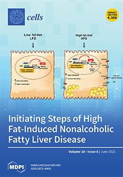
Nonalcoholic fatty liver disease (NAFLD) is a highly prevalent disease in the world and has become a serious public health concern. Here, we show that a high-fat diet (HFD) induces epigenetic modifications in the liver that increase the expression of peroxisome proliferator-activated receptor (PPARγ), as well as its target genes’ very low-density lipoprotein receptor (VLDLR) and differentiation cluster 36 (CD36), leading to excess lipid uptake accumulation. The study provides strong evidence that HFD-induced epigenetic modifications in pparγ promoter are a crucial initiating mechanism of regulation in NAFLD pathogenesis. View this paper
  • Issues are regarded as officially published after their release is announced to the table of contents alert mailing list.
  • You may sign up for e-mail alerts to receive table of contents of newly released issues.
  • PDF is the official format for papers published in both, html and pdf forms. To view the papers in pdf format, click on the "PDF Full-text" link, and use the free Adobe Reader to open them.
Order results
Result details
Section
Select all
Export citation of selected articles as:
15 pages, 2856 KiB  
Article
Advanced Maternal Age Deteriorates the Developmental Competence of Vitrified Oocytes in Mice
by Ju Hee Lee, Jae Kyun Park, Sook Young Yoon, Eun A Park, Jin Hyun Jun, Hyunjung J. Lim, Jayeon Kim and Haengseok Song
Cells 2021, 10(6), 1563; https://doi.org/10.3390/cells10061563 - 21 Jun 2021
Cited by 11 | Viewed by 3069
Abstract
Advanced maternal age (AMA) is known to be related to the decrease in the quality and quantity of oocytes. Oocyte vitrification is now considered an established assisted reproductive technology for fertility preservation. However, it remains unclear whether the oocytes in older women are [...] Read more.
Advanced maternal age (AMA) is known to be related to the decrease in the quality and quantity of oocytes. Oocyte vitrification is now considered an established assisted reproductive technology for fertility preservation. However, it remains unclear whether the oocytes in older women are more sensitive to various insults during vitrification. Thus, we evaluated whether AMA affects cellular and molecular features and developmental outcomes of oocytes after vitrification in mice. The oocytes were grouped as young fresh (YF), young vitrified/warmed (YV), aged fresh (AF), and aged vitrified/warmed (AV). The survival rate of AV oocytes was significantly lower than that of YV oocytes. The rates of fertilization, cleavage, and blastocyst formation of AV oocytes were significantly lower than those of other groups. AV oocytes were represented as aberrations in mitochondria distribution, microvacuole size, and autophagosome formation, leading to delayed embryo development in mice. This delay was associated with a reduced number of total cells and trophectoderm in the blastocyst developed from AV oocytes. Collectively, AMA exaggerates the vulnerability of oocytes to cryo-damage that occurs during vitrification in mice, suggesting that the current vitrification protocols optimized for oocytes from young females should be modified for oocytes from aged women. Full article
(This article belongs to the Section Reproductive Cells and Development)
Show Figures

Graphical abstract

19 pages, 11654 KiB  
Article
Eritoran Attenuates Hepatic Inflammation and Fibrosis in Mice with Chronic Liver Injury
by Yun-Cheng Hsieh, Kuei-Chuan Lee, Pei-Shan Wu, Teh-Ia Huo, Yi-Hsiang Huang, Ming-Chih Hou and Han-Chieh Lin
Cells 2021, 10(6), 1562; https://doi.org/10.3390/cells10061562 - 21 Jun 2021
Cited by 11 | Viewed by 3040
Abstract
Toll-like receptor 4 (TLR4) signaling plays a key role in liver inflammation and fibrosis. The therapeutic effects of eritoran, a TLR4 antagonist, in mice with chronic liver injury remained unclear. C57BL/6 mice were fed a fast-food diet (FFD) or treated with carbon tetrachloride [...] Read more.
Toll-like receptor 4 (TLR4) signaling plays a key role in liver inflammation and fibrosis. The therapeutic effects of eritoran, a TLR4 antagonist, in mice with chronic liver injury remained unclear. C57BL/6 mice were fed a fast-food diet (FFD) or treated with carbon tetrachloride (CCl4) to induce chronic liver injury. Eritoran (10 mg/kg) or a vehicle was randomly intraperitoneally administered to the FFD-fed mice and the CCl4-injured mice. Primary mouse liver cells were cultured with lipopolysaccharide (LPS) or eritoran. In both FFD and CCl4 mouse models, eritoran significantly reduced serum ALT levels and decreased hepatic inflammatory cell infiltration without altering hepatic steatosis. Additionally, eritoran attenuated liver fibrosis by decreasing hepatic stellate cells (HSCs) activation and the abundance of α-smooth muscle actin and transforming growth factor-β1. Hepatic TLR4 downstream signaling including MyD88 expression, NF-κB p65 nuclear translocation, p38 and JNK phosphorylation were successfully inhibited by eritoran. In the in vitro study, LPS-induced nuclear translocation of NF-κB in primary HSCs and Kupffer cells was significantly suppressed by eritoran. In conclusion, eritoran attenuated hepatic inflammation and fibrosis by inhibition of the TLR4 signaling pathway in mice with chronic liver injury. Eritoran may serve as a potential drug for chronic liver disease. Full article
(This article belongs to the Special Issue Molecular Mechanisms in Organ Fibrosis)
Show Figures

Graphical abstract

28 pages, 7458 KiB  
Article
The Timecourses of Functional, Morphological, and Molecular Changes Triggered by Light Exposure in Sprague–Dawley Rat Retinas
by Serena Riccitelli, Mattia Di Paolo, James Ashley, Silvia Bisti and Stefano Di Marco
Cells 2021, 10(6), 1561; https://doi.org/10.3390/cells10061561 - 21 Jun 2021
Cited by 13 | Viewed by 2489
Abstract
Retinal neurodegeneration can impair visual perception at different levels, involving not only photoreceptors, which are the most metabolically active cells, but also the inner retina. Compensatory mechanisms may hide the first signs of these impairments and reduce the likelihood of receiving timely treatments. [...] Read more.
Retinal neurodegeneration can impair visual perception at different levels, involving not only photoreceptors, which are the most metabolically active cells, but also the inner retina. Compensatory mechanisms may hide the first signs of these impairments and reduce the likelihood of receiving timely treatments. Therefore, it is essential to characterize the early critical steps in the neurodegenerative progression to design adequate therapies. This paper describes and correlates early morphological and biochemical changes in the degenerating retina with in vivo functional analysis of retinal activity and investigates the progression of neurodegenerative stages for up to 7 months. For these purposes, Sprague–Dawley rats were exposed to 1000 lux light either for different durations (12 h to 24 h) and examined seven days afterward (7d) or for a fixed duration (24 h) and monitored at various time points following the exposure (up to 210d). Flash electroretinogram (fERG) recordings were correlated with morphological and histological analyses to evaluate outer and inner retinal disruptions, gliosis, trophic factor release, and microglial activation. Twelve hours or fifteen hours of exposure to constant light led to a severe retinal dysfunction with only minor morphological changes. Therefore, early pathological signs might be hidden by compensatory mechanisms that silence retinal dysfunction, accounting for the discrepancy between photoreceptor loss and retinal functional output. The long-term analysis showed a transient functional recovery, maximum at 45 days, despite a progressive loss of photoreceptors and coincident increases in glial fibrillary acidic protein (GFAP) and basic fibroblast growth factor-2 (bFGF-2) expression. Interestingly, the progression of the disease presented different patterns in the dorsal and ventral retina. The information acquired gives us the potential to develop a specific diagnostic tool to monitor the disease’s progression and treatment efficacy. Full article
(This article belongs to the Special Issue The Retina in Health and Disease)
Show Figures

Figure 1

16 pages, 3689 KiB  
Article
TRPV1 Activation by Capsaicin Mediates Glucose Oxidation and ATP Production Independent of Insulin Signalling in Mouse Skeletal Muscle Cells
by Parisa Vahidi Ferdowsi, Kiran D. K. Ahuja, Jeffrey M. Beckett and Stephen Myers
Cells 2021, 10(6), 1560; https://doi.org/10.3390/cells10061560 - 21 Jun 2021
Cited by 21 | Viewed by 3869
Abstract
Background: Insulin resistance (IR), a key characteristic of type 2 diabetes (T2DM), is manifested by decreased insulin-stimulated glucose transport in target tissues. Emerging research has highlighted transient receptor potential cation channel subfamily V member (TRPV1) activation by capsaicin as a potential therapeutic target [...] Read more.
Background: Insulin resistance (IR), a key characteristic of type 2 diabetes (T2DM), is manifested by decreased insulin-stimulated glucose transport in target tissues. Emerging research has highlighted transient receptor potential cation channel subfamily V member (TRPV1) activation by capsaicin as a potential therapeutic target for these conditions. However, there are limited data on the effects of capsaicin on cell signalling molecules involved in glucose uptake. Methods: C2C12 cells were cultured and differentiated to acquire the myotube phenotype. The activation status of signalling molecules involved in glucose metabolism, including 5’ adenosine monophosphate-activated protein kinase (AMPK), calcium/calmodulin-dependent protein kinase 2 (CAMKK2), extracellular signal-regulated protein kinases 1 and 2 (ERK1/2), protein kinase B (AKT), and src homology phosphatase 2 (SHP2), was examined. Finally, activation of CAMKK2 and AMPK, and glucose oxidation and ATP levels were measured in capsaicin-treated cells in the presence or absence of TRPV1 antagonist (SB-452533). Results: Capsaicin activated cell signalling molecules including CAMKK2 and AMPK leading to increased glucose oxidation and ATP generation independent of insulin in the differentiated C2C12 cells. Pharmacological inhibition of TRPV1 diminished the activation of CAMKK2 and AMPK as well as glucose oxidation and ATP production. Moreover, we observed an inhibitory effect of capsaicin in the phosphorylation of ERK1/2 in the mouse myotubes. Conclusion: Our data show that capsaicin-mediated stimulation of TRPV1 in differentiated C2C12 cells leads to activation of CAMKK2 and AMPK, and increased glucose oxidation which is concomitant with an elevation in intracellular ATP level. Further studies of the effect of TRPV1 channel activation by capsaicin on glucose metabolism could provide novel therapeutic utility for the management of IR and T2DM. Full article
Show Figures

Figure 1

21 pages, 3321 KiB  
Article
Right Ventricle Remodeling Metabolic Signature in Experimental Pulmonary Hypertension Models of Chronic Hypoxia and Monocrotaline Exposure
by Thaïs Hautbergue, Fabrice Antigny, Angèle Boët, François Haddad, Bastien Masson, Mélanie Lambert, Amélie Delaporte, Jean-Baptiste Menager, Laurent Savale, Jérôme Le Pavec, Elie Fadel, Marc Humbert, Christophe Junot, François Fenaille, Benoit Colsch and Olaf Mercier
Cells 2021, 10(6), 1559; https://doi.org/10.3390/cells10061559 - 21 Jun 2021
Cited by 10 | Viewed by 3163
Abstract
Introduction: Over time and despite optimal medical management of patients with pulmonary hypertension (PH), the right ventricle (RV) function deteriorates from an adaptive to maladaptive phenotype, leading to RV failure (RVF). Although RV function is well recognized as a prognostic factor of PH, [...] Read more.
Introduction: Over time and despite optimal medical management of patients with pulmonary hypertension (PH), the right ventricle (RV) function deteriorates from an adaptive to maladaptive phenotype, leading to RV failure (RVF). Although RV function is well recognized as a prognostic factor of PH, no predictive factor of RVF episodes has been elucidated so far. We hypothesized that determining RV metabolic alterations could help to understand the mechanism link to the deterioration of RV function as well as help to identify new biomarkers of RV failure. Methods: In the current study, we aimed to characterize the metabolic reprogramming associated with the RV remodeling phenotype during experimental PH induced by chronic-hypoxia-(CH) exposure or monocrotaline-(MCT) exposure in rats. Three weeks after PH initiation, we hemodynamically characterized PH (echocardiography and RV catheterization), and then we used an untargeted metabolomics approach based on liquid chromatography coupled to high-resolution mass spectrometry to analyze RV and LV tissues in addition to plasma samples from MCT-PH and CH-PH rat models. Results: CH exposure induced adaptive RV phenotype as opposed to MCT exposure which induced maladaptive RV phenotype. We found that predominant alterations of arginine, pyrimidine, purine, and tryptophan metabolic pathways were detected on the heart (LV+RV) and plasma samples regardless of the PH model. Acetylspermidine, putrescine, guanidinoacetate RV biopsy levels, and cytosine, deoxycytidine, deoxyuridine, and plasmatic thymidine levels were correlated to RV function in the CH-PH model. It was less likely correlated in the MCT model. These pathways are well described to regulate cell proliferation, cell hypertrophy, and cardioprotection. These findings open novel research perspectives to find biomarkers for early detection of RV failure in PH. Full article
(This article belongs to the Special Issue Cellular Pathogenesis of Pulmonary Arterial Hypertension)
Show Figures

Figure 1

17 pages, 4384 KiB  
Article
Direct Interaction of Polar Scaffolding Protein Wag31 with Nucleoid-Associated Protein Rv3852 Regulates Its Polar Localization
by Rajni Garg, Chinmay Anand, Sohini Ganguly, Sandhya Rao, Rinkee Verma and Valakunja Nagaraja
Cells 2021, 10(6), 1558; https://doi.org/10.3390/cells10061558 - 20 Jun 2021
Cited by 1 | Viewed by 2824
Abstract
Rv3852 is a unique nucleoid-associated protein (NAP) found exclusively in Mycobacterium tuberculosis (Mtb) and closely related species. Although annotated as H-NS, we showed previously that it is very different from H-NS in its properties and is distinct from other NAPs, anchoring to cell [...] Read more.
Rv3852 is a unique nucleoid-associated protein (NAP) found exclusively in Mycobacterium tuberculosis (Mtb) and closely related species. Although annotated as H-NS, we showed previously that it is very different from H-NS in its properties and is distinct from other NAPs, anchoring to cell membrane by virtue of possessing a C-terminal transmembrane helix. Here, we investigated the role of Rv3852 in Mtb in organizing architecture or synthesis machinery of cell wall by protein–protein interaction approach. We demonstrated a direct physical interaction of Rv3852 with Wag31, an important cell shape and cell wall integrity determinant essential in Mtb. Wag31 localizes to the cell poles and possibly acts as a scaffold for cell wall synthesis proteins, resulting in polar cell growth in Mtb. Ectopic expression of Rv3852 in M. smegmatis resulted in its interaction with Wag31 orthologue DivIVAMsm. Binding of the NAP to Wag31 appears to be necessary for fine-tuning Wag31 localization to the cell poles, enabling complex cell wall synthesis in Mtb. In Rv3852 knockout background, Wag31 is mislocalized resulting in disturbed nascent peptidoglycan synthesis, suggesting that the NAP acts as a driver for localization of Wag31 to the cell poles. While this novel association between these two proteins presents one of the mechanisms to structure the elaborate multi-layered cell envelope of Mtb, it also exemplifies a new function for a NAP in mycobacteria. Full article
Show Figures

Figure 1

20 pages, 1680 KiB  
Review
From RNA World to SARS-CoV-2: The Edited Story of RNA Viral Evolution
by Zachary W. Kockler and Dmitry A. Gordenin
Cells 2021, 10(6), 1557; https://doi.org/10.3390/cells10061557 - 20 Jun 2021
Cited by 14 | Viewed by 5380
Abstract
The current SARS-CoV-2 pandemic underscores the importance of understanding the evolution of RNA genomes. While RNA is subject to the formation of similar lesions as DNA, the evolutionary and physiological impacts RNA lesions have on viral genomes are yet to be characterized. Lesions [...] Read more.
The current SARS-CoV-2 pandemic underscores the importance of understanding the evolution of RNA genomes. While RNA is subject to the formation of similar lesions as DNA, the evolutionary and physiological impacts RNA lesions have on viral genomes are yet to be characterized. Lesions that may drive the evolution of RNA genomes can induce breaks that are repaired by recombination or can cause base substitution mutagenesis, also known as base editing. Over the past decade or so, base editing mutagenesis of DNA genomes has been subject to many studies, revealing that exposure of ssDNA is subject to hypermutation that is involved in the etiology of cancer. However, base editing of RNA genomes has not been studied to the same extent. Recently hypermutation of single-stranded RNA viral genomes have also been documented though its role in evolution and population dynamics. Here, we will summarize the current knowledge of key mechanisms and causes of RNA genome instability covering areas from the RNA world theory to the SARS-CoV-2 pandemic of today. We will also highlight the key questions that remain as it pertains to RNA genome instability, mutations accumulation, and experimental strategies for addressing these questions. Full article
Show Figures

Figure 1

20 pages, 3453 KiB  
Article
DNA-RNA Hybrid (R-Loop): From a Unified Picture of the Mammalian Telomere to the Genome-Wide Profile
by Minoo Rassoulzadegan, Ali Sharifi-Zarchi and Leila Kianmehr
Cells 2021, 10(6), 1556; https://doi.org/10.3390/cells10061556 - 19 Jun 2021
Cited by 5 | Viewed by 3230
Abstract
Local three-stranded DNA/RNA hybrid regions of genomes (R-loops) have been detected either by binding of a monoclonal antibody (DRIP assay) or by enzymatic recognition by RNaseH. Such a structure has been postulated for mouse and human telomeres, clearly suggested by the identification of [...] Read more.
Local three-stranded DNA/RNA hybrid regions of genomes (R-loops) have been detected either by binding of a monoclonal antibody (DRIP assay) or by enzymatic recognition by RNaseH. Such a structure has been postulated for mouse and human telomeres, clearly suggested by the identification of the complementary RNA Telomeric repeat-containing RNA “TERRA”. However, the tremendous disparity in the information obtained with antibody-based technology drove us to investigate a new strategy. Based on the observation that DNA/RNA hybrids in a triplex complex genome co-purify with the double-stranded chromosomal DNA fraction, we developed a direct preparative approach from total protein-free cellular extract without antibody that allows their physical isolation and determination of their RNA nucleotide sequence. We then define in the normal mouse and human sperm genomes the notion of stable DNA associated RNA terminal R-loop complexes, including TERRA molecules synthesized from local promoters of every chromosome. Furthermore, the first strong evidence of all telomeric structures, applied additionally to the whole murine sperm genome compared to the testes, showed reproducible R-loop complexes of the whole genome and suggesting a defined profile in the sperm genome for the next generation. Full article
Show Figures

Figure 1

17 pages, 3427 KiB  
Article
New Stable Cell Lines Derived from the Proximal and Distal Intestine of Rainbow Trout (Oncorhynchus mykiss) Retain Several Properties Observed In Vivo
by Rolando Pasquariello, Nicole Verdile, Radmila Pavlovic, Sara Panseri, Kristin Schirmer, Tiziana A. L. Brevini and Fulvio Gandolfi
Cells 2021, 10(6), 1555; https://doi.org/10.3390/cells10061555 - 19 Jun 2021
Cited by 16 | Viewed by 5935
Abstract
We derived two novel cell lines from rainbow trout (RT) proximal (RTpi-MI) and distal intestine (RTdi-MI) and compared them with the previously established continuous cell line RTgutGC. Intestinal stem cells, differentiating and differentiated epithelial cells, and connective cells were found in all cell [...] Read more.
We derived two novel cell lines from rainbow trout (RT) proximal (RTpi-MI) and distal intestine (RTdi-MI) and compared them with the previously established continuous cell line RTgutGC. Intestinal stem cells, differentiating and differentiated epithelial cells, and connective cells were found in all cell lines. The cell lines formed a polarized barrier, which was not permeable to large molecules and absorbed proline and glucose. High seeding density induced their differentiation into more mature phenotypes, as indicated by the downregulation of intestinal stem cell-related genes (i.e., sox9, hopx and lgr5), whereas alkaline phosphatase activity was upregulated. Other enterocyte markers (i.e., sglt1 and pept1), however, were not regulated as expected. In all cell lines, the presence of a mixed population of epithelial and stromal cells was characterized for the first time. The expression by the stromal component of lgr5, a stem cell niche regulatory molecule, may explain why these lines proliferate stably in vitro. Although most parameters were conserved among the three cell lines, some significant differences were observed, suggesting that characteristics typical of each tract are partly conserved in vitro as well. Full article
Show Figures

Graphical abstract

23 pages, 1769 KiB  
Review
Multidimensional Regulation of Cardiac Mitochondrial Potassium Channels
by Bogusz Kulawiak, Piotr Bednarczyk and Adam Szewczyk
Cells 2021, 10(6), 1554; https://doi.org/10.3390/cells10061554 - 19 Jun 2021
Cited by 18 | Viewed by 5762
Abstract
Mitochondria play a fundamental role in the energetics of cardiac cells. Moreover, mitochondria are involved in cardiac ischemia/reperfusion injury by opening the mitochondrial permeability transition pore which is the major cause of cell death. The preservation of mitochondrial function is an essential component [...] Read more.
Mitochondria play a fundamental role in the energetics of cardiac cells. Moreover, mitochondria are involved in cardiac ischemia/reperfusion injury by opening the mitochondrial permeability transition pore which is the major cause of cell death. The preservation of mitochondrial function is an essential component of the cardioprotective mechanism. The involvement of mitochondrial K+ transport in this complex phenomenon seems to be well established. Several mitochondrial K+ channels in the inner mitochondrial membrane, such as ATP-sensitive, voltage-regulated, calcium-activated and Na+-activated channels, have been discovered. This obliges us to ask the following question: why is the simple potassium ion influx process carried out by several different mitochondrial potassium channels? In this review, we summarize the current knowledge of both the properties of mitochondrial potassium channels in cardiac mitochondria and the current understanding of their multidimensional functional role. We also critically summarize the pharmacological modulation of these proteins within the context of cardiac ischemia/reperfusion injury and cardioprotection. Full article
(This article belongs to the Special Issue Regulation and Function of Cardiac Ion Channels)
Show Figures

Figure 1

12 pages, 610 KiB  
Review
Neuron-Glia-Immune Triad and Cortico-Limbic System in Pathology of Pain
by Isabella Murray, Gayatri Bhanot and Aditi Bhargava
Cells 2021, 10(6), 1553; https://doi.org/10.3390/cells10061553 - 19 Jun 2021
Cited by 13 | Viewed by 4701
Abstract
Pain is an unpleasant sensation that alerts one to the presence of obnoxious stimuli or sensations. These stimuli are transferred by sensory neurons to the dorsal root ganglia-spinal cord and finally to the brain. Glial cells in the peripheral nervous system, astrocytes in [...] Read more.
Pain is an unpleasant sensation that alerts one to the presence of obnoxious stimuli or sensations. These stimuli are transferred by sensory neurons to the dorsal root ganglia-spinal cord and finally to the brain. Glial cells in the peripheral nervous system, astrocytes in the brain, dorsal root ganglia, and immune cells all contribute to the development, maintenance, and resolution of pain. Both innate and adaptive immune responses modulate pain perception and behavior. Neutrophils, microglial, and T cell activation, essential components of the innate and adaptive immune responses, can play both excitatory and inhibitory roles and are involved in the transition from acute to chronic pain. Immune responses may also exacerbate pain perception by modulating the function of the cortical-limbic brain regions involved in behavioral and emotional responses. The link between an emotional state and pain perception is larger than what is widely acknowledged. In positive psychological states, perception of pain along with other somatic symptoms decreases, whereas in negative psychological states, these symptoms may worsen. Sex differences in mechanisms of pain perception are not well studied. In this review, we highlight what is known, controversies, and the gaps in this field. Full article
(This article belongs to the Special Issue Sex Differences in Health and Disease: Mechanisms and Outcomes)
Show Figures

Figure 1

19 pages, 700 KiB  
Review
Impact of Aldosterone on the Failing Myocardium: Insights from Mitochondria and Adrenergic Receptors Signaling and Function
by Mariona Guitart-Mampel, Pedro Urquiza, Jordana I. Borges, Anastasios Lymperopoulos and Maria E. Solesio
Cells 2021, 10(6), 1552; https://doi.org/10.3390/cells10061552 - 19 Jun 2021
Cited by 12 | Viewed by 3161
Abstract
The mineralocorticoid aldosterone regulates electrolyte and blood volume homeostasis, but it also adversely modulates the structure and function of the chronically failing heart, through its elevated production in chronic human post-myocardial infarction (MI) heart failure (HF). By activating the mineralocorticoid receptor (MR), a [...] Read more.
The mineralocorticoid aldosterone regulates electrolyte and blood volume homeostasis, but it also adversely modulates the structure and function of the chronically failing heart, through its elevated production in chronic human post-myocardial infarction (MI) heart failure (HF). By activating the mineralocorticoid receptor (MR), a ligand-regulated transcription factor, aldosterone promotes inflammation and fibrosis of the heart, while increasing oxidative stress, ultimately induding mitochondrial dysfunction in the failing myocardium. To reduce morbidity and mortality in advanced stage HF, MR antagonist drugs, such as spironolactone and eplerenone, are used. In addition to the MR, aldosterone can bind and stimulate other receptors, such as the plasma membrane-residing G protein-coupled estrogen receptor (GPER), further complicating it signaling properties in the myocardium. Given the salient role that adrenergic receptor (ARs)—particularly βARs—play in cardiac physiology and pathology, unsurprisingly, that part of the impact of aldosterone on the failing heart is mediated by its effects on the signaling and function of these receptors. Aldosterone can significantly precipitate the well-documented derangement of cardiac AR signaling and impairment of AR function, critically underlying chronic human HF. One of the main consequences of HF in mammalian models at the cellular level is the presence of mitochondrial dysfunction. As such, preventing mitochondrial dysfunction could be a valid pharmacological target in this condition. This review summarizes the current experimental evidence for this aldosterone/AR crosstalk in both the healthy and failing heart, and the impact of mitochondrial dysfunction in HF. Recent findings from signaling studies focusing on MR and AR crosstalk via non-conventional signaling of molecules that normally terminate the signaling of ARs in the heart, i.e., the G protein-coupled receptor-kinases (GRKs), are also highlighted. Full article
(This article belongs to the Special Issue Renin–Angiotensin–Aldosterone System)
Show Figures

Figure 1

19 pages, 1890 KiB  
Review
Insights into the Interactions among Roots, Rhizosphere, and Rhizobacteria for Improving Plant Growth and Tolerance to Abiotic Stresses: A Review
by Naeem Khan, Shahid Ali, Muhammad Adnan Shahid, Adnan Mustafa, R. Z. Sayyed and José Alfredo Curá
Cells 2021, 10(6), 1551; https://doi.org/10.3390/cells10061551 - 19 Jun 2021
Cited by 111 | Viewed by 7629
Abstract
Abiotic stresses, such as drought, salinity, heavy metals, variations in temperature, and ultraviolet (UV) radiation, are antagonistic to plant growth and development, resulting in an overall decrease in plant yield. These stresses have direct effects on the rhizosphere, thus severely affect the root [...] Read more.
Abiotic stresses, such as drought, salinity, heavy metals, variations in temperature, and ultraviolet (UV) radiation, are antagonistic to plant growth and development, resulting in an overall decrease in plant yield. These stresses have direct effects on the rhizosphere, thus severely affect the root growth, and thereby affecting the overall plant growth, health, and productivity. However, the growth-promoting rhizobacteria that colonize the rhizosphere/endorhizosphere protect the roots from the adverse effects of abiotic stress and facilitate plant growth by various direct and indirect mechanisms. In the rhizosphere, plants are constantly interacting with thousands of these microorganisms, yet it is not very clear when and how these complex root, rhizosphere, and rhizobacteria interactions occur under abiotic stresses. Therefore, the present review attempts to focus on root–rhizosphere and rhizobacterial interactions under stresses, how roots respond to these interactions, and the role of rhizobacteria under these stresses. Further, the review focuses on the underlying mechanisms employed by rhizobacteria for improving root architecture and plant tolerance to abiotic stresses. Full article
(This article belongs to the Special Issue Photosynthesis under Biotic and Abiotic Environmental Stress)
Show Figures

Figure 1

10 pages, 485 KiB  
Review
The Role of Th17 Response in COVID-19
by Diana Martonik, Anna Parfieniuk-Kowerda, Magdalena Rogalska and Robert Flisiak
Cells 2021, 10(6), 1550; https://doi.org/10.3390/cells10061550 - 19 Jun 2021
Cited by 83 | Viewed by 6774
Abstract
COVID-19 is an acute infectious disease of the respiratory system caused by infection with the SARS-CoV-2 virus (Severe Acute Respiratory Syndrome Coronavirus 2). Transmission of SARS-CoV-2 infections occurs through droplets and contaminated objects. A rapid and well-coordinated immune system response is the first [...] Read more.
COVID-19 is an acute infectious disease of the respiratory system caused by infection with the SARS-CoV-2 virus (Severe Acute Respiratory Syndrome Coronavirus 2). Transmission of SARS-CoV-2 infections occurs through droplets and contaminated objects. A rapid and well-coordinated immune system response is the first line of defense in a viral infection. However, a disturbed and over-activated immune response may be counterproductive, causing damage to the body. Severely ill patients hospitalised with COVID-19 exhibit increased levels of many cytokines, including Interleukin (IL)-1β, IL-2, IL-6, IL-7, IL-8, IL-10, IL-17, granulocyte colony stimulating factor (G-CSF), monocyte chemoattractant protein 1 (MCP-1) and tumor necrosis factor (TNF). Increasing evidence suggests that Th17 cells play an important role in the pathogenesis of COVID-19, not only by activating cytokine cascade but also by inducing Th2 responses, inhibiting Th1 differentiation and suppressing Treg cells. This review focuses on a Th17 pathway in the course of the immune response in COVID-19, and explores plausible targets for therapeutic intervention. Full article
(This article belongs to the Collection Cellular Immunology and COVID-19)
Show Figures

Figure 1

24 pages, 4595 KiB  
Article
SUN-MKL1 Crosstalk Regulates Nuclear Deformation and Fast Motility of Breast Carcinoma Cells in Fibrillar ECM Microenvironment
by Ved P. Sharma, James Williams, Edison Leung, Joe Sanders, Robert Eddy, James Castracane, Maja H. Oktay, David Entenberg and John S. Condeelis
Cells 2021, 10(6), 1549; https://doi.org/10.3390/cells10061549 - 19 Jun 2021
Cited by 9 | Viewed by 3161
Abstract
Aligned collagen fibers provide topography for the rapid migration of single tumor cells (streaming migration) to invade the surrounding stroma, move within tumor nests towards blood vessels to intravasate and form distant metastases. Mechanisms of tumor cell motility have been studied extensively in [...] Read more.
Aligned collagen fibers provide topography for the rapid migration of single tumor cells (streaming migration) to invade the surrounding stroma, move within tumor nests towards blood vessels to intravasate and form distant metastases. Mechanisms of tumor cell motility have been studied extensively in the 2D context, but the mechanistic understanding of rapid single tumor cell motility in the in vivo context is still lacking. Here, we show that streaming tumor cells in vivo use collagen fibers with diameters below 3 µm. Employing 1D migration assays with matching in vivo fiber dimensions, we found a dependence of tumor cell motility on 1D substrate width, with cells moving the fastest and the most persistently on the narrowest 1D fibers (700 nm–2.5 µm). Interestingly, we also observed nuclear deformation in the absence of restricting extracellular matrix pores during high speed carcinoma cell migration in 1D, similar to the nuclear deformation observed in tumor cells in vivo. Further, we found that actomyosin machinery is aligned along the 1D axis and actomyosin contractility synchronously regulates cell motility and nuclear deformation. To further investigate the link between cell speed and nuclear deformation, we focused on the Linker of Nucleoskeleton and Cytoskeleton (LINC) complex proteins and SRF-MKL1 signaling, key regulators of mechanotransduction, actomyosin contractility and actin-based cell motility. Analysis of The Cancer Genome Atlas dataset showed a dramatic decrease in the LINC complex proteins SUN1 and SUN2 in primary tumor compared to the normal tissue. Disruption of LINC complex by SUN1 + 2 KD led to multi-lobular elongated nuclei, increased tumor cell motility and concomitant increase in F-actin, without affecting Lamin proteins. Mechanistically, we found that MKL1, an effector of changes in cellular G-actin to F-actin ratio, is required for increased 1D motility seen in SUN1 + 2 KD cells. Thus, we demonstrate a previously unrecognized crosstalk between SUN proteins and MKL1 transcription factor in modulating nuclear shape and carcinoma cell motility in an in vivo relevant 1D microenvironment. Full article
(This article belongs to the Special Issue Molecular, Cellular and Therapeutic Approaches to Cancer Metastasis)
Show Figures

Figure 1

42 pages, 1218 KiB  
Review
Neuroinflammation and the Kynurenine Pathway in CNS Disease: Molecular Mechanisms and Therapeutic Implications
by Mustafa N. Mithaiwala, Danielle Santana-Coelho, Grace A. Porter and Jason C. O’Connor
Cells 2021, 10(6), 1548; https://doi.org/10.3390/cells10061548 - 19 Jun 2021
Cited by 60 | Viewed by 10740
Abstract
Diseases of the central nervous system (CNS) remain a significant health, social and economic problem around the globe. The development of therapeutic strategies for CNS conditions has suffered due to a poor understanding of the underlying pathologies that manifest them. Understanding common etiological [...] Read more.
Diseases of the central nervous system (CNS) remain a significant health, social and economic problem around the globe. The development of therapeutic strategies for CNS conditions has suffered due to a poor understanding of the underlying pathologies that manifest them. Understanding common etiological origins at the cellular and molecular level is essential to enhance the development of efficacious and targeted treatment options. Over the years, neuroinflammation has been posited as a common link between multiple neurological, neurodegenerative and neuropsychiatric disorders. Processes that precipitate neuroinflammatory conditions including genetics, infections, physical injury and psychosocial factors, like stress and trauma, closely link dysregulation in kynurenine pathway (KP) of tryptophan metabolism as a possible pathophysiological factor that ‘fuel the fire’ in CNS diseases. In this study, we aim to review emerging evidence that provide mechanistic insights between different CNS disorders, neuroinflammation and the KP. We provide a thorough overview of the different branches of the KP pertinent to CNS disease pathology that have therapeutic implications for the development of selected and efficacious treatment strategies. Full article
(This article belongs to the Special Issue Studies around Neuroinflammation - Series 2)
Show Figures

Graphical abstract

17 pages, 3863 KiB  
Review
Pentose Phosphate Pathway Reactions in Photosynthesizing Cells
by Thomas D. Sharkey
Cells 2021, 10(6), 1547; https://doi.org/10.3390/cells10061547 - 18 Jun 2021
Cited by 28 | Viewed by 8352
Abstract
The pentose phosphate pathway (PPP) is divided into an oxidative branch that makes pentose phosphates and a non-oxidative branch that consumes pentose phosphates, though the non-oxidative branch is considered reversible. A modified version of the non-oxidative branch is a critical component of the [...] Read more.
The pentose phosphate pathway (PPP) is divided into an oxidative branch that makes pentose phosphates and a non-oxidative branch that consumes pentose phosphates, though the non-oxidative branch is considered reversible. A modified version of the non-oxidative branch is a critical component of the Calvin–Benson cycle that converts CO2 into sugar. The reaction sequence in the Calvin–Benson cycle is from triose phosphates to pentose phosphates, the opposite of the typical direction of the non-oxidative PPP. The photosynthetic direction is favored by replacing the transaldolase step of the normal non-oxidative PPP with a second aldolase reaction plus sedoheptulose-1,7-bisphosphatase. This can be considered an anabolic version of the non-oxidative PPP and is found in a few situations other than photosynthesis. In addition to the strong association of the non-oxidative PPP with photosynthesis metabolism, there is recent evidence that the oxidative PPP reactions are also important in photosynthesizing cells. These reactions can form a shunt around the non-oxidative PPP section of the Calvin–Benson cycle, consuming three ATP per glucose 6-phosphate consumed. A constitutive operation of this shunt occurs in the cytosol and gives rise to an unusual labeling pattern of photosynthetic metabolites while an inducible shunt in the stroma may occur in response to stress. Full article
Show Figures

Figure 1

15 pages, 3423 KiB  
Article
Analysis of the Role of Bradysia impatiens (Diptera: Sciaridae) as a Vector Transmitting Peanut Stunt Virus on the Model Plant Nicotiana benthamiana
by Marta Budziszewska, Patryk Frąckowiak and Aleksandra Obrępalska-Stęplowska
Cells 2021, 10(6), 1546; https://doi.org/10.3390/cells10061546 - 18 Jun 2021
Cited by 2 | Viewed by 3564
Abstract
Bradysia species, commonly known as fungus gnats, are ubiquitous in greenhouses, nurseries of horticultural plants, and commercial mushroom houses, causing significant economic losses. Moreover, the insects from the Bradysia genus have a well-documented role in plant pathogenic fungi transmission. Here, a study on [...] Read more.
Bradysia species, commonly known as fungus gnats, are ubiquitous in greenhouses, nurseries of horticultural plants, and commercial mushroom houses, causing significant economic losses. Moreover, the insects from the Bradysia genus have a well-documented role in plant pathogenic fungi transmission. Here, a study on the potential of Bradysia impatiens to acquire and transmit the peanut stunt virus (PSV) from plant to plant was undertaken. Four-day-old larvae of B. impatiens were exposed to PSV-P strain by feeding on virus-infected leaves of Nicotiana benthamiana and then transferred to healthy plants in laboratory conditions. Using the reverse transcription-polymerase chain reaction (RT-PCR), real-time PCR (RT-qPCR), and digital droplet PCR (RT-ddPCR), the PSV RNAs in the larva, pupa, and imago of B. impatiens were detected and quantified. The presence of PSV genomic RNA strands as well as viral coat protein in N. benthamiana, on which the viruliferous larvae were feeding, was also confirmed at the molecular level, even though the characteristic symptoms of PSV infection were not observed. The results have shown that larvae of B. impatiens could acquire the virus and transmit it to healthy plants. Moreover, it has been proven that PSV might persist in the insect body transstadially. Although the molecular mechanisms of virion acquisition and retention during insect development need further studies, this is the first report on B. impatiens playing a potential role in plant virus transmission. Full article
(This article belongs to the Collection Plant-Virus/Viroid-Vector Interactions)
Show Figures

Figure 1

19 pages, 5364 KiB  
Article
Mechanical Control of Cell Migration by the Metastasis Suppressor Tetraspanin CD82/KAI1
by Laura Ordas, Luca Costa, Anthony Lozano, Christopher Chevillard, Alexia Calovoulos, Diala Kantar, Laurent Fernandez, Lucie Chauvin, Patrice Dosset, Christine Doucet, Lisa Heron-Milhavet, Elena Odintsova, Fedor Berditchevski, Pierre-Emmanuel Milhiet and Christine Bénistant
Cells 2021, 10(6), 1545; https://doi.org/10.3390/cells10061545 - 18 Jun 2021
Cited by 8 | Viewed by 3285
Abstract
The plasma membrane is a key actor of cell migration. For instance, its tension controls persistent cell migration and cell surface caveolae integrity. Then, caveolae constituents such as caveolin-1 can initiate a mechanotransduction loop that involves actin- and focal adhesion-dependent control of the [...] Read more.
The plasma membrane is a key actor of cell migration. For instance, its tension controls persistent cell migration and cell surface caveolae integrity. Then, caveolae constituents such as caveolin-1 can initiate a mechanotransduction loop that involves actin- and focal adhesion-dependent control of the mechanosensor YAP to finely tune cell migration. Tetraspanin CD82 (also named KAI-1) is an integral membrane protein and a metastasis suppressor. Its expression is lost in many cancers including breast cancer. It is a strong inhibitor of cell migration by a little-known mechanism. We demonstrated here that CD82 controls persistent 2D migration of EGF-induced single cells, stress fibers and focal adhesion sizes and dynamics. Mechanistically, we found that CD82 regulates membrane tension, cell surface caveolae abundance and YAP nuclear translocation in a caveolin-1-dependent manner. Altogether, our data show that CD82 controls 2D cell migration using membrane-driven mechanics involving caveolin and the YAP pathway. Full article
(This article belongs to the Section Cell Motility and Adhesion)
Show Figures

Figure 1

23 pages, 1561 KiB  
Review
Stem Cells as a Source of Pancreatic Cells for Production of 3D Bioprinted Bionic Pancreas in the Treatment of Type 1 Diabetes
by Michał Wszoła, Daria Nitarska, Piotr Cywoniuk, Magdalena Gomółka and Marta Klak
Cells 2021, 10(6), 1544; https://doi.org/10.3390/cells10061544 - 18 Jun 2021
Cited by 11 | Viewed by 5243
Abstract
Type 1 diabetes (T1D) is the third most common autoimmune disease which develops due to genetic and environmental risk factors. Often, intensive insulin therapy is insufficient, and patients require a pancreas or pancreatic islets transplant. However, both solutions are associated with many possible [...] Read more.
Type 1 diabetes (T1D) is the third most common autoimmune disease which develops due to genetic and environmental risk factors. Often, intensive insulin therapy is insufficient, and patients require a pancreas or pancreatic islets transplant. However, both solutions are associated with many possible complications, including graft rejection. The best approach seems to be a donor-independent T1D treatment strategy based on human stem cells cultured in vitro and differentiated into insulin and glucagon-producing cells (β and α cells, respectively). Both types of cells can then be incorporated into the bio-ink used for 3D printing of the bionic pancreas, which can be transplanted into T1D patients to restore glucose homeostasis. The aim of this review is to summarize current knowledge about stem cells sources and their transformation into key pancreatic cells. Last, but not least, we comment on possible solutions of post-transplant immune response triggered stem cell-derived pancreatic cells and their potential control mechanisms. Full article
(This article belongs to the Special Issue Pluripotent Stem Cells for Regenerative Medicine)
Show Figures

Figure 1

17 pages, 1668 KiB  
Article
Profiling of Extracellular Small RNAs Highlights a Strong Bias towards Non-Vesicular Secretion
by Helena Sork, Mariana Conceicao, Giulia Corso, Joel Nordin, Yi Xin Fiona Lee, Kaarel Krjutskov, Jakub Orzechowski Westholm, Pieter Vader, Marie Pauwels, Roosmarijn E. Vandenbroucke, Matthew JA Wood, Samir EL Andaloussi and Imre Mäger
Cells 2021, 10(6), 1543; https://doi.org/10.3390/cells10061543 - 18 Jun 2021
Cited by 9 | Viewed by 3306
Abstract
The extracellular environment consists of a plethora of molecules, including extracellular miRNA that can be secreted in association with extracellular vesicles (EVs) or soluble protein complexes (non-EVs). Yet, interest in therapeutic short RNA carriers lies mainly in EVs, the vehicles conveying the great [...] Read more.
The extracellular environment consists of a plethora of molecules, including extracellular miRNA that can be secreted in association with extracellular vesicles (EVs) or soluble protein complexes (non-EVs). Yet, interest in therapeutic short RNA carriers lies mainly in EVs, the vehicles conveying the great majority of the biological activity. Here, by overexpressing miRNA and shRNA sequences in parent cells and using size exclusion liquid chromatography (SEC) to separate the secretome into EV and non-EV fractions, we saw that >98% of overexpressed miRNA was secreted within the non-EV fraction. Furthermore, small RNA sequencing studies of native miRNA transcripts revealed that although the abundance of miRNAs in EVs, non-EVs and parent cells correlated well (R2 = 0.69–0.87), quantitatively an outstanding 96.2–99.9% of total miRNA was secreted in the non-EV fraction. Nevertheless, though EVs contained only a fraction of secreted miRNAs, these molecules were stable at 37 °C in a serum-containing environment, indicating that if sufficient miRNA loading is achieved, EVs can remain delivery-competent for a prolonged period of time. This study suggests that the passive endogenous EV loading strategy might be a relatively wasteful way of loading miRNA to EVs, and active miRNA loading approaches are needed for developing advanced EV miRNA therapies in the future. Full article
(This article belongs to the Special Issue Extracellular Vesicle-Associated Non-Coding RNAs)
Show Figures

Figure 1

13 pages, 1359 KiB  
Review
Neuroprotective Effects of Physical Activity via the Adaptation of Astrocytes
by Grazia Maugeri, Velia D’Agata, Benedetta Magrì, Federico Roggio, Alessandro Castorina, Silvia Ravalli, Michelino Di Rosa and Giuseppe Musumeci
Cells 2021, 10(6), 1542; https://doi.org/10.3390/cells10061542 - 18 Jun 2021
Cited by 21 | Viewed by 5599
Abstract
The multifold benefits of regular physical exercise have been largely demonstrated in human and animal models. Several studies have reported the beneficial effects of physical activity, both in peripheral tissues and in the central nervous system (CNS). Regular exercise improves cognition, brain plasticity, [...] Read more.
The multifold benefits of regular physical exercise have been largely demonstrated in human and animal models. Several studies have reported the beneficial effects of physical activity, both in peripheral tissues and in the central nervous system (CNS). Regular exercise improves cognition, brain plasticity, neurogenesis and reduces the symptoms of neurodegenerative diseases, making timeless the principle of “mens sana in corpore sano” (i.e., a healthy mind in a healthy body). Physical exercise promotes morphological and functional changes in the brain, acting not only in neurons but also in astrocytes, which represent the most numerous glial cells in the brain. The multiple effects of exercise on astrocytes comprise the increased number of new astrocytes, the maintenance of basal levels of catecholamine, the increase in glutamate uptake, the major release of trophic factors and better astrocytic coverage of cerebral blood vessels. The purpose of this review is to highlight the effects of exercise on brain function, emphasize the role of astrocytes in the healthy CNS, and provide an update for a better understanding of the effects of physical exercise in the modulation of astrocyte function. Full article
Show Figures

Figure 1

17 pages, 1061 KiB  
Review
Oncolytic Herpes Simplex Virus-Based Therapies for Cancer
by Norah Aldrak, Sarah Alsaab, Aliyah Algethami, Deepak Bhere, Hiroaki Wakimoto, Khalid Shah, Mohammad N. Alomary and Nada Zaidan
Cells 2021, 10(6), 1541; https://doi.org/10.3390/cells10061541 - 18 Jun 2021
Cited by 24 | Viewed by 5449
Abstract
With the increased worldwide burden of cancer, including aggressive and resistant cancers, oncolytic virotherapy has emerged as a viable therapeutic option. Oncolytic herpes simplex virus (oHSV) can be genetically engineered to target cancer cells while sparing normal cells. This leads to the direct [...] Read more.
With the increased worldwide burden of cancer, including aggressive and resistant cancers, oncolytic virotherapy has emerged as a viable therapeutic option. Oncolytic herpes simplex virus (oHSV) can be genetically engineered to target cancer cells while sparing normal cells. This leads to the direct killing of cancer cells and the activation of the host immunity to recognize and attack the tumor. Different variants of oHSV have been developed to optimize its antitumor effects. In this review, we discuss the development of oHSV, its antitumor mechanism of action and the clinical trials that have employed oHSV variants to treat different types of tumor. Full article
Show Figures

Figure 1

20 pages, 4605 KiB  
Review
DEAD-Box RNA Helicases in Cell Cycle Control and Clinical Therapy
by Lu Zhang and Xiaogang Li
Cells 2021, 10(6), 1540; https://doi.org/10.3390/cells10061540 - 18 Jun 2021
Cited by 23 | Viewed by 4823
Abstract
Cell cycle is regulated through numerous signaling pathways that determine whether cells will proliferate, remain quiescent, arrest, or undergo apoptosis. Abnormal cell cycle regulation has been linked to many diseases. Thus, there is an urgent need to understand the diverse molecular mechanisms of [...] Read more.
Cell cycle is regulated through numerous signaling pathways that determine whether cells will proliferate, remain quiescent, arrest, or undergo apoptosis. Abnormal cell cycle regulation has been linked to many diseases. Thus, there is an urgent need to understand the diverse molecular mechanisms of how the cell cycle is controlled. RNA helicases constitute a large family of proteins with functions in all aspects of RNA metabolism, including unwinding or annealing of RNA molecules to regulate pre-mRNA, rRNA and miRNA processing, clamping protein complexes on RNA, or remodeling ribonucleoprotein complexes, to regulate gene expression. RNA helicases also regulate the activity of specific proteins through direct interaction. Abnormal expression of RNA helicases has been associated with different diseases, including cancer, neurological disorders, aging, and autosomal dominant polycystic kidney disease (ADPKD) via regulation of a diverse range of cellular processes such as cell proliferation, cell cycle arrest, and apoptosis. Recent studies showed that RNA helicases participate in the regulation of the cell cycle progression at each cell cycle phase, including G1-S transition, S phase, G2-M transition, mitosis, and cytokinesis. In this review, we discuss the essential roles and mechanisms of RNA helicases in the regulation of the cell cycle at different phases. For that, RNA helicases provide a rich source of targets for the development of therapeutic or prophylactic drugs. We also discuss the different targeting strategies against RNA helicases, the different types of compounds explored, the proposed inhibitory mechanisms of the compounds on specific RNA helicases, and the therapeutic potential of these compounds in the treatment of various disorders. Full article
Show Figures

Figure 1

19 pages, 3006 KiB  
Article
Molecular and Dynamic Evaluation of Proteins Related to Resistance to Neoadjuvant Treatment with Chemoradiotherapy in Circulating Tumor Cells of Patients with Locally Advanced Rectal Cancer
by Virgílio Souza e Silva, Emne Ali Abdallah, Bianca de Cássia Troncarelli Flores, Alexcia Camila Braun, Daniela de Jesus Ferreira Costa, Anna Paula Carreta Ruano, Vanessa Alves Gasparini, Maria Letícia Gobo Silva, Gustavo Gomes Mendes, Laura Carolina Lopez Claro, Vinicius Fernando Calsavara, Samuel Aguiar Junior, Celso Abdon Lopes de Mello and Ludmilla Thomé Domingos Chinen
Cells 2021, 10(6), 1539; https://doi.org/10.3390/cells10061539 - 18 Jun 2021
Cited by 7 | Viewed by 3282
Abstract
The heterogeneity of response to neoadjuvant chemoradiotherapy (NCRT) is still a challenge in locally advanced rectal cancer (LARC). The evaluation of thymidylate synthase (TYMS) and RAD23 homolog B (RAD23B) expression in circulating tumor cells (CTCs) provides complementary clinical information. CTCs were prospectively evaluated [...] Read more.
The heterogeneity of response to neoadjuvant chemoradiotherapy (NCRT) is still a challenge in locally advanced rectal cancer (LARC). The evaluation of thymidylate synthase (TYMS) and RAD23 homolog B (RAD23B) expression in circulating tumor cells (CTCs) provides complementary clinical information. CTCs were prospectively evaluated in 166 blood samples (63 patients) with LARC undergoing NCRT. The primary objective was to verify if the absence of RAD23B/TYMS in CTCs would correlate with pathological complete response (pCR). Secondary objectives were to correlate CTC kinetics before (C1)/after NCRT (C2), in addition to the expression of transforming growth factor-β receptor I (TGF-βRI) with survival rates. CTCs were isolated by ISET and evaluated by immunocytochemistry (protein expression). At C1, RAD23B was detected in 54.1% of patients with no pCR and its absence in 91.7% of patients with pCR (p = 0.014); TYMS was observed in 90% of patients with pCR and TYMS+ in 51.7% without pCR (p = 0.057). Patients with CTC2 > CTC1 had worse disease-free survival (DFS) (p = 0.00025) and overall survival (OS) (p = 0.0036) compared with those with CTC2 ≤ CTC1. TGF-βRI expression in any time correlated with worse DFS (p = 0.059). To conclude, RAD23B/TYMS and CTC kinetics may facilitate the personalized treatment of LARC. Full article
Show Figures

Figure 1

23 pages, 5229 KiB  
Article
A Robust Protocol for Decellularized Human Lung Bioink Generation Amenable to 2D and 3D Lung Cell Culture
by Mohammadhossein Dabaghi, Neda Saraei, Mabel Barreiro Carpio, Vibudha Nanduri, Julia Ungureanu, Mouhanad Babi, Abiram Chandiramohan, Alexander Noble, Spencer D. Revill, Boyang Zhang, Kjetil Ask, Martin Kolb, Yaron Shargall, Jose Moran-Mirabal and Jeremy Alexander Hirota
Cells 2021, 10(6), 1538; https://doi.org/10.3390/cells10061538 - 18 Jun 2021
Cited by 22 | Viewed by 4466
Abstract
Decellularization efforts must balance the preservation of the extracellular matrix (ECM) components while eliminating the nucleic acid and cellular components. Following effective removal of nucleic acid and cell components, decellularized ECM (dECM) can be solubilized in an acidic environment with the assistance of [...] Read more.
Decellularization efforts must balance the preservation of the extracellular matrix (ECM) components while eliminating the nucleic acid and cellular components. Following effective removal of nucleic acid and cell components, decellularized ECM (dECM) can be solubilized in an acidic environment with the assistance of various enzymes to develop biological scaffolds in different forms, such as sheets, tubular constructs, or three-dimensional (3D) hydrogels. Each organ or tissue that undergoes decellularization requires a distinct and optimized protocol to ensure that nucleic acids are removed, and the ECM components are preserved. The objective of this study was to optimize the decellularization process for dECM isolation from human lung tissues for downstream 2D and 3D cell culture systems. Following protocol optimization and dECM isolation, we performed experiments with a wide range of dECM concentrations to form human lung dECM hydrogels that were physically stable and biologically responsive. The dECM based-hydrogels supported the growth and proliferation of primary human lung fibroblast cells in 3D cultures. The dECM is also amenable to the coating of polyester membranes in Transwell™ Inserts to improve the cell adhesion, proliferation, and barrier function of primary human bronchial epithelial cells in 2D. In conclusion, we present a robust protocol for human lung decellularization, generation of dECM substrate material, and creation of hydrogels that support primary lung cell viability in 2D and 3D culture systems Full article
Show Figures

Figure 1

9 pages, 1486 KiB  
Review
Neurofilament Light Chain (NfL) in Blood—A Biomarker Predicting Unfavourable Outcome in the Acute Phase and Improvement in the Late Phase after Stroke
by Milos Pekny, Ulrika Wilhelmsson, Anna Stokowska, Turgut Tatlisumak, Katarina Jood and Marcela Pekna
Cells 2021, 10(6), 1537; https://doi.org/10.3390/cells10061537 - 18 Jun 2021
Cited by 18 | Viewed by 4237
Abstract
Increased sensitivity of methods assessing the levels of neurofilament light chain (NfL), a neuron-specific intermediate filament protein, in human plasma or serum, has in recent years led to a number of studies addressing the utility of monitoring NfL in the blood of stroke [...] Read more.
Increased sensitivity of methods assessing the levels of neurofilament light chain (NfL), a neuron-specific intermediate filament protein, in human plasma or serum, has in recent years led to a number of studies addressing the utility of monitoring NfL in the blood of stroke patients. In this review, we discuss that elevated blood NfL levels after stroke may reflect several different neurobiological processes. In the acute and post-acute phase after stroke, high blood levels of NfL are associated with poor clinical outcome, and later on, the blood levels of NfL positively correlate with secondary neurodegeneration as assessed by MRI. Interestingly, increased blood levels of NfL in individuals who survived stroke for more than 10 months were shown to predict functional improvement in the late phase after stroke. Whereas in the acute phase after stroke the injured axons are assumed to be the main source of blood NfL, synaptic turnover and secondary neurodegeneration could be major contributors to blood NfL levels in the late phase after stroke. Elevated blood NfL levels after stroke should therefore be interpreted with caution. More studies addressing the clinical utility of blood NfL assessment in stroke patients are needed before the inclusion of NfL in the clinical workout as a useful biomarker in both the acute and the chronic phase after stroke. Full article
(This article belongs to the Special Issue Organization and Function of Cellular Structural Networks)
Show Figures

Figure 1

19 pages, 4193 KiB  
Article
Prenatal Hyperhomocysteinemia Induces Glial Activation and Alters Neuroinflammatory Marker Expression in Infant Rat Hippocampus
by Anastasiia D. Shcherbitskaia, Dmitrii S. Vasilev, Yulia P. Milyutina, Natalia L. Tumanova, Anastasiia V. Mikhel, Irina V. Zalozniaia and Alexander V. Arutjunyan
Cells 2021, 10(6), 1536; https://doi.org/10.3390/cells10061536 - 18 Jun 2021
Cited by 14 | Viewed by 2399
Abstract
Maternal hyperhomocysteinemia is one of the common complications of pregnancy that causes offspring cognitive deficits during postnatal development. In this study, we investigated the effect of prenatal hyperhomocysteinemia (PHHC) on inflammatory, glial activation, and neuronal cell death markers in the hippocampus of infant [...] Read more.
Maternal hyperhomocysteinemia is one of the common complications of pregnancy that causes offspring cognitive deficits during postnatal development. In this study, we investigated the effect of prenatal hyperhomocysteinemia (PHHC) on inflammatory, glial activation, and neuronal cell death markers in the hippocampus of infant rats. Female Wistar rats received L-methionine (0.6 g/kg b.w.) by oral administration during pregnancy. On postnatal days 5 and 20, the offspring’s hippocampus was removed to perform histological and biochemical studies. After PHHC, the offspring exhibited increased brain interleukin-1β and interleukin-6 levels and glial activation, as well as reduced anti-inflammatory interleukin-10 level in the hippocampus. Additionally, the activity of acetylcholinesterase was increased in the hippocampus of the pups. Exposure to PHHC also resulted in the reduced number of neurons and disrupted neuronal ultrastructure. At the same time, no changes in the content and activity of caspase-3 were found in the hippocampus of the pups. In conclusion, our findings support the hypothesis that neuroinflammation and glial activation could be involved in altering the hippocampus cellular composition following PHHC, and these alterations could be associated with cognitive disorders later in life. Full article
(This article belongs to the Special Issue Studies around Neuroinflammation - Series 2)
Show Figures

Figure 1

18 pages, 3999 KiB  
Review
Mismatch Repair: From Preserving Genome Stability to Enabling Mutation Studies in Real-Time Single Cells
by Marina Elez
Cells 2021, 10(6), 1535; https://doi.org/10.3390/cells10061535 - 18 Jun 2021
Cited by 4 | Viewed by 3270
Abstract
Mismatch Repair (MMR) is an important and conserved keeper of the maintenance of genetic information. Miroslav Radman’s contributions to the field of MMR are multiple and tremendous. One of the most notable was to provide, along with Bob Wagner and Matthew Meselson, the [...] Read more.
Mismatch Repair (MMR) is an important and conserved keeper of the maintenance of genetic information. Miroslav Radman’s contributions to the field of MMR are multiple and tremendous. One of the most notable was to provide, along with Bob Wagner and Matthew Meselson, the first direct evidence for the existence of the methyl-directed MMR. The purpose of this review is to outline several aspects and biological implications of MMR that his work has helped unveil, including the role of MMR during replication and recombination editing, and the current understanding of its mechanism. The review also summarizes recent discoveries related to the visualization of MMR components and discusses how it has helped shape our understanding of the coupling of mismatch recognition to replication. Finally, the author explains how visualization of MMR components has paved the way to the study of spontaneous mutations in living cells in real time. Full article
Show Figures

Figure 1

5 pages, 628 KiB  
Comment
Introduction of Mouse Embryonic Fibroblasts into Early Embryos: From Confusion to Constructive Discussion. Comment on Savatier, P. Introduction of Mouse Embryonic Fibroblasts into Early Embryos Causes Reprogramming and (Con)fusion. Cells 2021, 10, 772
by Krystyna Żyżyńska-Galeńska, Jolanta Karasiewicz and Agnieszka Bernat
Cells 2021, 10(6), 1534; https://doi.org/10.3390/cells10061534 - 18 Jun 2021
Viewed by 2284
Abstract
We would like to address the issues raised by Pierre Savatier in “Introduction of Mouse Embryonic Fibroblasts into Early Embryos Causes Reprogramming and (Con)Fusion” [...] Full article
Show Figures

Figure 1

Previous Issue
Next Issue
Back to TopTop