Clinical Embryology and Reproductive Medicine

A special issue of Medicina (ISSN 1648-9144).

Deadline for manuscript submissions: closed (30 September 2019) | Viewed by 42615

Special Issue Editor


E-Mail Website
Guest Editor
1. Division of Morphology, Faculty of Medicine, School of Health Sciences, University of Thessaly, Volos, Greece
2. Assisted Reproduction Technology Unit (ART), Obstetrics and Gynaecology Clinic, University Hospital of Larisa, Larisa, Greece
Interests: quality of embryos; spermatozoa; IVF/ICSI; molecular embryology; IVF success/failure; assisted oocyte activation; time-lapse; pre-implantation genetic diagnosis (PGD); fertilization; ovarian stimulation; cryopreservation/vitrification; sperm/oocyte banking; fertility in oncology patients
Special Issues, Collections and Topics in MDPI journals

Special Issue Information

Dear Colleagues,

Clinical embryology covers the field of embryology in assisted reproductive techniques and involves sperm preparation and analysis, IVF/ICSI insemination procedures, the evaluation of the produced embryos either morphologically or with the help of advanced processes such as time-lapse imaging, metabolomics, the measurement of miRNAs with computational methods, the vitrification and thawing of gametes and embryos, and other aspects such as the quality control and assurance of laboratories. Today there is a need to assess the quality of transferred embryos, while the stimulation protocols used for the production of the best quality oocytes and subsequent embryos have been optimized according to each patient. Moreover, the field of clinical embryology and reproductive medicine covers bioethical issues, since embryology involves gametes and embryos at the pre-implantation stages.

Please consider contributing to this important area of study by submitting an article to this Special Issue on the state-of-the-art research on clinical embryology and reproductive medicine currently underway. By gathering our knowledge together, we aim to gain further common insights into the issues facing this field.

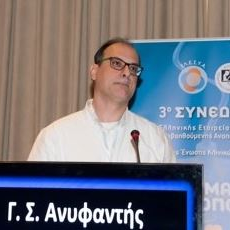
Assist. Prof. George Anifandis
Guest Editor

Manuscript Submission Information

Manuscripts should be submitted online at www.mdpi.com by registering and logging in to this website. Once you are registered, click here to go to the submission form. Manuscripts can be submitted until the deadline. All submissions that pass pre-check are peer-reviewed. Accepted papers will be published continuously in the journal (as soon as accepted) and will be listed together on the special issue website. Research articles, review articles as well as short communications are invited. For planned papers, a title and short abstract (about 100 words) can be sent to the Editorial Office for announcement on this website.

Submitted manuscripts should not have been published previously, nor be under consideration for publication elsewhere (except conference proceedings papers). All manuscripts are thoroughly refereed through a single-blind peer-review process. A guide for authors and other relevant information for submission of manuscripts is available on the Instructions for Authors page. Medicina is an international peer-reviewed open access monthly journal published by MDPI.

Please visit the Instructions for Authors page before submitting a manuscript. The Article Processing Charge (APC) for publication in this open access journal is 1800 CHF (Swiss Francs). Submitted papers should be well formatted and use good English. Authors may use MDPI's English editing service prior to publication or during author revisions.

Keywords

  • quality of embryos
  • spermatozoa
  • IVF/ICSI
  • molecular embryology
  • IVF success/failure
  • assisted oocyte activation
  • time-lapse
  • pre-implantation genetic diagnosis (PGD)
  • fertilization
  • ovarian stimulation
  • cryopreservation/vitrification
  • sperm/oocyte banking
  • fertility in oncology patients

Published Papers (12 papers)

Order results
Result details
Select all
Export citation of selected articles as:

Research

Jump to: Review, Other

11 pages, 523 KiB  
Article
Mature miR-99a Upregulation in the Amniotic Fluid Samples from Female Fetus Down Syndrome Pregnancies: A Pilot Study
by Anda-Cornelia Vizitiu, Danae Stambouli, Anca-Gabriela Pavel, Maria-Cezara Muresan, Diana Maria Anastasiu, Cristina Bejinar, Anda Alexa, Catalin Marian, Ioan Ovidiu Sirbu and Laurentiu Sima
Medicina 2019, 55(11), 728; https://doi.org/10.3390/medicina55110728 - 07 Nov 2019
Cited by 6 | Viewed by 2298
Abstract
Background and Objective: Although Down syndrome is the most frequent aneuploidy, its pathogenic molecular mechanisms are not yet fully understood. The aim of our study is to quantify—by qRT-PCR—the expression levels of both the mature forms and the pri-miRNAs of the microRNAs resident [...] Read more.
Background and Objective: Although Down syndrome is the most frequent aneuploidy, its pathogenic molecular mechanisms are not yet fully understood. The aim of our study is to quantify—by qRT-PCR—the expression levels of both the mature forms and the pri-miRNAs of the microRNAs resident on chromosome 21 (miR(21)) in the amniotic fluid samples from Down syndrome singleton pregnancies and to estimate the impact of the differentially expressed microRNAs on Down syndrome fetal heart and amniocytes transcriptomes. Materials and methods: We collected amniotic fluid samples harvested by trained obstetricians as part of the second trimester screening/diagnostic procedure for aneuploidies to assess the trisomy 21 status by QF-PCR and karyotyping. Next, we evaluated—by Taqman qRT-PCR—the expression levels of both the mature forms and the pri-miRNA precursors of the microRNAs resident on chromosome 21 in amniotic fluid samples from singleton Down syndrome and euploid pregnancies. Further, we combined miRWalk 3.0 microRNA target prediction with GEO DataSets analysis to estimate the impact of hsa-miR-99a abnormal expression on Down syndrome heart and amniocytes transcriptome. Results: We found a statistically significant up-regulation of the mature form of miR-99a, but not pri-miR-99a, in the amniotic fluid samples from Down syndrome pregnancies with female fetuses. GATHER functional enrichment analysis of miRWalk3.0-predicted targets from Down syndrome amniocytes and fetal hearts transcriptome GEODataSets outlined both focal adhesion and cytokine–cytokine receptor interaction signaling as novel signaling pathways impacted by miR-99a and associated with cardiac defects in female Down syndrome patients. Conclusions: The significant overexpression of miR-99a, but not pri-miR-99a, points towards an alteration of the post-transcriptional mechanisms of hsa-miR-99a maturation and/or stability in the female trisomic milieu, with a potential impact on signaling pathways important for proper development of the heart. Full article
(This article belongs to the Special Issue Clinical Embryology and Reproductive Medicine)
Show Figures

Figure 1

8 pages, 287 KiB  
Article
The Significance of Endometrial Scratching for Clinical Pregnancy Rate in Long Agonist and Antagonist Protocols
by Rimantas Gricius, Greta Balciuniene, Lina Jakubauskiene and Diana Ramasauskaite
Medicina 2019, 55(9), 527; https://doi.org/10.3390/medicina55090527 - 23 Aug 2019
Cited by 1 | Viewed by 1861
Abstract
Background and Objectives: Assisted reproductive techniques today have helped many infertile couples achieve their desired pregnancy. However, unsuccessful implantation is usually the key failure in in vitro fertilization cycles. Many factors are now being studied to improve the implantation rate, one being [...] Read more.
Background and Objectives: Assisted reproductive techniques today have helped many infertile couples achieve their desired pregnancy. However, unsuccessful implantation is usually the key failure in in vitro fertilization cycles. Many factors are now being studied to improve the implantation rate, one being endometrial artificial injury (endometrial scratching). The purpose of this study was to assess whether local endometrial artificial injury improved pregnancy rate, in long agonist and antagonist protocols. Materials and Methods: A retrospective analysis was carried out at the JSC Baltic American Clinic from January 1, 2016 to December 31, 2018. Women who were undergoing in vitro fertilization treatment were enrolled in the study. Medical data including demographic factors, menstrual history, and anamnesis of infertility were collected from medical records. Subjects were divided into intervention and control groups. The outcome of this study was clinical pregnancy rate (CPR). Results: A total of 137 women presenting with primary or secondary infertility were enrolled in the study. Clinical pregnancy was observed in 44/71 (61.9%) patients in the intervention group and 33/66 (50%) in the group without endometrial scratching (p value = 0.16). CPR in the intervention group was significantly higher in the patient group that had undergone ovarian stimulation with a long agonist protocol rather than the antagonist protocol (73.8% vs. 41.4%; p value = 0.006). In the multivariable logistic regression model, previously failed in vitro fertilization (IVF) (odds ratio (OR) = 0.07, 95% confidence interval (CI) 0.01–0.47), stimulation with the long agonist protocol (OR = 19.89, 95% CI 3.57–18.63), antagonist protocol (OR = 0.05, 95% CI 0.01–0.34), number of embryos (OR = 1.37, 95% CI 1.05–1.38), and number of blastocysts (OR = 2.96, 95% CI 1.43–8.36) remained important independent prognostic factors for clinical pregnancy. Conclusions: Our study findings indicate that randomized controlled studies are required to define patients for this procedure and to prove the effect and safety of local endometrial injury, before it is introduced to routine clinical practice. Full article
(This article belongs to the Special Issue Clinical Embryology and Reproductive Medicine)
16 pages, 8430 KiB  
Article
An Experimental Study to Evaluate the Protective Effects of Solanum lycopersicum Seed Essential Oil on Diabetes-Induced Testicular Injuries
by Javid Kermani, Nader Goodarzi and Mitra Bakhtiari
Medicina 2019, 55(8), 499; https://doi.org/10.3390/medicina55080499 - 19 Aug 2019
Cited by 12 | Viewed by 2795
Abstract
Background and Objectives: Diabetes is a chronic metabolic disorder that can effectively influences male reproductive performance. The present study was conducted to investigate the protective effects of Solanum lycopersicum essential oil (SL) on diabetes-induced testicular injuries. Materials and Methods: Adult male [...] Read more.
Background and Objectives: Diabetes is a chronic metabolic disorder that can effectively influences male reproductive performance. The present study was conducted to investigate the protective effects of Solanum lycopersicum essential oil (SL) on diabetes-induced testicular injuries. Materials and Methods: Adult male rats were randomly allocated into five groups (n = 8 in each group). 1: control; 2: diabetic; 3: diabetic + 30 mg/kg of SL essential oil; 4: diabetic + 90 mg/kg of SL essential oil; 5: diabetic + 270 mg/kg of SL essential oil extract. Diabetes was induced by a single dose of streptozotocin (55 mg/kg) intraperitoneally. Testicular changes were assessed quantitatively using stereological method followed by measuring antioxidant enzymes including catalase, superoxide dismutase, and glutathione peroxidase, and the serum testosterone level. Malondialdehyde (MDA) levels and Bcl-2expression were also evaluated in the tissue samples. Results: Diabetes resulted in significant deleterious changes in the structure of testicular tissue, suppressed antioxidant enzymes and testosterone levels, and increased lipid peroxidation. The expression of Bcl-2 was downregulated in diabetic testis and resulted in enhanced apoptosis. Following 8 weeks of treatment with SL essential oil, there were noticeable improvements in the structural changes of testis and the restoration of antioxidant defense and testosterone levels in testicular tissue, especially at higher doses. Conclusion: In conclusion, these findings reveal that the essential oil of Solanum lycopersicum has potent antioxidant properties and can attenuate the adverse effects of diabetes on male reproduction. Full article
(This article belongs to the Special Issue Clinical Embryology and Reproductive Medicine)
Show Figures

Figure 1

6 pages, 446 KiB  
Article
Human Papillomavirus and Infertility
by Vilma Jeršovienė, Živilė Gudlevičienė, Jolita Rimienė and Dalius Butkauskas
Medicina 2019, 55(7), 377; https://doi.org/10.3390/medicina55070377 - 15 Jul 2019
Cited by 32 | Viewed by 5205
Abstract
Background and objectives. Human papillomavirus (HPV) is the most commonly sexually transmitted infection. Recent evidence suggests that an HPV infection may affect fertility. The aim of the study was to determine the prevalence of HPV infections among couples undergoing in vitro fertilization (IVF) [...] Read more.
Background and objectives. Human papillomavirus (HPV) is the most commonly sexually transmitted infection. Recent evidence suggests that an HPV infection may affect fertility. The aim of the study was to determine the prevalence of HPV infections among couples undergoing in vitro fertilization (IVF) and to identify their awareness of HPV. Material and Methods. A total of 200 samples were collected from couples who received IVF treatment during 2017–2018 in Vilnius University Hospital Santaros Klinikos (VUH SK) Santaros Fertility Centre (SFC). For HPV detection, cervical swabs from women and sperm samples from men were taken and a real time polymerase chain reaction (RT-PCR) was used for the identification of 14 high-risk HPV types. Sperm parameters were evaluated according to World Health Organization (WHO) recommendations for 2010. Research subjects answered an anonymous questionnaire to ascertain their knowledge of HPV. Results. After testing of HPV in couples undergoing IVF, it was found that 33 out of 100 couples (33%) were HPV positive. Of these, 19% of women (19/100) and 20% of men (20/100) tested positive. Using Fisher’s exact test, a statistically significant difference was found between HPV infections and abnormal sperm quality parameters (p = 0.023). Conclusions. HPV may have an impact in spermatogenesis, because an HPV infection was more frequently detected in men with abnormal sperm parameters. High-risk HPV 52 was the most common genotype among couples undergoing IVF treatment. Full article
(This article belongs to the Special Issue Clinical Embryology and Reproductive Medicine)
Show Figures

Figure 1

10 pages, 288 KiB  
Article
Urotensin 2 and Oxidative Stress Levels in Maternal Serum in Pregnancies Complicated by Intrauterine Growth Restriction
by Esra Celik, Seyithan Taysi, Seyhun Sucu, Hasan Ulusal, Emin Sevincler and Ahmet Celik
Medicina 2019, 55(7), 328; https://doi.org/10.3390/medicina55070328 - 02 Jul 2019
Cited by 10 | Viewed by 2131
Abstract
Background and objectives: In this study, the aim was to investigate Urotensin 2 (U-II) levels and oxidant/antioxidant system parameters in pregnancies with intrauterine growth restriction (IUGR). Materials and Methods: A total of 36 healthy, pregnant women who had not been diagnosed with IUGR [...] Read more.
Background and objectives: In this study, the aim was to investigate Urotensin 2 (U-II) levels and oxidant/antioxidant system parameters in pregnancies with intrauterine growth restriction (IUGR). Materials and Methods: A total of 36 healthy, pregnant women who had not been diagnosed with IUGR and 36 pregnant women who had been diagnosed with IUGR at the Obstetrics and Gynecology Outpatient Clinic at Gaziantep University Hospital were enrolled in this study. The serum total antioxidant status (TAS), total oxidant status (TOS), thiol-disulfide levels, U-II measurements, and oxidative stress index (OSI) calculations were carried out at the biochemistry laboratory at Gaziantep University. Results: According to this study, there was no statistically significant difference between the group with IUGR and the control group of healthy, pregnant women in terms of total antioxidant status (TAS), total oxidant status (TOS), oxidative stress index (OSI), native thiol, total thiol, disulfide, disulfide/native thiol, disulfide/total thiol, native thiol/total thiol, and U-II values. There was, however, a positive linear correlation between TOS and total thiol levels in the group with IUGR (p = 0.021, r = 0.384), and a positive linear correlation between OSI and total thiol values in the control group (p = 0.049, r = 0.330). In addition, there was a negative correlation between disulfide levels and gestational weeks at birth in the group with IUGR (p = 0.027, r = 0.369). Conclusions: Consequently, there was no significant difference between the control group and the group with pregnancies complicated by idiopathic IUGR in terms of serum oxidant/antioxidant system parameters and U-II levels. It is necessary to conduct more extensive studies evaluating placental, maternal, and fetal oxidative stress in conjunction in order to investigate the role of oxidative stress in IUGR. Full article
(This article belongs to the Special Issue Clinical Embryology and Reproductive Medicine)
8 pages, 293 KiB  
Article
Cleavage Stage versus Blastocyst Stage Embryo Transfer in Oocyte Donation Cycles
by George Kontopoulos, Mara Simopoulou, Ioannis Zervomanolakis, Thomas Prokopakis, Kostas Dimitropoulos, Evaggelos Dedoulis, Stylianos Grigorakis, Kristi Agapitou, Eros Nikitos, Anna Rapani and Nikos Vlahos
Medicina 2019, 55(6), 293; https://doi.org/10.3390/medicina55060293 - 20 Jun 2019
Cited by 17 | Viewed by 3163
Abstract
Background and Objective: During the last few years, a trend has been noted towards embryos being transferred at the blastocyst stage, which has been associated with improved rates regarding implantation and clinical pregnancy in comparison to cleavage stage embryo transfers. There is a [...] Read more.
Background and Objective: During the last few years, a trend has been noted towards embryos being transferred at the blastocyst stage, which has been associated with improved rates regarding implantation and clinical pregnancy in comparison to cleavage stage embryo transfers. There is a limited number of studies investigating this notion in oocyte donation cycles employing cryopreserved embryos. The aim of this study is to evaluate the implantation potential and clinical pregnancy rates between the day 3 cleavage stage and blastocyst stage embryo transfers in oocyte donation cycles employing vitrified embryos. Methods: This is a retrospective evaluation of oocyte donation frozen–thawed transfers completed in our clinic from January 2017 to December 2017. Intracytoplasmic sperm injection was conducted for all oocytes. Following fertilization, all embryos were cryopreserved either at the cleavage or blastocyst stage. Embryo transfer of two embryos was performed under direct sonographic guidance in all cases. Results: Our results confirmed a 55.6% clinical pregnancy (CP) resulting from day 3 embryo transfers, a 68.8% CP from day 5, and 71.4% CP from day 6. Significantly improved pregnancy rates were related to embryo transfers at the blastocyst stage when compared to cleavage stage transfers (68.9% and 55.6% respectively, p = 0.016). The risk with regards to multiple pregnancies was similar. Conclusion: Our findings indicate that in oocyte donation cycles employing vitrified embryos, embryo transfer at the blastocyst stage is accompanied with a significant improvement in pregnancy rates and merits further investigation. Full article
(This article belongs to the Special Issue Clinical Embryology and Reproductive Medicine)
11 pages, 3244 KiB  
Article
Endometrial Vascularization Characterized by Optical Coherence Tomography and Immunohistochemistry in Women Undergoing In Vitro Fertilization-Embryo Transfer Treatment
by Tracy Sze Man Law, Wing Ching Cheung, Fangrong Wu, Ruizhe Zhang, Jacqueline Pui Wah Chung, Chi Chiu Wang, Xiaoyan Chen and Tin Chiu Li
Medicina 2019, 55(4), 81; https://doi.org/10.3390/medicina55040081 - 27 Mar 2019
Cited by 10 | Viewed by 3054
Abstract
Background and objective: Endometrial angiogenesis is a prerequisite for successful pregnancy. Optical coherence tomography (OCT) is a non-invasive physically optical imaging technique widely used in ophthalmology and cardiology. However, there is no study using OCT to evaluate endometrium. The aim of this study [...] Read more.
Background and objective: Endometrial angiogenesis is a prerequisite for successful pregnancy. Optical coherence tomography (OCT) is a non-invasive physically optical imaging technique widely used in ophthalmology and cardiology. However, there is no study using OCT to evaluate endometrium. The aim of this study was to use OCT and traditionally histological methods to investigate endometrial vascularization in women undergoing in vitro fertilization-embryo transfer (IVF-ET) treatment and to determine the association with the pregnancy outcome. Methods: A total of 47 women were included in this study. OCT was used to assess endometrial vascularization by determining the high signal areas precisely on the seventh day after luteinizing hormone surge in non-conception natural cycles. Endometrial biopsies were obtained following OCT and immunohistochemistry was used to determine micro vessel and expression of vascular endothelial growth factor-A (VEGF-A) in the luminal epithelium, glandular epithelium and stroma, separately. Micro vessel counting was performed and the result was expressed as micro vessel density (MVD). A semi-quantitative H-score was used to determine the staining intensity of VEGF-A. Results: In women who successfully conceived after embryo transfer, the proportion of extensive high signal area in the uterine body detected by OCT (80%, 8/10), MVD (median number of micro vessels/mm2 of 10, range 4–17) and stromal expression of VEGF-A (median H-score of 189, range 72–395) were found to be significantly higher than those of women who did not conceive after embryo transfer in the subsequent IVF-ET treatment (OCT: 30%, 3/10; MVD: median number of micro vessels/mm2 of 7, range 4–10; VEGF-A: median H-score of 125, range 86–299, respectively). In addition, a significantly higher stromal expression of VEGF-A (median H-score of 196, range 84–395) and MVD (median number of micro vessels/mm2 of 9, range 5–16) was found in women with extensive high signal area in uterine body, compared to those with focal or no high signal area (stromal VEGF-A: median H-score of 135, range 92–302; MVD: number of micro vessels/mm2 of 6, range 4-11). Conclusions: Both immunohistochemistry and OCT demonstrated significant difference in vascularization of the peri-implantation endometrium between subjects who did and did not conceive after IVF-ET treatment. Our findings also suggest OCT appears to be a promising non-invasive or minimally invasive alternative to study endometrial vascularity in women with reproductive failure. Full article
(This article belongs to the Special Issue Clinical Embryology and Reproductive Medicine)
Show Figures

Figure 1

14 pages, 869 KiB  
Article
Evaluation of the Second Follicular Wave Phenomenon in Natural Cycle Assisted Reproduction: A Key Option for Poor Responders through Luteal Phase Oocyte Retrieval
by Konstantinos Sfakianoudis, Mara Simopoulou, Evangelos Maziotis, Polina Giannelou, Petroula Tsioulou, Anna Rapani, Agni Pantou, Konstantina Petroutsou, Irene Angeli, Efthymios Deligeoroglou, Michael Koutsilieris and Konstantinos Pantos
Medicina 2019, 55(3), 68; https://doi.org/10.3390/medicina55030068 - 14 Mar 2019
Cited by 13 | Viewed by 4692
Abstract
Background: Emergence of Luteal Phase Oocyte Retrieval (LuPOR) may revolutionize the practice regarding the time-sensitive nature of poor responders ascertaining a higher number of oocytes, in a shorter amount of time. This may be especially important in view of employing the approach [...] Read more.
Background: Emergence of Luteal Phase Oocyte Retrieval (LuPOR) may revolutionize the practice regarding the time-sensitive nature of poor responders ascertaining a higher number of oocytes, in a shorter amount of time. This may be especially important in view of employing the approach of natural cycles for Poor Responders. We suggest the acronym LuPOR describing the clinical practice of luteal phase oocyte retrieval. The aim of the study is to offer insight regarding the identity of LuPOR, and highlight how this practice may improve management of the special subgroup of poor responders. Materials and Methods: The present retrospective observational clinical study includes the collection and statistical analysis of data from 136 poor responders who underwent follicular oocyte retrieval (FoPOR) and subsequent LuPOR in natural cycles, during their In Vitro Fertilization (IVF) treatment, from the time period of 2015 to 2018. All 136 participants were diagnosed with poor ovarian reserve (POR) according to Bologna criteria. The 272 cycles were categorized as follows: 136 natural cycles with only FoPORs (Control Group) and 136 natural cycles including both FoPORs and LuPORs. Results: Our primary results indicate no statistically significant differences with regards to the mean number of oocytes, the maturation status, and fertilization rate between FoPOR and LuPOR in natural cycles. Secondarily, we demonstrate a statistically significant higher yield of oocytes (2.50 ± 0.78 vs. 1.25 ± 0.53), better oocyte maturity status (1.93 ± 0.69 vs. 0.95 ± 0.59) and higher fertilization rate (1.31 ± 0.87 vs. 0.61 ± 0.60) in natural cycles including both FoPOR and LuPOR, when compared to cycles including only FoPOR. Conclusion: Our study may contribute towards the establishment of an efficient poor responders’ management through the natural cycle approach, paving a novel clinical practice and ascertaining the opportunity to employ oocytes and embryos originating from a luteal phase follicular wave. Full article
(This article belongs to the Special Issue Clinical Embryology and Reproductive Medicine)
Show Figures

Figure 1

9 pages, 4842 KiB  
Article
Changes in Morphology and Presence of Pinopodes in Endometrial Cells during the Luteal Phase in Women with Infertility Problems: A Pilot Study
by Marina Aunapuu, Piret Kibur, Tõnu Järveots and Andres Arend
Medicina 2018, 54(5), 69; https://doi.org/10.3390/medicina54050069 - 10 Oct 2018
Cited by 16 | Viewed by 3525
Abstract
Objective: To investigate morphological changes in the endometrial epithelial cells of patients with infertility problems. Materials and methods: Endometrial biopsies were obtained from 10 women who have undergone several unsuccessful in vitro fertilisation (IVF) procedures. Endometrial biopsies were performed between luteinizing hormone surge [...] Read more.
Objective: To investigate morphological changes in the endometrial epithelial cells of patients with infertility problems. Materials and methods: Endometrial biopsies were obtained from 10 women who have undergone several unsuccessful in vitro fertilisation (IVF) procedures. Endometrial biopsies were performed between luteinizing hormone surge days LH+6 to +10 of the natural menstrual cycle. Each sample was divided into three parts, which were processed for histological, transmission (TEM), and scanning electron microscopy (SEM) investigations. Results: Histological investigations demonstrated significant alterations in the apical part of epithelial cells of one patient; in four patients, the gland maturity was low, not matching the cycle day, and thus a phase lag had developed. By TEM examination, we ascertained changes in secretory and ciliated cells in three patients (decreased amount or missing microvilli, irregular cilia in ciliated cells). SEM examination found pinopodes in five patients: three samples contained fully developed pinopodes—larger and completely smooth, with only some wrinkles; one sample contained regressing small pinopodes, with wrinkled surfaces; and one sample had both developed and regressing pinopodes. Conclusions: To conclude, our study shows that the endometrium of patients with poor IVF outcome has either significant changes in the morphology or the endometrial maturation is inhibited and a phase lag often develops. Our study shows that endometrial pinopodes are found throughout the mid-luteal phase up to day LH+10. Full article
(This article belongs to the Special Issue Clinical Embryology and Reproductive Medicine)
Show Figures

Figure 1

Review

Jump to: Research, Other

11 pages, 1439 KiB  
Review
Association between the FAS/FASL Variants and Risk of Male Infertility in Asian Populations; A Systematic Review and Meta-Analysis
by Rezvan Asgari, Kamran Mansouri, Mitra Bakhtiari, Hadi Mozafari and Shiva Roshankhah
Medicina 2019, 55(6), 247; https://doi.org/10.3390/medicina55060247 - 05 Jun 2019
Cited by 3 | Viewed by 2394
Abstract
Background and Objectives: Studies suggest that FAS/FASL polymorphisms are associated with male infertility; however, their results are still inconclusive. Therefore, this systematic review and meta-analysis aimed to summarize and clarify the overall association of FAS/FASL polymorphisms and risk of male infertility. Materials [...] Read more.
Background and Objectives: Studies suggest that FAS/FASL polymorphisms are associated with male infertility; however, their results are still inconclusive. Therefore, this systematic review and meta-analysis aimed to summarize and clarify the overall association of FAS/FASL polymorphisms and risk of male infertility. Materials and Methods: Our search was conducted on the databases of Science Direct, PubMed and Google Scholar. For performing the meta-analysis, pooled odds ratio (OR) values with 95% confidence interval (CI) was applied in order to analyze the strength of association between the FAS/FASL polymorphisms and risk of male infertility. A total of seven relevant studies published up to September 2018 were considered. Results: FASL-844C/T genotype results of 559 patients and 623 healthy individuals were included in our study. For FAS-670A/G genotype effect, 751 patients and 821 healthy individuals were explored. Results showed that all analysis models including dominant, recessive and allelic models of FASL-844C/T and FAS-670A/G polymorphism had no significant effect on infertility in men (p > 0.05 and p > 0.05, respectively). According to sensitivity analysis, our results were stable. Conclusion: We demonstrated that FAS/FASL polymorphisms might not be an effective factor on male reproductive health. For precise determination of FAS/FASL polymorphisms effects on male infertility, large-scale case-control studies should be performed. Full article
(This article belongs to the Special Issue Clinical Embryology and Reproductive Medicine)
Show Figures

Figure 1

19 pages, 1592 KiB  
Review
Postponing Pregnancy Through Oocyte Cryopreservation for Social Reasons: Considerations Regarding Clinical Practice and the Socio-Psychological and Bioethical Issues Involved
by Mara Simopoulou, Konstantinos Sfakianoudis, Panagiotis Bakas, Polina Giannelou, Christina Papapetrou, Theodoros Kalampokas, Anna Rapani, Ekaterini Chatzaki, Maria Lambropoulou, Chrysoula Lourida, Efthymios Deligeoroglou, Konstantinos Pantos and Michael Koutsilieris
Medicina 2018, 54(5), 76; https://doi.org/10.3390/medicina54050076 - 25 Oct 2018
Cited by 12 | Viewed by 6348
Abstract
Oocyte freezing for ‘social reasons’ refers to women of reproductive age who are aiming to prolong, protect and secure their fertility. The term emerged to describe application of the highly promising technique, namely vitrification on oocytes retrieved through controlled ovarian stimulation (COS) from [...] Read more.
Oocyte freezing for ‘social reasons’ refers to women of reproductive age who are aiming to prolong, protect and secure their fertility. The term emerged to describe application of the highly promising technique, namely vitrification on oocytes retrieved through controlled ovarian stimulation (COS) from women intending to preserve their fertility for social reasons. These women opt to cryopreserve their oocytes at a point in their life when they need to postpone childbearing on the grounds of so called ‘social’ reasons. These reasons may include a highly driven career, absence of an adequate partner, financial instability, or personal reasons that make them feel unprepared for motherhood. This is a sensitive and multifaceted issue that entails medical, bioethical and socio-psychological components. The latest trend and the apparent increase noted on oocyte freezing for ‘social reasons’ has prompted our team of fertility specialists, embryologists, obstetricians, gynecologists and psychologists to proceed with a thorough, critical and all-inclusive comprehensive analysis. The wide range of findings of this analysis involve concerns of embryology and epigenetics that shape decisions made in the IVF laboratory, issues regarding obstetric and perinatal concerns on the pregnancy concluding from these oocytes and the respective delivery management and neonatal data, to the social and bioethical impact of this trend’s application. This literature review refers to matters rising from the moment the ‘idea’ of this option is ‘birthed’ in a woman’s thoughts, to proceeding and executing it clinically, up until the point of the pediatric follow up of the children born. We aim to shed light to the controversial issue of oocyte freezing, while objectively exhibit all aspects regarding this complex matter, as well as to respectfully approach how could the prospect of our future expectations be shaped from the impact of its application. Full article
(This article belongs to the Special Issue Clinical Embryology and Reproductive Medicine)
Show Figures

Figure 1

Other

Jump to: Research, Review

6 pages, 10403 KiB  
Case Report
Case Report of Misleading Features of a Rare Sertoli Cell Testicular Tumor
by Marius Anglickis, Rokas Stulpinas, Giedrė Anglickienė, Justinas Gabrilevičius and Arūnas Jaškevičius
Medicina 2019, 55(5), 170; https://doi.org/10.3390/medicina55050170 - 20 May 2019
Cited by 1 | Viewed by 2612
Abstract
Testicular Sertoli cell tumors are extremely rare. Generally, they are benign neoplasms, which belong to a group called sex cord–stromal tumors. In this article, we present a case report of a Sertoli cell tumor, which was accidentally discovered during a urological consultation of [...] Read more.
Testicular Sertoli cell tumors are extremely rare. Generally, they are benign neoplasms, which belong to a group called sex cord–stromal tumors. In this article, we present a case report of a Sertoli cell tumor, which was accidentally discovered during a urological consultation of a 42-year-old male. An ultrasound showed a 2.1 × 2.2 cm hypoechogenic, hypervascular tumor in the middle third of the left testicle. Serum tumor markers (α-fetoprotein, alkaline phosphatase, β-human chorionic gonadotropin, and lactic dehydrogenase) were all within the normal range. Rapid microscopic evaluation of fresh frozen sections during the operation was inconclusive, which led to a decision not to perform a radical orchiectomy immediately. On formalin-fixed paraffin-embedded (FFPE) sections, the tumor histology showed atypical patterns, and immunohistochemical analysis was performed in order to determine the type of neoplasm and differentiate it from other types of testicular tumors, so as to assign the further course of treatment. Radical inguinal orchiectomy was performed. The final pathology report showed a tumor with no predictive signs of aggressive behavior, which most closely resembled a Sertoli cell tumor. Full article
(This article belongs to the Special Issue Clinical Embryology and Reproductive Medicine)
Show Figures

Figure 1

Back to TopTop