


Int. J. Mol. Sci. 2022, 23(16), 9277; https://doi.org/10.3390/ijms23169277 - 17 Aug 2022
Cited by 10 | Viewed by 2151
Abstract
►
Show Figures
Electromagnetic pulse (EMP) radiation was reported to be harmful to hippocampal neurons. However, the mechanism underlying EMP-induced neuronal damage remains unclear. In this paper, for the first time, we attempted to investigate the involvement of ferroptosis in EMP-induced neuronal damage and its underlying
[...] Read more.
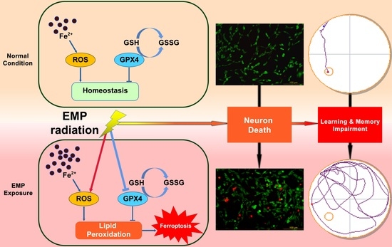
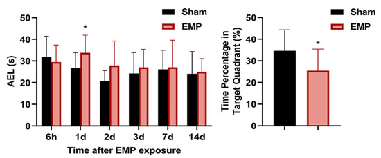
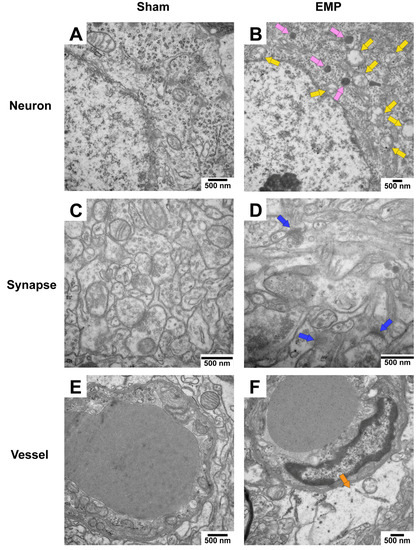
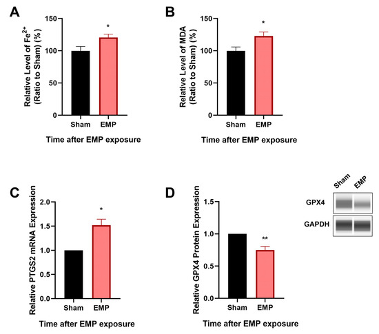
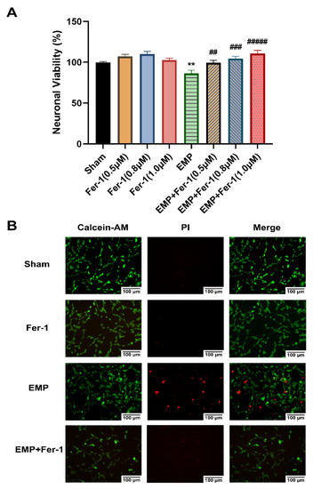
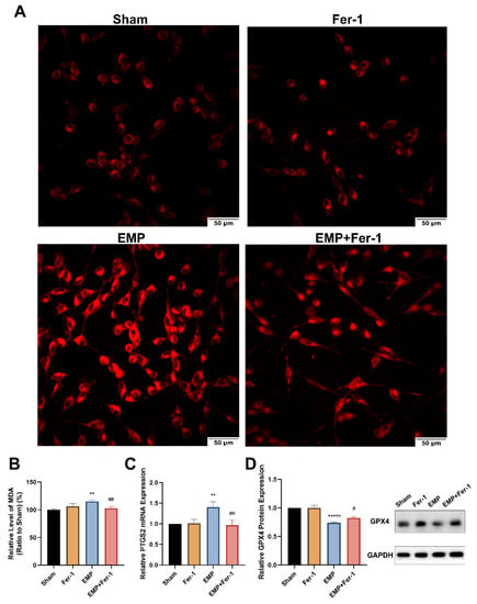
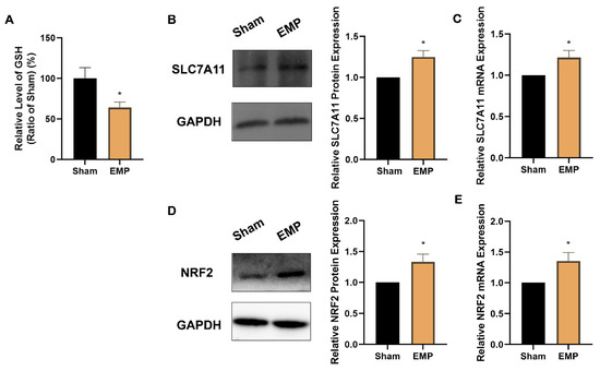
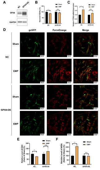
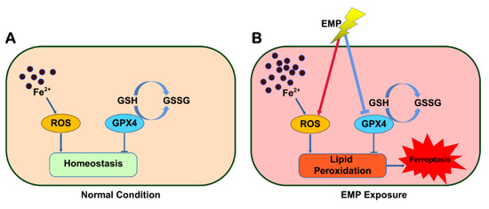
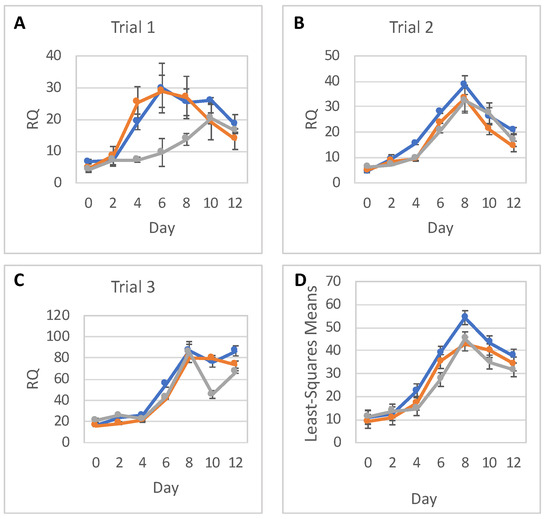
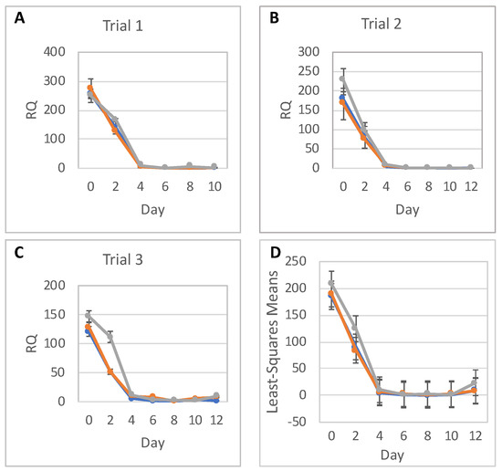
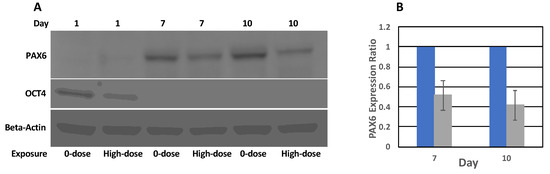
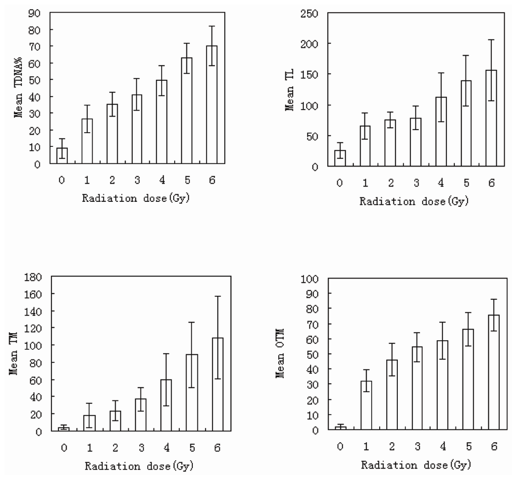
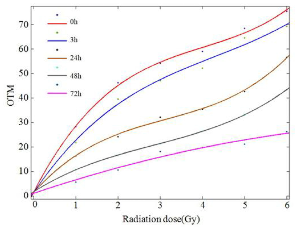
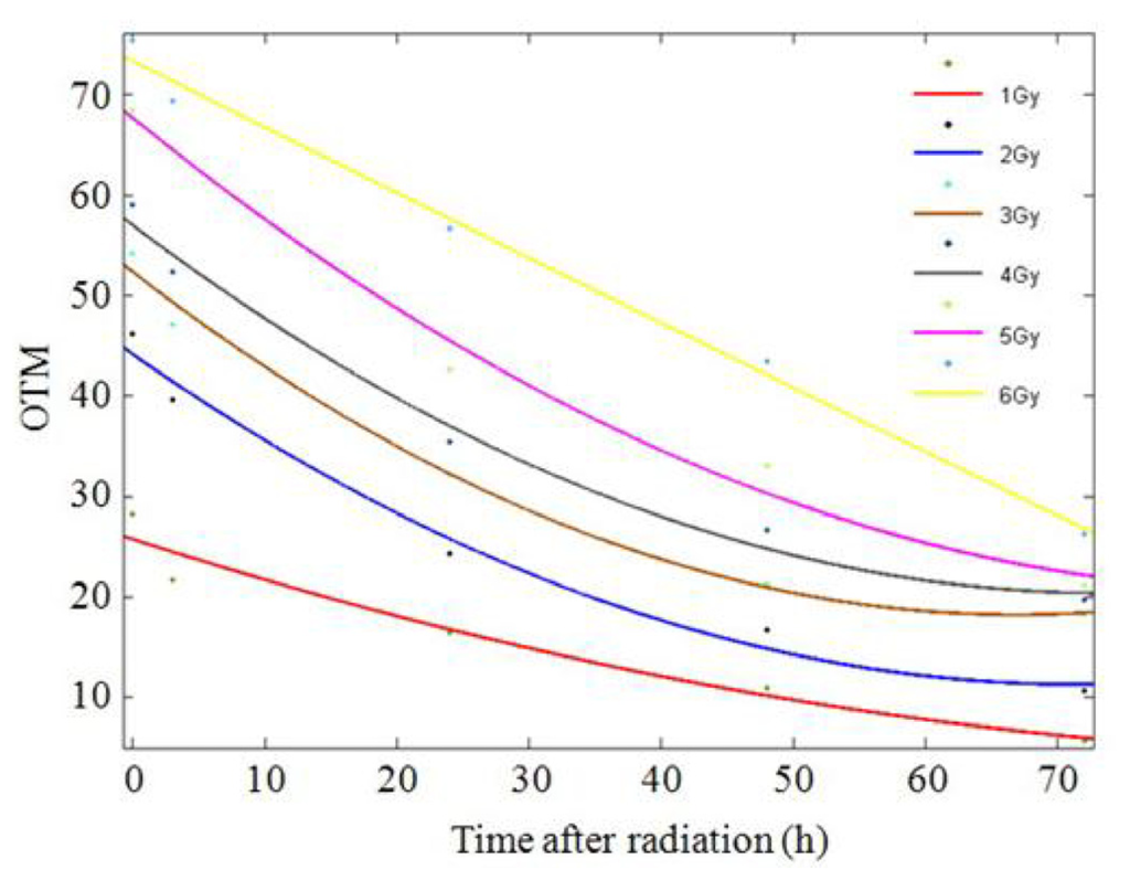
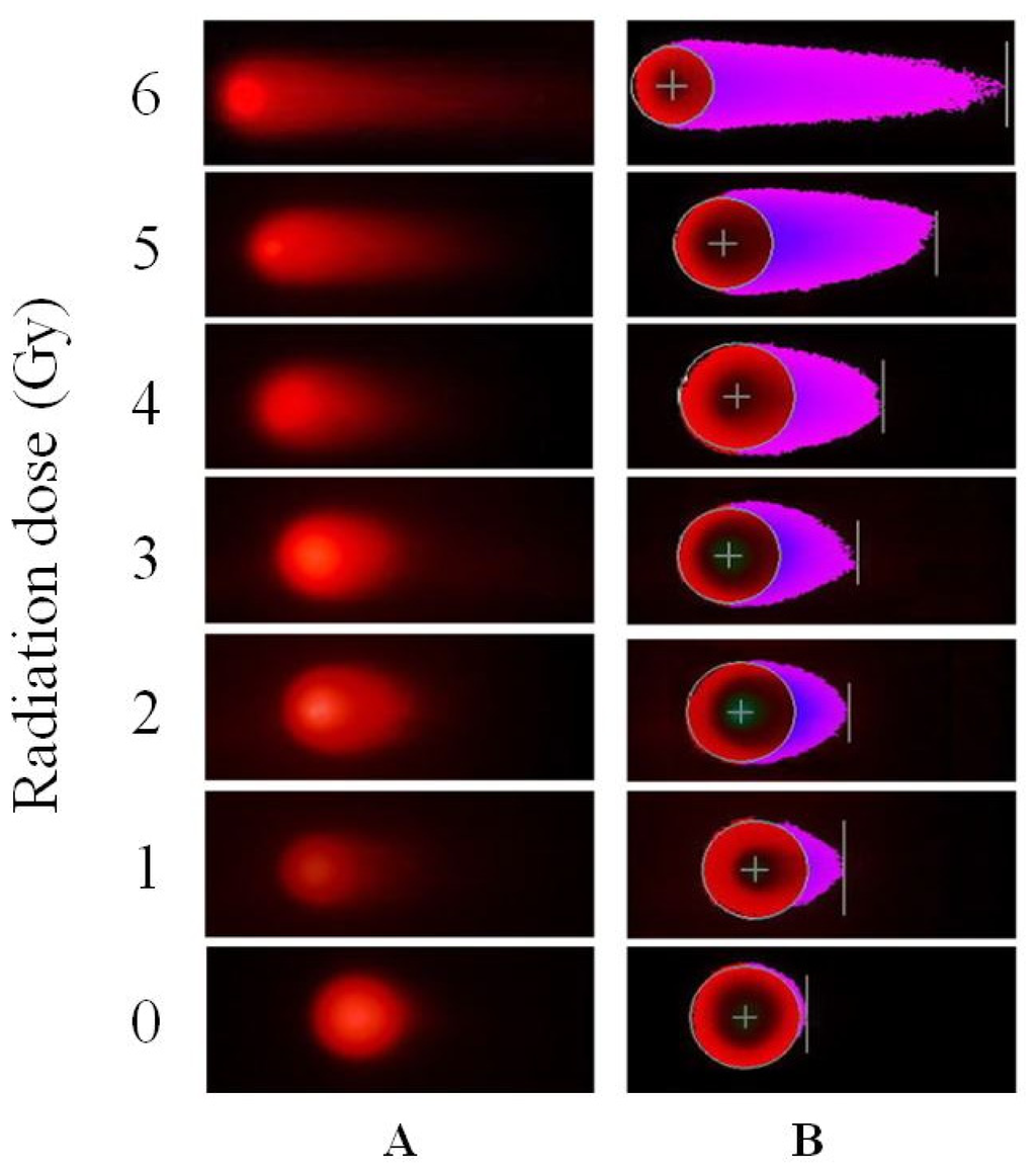
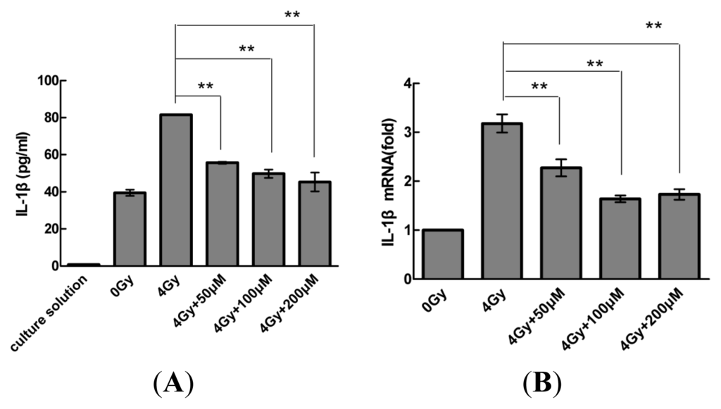
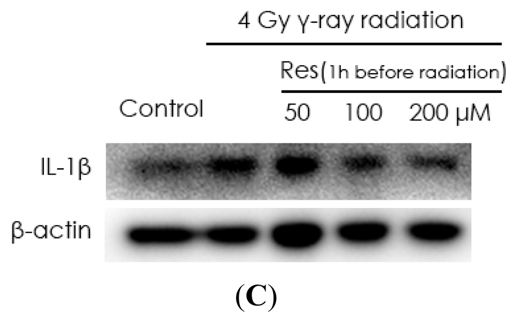
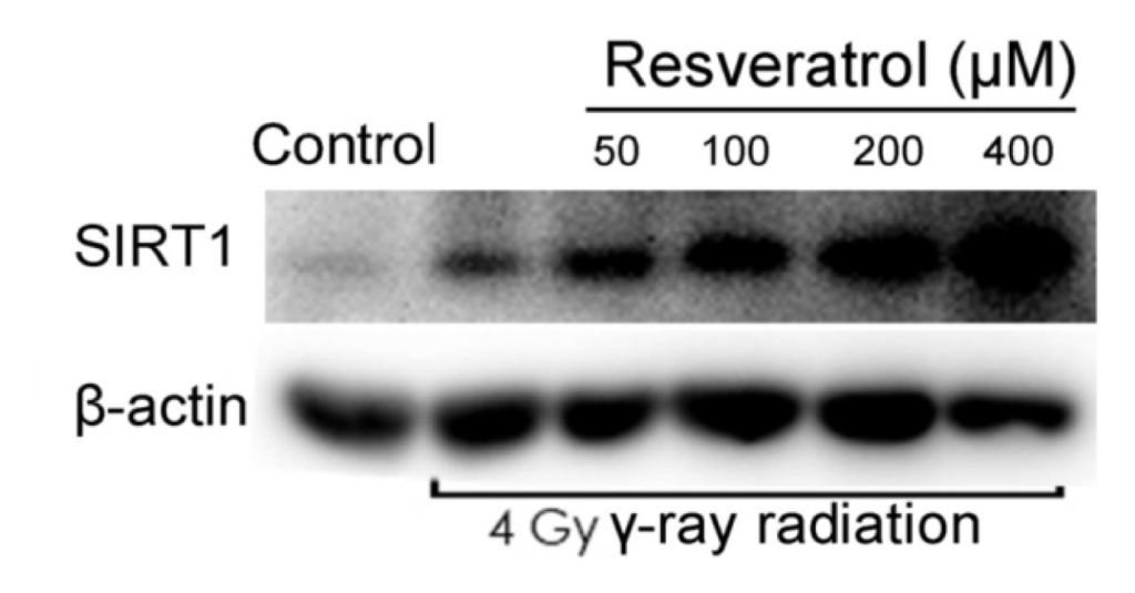
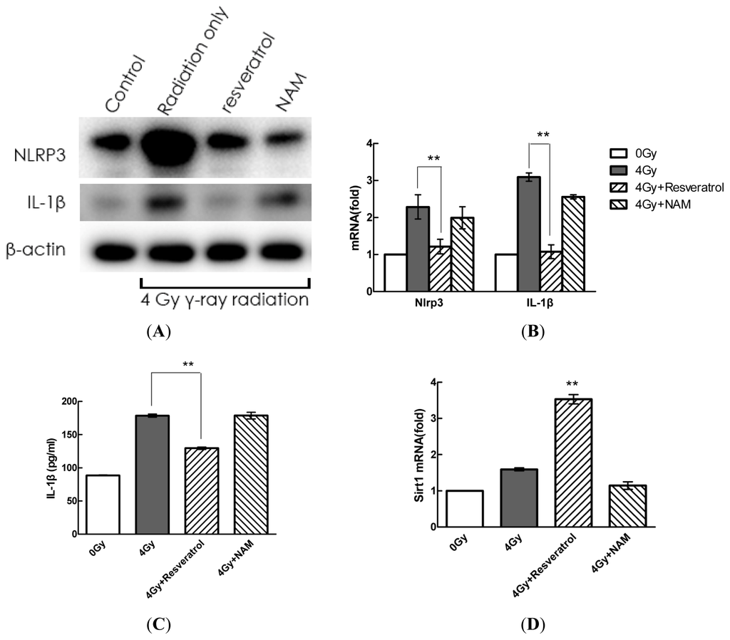
Electromagnetic pulse (EMP) radiation was reported to be harmful to hippocampal neurons. However, the mechanism underlying EMP-induced neuronal damage remains unclear. In this paper, for the first time, we attempted to investigate the involvement of ferroptosis in EMP-induced neuronal damage and its underlying mechanism. In vivo studies were conducted with a rat model to examine the association of ferroptosis and EMP-induced hippocampal neuronal damage. Moreover, in vitro studies were conducted with HT22 neurons to investigate the underlying mechanism of EMP-induced neuronal ferroptosis. In vivo results showed that EMP could induce learning and memory impairment of rats, ferroptotic morphological damages to mitochondria, accumulation of malonaldehyde (MDA) and iron, overexpression of prostaglandin-endoperoxide synthase 2 (PTGS2) mRNA, and downregulation of GPX4 protein in rat hippocampus. In vitro results showed that EMP could induce neuronal death, MDA accumulation, iron overload, PTGS2 overexpression, and GPX4 downregulation in HT22 neurons. These adverse effects could be reversed by either lipid peroxides scavenger ferrostatin-1 or overexpression of GPX4. These results suggest that EMP radiation can induce ferroptosis in hippocampal neurons via a vicious cycle of lipid peroxides accumulation and GSH/GPX4 axis downregulation. Lipid peroxides and the GSH/GPX4 axis provide potential effective intervention targets to EMP-induced hippocampal neuronal damage.
Full article
Graphical abstract


