Advances in Dental Materials: A Look inside Digital Workflows

A special issue of Applied Sciences (ISSN 2076-3417). This special issue belongs to the section "Applied Dentistry and Oral Sciences".

Deadline for manuscript submissions: closed (10 June 2023) | Viewed by 61047

Special Issue Editor


E-Mail Website
Guest Editor
Department of Surgical Sciences, Dental School, University of Turin, 10124 Turin, Italy
Interests: ental materials and prosthetic technologies; restorative dentistry; adhesion; oral diseases; aesthetic dentistry; endodontics; teeth; periodontics; dentistry; adhesives; operative dentistry; dental materials; composite resins; dental caries; clinical dentistry; esthetic dentistry; dental education
Special Issues, Collections and Topics in MDPI journals

Special Issue Information

Dear Colleagues,

In recent years, we have witnessed an increasingly rapid and decisive affirmation of digital dentistry, which has seen an increase in its quality levels and, consequently, in its fields of application. On the basis of this evolution, there is a constant development of materials related to digital dentistry, which are giving dentistry other ways of seeing and thinking about treatments.

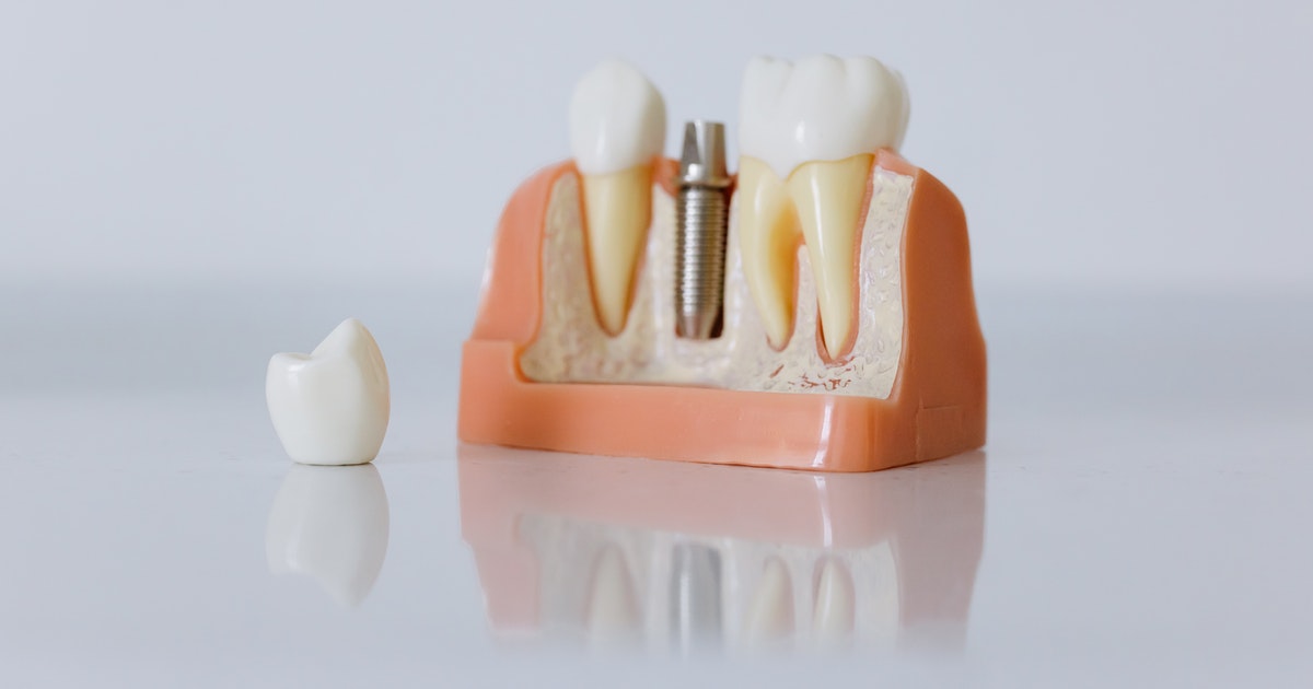
This Special Issue will focus on dental materials and techniques that revolve around digital workflows. What are the latest developments in CAD-CAM hybrid materials? Do we have reliable protocols for the cementation of the latest generation of zirconia? Which adhesive approaches allow us to reliably lute CAD-CAM materials? What stage have we reached on surgical guides and implant-prosthetic rehabilitations? What do we know about fiber-reinforced resinous materials or the reliability and precision of 3D printing in oral rehabilitations? Can intraoral scanner completely substitute “analogical” impressions?

Many questions require updated answers in this field: scientific research often finds itself chasing digital progress due to the rapid evolution of everything that revolves around the fascinating and risky digital dentistry.

Prof. Dr. Nicola Scotti
Guest Editor

Manuscript Submission Information

Manuscripts should be submitted online at www.mdpi.com by registering and logging in to this website. Once you are registered, click here to go to the submission form. Manuscripts can be submitted until the deadline. All submissions that pass pre-check are peer-reviewed. Accepted papers will be published continuously in the journal (as soon as accepted) and will be listed together on the special issue website. Research articles, review articles as well as short communications are invited. For planned papers, a title and short abstract (about 100 words) can be sent to the Editorial Office for announcement on this website.

Submitted manuscripts should not have been published previously, nor be under consideration for publication elsewhere (except conference proceedings papers). All manuscripts are thoroughly refereed through a single-blind peer-review process. A guide for authors and other relevant information for submission of manuscripts is available on the Instructions for Authors page. Applied Sciences is an international peer-reviewed open access semimonthly journal published by MDPI.

Please visit the Instructions for Authors page before submitting a manuscript. The Article Processing Charge (APC) for publication in this open access journal is 2400 CHF (Swiss Francs). Submitted papers should be well formatted and use good English. Authors may use MDPI's English editing service prior to publication or during author revisions.

Keywords

  • digital dentistry
  • CAD-CAM
  • luting
  • implant-supported prosthodontics
  • intraoral scanner
  • 3D printing
  • fiber-supported resins
  • adhesive system

Published Papers (20 papers)

Order results
Result details
Select all
Export citation of selected articles as:

Research

Jump to: Review, Other

12 pages, 1807 KiB  
Article
Reliability of Polyetherketoneketone as Definitive Implant-Supported Bridges in the Posterior Region—An In Vitro Study of the Ultimate Fracture Load and Vertical Marginal Discrepancy after Artificial Aging
by Surakit Visuttiwattanakorn, Apitchaya Suthamwat, Somchai Urapepon and Sirichai Kiattavorncharoen
Appl. Sci. 2022, 12(22), 11454; https://doi.org/10.3390/app122211454 - 11 Nov 2022
Cited by 1 | Viewed by 954
Abstract
Purpose: This study aims to investigate the ultimate fractural strength and marginal integrity of a three-unit implant-supported fixed partial denture (FPD) framework fabricated of polyetherketoneketone (PEKK) after simulated five-year clinical aging. Materials and Methods: Twelve FPD frameworks were milled (n = 6 per [...] Read more.
Purpose: This study aims to investigate the ultimate fractural strength and marginal integrity of a three-unit implant-supported fixed partial denture (FPD) framework fabricated of polyetherketoneketone (PEKK) after simulated five-year clinical aging. Materials and Methods: Twelve FPD frameworks were milled (n = 6 per group). All experimental frameworks were cemented on identical stainless-steel abutment models and subjected to five years of clinically simulated thermomechanical aging. The vertical marginal gap values were analyzed using a scanning electron microscope before and after being subjected to each aging condition. A universal testing machine was used to evaluate the ultimate fracture load. Results: A significant increase in marginal gap values of the PEKK group was observed after five years of simulation aging (p < 0.001), while no significant difference was seen in the titanium group (p = 0.071). After thermocycling, the PEKK group showed a higher statistically significant mean marginal gap value (84.99 + 44.28 μm) than before (81.75 + 44.53 μm). The titanium group exhibited superior mechanical strength, with a fracture load significantly higher than that of the PEKK group (3050 + 385.30 and 1359.14 + 205.49 N, respectively). Conclusions: Thermocycling affects the marginal gap discrepancy of PEKK restoration. However, the mean vertical marginal gap values in PEKK and titanium groups after a five-year clinical aging simulation were clinically acceptable. The ultimate fracture load values were higher than the maximum bite force reported in the posterior region. Thus, PEKK could serve as a suitable alternative material to metal in the framework of fixed dental prostheses. Full article
(This article belongs to the Special Issue Advances in Dental Materials: A Look inside Digital Workflows)
Show Figures

Figure 1

11 pages, 1169 KiB  
Article
Assessment of Communication Quality through Work Authorization between Dentists and Dental Technicians in Fixed and Removable Prosthodontics
by Hadiqa Afzal, Naseer Ahmed, Abhishek Lal, Khulud A. Al-Aali, Mohammed Alrabiah, Mai M. Alhamdan, Ahmed Albahaqi, Abdulaziz Sharaf, Fahim Vohra and Tariq Abduljabbar
Appl. Sci. 2022, 12(12), 6263; https://doi.org/10.3390/app12126263 - 20 Jun 2022
Cited by 3 | Viewed by 2801
Abstract
Fabrication of a clinically successful dental prosthesis requires clear and effective communication between dentists and dental technicians. Adequate completion of work authorization by dentists provides a means for increased professional quality assurance and satisfaction in dental prosthesis. The aim of this study was [...] Read more.
Fabrication of a clinically successful dental prosthesis requires clear and effective communication between dentists and dental technicians. Adequate completion of work authorization by dentists provides a means for increased professional quality assurance and satisfaction in dental prosthesis. The aim of this study was to evaluate the quality of communication between dentists and dental technicians via work authorization for fixed and removable dental prosthesis in Pakistan. This cross-sectional study was conducted for a period of 6 months to assess the quality of communication between dentists and dental technicians. A well-constructed questionnaire regarding work authorization of removable and fixed prostheses was used as a tool to collect data from 453 dentists. A linear regression analysis was performed to evaluate the relationship of the independent and dependent variables. A p value of ≤0.05 was considered statistically significant. It was observed that 92.1% of the dentists leave removable partial denture design to the dental technicians. About 56.5% of the dentists indicated information about base retention of partial dentures in work authorization. The majority of the dentists (56.5%) mentioned the material to be used for the construction of casting partial dentures. Nearly 66% of the dentists indicated the design of margins, and 44.6% of the dentists did not select the shade for fixed prostheses. Nearly 43% of the dentists did not draw the design of restoration, 21.9% chose to draw the design on paper, and 32.2% of the dentists drew the design on the cast. Meanwhile, 76.8% of the participants chose to directly communicate with their dental technicians personally. Nearly 41% of the dentists were satisfied with the restoration design, and 13.5% of the participants were not satisfied with the designs. Poor communication between dentists and technicians was observed, as the majority of the design decisions were left to the dental technicians. Therefore, communication between dentists and dental technicians should be improved by conducting combined workshops for the successful construction of dental prostheses. Full article
(This article belongs to the Special Issue Advances in Dental Materials: A Look inside Digital Workflows)
Show Figures

Figure 1

9 pages, 2237 KiB  
Article
Accuracy of Additively Manufactured Dental Casts Compared with That of Virtual Scan Data Obtained with an Intraoral Scanner: An In Vitro Study
by Gi Youn Kim, Jiayi Li, Hong Seok Moon and Kyung Chul Oh
Appl. Sci. 2022, 12(9), 4348; https://doi.org/10.3390/app12094348 - 25 Apr 2022
Cited by 1 | Viewed by 1321
Abstract
The study aimed to evaluate the time-related accuracy of additively manufactured dental casts and to compare it with scan data obtained with an intraoral scanner in vitro. Twenty-eight markers were attached to a set of dentiforms as reference model, and the distances between [...] Read more.
The study aimed to evaluate the time-related accuracy of additively manufactured dental casts and to compare it with scan data obtained with an intraoral scanner in vitro. Twenty-eight markers were attached to a set of dentiforms as reference model, and the distances between the markers were measured using a digital caliper. An intraoral scanner was used to obtain the virtual scan data of the reference model with a total of 30 scans per arch. The distances between markers were measured using a three-dimensional inspection software for all scans (group IOS). Scan data were additively manufactured using a 3D printer, and the distances between markers were measured as in the reference model immediately after post-polymerization (group PPIA), 1 day (group PP1D), 7 days (group PP7D), and 30 days after post-polymerization (group PP30D). The linear deviation in group IOS was 199.74 ± 11.14 μm, PPIA was 242.88 ± 49 μm, PP1D was 259.9 ± 42.59 μm, PP7D was 289.82 ± 39.74 μm, and PP30D was 315.8 ± 33.28 μm, in comparison with the reference model, with significant differences among all groups (all p < 0.05). When additively manufacturing casts from scan data to verify the quality of dental prostheses designed virtually, the prostheses should be adapted to casts manufactured within one week. Full article
(This article belongs to the Special Issue Advances in Dental Materials: A Look inside Digital Workflows)
Show Figures

Figure 1

13 pages, 2203 KiB  
Article
Accuracy of 3D-Printed Master Cast Workflow Using a Digital Light Processing Printer
by Saskia Berndt, Hannah Herstell, Stefan Raith, Christina Kühne and Sven Reich
Appl. Sci. 2022, 12(5), 2619; https://doi.org/10.3390/app12052619 - 03 Mar 2022
Cited by 2 | Viewed by 2192
Abstract
This in vitro study was designed to investigate whether conventionally produced casts and printed casts for prosthodontic purposes show comparable full-arch accuracy; a ceramic reference cast with inlay and crown preparations was fabricated. Ten gypsum casts were fabricated from conventional silicone elastomeric impressions. [...] Read more.
This in vitro study was designed to investigate whether conventionally produced casts and printed casts for prosthodontic purposes show comparable full-arch accuracy; a ceramic reference cast with inlay and crown preparations was fabricated. Ten gypsum casts were fabricated from conventional silicone elastomeric impressions. Ten digital impressions [IOS] of the reference cast were obtained by an intraoral scanner to fabricate 3D-printed resin casts. The ceramic reference cast, the gypsum, and the printed casts were digitized by an industrial structured light scanner (ILS) and provided as stl files. To evaluate absolute mean trueness values, the digitized gypsum casts [CON], digitized printed casts [PRINT], and [IOS] were superimposed with the digitized ceramic reference cast [REF]. Additionally, each [IOS] scan was compared with its corresponding [PRINT]. The precision was calculated for [CON], [IOS], and [PRINT]. The Mann–Whitney U test for independent samples and the Wilcoxon test for connected samples were performed (p ≤ 0.05). As absolute mean deviation trueness values were obtained: 69 ± 24 µm for [REF]-[CON], 33 ± 4 µm for [REF]-[PRINT], and 19 ± 3 µm for [REF]-[IOS]. The superimposition [IOS]-[PRINT] revealed 38 ± 6 µm. The precision was 74 ± 22 µm for [CON], 32 ± 10 µm for [PRINT], and 15 ± 4 µm for [IOS]. With respect to the workflow, the trueness values of [REF]-[CON] and [REF]-[PRINT] differed significantly. Within the digital workflow, [REF]-[PRINT], [REF]-[IOS], and [IOS]-[PRINT], all values differed significantly. Within the limitations of the study, digital impression and printed cast fabrication were more accurate and reproducible than the conventional workflow. Full article
(This article belongs to the Special Issue Advances in Dental Materials: A Look inside Digital Workflows)
Show Figures

Figure 1

10 pages, 1803 KiB  
Article
Comparison of the Accuracy of Intraoral Scanners Based on the Type of Tooth Preparation for a Single Crown
by Dong-Geun Lee, Keunbada Son and Kyu-Bok Lee
Appl. Sci. 2021, 11(20), 9399; https://doi.org/10.3390/app11209399 - 10 Oct 2021
Cited by 4 | Viewed by 2444
Abstract
The purpose of this study was to evaluate the accuracy of intraoral scanners in 10 abutments (five premolars and five molars) obtained in a dental clinic and to analyze the impacts of the volume and area of abutments on scanning accuracy. Abutment casts [...] Read more.
The purpose of this study was to evaluate the accuracy of intraoral scanners in 10 abutments (five premolars and five molars) obtained in a dental clinic and to analyze the impacts of the volume and area of abutments on scanning accuracy. Abutment casts were scanned five times with a 3D contact scanner (DS10; Renishaw plc). The five scan files were lined up and then merged, and one high-resolution computer-aided design reference model (CRM) was obtained. To obtain a computer-aided design test model (CTM), three types of intraoral scanners (CS3600 (Carestream Dental), i500 (Medit), and EZIS PO (DDS)) and one type of laboratory scanner (E1; 3Shape) were employed. Using 3D analysis software (Geomagic control X; 3D Systems), the accuracy of the scanners was evaluated, including optimal overlap by optimal alignment. The conformity of the overlapped data was calculated by the root mean square (RMS) value, using the 3D compare function for evaluation. As for statistical analysis, testing was conducted, using one-way and two-way ANOVA and the Tukey HSD test (α = 0.05) for the comparison of the groups. To analyze the correlations of the volume and area of the abutments with accuracy, Pearson’s correlation analysis was conducted (α = 0.00625). Both premolar and molar abutments showed a lower RMS value on the laboratory scanner than on the intraoral scanners, and the RMS value was lower in premolars than in molars (p < 0.001). In the intraoral scanner group, CS3600 showed the best accuracy (p < 0.001). There were significant positive correlations for the volume and area of the abutments with accuracy (p < 0.001). The type, volume, and area of the clinically applicable abutments may affect the accuracy of intraoral scanners; however, the scanners used in the present study showed a clinically acceptable accuracy range, regardless of the type of abutment. Full article
(This article belongs to the Special Issue Advances in Dental Materials: A Look inside Digital Workflows)
Show Figures

Figure 1

14 pages, 5547 KiB  
Article
Measurement of Tooth Wear by Means of Digital Impressions: An In-Vitro Evaluation of Three Intraoral Scanning Systems
by Christina Kühne, Ulrich Lohbauer, Stefan Raith and Sven Reich
Appl. Sci. 2021, 11(11), 5161; https://doi.org/10.3390/app11115161 - 02 Jun 2021
Cited by 4 | Viewed by 2440
Abstract
This in-vitro study aimed to investigate whether intraoral scanners (IOS) are suitable for wear measurement compared to optical profilometry (WLP). A zirconia cast representing the teeth (24–28) was fabricated. It was digitized six times using three different intraoral scanners, Cerec Omnicam AC (OC), [...] Read more.
This in-vitro study aimed to investigate whether intraoral scanners (IOS) are suitable for wear measurement compared to optical profilometry (WLP). A zirconia cast representing the teeth (24–28) was fabricated. It was digitized six times using three different intraoral scanners, Cerec Omnicam AC (OC), Trios 3 (Tr3), and True Definition (TD). The scans were conducted at baseline (t0) and at three different stages of simulated wear (t1–t3), each at one wear-facet on FDI 26 and FDI 27. WLP was used as a reference method. Within each acquisition system, the maximum wear at each facet was analyzed by superimposing the STL data of t0 with t1–t3. A power analysis was performed (G*Power), and the Wilcoxon-signed-rank-test was used to evaluate whether there were statistically significant differences between the groups (Bonferroni corrected) (α = 0.05). At wear-facet FDI 27, differences from +4% t1 TD up to +19% t2 OC, corresponding to a metric value of 8 µm and 45 µm, were measured. At FDI 26 deviations between −2% t1 Tr3, and +10% OC and Tr3, were observed. Considering some limitations, the IOS are a promising alternative to wear measurement based on WLP due to its simple application to capture surface changes in a reasonable and quick way. Full article
(This article belongs to the Special Issue Advances in Dental Materials: A Look inside Digital Workflows)
Show Figures

Figure 1

9 pages, 1644 KiB  
Article
Digital Procedures Compared to Conventional Gypsum Casts in the Manufacturing of CAD/CAM Adhesive Restorations: 3D Surface Trueness and Interfacial Adaptation Analysis
by Andrea Baldi, Allegra Comba, Edoardo Alberto Vergano, Michail L. Vakalis, Mario Alovisi, Damiano Pasqualini, Giorgio Ferrero, Edoardo Italia, Riccardo Michelotto Tempesta, Domenico Baldi and Nicola Scotti
Appl. Sci. 2021, 11(11), 5060; https://doi.org/10.3390/app11115060 - 30 May 2021
Cited by 3 | Viewed by 2018
Abstract
Objective. The purpose of this ex vivo study was to compare the trueness of traditional and digital workflows and to analyze the interfacial fit of CAD/CAM restorations on gypsum and 3D-printed casts (3DC). Methods: Forty patients underwent indirect posterior adhesive restorations. After tooth [...] Read more.
Objective. The purpose of this ex vivo study was to compare the trueness of traditional and digital workflows and to analyze the interfacial fit of CAD/CAM restorations on gypsum and 3D-printed casts (3DC). Methods: Forty patients underwent indirect posterior adhesive restorations. After tooth preparation, both traditional and chairside procedures were followed. Obtained models were scanned to generate STL files of the intraoral impression (IOS), the conventional cast (RS), and the 3D-printed cast (3DCS). Superimposition of the casts was performed to evaluate trueness. Then, for each preparation, two identical CAD/CAM restorations were milled and luted on RS and 3DC. Micro-CT scan was performed to evaluate 3D interfacial fit. Results. Surface trueness analysis showed no significant differences among groups (p > 0.05), with average trueness ranging from 11.56 to 17.01 µm. Micro-CT analysis showed significant differences between gypsum casts (average ranging from 135.78 to 212.31 µm) and 3DC (average ranging from 57.63 to 144.55 µm) for both marginal and internal fit. Conclusions. In adhesive restorations manufacturing, digital and conventional procedures generate casts that are not significantly different. Marginal fit of adhesive restorations is similar to conventional crown design and clinically acceptable. It is assumable that a direct digital workflow could benefit from the usage of 3DC. Full article
(This article belongs to the Special Issue Advances in Dental Materials: A Look inside Digital Workflows)
Show Figures

Figure 1

13 pages, 2450 KiB  
Article
Bond Strength Stability of Different Dual-Curing Adhesive Cements towards CAD-CAM Resin Nanoceramic: An In Vitro Study
by Edoardo Alberto Vergano, Andrea Baldi, Allegra Comba, Edoardo Italia, Giorgio Ferrero, Rossella Femiano, Felice Femiano and Nicola Scotti
Appl. Sci. 2021, 11(9), 3971; https://doi.org/10.3390/app11093971 - 27 Apr 2021
Viewed by 1566
Abstract
Background: To evaluate different adhesive luting procedures on coronal dentin bond-strength of Cerasmart CAD-CAM blocks with μTBS test. Methods: 36 molar crowns were flattened in order to expose sound dentin and a standardized smear layer was created with 600 grit paper. Specimens were [...] Read more.
Background: To evaluate different adhesive luting procedures on coronal dentin bond-strength of Cerasmart CAD-CAM blocks with μTBS test. Methods: 36 molar crowns were flattened in order to expose sound dentin and a standardized smear layer was created with 600 grit paper. Specimens were divided into six groups according to the luting cement employed (n = 12 each): G1: Panavia V5 (Kuraray, Japan); G2: Bifix QM (Voco, Germany); G3: Estecem (Tokuyama, Japan). CAD-CAM blocks (Cerasmart, GC), shade A2LT, size 14, were sectioned with a diamond saw to obtain 4 mm high specimens, which were then luted on the coronal dentin, following the manufacturer instructions. Specimens were serially sectioned to obtain 1 mm thick beams in accordance with the μTBS test technique. Half of the beams were stressed to failure after 24 h (t = 0), while the other half were stored in artificial saliva for 12 months, at 37 °C, for ageing before stressing to failure (t = 12). Results: two-way ANOVA test showed significant difference for the factor “luting cement” (p = 0.0002), while the factor “time of storage” (p = 0.0991) had no significant effect on µTBS. Conclusions: PanaviaV5 seems to have better µTBS values at T0 than QM and ES and 1 year aging doesn’t seem to affect the bonding strength of tested systems. Full article
(This article belongs to the Special Issue Advances in Dental Materials: A Look inside Digital Workflows)
Show Figures

Figure 1

9 pages, 832 KiB  
Article
Effects of Veneering Ceramic and Methods on Failure Load of Veneered Zirconia
by Hattanas Kumchai, Patrapan Juntavee, Arthur F. Sun and Dan Nathanson
Appl. Sci. 2021, 11(5), 2129; https://doi.org/10.3390/app11052129 - 27 Feb 2021
Cited by 2 | Viewed by 2273
Abstract
Background: A variety of veneering options to zirconia frameworks are now available. The purpose of this study is to evaluate the effect of veneer materials, veneering methods, cement materials, and aging on the failure load of bilayered veneer zirconia. Material and methods: Zirconia [...] Read more.
Background: A variety of veneering options to zirconia frameworks are now available. The purpose of this study is to evaluate the effect of veneer materials, veneering methods, cement materials, and aging on the failure load of bilayered veneer zirconia. Material and methods: Zirconia bars (20 × 4 × 1 mm) were veneered to 2 mm total thickness (n = 10/group). Veneering method groups included: 1. Hand-layered feldsparthic porcelain (VM = Vita VM9, Vident) and fluorapatite glass–ceramic (CR = IPS e.max Ceram, IvoclarVivadent); 2. Pressed feldspathic porcelain (PM = Vita PM9, Vident) and fluorapatite glass–ceramic (ZP = IPS e.max ZirPress, IvoclarVivadent); 3. CAD-/CAM-milled feldspathic ceramic (TF = Vitablocs Triluxe Forte, Vident) and lithium-disilicate glass–ceramic (CAD = IPS e.max CAD, IvoclarVivadent). CAD/CAM veneers were either cemented with resin cements (P = Panavia21, KurarayDental), (R = RelyX Ultimate, 3M ESPE), (M = Multilink Automix, Ivoclar Vivadent) or fused with fusion glass–ceramic (C = CrystalConnect, IvoclarVivadent). A three-point bending test (15 mm span, zirconia on tension side) was performed on Instron universal testing machine (ISO 6872) recording load-to-failure (LTF) of first veneer cracks or catastrophic failure. For group VM, PM, TF-M, TF-C, CAD-M, CAD-C, ten more bars were prepared and aged with cyclic loading (100,000 cycles, 50% LTF) and thermocycling (2000 cycles) before testing. Data were analyzed by ANOVA, Tukey HSD post hoc tests, and t-test (α = 0.05). Zirconia veneered with IPS e.max CAD by fusing had significantly higher failure load compared with zirconia veneered with other veneering materials (p ≤ 0.05). For cemented veneers, the cement type had a significant effect on the failure load of the veneer zirconia specimens. Specimens cemented with Panavia 21 had a lower resistance to loading than other cements. The aging experiment revealed a significant difference in failure load between non-aged and aged bars in groups VM and PM, but not in the groups with CAD-/CAM-milled veneers. In conclusion, veneer materials, veneering methods, and cement materials have a significant effect on the failure load of bilayered veneer zirconia. CAD-/CAM-milled veneer zirconia is not susceptible to aging performed in this study. Full article
(This article belongs to the Special Issue Advances in Dental Materials: A Look inside Digital Workflows)
Show Figures

Figure 1

13 pages, 2802 KiB  
Article
Assessment of Dental Arch Reproduction Quality by Using Traditional and Digital Methods
by Mateusz Wójcik, Dariusz Skaba, Małgorzata Skucha-Nowak, Marta Tanasiewicz and Rafał Wiench
Appl. Sci. 2021, 11(3), 1263; https://doi.org/10.3390/app11031263 - 30 Jan 2021
Viewed by 2026
Abstract
Background: There exists few scientific reports on the quality of digitally reproduced dental arches, even though digital devices have been used in dentistry for many years. This study assesses the accuracy of the standard dental arch model reproduction using both traditional and digital [...] Read more.
Background: There exists few scientific reports on the quality of digitally reproduced dental arches, even though digital devices have been used in dentistry for many years. This study assesses the accuracy of the standard dental arch model reproduction using both traditional and digital methods. Methods: The quality of the full upper dental arch standard model reproduction by physical models obtained through traditional and digital methods was compared: gypsum models (SGM) and models printed from data obtained using an intraoral scanner (TPM) (n = 20). All models were scanned with a reference scanner. Comparisons were made using Geomagic Control X program by measuring deviations of the models relative to the standard model and analyzing linear dimensions deviations. Results: The average error of reproduction accuracy of the standard model ranged from 0.0424 ± 0.0102 millimeters (mm) (SGM) to 0.1059 ± 0.0041 mm (TPM). In digital methods, all analyzed linear dimensions were shortened to a statistically significantly degree compared to traditional. The SGM method provided the smallest deviations to a significant degree of linear dimensions from the pattern, and TPM the largest. The intercanine dimension was reproduced with the lowest accuracy, and the intermolar the highest in each method. Conclusions: Traditional methods provided the highest reproduction trueness of the full dental arch and all analyzed linear dimensions. The intercanine dimension was reproduced with the lowest accuracy, and the intermolar the highest in each method, where digital methods shortened all analyzed linear dimensions. Full article
(This article belongs to the Special Issue Advances in Dental Materials: A Look inside Digital Workflows)
Show Figures

Figure 1

14 pages, 2392 KiB  
Article
Accuracy Improvement of Intraoral Scanning and Buccal Bite Registration Using Healing Abutment as Landmarks: An In Vitro Study
by Gan Jin, Jong-Eun Kim, Na-Eun Nam, Seung-Ho Shin and June-Sung Shim
Appl. Sci. 2021, 11(1), 318; https://doi.org/10.3390/app11010318 - 30 Dec 2020
Cited by 4 | Viewed by 2325
Abstract
This study aimed to determine the influence of the healing abutment (HA), placed at the implant placement site, on the accuracy of intraoral scanning and buccal bite registration in quadrant maxillary and mandibular models when using three types of intraoral scanner (IOS) and [...] Read more.
This study aimed to determine the influence of the healing abutment (HA), placed at the implant placement site, on the accuracy of intraoral scanning and buccal bite registration in quadrant maxillary and mandibular models when using three types of intraoral scanner (IOS) and elucidate the distribution of arch distortion. Six experimental groups based on whether the HA was connected and the location of missing teeth were digitized using one laboratory scanner (Identica T500) and three IOSs (Trios 3, CS3600, and i500). Three-way ANOVA was used to analyze the effects of the scanner, location and number of missing teeth, and HA using Tukey investigation analyses for multiple comparisons (α = 0.05). Linear distances between hemispheres from the upper and lower arches were analyzed using the Jonckheere–Terpstra trend test to investigate the tendency of distortion from anterior to posterior tooth positions (α = 0.05). The accuracy of IOSs in groups with HAs was found significantly more accurate than groups without HAs. There was a significant trend in distortion from anterior to posterior tooth positions when using the Trios 3 and i500 IOSs. The scanning accuracy was affected by the presence of an HA, and the distortion along with the arch span may vary with the IOS utilized. Full article
(This article belongs to the Special Issue Advances in Dental Materials: A Look inside Digital Workflows)
Show Figures

Figure 1

13 pages, 2184 KiB  
Article
Postoperative Quality of Life after Single-Visit Root Canal Treatment Performed with Reciprocating Shaping Systems: An Observational Study
by Stefania Multari, Mario Alovisi, Elio Berutti, Stefano Corbella, Silvio Taschieri, Giorgia Carpegna, Nicola Scotti, Allegra Comba and Damiano Pasqualini
Appl. Sci. 2021, 11(1), 273; https://doi.org/10.3390/app11010273 - 30 Dec 2020
Cited by 4 | Viewed by 2108
Abstract
Postoperative pain is a frequent complication of root canal treatment. It could worse patients’ quality of life (QoL) and it may be associated to several factors, including the shaping technique. The aim of the study was to compare the impact of WaveOne Gold [...] Read more.
Postoperative pain is a frequent complication of root canal treatment. It could worse patients’ quality of life (QoL) and it may be associated to several factors, including the shaping technique. The aim of the study was to compare the impact of WaveOne Gold (WOG) and WaveOne Classic (WOC) reciprocating instrumentation on postoperative QoL after single-visit primary root canal treatment. Healthy subjects with pulp necrosis on multirooted teeth were observed. Canal shaping was performed with WaveOne Gold Primary (n = 25) or WaveOne Classic Primary (n = 29) and canal filling was completed with a carrier-based technique. Mean and maximum scores for postoperative pain were assessed through a Visual Analogue Scale (VAS) and QoL indicators were evaluated with a self-assessment questionnaire based on a Likert scale. Postoperative pain curves were similar in both groups (mean pain p = 0.43; maximum pain p = 0.27) and quality of life indicators showed no significant differences (p > 0.05). There was a more favourable trend of QoL values in the WOG group, reaching statistical significance on day six posttreatment (p = 0.021). Within the limitations of the study, reciprocating instrumentation may have an impact on patients’ QoL, but the innovative geometrical and alloy properties of the WaveOne Gold seemed to induce a faster resolution of the postoperative symptoms. Full article
(This article belongs to the Special Issue Advances in Dental Materials: A Look inside Digital Workflows)
Show Figures

Figure 1

10 pages, 793 KiB  
Article
A New Combined Protocol to Treat the Dentin Hypersensitivity Associated with Non-Carious Cervical Lesions: A Randomized Controlled Trial
by Felice Femiano, Rossella Femiano, Luigi Femiano, Ludovica Nucci, Giuseppe Minervini, Alessandro Antonelli, Francesco Bennardo, Selene Barone, Nicola Scotti, Vittoria Sorice and Rossella Sorice
Appl. Sci. 2021, 11(1), 187; https://doi.org/10.3390/app11010187 - 28 Dec 2020
Cited by 19 | Viewed by 3254
Abstract
The goal of this research is to assess the desensitizing effect of a diode laser on non-carious cervical lesions (NCCLs) responsible for dentin hypersensitivity (DH) in two separate output forces implemented both independently and in sequential combination modalities. A randomized controlled trial for [...] Read more.
The goal of this research is to assess the desensitizing effect of a diode laser on non-carious cervical lesions (NCCLs) responsible for dentin hypersensitivity (DH) in two separate output forces implemented both independently and in sequential combination modalities. A randomized controlled trial for this study was applied. Sixty-nine NCCLs responsible for DH pain with severity between 6 and 9 on the VAS scale were considered. Three study groups were developed using just one lesion from three different quadrants of the oral cavity of each patient. All treatment procedures were conducted using a laser diode (810 nm, 5 W) with varying power outputs used separately or in combination. The pain by DH was evaluated at baseline, at treatment completion, and at 15 days and 3 months after each laser procedure. Data analysis was performed using a Wilcoxon test for paired samples, a one-way ANOVA test, and an unpaired t-test. The significant reduction of the mean VAS score was estimated in each study group immediately and at 15 days and 3 months after the end of treatment and compared with the baseline mean VAS score (p-value < 0.0001). The best result concerning the improvement of DH symptomatology was assessed when a combined protocol of two different output powers of the diode laser was used. The authors conclude that the diode laser (810 nm) therapy procedure combining two separate output forces (low and high power) can improve the painful symptoms of DH from NCCLs. Full article
(This article belongs to the Special Issue Advances in Dental Materials: A Look inside Digital Workflows)
Show Figures

Figure 1

16 pages, 2159 KiB  
Article
Comparison between Occlusal Errors of Single Posterior Crowns Adjusted Using Patient Specific Motion or Conventional Methods
by Ye-Chan Lee, Chunui Lee, June-Sung Shim, Ji-Man Park, Yooseok Shin, Jong-Eun Kim and Keun-Woo Lee
Appl. Sci. 2020, 10(24), 9140; https://doi.org/10.3390/app10249140 - 21 Dec 2020
Cited by 4 | Viewed by 3910
Abstract
Recently, digital technology has been used in dentistry to enhance accuracy and to reduce operative time. Due to advances in digital technology, the integration of individual mandibular motion into the mapping of the occlusal surface is being attempted. The Patient Specific Motion (PSM) [...] Read more.
Recently, digital technology has been used in dentistry to enhance accuracy and to reduce operative time. Due to advances in digital technology, the integration of individual mandibular motion into the mapping of the occlusal surface is being attempted. The Patient Specific Motion (PSM) is one such method. However, it is not clear whether the occlusal design that is adjusted using PSM could clinically show reduced occlusal error compared to conventional methods based on static occlusion. In this clinical comparative study including fifteen patients with a single posterior zirconia crown treatment, the occlusal surface after a clinical adjustment was compared to no adjustment (NA; design based on static occlusion), PSM (adjusted using PSM), and adjustment using a semi-adjustable articulator (SA) for the assessment of occlusal error. The root mean square (RMS; μm), average deviation value (±AVG; μm), and proportion inside the tolerance (in Tol; %) were calculated using the entire, subdivided occlusal surface and the out of tolerance area. Using a one-way ANOVA, the RMS and +AVG from the out of tolerance area showed a statistical difference between PSM (202.3 ± 39.8 for RMS, 173.1 ± 31.3 for +AVG) and NA (257.0 ± 73.9 for RMS, 210.9 ± 48.6 for +AVG). For the entire and subdivided occlusal surfaces, there were no significant differences. In the color-coded map analysis, PSM demonstrated a reduced occlusal error compared to NA. In conclusion, adjustment occlusal design using PSM is a simple and effective method for reducing occlusal errors that are difficult to identify in a current computer-aided design (CAD) workflow with static occlusion. Full article
(This article belongs to the Special Issue Advances in Dental Materials: A Look inside Digital Workflows)
Show Figures

Figure 1

9 pages, 672 KiB  
Article
Upper Second Molar Distalization with Clear Aligners: A Finite Element Study
by Gabriele Rossini, Matteo Schiaffino, Simone Parrini, Ambra Sedran, Andrea Deregibus and Tommaso Castroflorio
Appl. Sci. 2020, 10(21), 7739; https://doi.org/10.3390/app10217739 - 01 Nov 2020
Cited by 17 | Viewed by 6920
Abstract
Among orthodontists and scientists, in the last years, upper molar distalization has been a debated topic in the orthodontic aligner field. However, despite that few clinical studies have been published, no insights on aligners’ biomechanics regarding this movement are available. The aim of [...] Read more.
Among orthodontists and scientists, in the last years, upper molar distalization has been a debated topic in the orthodontic aligner field. However, despite that few clinical studies have been published, no insights on aligners’ biomechanics regarding this movement are available. The aim of this study was to assess, through finite element analysis, the force system resulting in the upper arch during second maxillary molar distalization with clear aligners and variable attachments settings. The average tooth distalization was found to be 0.029, with buccal flaring of the upper incisors in all attachment configurations. The mesial deformation of the aligner was registered to be 0.2 mm on average. Different pressure areas on the interface between aligners and upper molars were registered, with the mesial attachment surface to be directly involved when present. Periodontal ligament pressure was reported to range between 67 g/cm2 and 132 g/cm2. Configurations with rectangular attachments from second molar-to-canine and from first molar-to-canine present, in an in silico environment, almost equal efficiency in distalizing the upper second molar. However, attachments from the second molar to the canine are suggested to be adopted in clinical environments due to greater feasibility in everyday practice. Full article
(This article belongs to the Special Issue Advances in Dental Materials: A Look inside Digital Workflows)
Show Figures

Figure 1

7 pages, 1043 KiB  
Article
Digital Intraoral Scanners and Alginate Impressions in Reproducing Full Dental Arches: A Comparative 3D Assessment
by Kyungmin Clara Lee and Seong-Joon Park
Appl. Sci. 2020, 10(21), 7637; https://doi.org/10.3390/app10217637 - 29 Oct 2020
Cited by 9 | Viewed by 3451
Abstract
The purpose of this in vivo study was to compare in vivo full arch intraoral scans obtained using two intraoral scanners and conventional impression. Twenty patients were scanned using TRIOS and iTero scanners, as well as conventional impression. Dental models obtained from alginate [...] Read more.
The purpose of this in vivo study was to compare in vivo full arch intraoral scans obtained using two intraoral scanners and conventional impression. Twenty patients were scanned using TRIOS and iTero scanners, as well as conventional impression. Dental models obtained from alginate impression were scanned with a laboratory desktop scanner. Individual intraoral scan data were compared with corresponding model scans using three-dimensional (3D) surface analysis. The average surface deviations were calculated for quantitative evaluation, and these values were compared between two intraoral scanners using the paired t-test. In the 3D surface analysis, most deviations between intraoral scans and model scans presented on the posterior teeth. The average surface deviations were less than 0.10 ± 0.03 mm. The results of 3D surface analysis indicated that there was 0.10 mm of overall deviation between conventional alginate impressions and in vivo full dental arch intraoral scans. Clinicians should take this into consideration when performing intraoral scanning for full dental arches. Full article
(This article belongs to the Special Issue Advances in Dental Materials: A Look inside Digital Workflows)
Show Figures

Figure 1

Review

Jump to: Research, Other

20 pages, 1391 KiB  
Review
Digital Workflow in Maxillofacial Prosthodontics—An Update on Defect Data Acquisition, Editing and Design Using Open-Source and Commercial Available Software
by Corina Marilena Cristache, Ioana Tudor, Liliana Moraru, Gheorghe Cristache, Alessandro Lanza and Mihai Burlibasa
Appl. Sci. 2021, 11(3), 973; https://doi.org/10.3390/app11030973 - 21 Jan 2021
Cited by 22 | Viewed by 9574
Abstract
Background: A maxillofacial prosthesis, an alternative to surgery for the rehabilitation of patients with facial disabilities (congenital or acquired due to malignant disease or trauma), are meant to replace parts of the face or missing areas of bone and soft tissue and restore [...] Read more.
Background: A maxillofacial prosthesis, an alternative to surgery for the rehabilitation of patients with facial disabilities (congenital or acquired due to malignant disease or trauma), are meant to replace parts of the face or missing areas of bone and soft tissue and restore oral functions such as swallowing, speech and chewing, with the main goal being to improve the quality of life of the patients. The conventional procedures for maxillofacial prosthesis manufacturing involve several complex steps, are very traumatic for the patient and rely on the skills of the maxillofacial team. Computer-aided design and computer-aided manufacturing have opened a new approach to the fabrication of maxillofacial prostheses. Our review aimed to perform an update on the digital design of a maxillofacial prosthesis, emphasizing the available methods of data acquisition for the extraoral, intraoral and complex defects in the maxillofacial region and assessing the software used for data processing and part design. Methods: A search in the PubMed and Scopus databases was done using the predefined MeSH terms. Results: Partially and complete digital workflows were successfully applied for extraoral and intraoral prosthesis manufacturing. Conclusions: To date, the software and interface used to process and design maxillofacial prostheses are expensive, not typical for this purpose and accessible only to very skilled dental professionals or to computer-aided design (CAD) engineers. As the demand for a digital approach to maxillofacial rehabilitation increases, more support from the software designer or manufacturer will be necessary to create user-friendly and accessible modules similar to those used in dental laboratories. Full article
(This article belongs to the Special Issue Advances in Dental Materials: A Look inside Digital Workflows)
Show Figures

Figure 1

Other

Jump to: Research, Review

11 pages, 28859 KiB  
Case Report
Digital Full-Mouth Reconstruction Assisted by Facial and Intraoral Scanners: A Case Report and Technique Description
by Jorge Garaicoa, Carlos A. Jurado, Kelvin I. Afrashtehfar, Abdulaziz Alhotan and Nicholas G. Fischer
Appl. Sci. 2023, 13(3), 1917; https://doi.org/10.3390/app13031917 - 02 Feb 2023
Viewed by 2517
Abstract
This report describes a technique integrating facial and dental scanners for treatment planning and execution of a tooth-borne full-mouth reconstruction (FMR) with zirconia fixed prostheses. A partially edentulous female adult presented generalized worn dentition in the incisal and occlusal surfaces. The patient accepted [...] Read more.
This report describes a technique integrating facial and dental scanners for treatment planning and execution of a tooth-borne full-mouth reconstruction (FMR) with zirconia fixed prostheses. A partially edentulous female adult presented generalized worn dentition in the incisal and occlusal surfaces. The patient accepted the option for treatment with a fixed FMR solution using a fully digital workflow. An intraoral scan (IoS) for both arches and facial scans collected the initial clinical situation. A digital diagnostic wax-up was generated as part of the treatment plan, and the models were printed. The intervention included crown lengthening of the maxillary teeth aided by a printed guide, installing milled provisional restorations, and delivering permanent milled zirconia partial restorations, single crowns (SCs), and fixed dental prostheses (FDPs). The functional extraoral scanner permitted designing an FMR that created a harmonious dental, gingival, and facial relationship with the patient’s esthetic consent approval. IoS captured the initial clinical situation to design (CAD) and fabricate (CAM) the PMMA provisional and zirconia final partial restorations, SCs, and FDPs. Facial and intraoral scanners are technologies that can synergistically assist oral health providers in the diagnosis, planning, and execution of fixed FMR using an entirely digital workflow. Full article
(This article belongs to the Special Issue Advances in Dental Materials: A Look inside Digital Workflows)
Show Figures

Figure 1

11 pages, 22376 KiB  
Case Report
Using a Preoperative Scan Digital Impression and a Digital Index to Build Immediate Interim Full-Arch Implant-Supported Prosthesis. A Case Report and Proof of Concept
by Massimo Lorenzetti, Virginia Lorenzetti, Massimo Carossa, Davide Cavagnetto and Federico Mussano
Appl. Sci. 2021, 11(3), 996; https://doi.org/10.3390/app11030996 - 22 Jan 2021
Cited by 2 | Viewed by 2178
Abstract
The transfer of information such esthetics and occlusion from the preoperative to the postoperative digital impression could reduce the time needed to build an immediately loaded full-arch not-guided rehabilitation and improve the quality of the immediate interim restoration. Based on the digital technology [...] Read more.
The transfer of information such esthetics and occlusion from the preoperative to the postoperative digital impression could reduce the time needed to build an immediately loaded full-arch not-guided rehabilitation and improve the quality of the immediate interim restoration. Based on the digital technology advances of the last years, the purpose of the present clinical report is to describe a novel digital workflow based on computer-aided design and computer-aided manufacturing aimed at fabricating an interim fixed full-arch restoration. The protocol entails recording preoperative information such as esthetics, the occlusal plane and the intermaxillary relationship in implant-supported complete rehabilitations before the surgical insertion of the fixtures. Then, the information is transferred to the postoperative impression using a digital index in the lower jaw and the palatal rugae in the upper jaw. Within the inherent limitations of a case report, the workflow was accurate, predictable, without errors from conventional protocols and was apparently characterized by low biological costs. Full article
(This article belongs to the Special Issue Advances in Dental Materials: A Look inside Digital Workflows)
Show Figures

Figure 1

7 pages, 2188 KiB  
Technical Note
A Novel Method for Precise Guided Hole Fabrication of Dental Implant Surgical Guide Fabricated with 3D Printing Technology
by Keunbada Son and Kyu-Bok Lee
Appl. Sci. 2021, 11(1), 49; https://doi.org/10.3390/app11010049 - 23 Dec 2020
Cited by 9 | Viewed by 3022
Abstract
A dental implant surgical guide fabricated by 3-dimensional (3D) printing technology is widely used in clinical practice due to its convenience and fast fabrication. However, the 3D printing technology produces an incorrect guide hole due to the shrinkage of the resin materials, and [...] Read more.
A dental implant surgical guide fabricated by 3-dimensional (3D) printing technology is widely used in clinical practice due to its convenience and fast fabrication. However, the 3D printing technology produces an incorrect guide hole due to the shrinkage of the resin materials, and in order to solve this, the guide hole is adjusted using a trimmer or a metal sleeve is attached to the guide hole. These methods can lead to another inaccuracy. The present method reports a technique to compensate for a decreased guide hole caused by shrinkage that can occur when a computer-guided implant surgical guide is fabricated with a 3D printer. The present report describes a technique to adjust the size of the guide hole using a free software program to identify the optimized guide hole size that is fabricated with the 3D printer. Full article
(This article belongs to the Special Issue Advances in Dental Materials: A Look inside Digital Workflows)
Show Figures

Figure 1

Back to TopTop