Next Issue
Volume 11, November
Previous Issue
Volume 11, September


Pathogens, Volume 11, Issue 10 (October 2022) – 152 articles

Cover Story (view full-size image): 
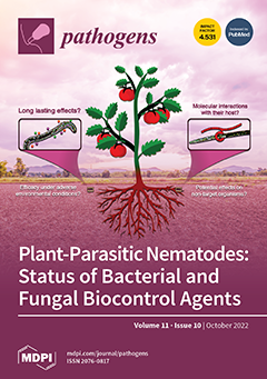
Plant-parasitic nematodes (PPNs) are among the most notorious and underrated threats to food security and plant health worldwide, compromising crop yields and causing billions of dollars of losses annually. Chemical control strategies rely heavily on synthetic chemical nematicides to reduce PPN population densities, but their use is being progressively restricted due to environmental and human health concerns, so alternative control methods are urgently needed. Here, we review the potential of bacterial and fungal agents to suppress the most important PPNs, namely Aphelenchoides besseyi, Bursaphelenchus xylophilus, Ditylenchus dipsaci, Globodera spp., Heterodera spp., Meloidogyne spp., Nacobbus aberrans, Pratylenchus spp., Radopholus similis, Rotylenchulus reniformis, and Xiphinema index. View this paper
  • Issues are regarded as officially published after their release is announced to the table of contents alert mailing list.
  • You may sign up for e-mail alerts to receive table of contents of newly released issues.
  • PDF is the official format for papers published in both, html and pdf forms. To view the papers in pdf format, click on the "PDF Full-text" link, and use the free Adobe Reader to open them.
Order results
Result details
Section
Select all
Export citation of selected articles as:
20 pages, 8980 KiB  
Article
Powassan Virus Induces Structural Changes in Human Neuronal Cells In Vitro and Murine Neurons In Vivo
by Jacob Nelson, Lorenzo Ochoa, Paula Villareal, Tiffany Dunn, Ping Wu, Gracie Vargas and Alexander N. Freiberg
Pathogens 2022, 11(10), 1218; https://doi.org/10.3390/pathogens11101218 - 21 Oct 2022
Cited by 2 | Viewed by 2502
Abstract
Powassan virus (POWV) is a tick-borne flavivirus (TBFV) that can cause severe encephalitis in humans with a case–fatality rate as high as 11%. Patients who survive severe encephalitic disease can develop long-term neurological sequelae that can be debilitating and life-long. In this study, [...] Read more.
Powassan virus (POWV) is a tick-borne flavivirus (TBFV) that can cause severe encephalitis in humans with a case–fatality rate as high as 11%. Patients who survive severe encephalitic disease can develop long-term neurological sequelae that can be debilitating and life-long. In this study, we have sought to characterize a primary human fetal brain neural stem cell system (hNSC), which can be differentiated into neuron and astrocyte co-cultures, to serve as a translational in vitro system for infection with POWV and a comparative mosquito-borne flavivirus (MBFV), West Nile virus (WNV). We found that both viruses are able to infect both cell types in the co-culture and that WNV elicits a strong inflammatory response characterized by increased cytokines IL-4, IL-6, IL-8, TNF-α and IL-1β and activation of apoptosis pathways. POWV infection resulted in fewer cytokine responses, as well as less detectable apoptosis, while neurons infected with POWV exhibited structural aberrations forming in the dendrites. These anomalies are consistent with previous findings in which tick-borne encephalitis virus (TBEV) infected murine primary neurons formed laminal membrane structures (LMS). Furthermore, these structural aberrations are also recapitulated in brain tissue from infected mice. Our findings indicate that POWV is capable of infecting human primary neurons and astrocytes without causing apparent widespread apoptosis, while forming punctate structures reminiscent with LMS in primary human neurons and in vivo. Full article
(This article belongs to the Special Issue Current Research on Arboviral Encephalitis)
Show Figures

Figure 1

8 pages, 267 KiB  
Communication
Prevalence of Toxoplasma gondii Antibodies and Risk Factor Investigation in Portuguese Veterinarians: A Matched Case–Control Study
by Daniela Almeida, Sérgio Santos-Silva, Maria Aires Pereira, Carla Santos, Cristina Mega, Catarina Coelho, Carmen Nóbrega, Fernando Esteves, Rita Cruz, Helena Vala and João R. Mesquita
Pathogens 2022, 11(10), 1217; https://doi.org/10.3390/pathogens11101217 - 21 Oct 2022
Cited by 1 | Viewed by 1481
Abstract
(1) Background: Toxoplasma gondii is a widespread zoonotic agent that greatly impacts Public Health, being responsible for one of the most important parasitic zoonosis worldwide. T. gondii has a heteroxenous life cycle, with cats being the definitive hosts and all warm-blooded animals, including [...] Read more.
(1) Background: Toxoplasma gondii is a widespread zoonotic agent that greatly impacts Public Health, being responsible for one of the most important parasitic zoonosis worldwide. T. gondii has a heteroxenous life cycle, with cats being the definitive hosts and all warm-blooded animals, including humans, being intermediate hosts. Veterinary practitioners (VP) may be at a higher risk than the general population for T. gondii infection, as they have direct and daily contact with many animal species. The aim of the present study was to ascertain if VP were more likely to be anti-T. gondii IgG seropositive than the general population, as well as to understand if age, accidents with blood-contaminated sharps (cross-blood contamination), gender, working years, and geographic regions play a role as risk factors for T. gondii infection. For this purpose, a case–control study using archived samples was performed. (2) Methods: A total of 350 veterinary practitioners were tested using a commercial semiquantitative enzyme immunoassay for anti-T. gondii IgG. From the general population, 175 anonymous volunteers (matched with cases by region, age, and gender) were studied for anti-T. gondii IgG. (3) Results: There was no statistical difference found between the presence of anti-T. gondii IgG in practitioners (26%; CI = 21.40–30.60%) and the general population (33.14%; CI = 26.17–40.12%) (p = 0.108). Univariate and multivariate analysis showed that only age (older groups) was found to be associated with a higher prevalence of anti-T. gondii IgG, with significant p values (p < 0.05) for both univariate and multivariate analysis. (4) Conclusions: To the best of our knowledge, this is the first case–control study fully focused on the prevalence of anti-T. gondii IgG in VP in Portugal, showing that there was no significant risk for T. gondii infection in veterinarians exposed daily and repeatedly to different species of animals. Full article
(This article belongs to the Special Issue Toxoplasma)
9 pages, 1069 KiB  
Article
Antimicrobial Resistance and Virulence Factors of Proteus mirabilis Isolated from Dog with Chronic Otitis Externa
by Jun Kwon, Myoung-Hwan Yang, Hyoung-Joon Ko, Sang-Guen Kim, Chul Park and Se-Chang Park
Pathogens 2022, 11(10), 1215; https://doi.org/10.3390/pathogens11101215 - 21 Oct 2022
Cited by 7 | Viewed by 2556
Abstract
Otitis externa is among the most prevalent diseases in dogs. If the underlying cause is not addressed, bacterial reinfection becomes frequent, necessitating antibiotic administration for an extended period of time. Prolonged treatment promotes the emergence of antibiotic-resistant bacteria and increases the risk of [...] Read more.
Otitis externa is among the most prevalent diseases in dogs. If the underlying cause is not addressed, bacterial reinfection becomes frequent, necessitating antibiotic administration for an extended period of time. Prolonged treatment promotes the emergence of antibiotic-resistant bacteria and increases the risk of their transmission from animals to humans. This study aimed to analyze the antibiotic resistance pattern of the emerging pathogen Proteus mirabilis to identify bacterial virulence and antibiotic selection. Samples were collected from randomly encountered dogs with chronic otitis externa. Thirty-two strains of P. mirabilis were isolated and identified, using MALDI-TOF. The Kirby-Bauer disk diffusion method was used to assess the antibiotic susceptibility of P. mirabilis to 11 antibiotics. The isolates (n = 32) were most resistant to cefazolin (75%), trimethoprim–sulfamethoxazole (72%), chloramphenicol (72%), amoxicillin–clavulanate (63%), ampicillin (59%), cefepime (56%), ciprofloxacin (53%), aztreonam (50%), ceftazidime avibactam (50%), gentamicin (22%), and amikacin (16%). Moreover, 75% of isolates were found to be multidrug-resistant bacteria. P. mirabilis was found to have a high resistance-pattern ratio. Although the exact cause is unknown, continuous antibiotic use is thought to be a major factor. We concluded that antibiotic use must be prudent and selective to prevent antibiotic resistance. Full article
(This article belongs to the Special Issue Detection and Characterization of Drug-Resistant Organisms)
Show Figures

Figure 1

11 pages, 720 KiB  
Article
Evidence of Dirofilaria immitis in Felids in North-Eastern Italy
by Marika Grillini, Antonio Frangipane di Regalbono, Cinzia Tessarin, Paola Beraldo, Rudi Cassini, Erica Marchiori and Giulia Simonato
Pathogens 2022, 11(10), 1216; https://doi.org/10.3390/pathogens11101216 - 20 Oct 2022
Cited by 3 | Viewed by 1725
Abstract
Dirofilaria immitis is a mosquito-borne nematode, causing heartworm (HW) disease in wild and domestic canids. HW can also affect felids with different clinical patterns from asymptomatic pictures to sudden death, making the monitoring and diagnosis complicated. Canine HW is endemic in North-eastern Italy; [...] Read more.
Dirofilaria immitis is a mosquito-borne nematode, causing heartworm (HW) disease in wild and domestic canids. HW can also affect felids with different clinical patterns from asymptomatic pictures to sudden death, making the monitoring and diagnosis complicated. Canine HW is endemic in North-eastern Italy; however, very little information has been recorded for felids. This study aims to provide new information on HW in felids in North-eastern Italy. Two hundred and six domestic cats from Veneto, Friuli-Venezia Giulia, Trentino Alto-Adige regions (North-eastern Italy), nine captive felids from zoological parks from Veneto, and nineteen European wildcats from Friuli Venezia Giulia were recruited. Sera/plasma was analysed for the detection of anti-HW antibodies (Ab) and HW antigens (Ag); positive blood samples were molecularly analysed, targeting the HW DNA (5S-rRNA gene). Twelve out of two hundred and six (5.8%) cats presented with Ab, and three out of two hundred and six (1.5%) presented with Ag, mainly those from the Veneto region, already known as a canine HW-endemic area. Among Ab-positive cats, two were from Belluno, a mountain province previously considered free, suggesting the expansion of HW into the northern areas. No cats were positive for both Ab and Ag. Three out of nineteen (15.8%) wildcats were Ag-positive, constituting the first HW report in Italy. No captive felids were positive. Dirofilaria immitis DNA was not amplified in positive samples, suggesting the low sensitivity of PCR on blood. This study provides new data on the occurrence of HW in domestic cats and wildcats in North-eastern Italy. Full article
(This article belongs to the Special Issue Arthropod- and Gastropod-Borne Diseases in a One Health Perspective)
Show Figures

Figure 1

13 pages, 1214 KiB  
Article
Inflammatory Response in Caco-2 Cells Stimulated with Anisakis Messengers of Pathogenicity
by Ilaria Bellini, Daniela Scribano, Meysam Sarshar, Cecilia Ambrosi, Antonella Pizzarelli, Anna Teresa Palamara, Stefano D’Amelio and Serena Cavallero
Pathogens 2022, 11(10), 1214; https://doi.org/10.3390/pathogens11101214 - 20 Oct 2022
Cited by 2 | Viewed by 2357
Abstract
Background: Anisakis spp. third-stage larvae (L3) are the causative agents of human zoonosis called anisakiasis. The accidental ingestion of L3 can cause acute and chronic inflammation at the gastric, intestinal, or ectopic levels. Despite its relevance in public health, studies on pathogenetic [...] Read more.
Background: Anisakis spp. third-stage larvae (L3) are the causative agents of human zoonosis called anisakiasis. The accidental ingestion of L3 can cause acute and chronic inflammation at the gastric, intestinal, or ectopic levels. Despite its relevance in public health, studies on pathogenetic mechanisms and parasite-human interplay are scarce. The aim of this study was to investigate the human inflammatory response to different Anisakis vehicles of pathogenicity. Methods: Human colorectal adenocarcinoma (Caco-2) cells were exposed to Anisakis L3 (the initial contact with the host), extracellular vesicles (EVs, Anisakis–host communication), and crude extract (CE, the larval dying). The protein quantity and gene expression of two pro-inflammatory cytokines (IL-6 and IL-8) were investigated using an ELISA test (6 h and 24 h) and a qReal-Time PCR (1 h, 6 h, and 24 h), respectively. Results: The L3 and EVs induced a downregulation in both the Il-6 and Il-8 gene expression and protein quantity. On the contrary, the CE stimulated IL-6 gene expression and its protein release, not affecting IL-8. Conclusions: The Caco-2 cells seemed to not react to the exposure to the L3 and EVs, suggesting a parasite’s immunomodulating action to remain alive in an inhospitable niche. Conversely, the dying larva (CE) could induce strong activation of the immune strategy of the host that, in vivo, would lead to parasite expulsion, eosinophilia, and/or granuloma formation. Full article
(This article belongs to the Special Issue Anisakiasis and Anisakidae)
Show Figures

Graphical abstract

13 pages, 3175 KiB  
Article
Mammalian and Avian Larval Schistosomatids in Bangladesh: Molecular Characterization, Epidemiology, Molluscan Vectors, and Occurrence of Human Cercarial Dermatitis
by Sharmin Shahid Labony, Md. Shahadat Hossain, Takeshi Hatta, Anita Rani Dey, Uday Kumar Mohanta, Ausraful Islam, Md. Shahiduzzaman, Muhammad Mehedi Hasan, Md. Abdul Alim, Naotoshi Tsuji and Anisuzzaman
Pathogens 2022, 11(10), 1213; https://doi.org/10.3390/pathogens11101213 - 20 Oct 2022
Cited by 3 | Viewed by 1762
Abstract
Schistosomiasis is a neglected tropical disease (NTD) caused by blood flukes (Schistosoma spp.). Schistosomatids affect a wide array of vertebrate hosts, including humans. In the present study, multiple species of schistosomatids were identified by isolating schistosomatid cercariae (SC) from naturally infected snails. [...] Read more.
Schistosomiasis is a neglected tropical disease (NTD) caused by blood flukes (Schistosoma spp.). Schistosomatids affect a wide array of vertebrate hosts, including humans. In the present study, multiple species of schistosomatids were identified by isolating schistosomatid cercariae (SC) from naturally infected snails. We also described different biotic and abiotic factors influencing SC infections in snails and reported human cercarial dermatitis (HCD) for the first time in Bangladesh. A total of 22,012 snails of seven species: Lymnaea auricularia, L. luteola, Indoplanorbis exustus, Physa acuta, Viviparus bengalensis, Brotia spp., and Thiara spp., were collected and examined. Among these snails, 581 (2.6%) belonging to five species: L. luteola, L. auricularia, P. acuta, I. exustus, and V. bengalensis, were infected with SC. The rate of infection was the highest for L. luteola (11.1%), followed by L. auricularia (5.3%), and was the lowest for V. bengalensis (0.4%). Prevalence in snails was the highest in September (16.8%), followed by October (9.5%) and November (8.8%), and was the lowest in colder months, such as January (1.8%) and February (2.1%). Infections with schistosomatids were more common in larger snails and snails collected from sunny areas. We confirmed the presence of Schistosoma indicum, S. incognitum, S. nasale, S. spindale, and Trichobilharzia szidati by PCR and sequencing. Through a questionnaire survey, we detected HCD in 214 (53.5%) individuals, and the infection rate was almost equally distributed across all professions. Collectively, the present results suggest that lymnaeid snails are the main vector for Schistosoma spp. prevalent in Bangladesh, and schistosomatids with zoonotic potential are also prevalent. Full article
(This article belongs to the Special Issue Arthropod- and Gastropod-Borne Diseases in a One Health Perspective)
Show Figures

Figure 1

13 pages, 1003 KiB  
Review
Human Neurocysticercosis: An Overview
by Oscar H. Del Brutto
Pathogens 2022, 11(10), 1212; https://doi.org/10.3390/pathogens11101212 - 20 Oct 2022
Cited by 12 | Viewed by 4179
Abstract
Human cysticercosis is caused by ingestion of T. solium eggs from taenia carriers. Neurocysticercosis (NCC), defined as the infection of the CNS and the meninges by the larval stage of Taenia solium, is the most common helminthic infection of the CNS worldwide. [...] Read more.
Human cysticercosis is caused by ingestion of T. solium eggs from taenia carriers. Neurocysticercosis (NCC), defined as the infection of the CNS and the meninges by the larval stage of Taenia solium, is the most common helminthic infection of the CNS worldwide. Parasites may lodge in brain parenchyma, subarachnoid space, ventricular system, or spinal cord, causing pathological changes that account for the pleomorphism of this disease. Seizures/epilepsy are the most common clinical manifestation, but other patients present with headache, focal deficits, intracranial hypertension, or cognitive decline. Accurate diagnosis of NCC is possible after interpretation of clinical data together with findings of neuroimaging studies and results of immunological tests. However, neuroimaging studies are fundamental for diagnosis because immunological test and clinical manifestations only provide circumstantial evidence of NCC. The introduction of cysticidal drugs changed the prognosis of most NCC patients. These drugs have been shown to reduce the burden of infection and to improve the clinical course of the disease in many patients. Efforts should be directed to eradicate the disease through the implementation of control programs against all the steps in the life cycle of T. solium, including carriers of the adult tapeworm, infected pigs, and eggs in the environment. Full article
(This article belongs to the Special Issue Recent Advances in Taeniasis and Cysticercosis)
Show Figures

Figure 1

6 pages, 522 KiB  
Commentary
CRISPR-Based Diagnostics: A Potential Tool to Address the Diagnostic Challenges of Tuberculosis
by Yong Qi, Kun Li, Yuxi Li, Donglin Guo, Jing Xu, Yuexi Li and Wenping Gong
Pathogens 2022, 11(10), 1211; https://doi.org/10.3390/pathogens11101211 - 20 Oct 2022
Cited by 6 | Viewed by 2086
Abstract
Tuberculosis (TB) is an infectious disease caused by Mycobacterium tuberculosis, which infects more than 23% of the world’s population. With the emergence of drug-resistant TB (DR-TB) and latent TB infection (LTBI), rapid diagnosis of DR-TB and LTBI has become a challenge for [...] Read more.
Tuberculosis (TB) is an infectious disease caused by Mycobacterium tuberculosis, which infects more than 23% of the world’s population. With the emergence of drug-resistant TB (DR-TB) and latent TB infection (LTBI), rapid diagnosis of DR-TB and LTBI has become a challenge for the prevention and control of TB. Herein, we highlight these challenges and discuss emerging clustered regularly interspaced short palindromic repeats (CRISPR)-based diagnostics in TB detection. Currently, the clinical diagnosis of M. tuberculosis infection mainly depends on pathogenic and molecular biological methods, including sputum smear, sputum culture, and Xpert. Although CRISPR-based diagnostics have not been applied to the clinical diagnosis of TB, they have shown exciting preponderances in TB diagnosis compared with traditional methods, including higher sensitivity, less sample input, and shorter turnaround time. CRISPR-based diagnostics represent a potential tool to address the challenges and natural weaknesses associated with traditional TB diagnosis methods. Based on the currently available data, we suggest that future CRISPR-based TB diagnostics should be developed in the direction of automation, modularization, diversification, and intelligence. By combining the CRISPR platform with various systems, such as microfluidic chips, droplet microfluidics, electrochemical techniques, and optical systems, the specificity and sensitivity of TB diagnosis may be revolutionized. Full article
(This article belongs to the Section Bacterial Pathogens)
Show Figures

Figure 1

10 pages, 2996 KiB  
Communication
High Rates of Detection and Molecular Characterization of Porcine Adenovirus Serotype 5 (Porcine mastadenovirus C) from Diarrheic Pigs
by Kerry Gainor, Yussaira Castillo Fortuna, Angeline Steny Alakkaparambil, Wendy González, Yashpal Singh Malik and Souvik Ghosh
Pathogens 2022, 11(10), 1210; https://doi.org/10.3390/pathogens11101210 - 20 Oct 2022
Cited by 4 | Viewed by 1491
Abstract
Since the first report on isolation of porcine adenovirus serotype 5 (PAdV-5, species Porcine mastadenovirus C (PAdV-C)) from pigs with respiratory illness in Japan in 1987, PAdV-5 have been detected in a few fecal samples from healthy pigs and in some [...] Read more.
Since the first report on isolation of porcine adenovirus serotype 5 (PAdV-5, species Porcine mastadenovirus C (PAdV-C)) from pigs with respiratory illness in Japan in 1987, PAdV-5 have been detected in a few fecal samples from healthy pigs and in some environmental samples. To date, only a single PAdV-5 strain (isolate HNF-70 from 1987) has been analyzed for the complete genome. We report here high detection rates of PAdV-5 (25.74%, 26/101 fecal samples) in diarrheic pigs at 3 different farms in the Caribbean country of Dominican Republic. After a long gap, the complete deduced amino acid sequences of the DNA-dependent DNA polymerase (pol) and hexon of two PAdV-5 strains (GES7 and Z11) were determined, revealing >99% sequence identities between PAdV-5 strains (HNF-70, GES7 and Z11) detected in different parts of the world and during different time periods (1987, and 2020–2021). By phylogenetic analysis, the putative hexon and pol of HNF-70, GES7 and Z11 exhibited similar clustering patterns, with the PAdV-5 strains forming a tight cluster near ruminant AdVs, distinct from the species PAdV-A and -B. GES7 and Z11 retained the various conserved features present in the putative pol and major late promoter region of HNF-70. Considering the paucity of data on current epidemiological status and genetic diversity of PAdV in porcine populations, our findings warrant similar studies on PAdV-5 and other PAdVs in clinically ill and healthy pigs. To our knowledge, this is the first report on detection and molecular characterization of PAdV-5 (PAdV-C) from diarrheic pigs. Full article
(This article belongs to the Special Issue 10th Anniversary of Pathogens—Feature Papers)
Show Figures

Figure 1

15 pages, 1082 KiB  
Article
Unlicensed Molnupiravir is an Effective Rescue Treatment Following Failure of Unlicensed GS-441524-like Therapy for Cats with Suspected Feline Infectious Peritonitis
by Meagan Roy, Nicole Jacque, Wendy Novicoff, Emma Li, Rosa Negash and Samantha J. M. Evans
Pathogens 2022, 11(10), 1209; https://doi.org/10.3390/pathogens11101209 - 20 Oct 2022
Cited by 11 | Viewed by 23197
Abstract
Feline infectious peritonitis (FIP) is a complex and historically fatal disease, though recent advances in antiviral therapy have uncovered potential treatments. A newer therapeutic option, unlicensed molnupiravir, is being used as a first-line therapy for suspect FIP and as a rescue therapy to [...] Read more.
Feline infectious peritonitis (FIP) is a complex and historically fatal disease, though recent advances in antiviral therapy have uncovered potential treatments. A newer therapeutic option, unlicensed molnupiravir, is being used as a first-line therapy for suspect FIP and as a rescue therapy to treat cats who have persistent or relapsed clinical signs of FIP after GS-441524 and/or GC376 therapy. Using owner-reported data, treatment protocols for 30 cats were documented. The 26 cats treated with unlicensed molnupiravir as a rescue therapy were treated with an average starting dosage of 12.8 mg/kg and an average ending dosage of 14.7 mg/kg twice daily for a median of 12 weeks (IQR = 10–15). In total, 24 of 26 cats were still living disease-free at the time of writing. One cat was euthanized after completing treatment due to a prolonged seizure, and the other cat underwent retreatment for relapsed clinical signs. Few adverse effects were reported, with the most notable—folded ears (1), broken whiskers (1), and severe leukopenia (1)—seen at dosages above 23 mg/kg twice daily. This study provides a proof of principle for the use of molnupiravir in cats and supports the need for future studies to further evaluate molnupiravir as a potentially safe and effective therapy for FIP. Full article
(This article belongs to the Special Issue Advances on Feline Coronavirus Infection)
Show Figures

Figure 1

14 pages, 10225 KiB  
Article
Inhibition Effects of Nippostrongylus brasiliensis and Its Derivatives against Atherosclerosis in ApoE-/- Mice through Anti-Inflammatory Response
by Yougui Yang, Xin Ding, Fuzhong Chen, Xiaomin Wu, Yuying Chen, Qiang Zhang, Jun Cao, Junhong Wang and Yang Dai
Pathogens 2022, 11(10), 1208; https://doi.org/10.3390/pathogens11101208 - 20 Oct 2022
Viewed by 3298
Abstract
Atherosclerosis (AS) is a dominant and growing cause of death and disability worldwide that involves inflammation from its inception to the emergence of complications. Studies have demonstrated that intervention with helminth infections or derived products could modulate the host immune response and effectively [...] Read more.
Atherosclerosis (AS) is a dominant and growing cause of death and disability worldwide that involves inflammation from its inception to the emergence of complications. Studies have demonstrated that intervention with helminth infections or derived products could modulate the host immune response and effectively prevent or mitigate the onset and progression of inflammation-related diseases. Therefore, to understand the molecular mechanisms underlying the development of atherosclerosis, we intervened in ApoE-/- mice maintained on a high-fat diet with Nippostrongylus brasiliensis (N. brasiliensis) infection and immunized with its derived products. We found that N. brasiliensis infection and its derived proteins had suitable protective effects both in the initial and progressive stages of atherosclerosis, effectively reducing aortic arch plaque areas and liver lipid contents and downregulating serum LDL levels, which may be associated with the significant upregulation of serum anti-inflammatory cytokines (IL-10 and IL-4) and the down-regulation of proinflammatory cytokines (TNF-α and IFN-γ) in the serum. In conclusion, these data highlighted the effective regulatory role of N. brasiliensis and its derived proteins in the development and progression of atherosclerosis. This could provide a promising new avenue for the prevention and treatment of atherosclerosis. Full article
(This article belongs to the Special Issue State-of-Art and Perspectives of Parasites in China)
Show Figures

Figure 1

8 pages, 1286 KiB  
Brief Report
In Vitro Assessment of Berberine against Ichthyophthirius multifiliis in Goldfish
by Ke Huang, Guangran Hu, Runqiu Wang, Qingwen Zeng, Wenxiang Li, Hong Zou, Shangong Wu, Guitang Wang and Ming Li
Pathogens 2022, 11(10), 1207; https://doi.org/10.3390/pathogens11101207 - 20 Oct 2022
Cited by 5 | Viewed by 2103
Abstract
Ichthyophthirius multifiliis is a pathogenic ciliate parasite, which infects almost all freshwater teleost fish and leads to significant economic losses. The present study aimed to evaluate the acute toxicity of berberine to the free-living stages of I. multifiliis, that is, theronts and [...] Read more.
Ichthyophthirius multifiliis is a pathogenic ciliate parasite, which infects almost all freshwater teleost fish and leads to significant economic losses. The present study aimed to evaluate the acute toxicity of berberine to the free-living stages of I. multifiliis, that is, theronts and tomonts. Our results indicated that 99.30% of I. multifiliis theronts were killed by a concentration of 15 mg/L berberine during the 4 h exposure time, while berberine had no effect on protomonts. Nevertheless, berberine at a concentration of 5 mg/L could effectively reduce the release of theronts from tomonts treated for 4 h. Additionally, according to the transmission electron microscopy results, berberine at 15 mg/L could strongly change the shape of protomonts, destroy their organelles, and significantly decrease the number of ribosomes. The median lethal concentration (LC50) of berberine for goldfish at 96 h was 528.44 mg/L, which was almost 67 times the median effective concentration (EC50) of berberine for killing theronts (7.86 mg/L). The results demonstrated that berberine could be an effective and safe potential parasiticide for killing I. multifiliis. Full article
Show Figures

Figure 1

15 pages, 1732 KiB  
Article
The Prevalence of Pathogens among Ticks Collected from Livestock in Kazakhstan
by Kulyaisan T. Sultankulova, Gaukhar O. Shynybekova, Aisha U. Issabek, Nazym N. Mukhami, Aibarys M. Melisbek, Olga V. Chervyakova, Nurlan S. Kozhabergenov, Sabyrkhan M. Barmak, Arailym K. Bopi, Zamira D. Omarova, Dana A. Alibekova, Takhmina U. Argimbayeva, Aidar M. Namet, Ivan A. Zuban and Mukhit B. Orynbayev
Pathogens 2022, 11(10), 1206; https://doi.org/10.3390/pathogens11101206 - 19 Oct 2022
Cited by 4 | Viewed by 2215
Abstract
Ticks carry and transmit a wide variety of pathogens (bacteria, viruses and protozoa) that pose a threat to humans and animals worldwide. The purpose of this work was to study ticks collected in different regions of Kazakhstan for the carriage of various pathogens. [...] Read more.
Ticks carry and transmit a wide variety of pathogens (bacteria, viruses and protozoa) that pose a threat to humans and animals worldwide. The purpose of this work was to study ticks collected in different regions of Kazakhstan for the carriage of various pathogens. The collected ticks were examined by PCR for the carriage of various pathogens. A total of 3341 tick samples parasitizing three animal species (cattle, sheep and horses) were collected at eight regions of Kazakhstan. Eight tick species were found infesting animals: Dermacentor marginatus (28.08%), Hyalomma asiaticum (21.28%), Hyalomma anatolicum (17.18%), Dermacentor reticulatus (2.01%), Ixodes ricinus (3.35%), Ixodes persulcatus (0.33%), Hyalomma scupense (12.87%) and Hyalomma marginatum (14.90%). Ticks collected from livestock animals were examined for the pathogen spectrum of transmissible infections to determine the degree of their infection. Four pathogen DNAs (lumpy skin disease virus (LSDV), Coxiella burnetti, Teileria annulata, and Babesia caballi) were detected by PCR in Dermacentor marginatus, Hyalomma asiaticum, Hyalomma scupense, Hyalomma anatolicum. The infection of ticks Dermacentor marginatus and Hyalomma asiaticum collected on cattle in the West Kazakhstan region with LSDV was 14.28% and 5.71%, respectively. Coxiella burnetti was found in the ticks Dermacentor marginatus (31.91%) in the Turkestan region and Hyalomma anatolicum (52.63%) in the Zhambyl region. Theileria annulata was found in ticks Hyalomma scupense (7.32%) and Dermacentor marginatus (6.10%) from cattle in the Turkestan region. Babesia caballi was isolated only from the species Hyalomma scupense (17.14%) in the Turkestan region. There were no PCR-positive tick samples collected from sheep. RNA/DNAs of tick-borne encephalitis virus (TBEV), African swine fever virus (ASFV), Hantavirus hemorrhagic fever with renal syndrome (HFRS), and chlamydia pathogens were not found in ticks. The new data give a better understanding of the epidemiology of tick-borne pathogens and the possibility of the emergence of tick-borne animal diseases in Kazakhstan. Full article
(This article belongs to the Special Issue Ticks and Tick-Borne Diseases―Pathogens, Parasites and People)
Show Figures

Figure 1

14 pages, 2414 KiB  
Article
Kinetoplastid Species Maintained by a Small Mammal Community in the Pantanal Biome
by Filipe Martins Santos, Nayara Yoshie Sano, Sany Caroline Liberal, Maria Augusta Dario, Wesley Arruda Gimenes Nantes, Fernanda Moreira Alves, Alanderson Rodrigues da Silva, Carina Elisei De Oliveira, André Luiz Rodrigues Roque, Heitor Miraglia Herrera and Ana Maria Jansen
Pathogens 2022, 11(10), 1205; https://doi.org/10.3390/pathogens11101205 - 19 Oct 2022
Cited by 3 | Viewed by 1551
Abstract
Kinetoplastids include species economically important in agriculture, livestock, and human health. We evaluated the richness of kinetoplastids that infect small mammals in patches of unflooded forests in the Pantanal biome, an area where we hypothesize that its diversity is higher than currently recognized. [...] Read more.
Kinetoplastids include species economically important in agriculture, livestock, and human health. We evaluated the richness of kinetoplastids that infect small mammals in patches of unflooded forests in the Pantanal biome, an area where we hypothesize that its diversity is higher than currently recognized. Hemocultures (HC) and Next Generation Sequencing (NGS) targeting the 18S rDNA gene were employed for the detection of kinetoplastids. We grouped the positive samples into pools for each small mammal species (Monodelphis domestica, Thylamys macrurus, Oecomys mamorae, Thrichomys fosteri, Clyomys laticeps, and Holochilus chacarius). Eight parasite species were identified: Leishmania amazonensis, L. infantum; Trypanosoma cascavelli (HC + NGS), T. cruzi, T. lainsoni, T. rangeli (HC + NGS), Trypanosoma sp. DID, and Neobodo sp. The use of a tool as sensitive as NGS has increased our awareness of the diversity of kinetoplastids, as well as their host range, with emphasis on the species O. mamorae (seven kinetoplastid species, excepting T. cascavelli in a pool of nine individuals) and T. macrurus (four kinetoplastid species in a single individual). Furthermore, L. infantum and L. amazonensis infections were described in small mammals from this region for the first time. These findings make it mandatory to revisit the kinetoplastids/host associations proposed so far. Full article
(This article belongs to the Special Issue Biology of Parasitism)
Show Figures

Graphical abstract

12 pages, 2309 KiB  
Article
First Record of Colletotrichum anthrisci Causing Anthracnose on Avocado Fruits in Chile
by Marcelo I. Bustamante, Claudio Osorio-Navarro, Ysadora Fernández, Tyler B. Bourret, Alan Zamorano and José Luis Henríquez-Sáez
Pathogens 2022, 11(10), 1204; https://doi.org/10.3390/pathogens11101204 - 19 Oct 2022
Cited by 3 | Viewed by 2879
Abstract
Anthracnose caused by Colletotrichum species is one of the most frequent and damaging fungal diseases affecting avocado fruits (Persea americana Mill.) worldwide. In Chile, the disease incidence has increased over the last decades due to the establishment of commercial groves in more [...] Read more.
Anthracnose caused by Colletotrichum species is one of the most frequent and damaging fungal diseases affecting avocado fruits (Persea americana Mill.) worldwide. In Chile, the disease incidence has increased over the last decades due to the establishment of commercial groves in more humid areas. Since 2018, unusual symptoms of anthracnose have been observed on Hass avocado fruits, with lesions developing a white to gray sporulation. Morphological features and multi-locus phylogenetic analyses using six DNA barcodes (act, chs-1, gapdh, his3, ITS, and tub2) allowed the identification of the causal agent as Colletotrichum anthrisci, a member of the dematium species complex. Pathogenicity was confirmed by inoculating healthy Hass avocado fruits with representative isolates, reproducing the same symptoms initially observed, and successfully reisolating the same isolates from the margin of the necrotic pulp. Previously, several Colletotrichum species belonging to other species complexes have been associated with avocado anthracnose in other countries. To our knowledge, this is the first record of C. anthrisci and of a species of the dematium species complex causing anthracnose on avocado fruits in Chile and worldwide. Full article
(This article belongs to the Special Issue Plant Pathogenic Fungi)
Show Figures

Figure 1

6 pages, 1071 KiB  
Case Report
Bacillus simplex as the Most Probable Culprit of Penetrating Trauma Infection: A Case Report
by Panagiota Xaplanteri, Dimitrios S. Serpanos, Ellie Dorva, Tatiana Beqo-Rokaj, Eleni Papadogeorgaki and Alexandra Lekkou
Pathogens 2022, 11(10), 1203; https://doi.org/10.3390/pathogens11101203 - 18 Oct 2022
Cited by 1 | Viewed by 2772
Abstract
Bacillus simplex is an environmental microorganism found in soil. Herein, we present the case of a 69-year-old Greek male patient who attended the Emergency Department at our hospital. The patient complained of lower right extremity swelling and pain, after suffering penetrating trauma whilst [...] Read more.
Bacillus simplex is an environmental microorganism found in soil. Herein, we present the case of a 69-year-old Greek male patient who attended the Emergency Department at our hospital. The patient complained of lower right extremity swelling and pain, after suffering penetrating trauma whilst doing farm work in a rural area. Swab aerobic cultures revealed Bacillus simplex as identified by MALDI-TOF Mass Spectrometry. The strain was susceptible to vancomycin, imipenem, clindamycin, and ciprofloxacin. Our patient refused hospitalization; therefore, both ciprofloxacin and clindamycin were registered for a total of 19 days. No complications were experienced, and he recovered fully. In our case, the thorough cleaning of the ulcer bed prior to sample collection, the fact that it was the only microorganism isolated, and the wound’s aggravating mechanism led the authors of the present study to the conclusion that B. simplex was the most probable culprit of the infection. To our knowledge, this is the second probable case of B. simplex infection described worldwide, and the first in Greece. Full article
(This article belongs to the Special Issue 10th Anniversary of Pathogens—Feature Papers)
Show Figures

Figure 1

9 pages, 1728 KiB  
Article
Clonal Dissemination of Multidrug-Resistant and Hypervirulent Klebsiella pneumoniae Clonal Complex in a Chinese Hospital
by Yi Wang, Mingxi Hua, Jingqiao Wang, Wen Xing, Jiatian Chen, Jingyuan Liu and Pengcheng Du
Pathogens 2022, 11(10), 1202; https://doi.org/10.3390/pathogens11101202 - 18 Oct 2022
Cited by 2 | Viewed by 1392
Abstract
The emergence of high antimicrobial-resistant and hypervirulent Klebsiella pneumoniae (hvKp) clones in clinics has become a cause of concern in recent years. Despite the global spread of the clonal complex (CC) 258, hvKp of other non-CC258 subgroups also emerged. Here, by performing a [...] Read more.
The emergence of high antimicrobial-resistant and hypervirulent Klebsiella pneumoniae (hvKp) clones in clinics has become a cause of concern in recent years. Despite the global spread of the clonal complex (CC) 258, hvKp of other non-CC258 subgroups also emerged. Here, by performing a retrospective study from July 2019 to August 2020 in a Chinese hospital, we obtained 25 K. pneumoniae isolates belonging to CC15. By antimicrobial susceptibility testing and whole genome sequencing and analysis, we obtained the resistant phenotypes and genotypes of these isolates. Twenty-one isolates (84%) were carbapenem-resistant, and eighteen were blaKPC-2 positive. In addition, ten isolates were identified as putative hvKp and seven were carbapenem-resistant hvKp. Nine isolates carried the pLVPK-like virulence plasmid, which contains the fragment including rmpA2, peg-589, iutA, and iucABCD. Another isolate carried iucA. Phylogenetic analysis revealed that the isolates belonged to four lineages, and the putative hvKp isolates were identified in three of these. Two independent sublineages of putative hvKp were caused by the acquisition of pLVPK-like virulence plasmid. Based on comparative genomic analysis, the number of pairwise single nucleotide polymorphisms amongst the four sublineages, Lineage 1a, 1b, 2a, and 2b, were 1–43, 2–13, 129–279, and 3–4, respectively, indicating clonal transmission of Lineage 1a, 1b, and 2b. These results indicate that multiple lineages of CC15 carbapenem-resistant hvKp have emerged in the hospital and caused nosocomial transmission, and that the spreading of virulence plasmids among classic K. pneumoniae subtypes might become more common and happen more easily. These findings highlight the importance of surveillance of local epidemics of non-CC258 subgroups in hospitals. Full article
(This article belongs to the Section Bacterial Pathogens)
Show Figures

Figure 1

15 pages, 311 KiB  
Review
COVID-19 Treatment—Current Status, Advances, and Gap
by Chian Ho and Ping-Chin Lee
Pathogens 2022, 11(10), 1201; https://doi.org/10.3390/pathogens11101201 - 18 Oct 2022
Cited by 8 | Viewed by 2179
Abstract
COVID-19, which emerged in December 2019, was declared a global pandemic by the World Health Organization (WHO) in March 2020. The disease was caused by severe acute respiratory syndrome coronavirus 2 (SARS-CoV-2). It has caused millions of deaths worldwide and caused social and [...] Read more.
COVID-19, which emerged in December 2019, was declared a global pandemic by the World Health Organization (WHO) in March 2020. The disease was caused by severe acute respiratory syndrome coronavirus 2 (SARS-CoV-2). It has caused millions of deaths worldwide and caused social and economic disruption. While clinical trials on therapeutic drugs are going on in an Accelerating COVID-19 Therapeutic Interventions and Vaccines (ACTIV) public–private partnership collaboration, current therapeutic approaches and options to counter COVID-19 remain few. Therapeutic drugs include the FDA-approved antiviral drugs, Remdesivir, and an immune modulator, Baricitinib. Hence, therapeutic approaches and alternatives for COVID-19 treatment need to be broadened. This paper discusses efforts in approaches to find treatment for COVID-19, such as inhibiting viral entry and disrupting the virus life cycle, and highlights the gap that needs to be filled in these approaches. Full article
(This article belongs to the Special Issue Antiviral Drugs in the Time of COVID-19)
15 pages, 1175 KiB  
Review
Tight Junctions, the Key Factor in Virus-Related Disease
by Guofei Ding, Qingyuan Shao, Haiyan Yu, Jiaqi Liu, Yingchao Li, Bin Wang, Haotian Sang, Dexin Li, Aiying Bing, Yanmeng Hou and Yihong Xiao
Pathogens 2022, 11(10), 1200; https://doi.org/10.3390/pathogens11101200 - 18 Oct 2022
Cited by 7 | Viewed by 2084
Abstract
Tight junctions (TJs) are highly specialized membrane structural domains that hold cells together and form a continuous intercellular barrier in epithelial cells. TJs regulate paracellular permeability and participate in various cellular signaling pathways. As physical barriers, TJs can block viral entry into host [...] Read more.
Tight junctions (TJs) are highly specialized membrane structural domains that hold cells together and form a continuous intercellular barrier in epithelial cells. TJs regulate paracellular permeability and participate in various cellular signaling pathways. As physical barriers, TJs can block viral entry into host cells; however, viruses use a variety of strategies to circumvent this barrier to facilitate their infection. This paper summarizes how viruses evade various barriers during infection by regulating the expression of TJs to facilitate their own entry into the organism causing infection, which will help to develop drugs targeting TJs to contain virus-related disease. Full article
(This article belongs to the Special Issue Host-Virus Interactions in Viral Infectious Diseases)
Show Figures

Figure 1

19 pages, 1703 KiB  
Review
Distribution and Current State of Molecular Genetic Characterization in Pathogenic Free-Living Amoebae
by Alejandro Otero-Ruiz, Leobardo Daniel Gonzalez-Zuñiga, Libia Zulema Rodriguez-Anaya, Luis Fernando Lares-Jiménez, Jose Reyes Gonzalez-Galaviz and Fernando Lares-Villa
Pathogens 2022, 11(10), 1199; https://doi.org/10.3390/pathogens11101199 - 18 Oct 2022
Cited by 10 | Viewed by 3977
Abstract
Free-living amoebae (FLA) are protozoa widely distributed in the environment, found in a great diversity of terrestrial biomes. Some genera of FLA are linked to human infections. The genus Acanthamoeba is currently classified into 23 genotypes (T1-T23), and of these some (T1, T2, [...] Read more.
Free-living amoebae (FLA) are protozoa widely distributed in the environment, found in a great diversity of terrestrial biomes. Some genera of FLA are linked to human infections. The genus Acanthamoeba is currently classified into 23 genotypes (T1-T23), and of these some (T1, T2, T4, T5, T10, T12, and T18) are known to be capable of causing granulomatous amoebic encephalitis (GAE) mainly in immunocompromised patients while other genotypes (T2, T3, T4, T5, T6, T10, T11, T12, and T15) cause Acanthamoeba keratitis mainly in otherwise healthy patients. Meanwhile, Naegleria fowleri is the causative agent of an acute infection called primary amoebic meningoencephalitis (PAM), while Balamuthia mandrillaris, like some Acanthamoeba genotypes, causes GAE, differing from the latter in the description of numerous cases in patients immunocompetent. Finally, other FLA related to the pathologies mentioned above have been reported; Sappinia sp. is responsible for one case of amoebic encephalitis; Vermamoeba vermiformis has been found in cases of ocular damage, and its extraordinary capacity as endocytobiont for microorganisms of public health importance such as Legionella pneumophila, Bacillus anthracis, and Pseudomonas aeruginosa, among others. This review addressed issues related to epidemiology, updating their geographic distribution and cases reported in recent years for pathogenic FLA. Full article
(This article belongs to the Special Issue Genomics and Epidemiology of Protozoan Parasites)
Show Figures

Figure 1

21 pages, 2959 KiB  
Review
Multidistrict Host–Pathogen Interaction during COVID-19 and the Development Post-Infection Chronic Inflammation
by Marialaura Fanelli, Vita Petrone, Margherita Buonifacio, Elisabetta Delibato, Emanuela Balestrieri, Sandro Grelli, Antonella Minutolo and Claudia Matteucci
Pathogens 2022, 11(10), 1198; https://doi.org/10.3390/pathogens11101198 - 18 Oct 2022
Cited by 3 | Viewed by 3202
Abstract
Due to the presence of the ACE2 receptor in different tissues (nasopharynx, lung, nervous tissue, intestine, liver), the COVID-19 disease involves several organs in our bodies. SARS-CoV-2 is able to infect different cell types, spreading to different districts. In the host, an uncontrolled [...] Read more.
Due to the presence of the ACE2 receptor in different tissues (nasopharynx, lung, nervous tissue, intestine, liver), the COVID-19 disease involves several organs in our bodies. SARS-CoV-2 is able to infect different cell types, spreading to different districts. In the host, an uncontrolled and altered immunological response is triggered, leading to cytokine storm, lymphopenia, and cellular exhaustion. Hence, respiratory distress syndrome (ARDS) and systemic multi-organ dysfunction syndrome (MODS) are established. This scenario is also reflected in the composition of the microbiota, the balance of which is regulated by the interaction with the immune system. A change in microbial diversity has been demonstrated in COVID-19 patients compared with healthy donors, with an increase in potentially pathogenic microbial genera. In addition to other symptoms, particularly neurological, the occurrence of dysbiosis persists after the SARS-CoV-2 infection, characterizing the post-acute COVID syndrome. This review will describe and contextualize the role of the immune system in unbalance and dysbiosis during SARS-CoV-2 infection, from the acute phase to the post-COVID-19 phase. Considering the tight relationship between the immune system and the gut–brain axis, the analysis of new, multidistrict parameters should be aimed at understanding and addressing chronic multisystem dysfunction related to COVID-19. Full article
(This article belongs to the Special Issue Role of Pathogens in Chronic Inflammatory Diseases and Cancer)
Show Figures

Figure 1

8 pages, 1397 KiB  
Article
Fluorescent Light Energy in the Management of Multi Drug Resistant Canine Pyoderma: A Prospective Exploratory Study
by Andrea Marchegiani, Alessandro Fruganti, Marilena Bazzano, Matteo Cerquetella, Fabrizio Dini and Andrea Spaterna
Pathogens 2022, 11(10), 1197; https://doi.org/10.3390/pathogens11101197 - 18 Oct 2022
Cited by 3 | Viewed by 1936
Abstract
The increase in prevalence of staphylococcal antimicrobial resistance has been also associated with pyoderma in dogs, and prolonged antibiotic treatment, as often needed in severe cases of pyoderma, has been related to influencing possible development of multidrug resistance (MDR). Fluorescent light energy (FLE) [...] Read more.
The increase in prevalence of staphylococcal antimicrobial resistance has been also associated with pyoderma in dogs, and prolonged antibiotic treatment, as often needed in severe cases of pyoderma, has been related to influencing possible development of multidrug resistance (MDR). Fluorescent light energy (FLE) has been indicated to improve pyoderma lesions as adjunct therapy to systemic antibiotics. In the present study, we evaluated the effect of FLE on clinical signs of MDR canine deep pyoderma (CDP) and interdigital furunculosis (CIF) when administered as solely management. Sixteen client-owned dogs affected by CIF (five dogs) and CDP (eleven dogs) were scored using a dedicated scoring system and received a single FLE applications twice weekly, until clinical resolution was achieved. Mean time to achieve complete resolution was 5.20 ± 3.56 weeks (median 3 weeks) for CIF cases and 4.18 ± 1.47 weeks (median 4 weeks) for CDP ones. FLE shows promise as an aid to managing clinical signs while reducing reliance on antibiotics for MDR CDP and CIF. In this study, FLE was responsible for the decrease in lesion scores and resolution of MDR pyoderma infection without any adjunct therapy, having a potential useful role to play in antibiotic stewardship programs, efficiently promoting complete clinical resolution of MDR lesions while optimizing the use of antibiotics. Full article
(This article belongs to the Special Issue 10th Anniversary of Pathogens—Feature Papers)
Show Figures

Figure 1

11 pages, 287 KiB  
Article
Genetic Correlation of Virulent Salmonella Serovars (Extended Spectrum β-Lactamases) Isolated from Broiler Chickens and Human: A Public Health Concern
by Ahmed Orabi, Wagih Armanious, Ismail A. Radwan, Zeinab M. S. A. Girh, Enas Hammad, Mohamed S. Diab and Ahmed R. Elbestawy
Pathogens 2022, 11(10), 1196; https://doi.org/10.3390/pathogens11101196 - 17 Oct 2022
Cited by 5 | Viewed by 1354
Abstract
This study aimed to detect the virulent Salmonella serovars (including ESBLs producing) isolated from broiler chickens and humans. Three hundred broilers and sixty human fecal samples were bacteriologically examined. Thirty (10%) and fourteen (23.4%) Salmonella isolates were recovered from broiler and human samples, [...] Read more.
This study aimed to detect the virulent Salmonella serovars (including ESBLs producing) isolated from broiler chickens and humans. Three hundred broilers and sixty human fecal samples were bacteriologically examined. Thirty (10%) and fourteen (23.4%) Salmonella isolates were recovered from broiler and human samples, respectively. The most predominant serovar was S. enteritidis and S. typhimurium. All Salmonella isolates were confirmed by conventional PCR-based invA and ompA genes. Multidrug resistant (MDR) isolates were screened for the detection of adrA and csgD biofilm-associated genes, which were found in all isolated serovars except one S. typhimurium and 2 S. infantis of chicken isolates that were devoid of the adrA gene. Moreover, MDR isolates were screened for detection of seven resistance genes including ESBLs and other classes of resistance genes. Chicken isolates harbored blaTEM, int1, blaCTX and qnrS genes as 100, 27.8, 11.1 and 11.1%, respectively, while all human isolates harbored blaTEM, int1 and int3 genes. The genetic correlations between virulent Salmonella serovars (including antimicrobial resistance) avian and human origins were compared. In conclusion, the high prevalence of virulent ESBL producing Salmonella serovars in broilers and humans with genetic correlations between them might be zoonotic and public health hazards. Full article
(This article belongs to the Special Issue Detection of Foodborne Pathogens by Means of Omics Technologies)
18 pages, 4803 KiB  
Article
Antagonism of Rhizosphere Streptomyces yangpuensis CM253 against the Pathogenic Fungi Causing Corm Rot in Saffron (Crocus sativus L.)
by Li Tian, Shuang Hu, Xingxing Wang, Yingqiu Guo, Luyang Huang, Lili Wang and Wankui Li
Pathogens 2022, 11(10), 1195; https://doi.org/10.3390/pathogens11101195 - 16 Oct 2022
Cited by 2 | Viewed by 2338
Abstract
Plant diseases lead to a significant decline in the output and quality of Chinese herbal medicines. Actinomycetes play a vital role in the rhizosphere ecosystem. This is especially true for Streptomyces, which have become a valuable biological control resource because of their [...] Read more.
Plant diseases lead to a significant decline in the output and quality of Chinese herbal medicines. Actinomycetes play a vital role in the rhizosphere ecosystem. This is especially true for Streptomyces, which have become a valuable biological control resource because of their advantages in producing various secondary metabolites with novel structures and remarkable biological activities. The purpose of this study was to isolate an effective antagonistic actinomycete against the pathogen of corm rot in saffron. An antagonistic actinomycete, CM253, was screened from the rhizosphere soil samples of Crocus sativus, by plate co-culture with four pathogenic fungi (Fusarium oxysporum, Fusarium solani, Penicillium citreosulfuratum, and Penicillium citrinum). CM253 inhibited the growth and development of F. oxysporum hyphae by scanning electron microscopy (SEM) and transmission electron microscopy (TEM). Furthermore, by analyzing the degrading enzyme, the growth-promoting performance, and the whole genome of strain CM253, it was identified as Streptomyces yangpuensis, which produces NH3, protease, glucanase, cellulase, IAA, and ACC deaminase. In addition, 24 secondary metabolite synthesis gene clusters were predicted in antiSMASH. We identified genes encoding 2,3-butanediol; methionine; isoprene (metH, mmuM, ispEFH, gcpE, idi, and ilvABCDEH); biofilm formation; and colonization (upp, rfbBC, efp, aftA, pssA, pilD, fliA, and dhaM). Above all, S. yangpuensis CM253 showed the potential for future development as a biocontrol agent. Full article
(This article belongs to the Special Issue Biological Control of Phytopathogens: Mechanisms and Applications)
Show Figures

Figure 1

15 pages, 1512 KiB  
Article
Molecular Epidemiological Investigation of Piroplasms and Anaplasmataceae Bacteria in Egyptian Domestic Animals and Associated Ticks
by Sobhy Abdel-Shafy, Hend H. A. M. Abdullah, Mohamed K. Elbayoumy, Bassma S. M. Elsawy, Mohamed R. Hassan, Mona S. Mahmoud, Ahmed G. Hegazi and Eman H. Abdel-Rahman
Pathogens 2022, 11(10), 1194; https://doi.org/10.3390/pathogens11101194 - 16 Oct 2022
Cited by 3 | Viewed by 1975
Abstract
Piroplasmosis and anaplasmosis are serious tick-borne diseases (TBDs) that are concerning for the public and animal health. This study aimed to detect the molecular prevalence and epidemiological risk factors of Piroplasma and Anaplasma species in animal hosts and their associated ticks in Egypt. [...] Read more.
Piroplasmosis and anaplasmosis are serious tick-borne diseases (TBDs) that are concerning for the public and animal health. This study aimed to detect the molecular prevalence and epidemiological risk factors of Piroplasma and Anaplasma species in animal hosts and their associated ticks in Egypt. A total of 234 blood samples and 95 adult ticks were collected from animal hosts (112 cattle, 38 sheep, 28 goats, 26 buffaloes, 22 donkeys, and 8 horses) from six provinces of Egypt (AL-Faiyum, AL-Giza, Beni-Suef, Al-Minufia, Al-Beheira, and Matruh). Blood and tick samples were investigated by polymerase chain reaction coupled with sequencing targeting 18S and 16S RNA genes for Piroplasma and anaplasmataceae, respectively. Statistical analysis was conducted on the potential epidemiological factors. Of the 234 animals examined, 54 (23.08%) were positive for pathogens DNA distributed among the six provinces, where 10 (4.27%) were positive for Piroplasma, 44 (18.80%) for anaplasmataceae, and 5 (2.14%) were co-infected. Co-infections were observed only in cattle as Theileria annulata and Anaplasma marginale plus Babesia bigemina, A. marginale plus B. bigemina, and T. annulata plus B. bigemina. Piroplasmosis was recorded in cattle, with significant differences between their prevalence in their tick infestation factors. Animal species, age, and tick infestation were the potential risk factors for anaplasmosis. All ticks were free from piroplasms, but they revealed high prevalence rates of 72.63% (69/95) with anaplasmataceae. We identified T. annulata, B. bigemina, and A. marginale in cattle; A. platys in buffaloes; A. marginale and A. ovis in sheep; for the first time, A. ovis in goats; and Ehrlichia sp. in Rhipicephalus annulatus ticks. Our findings confirm the significant prevalence of piroplasmosis and anaplasmosis among subclinical and carrier animals in Egypt, highlighting the importance of the government developing policies to improve animal and public health security. Full article
(This article belongs to the Special Issue Tick-Borne Bacteria in Africa: From Diagnosis to Control)
Show Figures

Figure 1

16 pages, 1626 KiB  
Article
The Enhanced Recovery after Surgery (ERAS) Pathway Is a Safe Journey for Kidney Transplant Recipients during the “Extended Criteria Donor” Era
by Roberta Angelico, Francesca Romano, Camilla Riccetti, Marco Pellicciaro, Luca Toti, Evaldo Favi, Roberto Cacciola, Tommaso Maria Manzia and Giuseppe Tisone
Pathogens 2022, 11(10), 1193; https://doi.org/10.3390/pathogens11101193 - 16 Oct 2022
Cited by 5 | Viewed by 2039
Abstract
Enhanced recovery after surgery (ERAS) protocols are still underused in kidney transplantation (KT) due to recipients’ “frailty” and risk of postoperative complications. We aimed to evaluate the feasibility and safety of ERAS in KT during the “extended-criteria donor” era, and to identify the [...] Read more.
Enhanced recovery after surgery (ERAS) protocols are still underused in kidney transplantation (KT) due to recipients’ “frailty” and risk of postoperative complications. We aimed to evaluate the feasibility and safety of ERAS in KT during the “extended-criteria donor” era, and to identify the predictive factors of prolonged hospitalization. In 2010–2019, all patients receiving KT were included in ERAS program targeting a discharge home within 5 days of surgery. Recipient, transplant, and outcomes data were analyzed. Of 454 KT [male: 280, 63.9%; age: 57 (19–77) years], 212 (46.7%) recipients were discharged within the ERAS target (≤5 days), while 242 (53.3%) were discharged later. Patients within the ERAS target (≤5 days) had comparable recipient and transplant characteristics to those with longer hospital stays, and they had similar post-operative complications, readmission rates, and 5 year graft/patient survival. In the multivariate analysis, DGF (HR: 2.16, 95% CI: 1.08–4.34, p < 0.030) and in-hospital dialysis (HR: 3.68, 95% CI: 1.73–7.85, p < 0.001) were the only predictive factors for late discharge. The ERAS approach is feasible and safe in all KT candidates, and its failure is primarily related to the postoperative graft function, rather than the recipient’s clinical status. ERAS pathways, integrated with strict collaboration with local nephrologists, allow early discharge after KT, with clinical benefits. Full article
Show Figures

Figure 1

18 pages, 4612 KiB  
Article
Microbial Genetics and Clonal Dissemination of Salmonella enterica Serotype Javiana Isolated from Human Populations in Arkansas, USA
by Yasser M. Sanad, Nesreen H. Aljahdali, Bijay K. Khajanchi, Rajesh Nayak, Ashraf Khan and Steven L. Foley
Pathogens 2022, 11(10), 1192; https://doi.org/10.3390/pathogens11101192 - 16 Oct 2022
Viewed by 2037
Abstract
Salmonella is estimated to cause over a million infections and ~400 deaths annually in the U.S. Salmonella enterica serotype Javiana strains (n = 409) that predominantly originated from the State of Arkansas over a six-year period (2003 to 2008) were studied. This period [...] Read more.
Salmonella is estimated to cause over a million infections and ~400 deaths annually in the U.S. Salmonella enterica serotype Javiana strains (n = 409) that predominantly originated from the State of Arkansas over a six-year period (2003 to 2008) were studied. This period coincided with a rapid rise in the incidence of S. Javiana infections in the U.S. Children under the age of 10 displayed the highest prevalence of S. Javiana infections, regardless of sex or year of detection. Antimicrobial susceptibility to 15 different antimicrobials was assessed and 92% (n = 375) were resistant to at least one of the antimicrobials. Approximately 89% of the isolates were resistant to sulfisoxazole alone and 3% (n = 11) were resistant to different antimicrobials, including gentamicin, ciprofloxacin or ceftiofur. The pulsed-field gel electrophoresis (PFGE) analyses assessed the genotypic diversity and distribution of S. Javiana strains using XbaI restriction. Nine major clusters were identified and isolates from each group were digested with the restriction enzyme AvrII. Isolates with identical profiles of XbaI and AvrII were found to be disseminated in human populations. These distinct “types” of S. Javiana were persistent in human populations for multiple years. A subset of isolates (n = 19) with unique resistance phenotypes underwent plasmid and incompatibility (Inc) type analyses and the isolates resistant to more than one antimicrobial harbored multiple plasmids (<3 to 165 kb). Furthermore, these strains possessed 14 virulence genes, including pagC, cdtB, and iroN. The whole genome sequences (WGS) of 18 isolates that mostly originated from Arkansas from 2003 to 2011 were compared with isolates collected from different areas in the U.S. in 1999, indicating the perseverance of S. Javiana in disseminating antimicrobial resistance and virulence genes. Full article
Show Figures

Figure 1

10 pages, 604 KiB  
Article
Markers of Infection-Mediated Cardiac Damage in Influenza and COVID-19
by Francesco Robert Burkert, Lukas Lanser, Alex Pizzini, Rosa Bellmann-Weiler and Günter Weiss
Pathogens 2022, 11(10), 1191; https://doi.org/10.3390/pathogens11101191 - 16 Oct 2022
Cited by 1 | Viewed by 1242
Abstract
Introduction: Influenza and the coronavirus disease 2019 (COVID-19) are two potentially severe viral infections causing significant morbidity and mortality. The causative viruses, influenza A/B and the severe acute respiratory syndrome coronavirus 2 (SARS-CoV2) can cause both pulmonary and extra-pulmonary disease, including cardiovascular involvement. [...] Read more.
Introduction: Influenza and the coronavirus disease 2019 (COVID-19) are two potentially severe viral infections causing significant morbidity and mortality. The causative viruses, influenza A/B and the severe acute respiratory syndrome coronavirus 2 (SARS-CoV2) can cause both pulmonary and extra-pulmonary disease, including cardiovascular involvement. The objective of this study was to determine the levels of cardiac biomarkers in hospitalized patients infected with influenza or COVID-19 and their correlation with secondary outcomes. Methods: We performed a retrospective comparative analysis of cardiac biomarkers in patients hospitalized at our department with influenza or COVID-19 by measuring high-sensitivity troponin-T (hs-TnT) and creatinine kinase (CK) in plasma. Secondary outcomes were intensive care unit (ICU) admission and all-cause in-hospital mortality. Results: We analyzed the data of 250 influenza patients and 366 COVID-19 patients. 58.6% of patients with influenza and 46.2% of patients with COVID-19 presented with increased hs-TnT levels. Patients of both groups with increased hs-TnT levels were significantly more likely to require ICU treatment or to die during their hospital stay. Compared with COVID-19, cardiac biomarkers were significantly higher in patients affected by influenza of all age groups, regardless of pre-existing cardiovascular disease. In patients aged under 65 years, no significant difference in ICU admission and mortality was detected between influenza and COVID-19, whereas significantly more COVID-19 patients 65 years or older died or required intensive care treatment. Conclusions: Our study shows that increased cardiac biomarkers are associated with higher mortality and ICU admission in both, influenza and SARS-CoV-2-infected patients. Cardiac biomarkers are higher in the influenza cohort; however, this does not translate into worse outcomes when compared with the COVID-19 cohort. Full article
(This article belongs to the Special Issue Fighting Flu—Recent Advances in Influenza Research)
Show Figures

Figure 1

19 pages, 758 KiB  
Review
Review of the Pig-Adapted African Swine Fever Viruses in and Outside Africa
by Mary-Louise Penrith, Juanita Van Heerden, Livio Heath, Edward Okoth Abworo and Armanda D. S. Bastos
Pathogens 2022, 11(10), 1190; https://doi.org/10.3390/pathogens11101190 - 16 Oct 2022
Cited by 10 | Viewed by 2779
Abstract
The region in eastern, central and southern Africa (ECSA) where African swine fever (ASF) originated in a sylvatic cycle is home to all the p72 genotypes of ASF virus identified so far. While 20 of the 24 genotypes have been isolated from outbreaks [...] Read more.
The region in eastern, central and southern Africa (ECSA) where African swine fever (ASF) originated in a sylvatic cycle is home to all the p72 genotypes of ASF virus identified so far. While 20 of the 24 genotypes have been isolated from outbreaks in domestic pigs in the region, only five of the genotypes (I, II, VIII, IX, X) have an extended field presence associated with domestic pigs. Of the genotypes that appear to be strongly adapted to domestic pigs, two have spread beyond the African continent and have been the focus of efforts to develop vaccines against ASF. Most of the experimental ASF vaccines described do not protect against a wider spectrum of viruses and may be less useful in the event of incursions of different strains or where multiple genotypes co-exist. The other three pig-adapted strains that are currently restricted to the ECSA region might spread, and priority should be given to understanding not only the genetic and antigenic characteristics of these viruses but also their history. We review historic and current knowledge of the distribution of these five virus genotypes, and note that as was the case for genotype II, some pig-associated viruses have the propensity for geographical range expansion. These features are valuable for prioritizing vaccine-development efforts to ensure a swift response to virus escape. However, whilst ASF vaccines are critical for high-production systems, global food security relies on parallel efforts to improve biosecurity and pig production in Africa and on continued ASFV surveillance and characterisation in the ECSA region. Full article
Show Figures

Figure 1

11 pages, 258 KiB  
Review
Preventive Therapy for Contacts of Drug-Resistant Tuberculosis
by Yousra Kherabi, Simone Tunesi, Alexander Kay and Lorenzo Guglielmetti
Pathogens 2022, 11(10), 1189; https://doi.org/10.3390/pathogens11101189 - 15 Oct 2022
Cited by 7 | Viewed by 2619
Abstract
Preventing the progression of a drug-resistant tuberculosis (DR-TB) infection to disease is an important pillar of the DR-TB elimination strategy. International guidelines have recently proposed fluoroquinolones for tuberculosis preventive therapy (TPT) in DR-TB contacts, although the available evidence is low quality. The pooled [...] Read more.
Preventing the progression of a drug-resistant tuberculosis (DR-TB) infection to disease is an important pillar of the DR-TB elimination strategy. International guidelines have recently proposed fluoroquinolones for tuberculosis preventive therapy (TPT) in DR-TB contacts, although the available evidence is low quality. The pooled data from small observational studies suggest that a fluoroquinolone-based TPT is safe, effective and cost-effective as a preventive treatment in DR-TB contacts. Three clinical trials are currently ongoing to generate higher quality evidence on the efficacy of levofloxacin and delamanid as a DR-TB preventive therapy. Additional evidence is also needed, regarding TPT treatment in fluoroquinolone-resistant-TB contacts, patient and health care worker perceptions on DR-TB preventive therapy for contacts, and the service delivery models to increase DR-TPT access. This state-of-the-art review presents the current literature on TPT for contacts of DR-TB cases, focusing on the available evidence and international guidelines. Full article
Previous Issue
Next Issue
Back to TopTop