Article

Inflammatory Response in Caco-2 Cells Stimulated with Anisakis Messengers of Pathogenicity

1
Department of Public Health and Infectious Diseases, Sapienza University of Rome, 00185 Rome, Italy
2
Research Laboratories, Bambino Gesù Children’s Hospital, IRCCS, 00146 Rome, Italy
3
Human Sciences and Promotion of the Quality of Life, San Raffaele Open University, IRCCS, 00166 Rome, Italy
4
Laboratory Affiliated to Institute Pasteur Italia—Cenci Bolognetti Foundation, Department of Public Health and Infectious Diseases, Sapienza University of Rome, 00185 Rome, Italy
5
Department of Infectious Diseases, National Institute of Health, 00185 Rome, Italy
*
Author to whom correspondence should be addressed.
Pathogens 2022, 11(10), 1214; https://doi.org/10.3390/pathogens11101214
Submission received: 19 September 2022 / Revised: 13 October 2022 / Accepted: 18 October 2022 / Published: 20 October 2022
(This article belongs to the Special Issue Anisakiasis and Anisakidae)

Abstract

:
Background: Anisakis spp. third-stage larvae (L3) are the causative agents of human zoonosis called anisakiasis. The accidental ingestion of L3 can cause acute and chronic inflammation at the gastric, intestinal, or ectopic levels. Despite its relevance in public health, studies on pathogenetic mechanisms and parasite-human interplay are scarce. The aim of this study was to investigate the human inflammatory response to different Anisakis vehicles of pathogenicity. Methods: Human colorectal adenocarcinoma (Caco-2) cells were exposed to Anisakis L3 (the initial contact with the host), extracellular vesicles (EVs, Anisakis–host communication), and crude extract (CE, the larval dying). The protein quantity and gene expression of two pro-inflammatory cytokines (IL-6 and IL-8) were investigated using an ELISA test (6 h and 24 h) and a qReal-Time PCR (1 h, 6 h, and 24 h), respectively. Results: The L3 and EVs induced a downregulation in both the Il-6 and Il-8 gene expression and protein quantity. On the contrary, the CE stimulated IL-6 gene expression and its protein release, not affecting IL-8. Conclusions: The Caco-2 cells seemed to not react to the exposure to the L3 and EVs, suggesting a parasite’s immunomodulating action to remain alive in an inhospitable niche. Conversely, the dying larva (CE) could induce strong activation of the immune strategy of the host that, in vivo, would lead to parasite expulsion, eosinophilia, and/or granuloma formation.

Graphical Abstract

1. Introduction

Nematodes of the genus Anisakis are cosmopolitan parasites that depend on numerous aquatic hosts to successfully complete their life cycle [1]. Their occurrence as third-stage infective larvae (L3) in marine products such as fish and cephalopods intended for human consumption is of economic and medical concern [2], enough to define Anisakis as an emerging pathogen. Despite that Anisakis L3 cannot reach the adult stage in humans, the ingestion of a single L3 can accidentally determine a fish-borne zoonosis called anisakiasis. Human anisakiasis is a disease characterized by a range of unspecific symptoms leading to acute or chronic forms [3], often associated with clinical signs similar to those observed with bacterial or viral gastroenteritis (diarrhea, nausea, and abdominal pain). It shows mild to severe gastric and intestinal signs and/or allergic reactions (urticaria, rhinitis, and anaphylaxis) [4]. These are a result of the combined action of inflammatory/immunological responses, mechanical damage caused by the larval migratory behavior through the gastro-intestinal mucosa [5], and the release of secreted serine and metalloproteases that degrade collagen and glycoproteins [6]. If not removed or expelled, the larva dies a few weeks after ingestion [7]. Therefore, its dying involves the release of somatic antigens [8,9] able to cause chronic lesions such as granulomas, persistent inflammation, and eosinophilic infiltration [7].
The incidence of anisakiasis is strictly related to the traditional food habit of eating raw fish, and Japan alone accounts for around 90% of the total reported cases [10]. The remaining cases occur in countries such as Korea, China, Peru, the Netherlands, Germany, France, Spain, Croatia, and Italy [11,12,13,14]. In Italy, around 400 symptomatic cases have been reported in hospital discharge records in a decade [15]. Given several not-exclusive factors such as the large occurrence of L3 in numerous fishes, the emerging trend of exotic dishes, the increasing fish consumption worldwide, the difficulty in diagnosis, and the asymptomatic cases, anisakiasis remains largely underestimated [16].
In addition, the association between Anisakis and tumorigenic potential is also not completely understood. Helminths such as flukes (among several, including Schistosoma haematobium, Opisthorchis viverrini, and Clonorchis sinensis) are capable of significant tumor-promoting activity [17,18,19]. Reports of tumor co-localization with Anisakis L3 and L3 mimicking metastatic lesions are increasing [20,21,22] and, interestingly, cases have been reported mainly in countries with a higher prevalence of anisakiasis [23,24]. Despite that it is still unclear if anisakiasis and carcinoma are causatively related or accidental incidences [25], neoplasia and embedded larvae share a common site in all of the reported cases [26]. Nonetheless, exposure to Anisakis is suggested as a risk factor for gastric or colon adenocarcinoma [27] but to date, data about the pathogenetic mechanisms are very scarce, and in this scenario, investigations about human-Anisakis interactions have acquired even greater importance.
Among the parasitic aspects involved in human-Anisakis interplay, three main factors can be identified: the live larvae itself, the entire antigenic content of the whole parasite (namely the crude extract), and the excreted/secreted molecules (E/S), including extracellular vesicles (EVs), in particular, exosomes. EVs have been recently detected in helminths, including nematodes [28,29,30], revealing a new paradigm in host–parasite relationships. EVs may represent a new potential vehicle of pathogenicity, whose content is characterized by transporting key elements such as miRNAs that are involved in a complex regulatory network modulating gene expression [31]. This packaged content suggests a strong immunomodulatory potential, able to suppress the host’s Type 2 innate immune response in vivo [32] to ameliorate host inflammatory pathways [33] and to modulate the transcription of related target genes, affecting the human immune system [30]. To date, most of the data about anisakiasis are based on in vitro models that do not fully resemble the microenvironment in which the parasite acts. However, available studies using human fibroblasts [34], human dendritic cells [35], and human epithelial colonic cancer cells (Caco-2) [36] described a modulatory activity exerted by Anisakis parasitic products, consisting of the upregulation of oxidative stress, inhibition of apoptosis-related biomarkers, and inflammatory response.
Given this scarce availability of information on the disease and its potential consequences, the present study attempts to extend the knowledge in this field using Caco-2, a suitable in vitro model, to recreate the parasite–host interactions, given its ability to differentiate into a monolayer characterized by many typical properties of the large intestines, despite that they originally derived from colon carcinoma [37]. According to that, this model has been widely used for viral, bacterial [38,39], and parasitic infections [40,41,42].
Here, Caco-2 cells have been exposed to three Anisakis-derived biological samples: (i) the active live infective larva (=L3), as a model for the initial contact with the intestinal epithelium; (ii) the crude extract (=CE), representing the whole body of senescent larvae, mimicking the moment in which the parasite, failing to complete its life-cycle, and eventually dying, and (iii) the extracellular vesicles enriched fraction (=EVs), as the potential vehicle of pathogenicity. In particular, the gene expression and the protein product abundance of two crucial pro-inflammatory cytokines (IL-8 and IL-6) have been assessed by a quantitative real-time PCR (qRT-PCR) and a sandwiched enzyme-linked immunosorbent assay (ELISA), respectively. IL-8 is a potent chemokine secreted by different cell types including blood monocytes, fibroblasts, and epithelial and endothelial cells, and it is involved in the recruitment of neutrophils and granulocytes to the site of damage [43]. IL-6, as a pleiotropic cytokine with a critical role in inflammation and hematopoiesis, acts as a link between the innate and acquired immune response, and it is often involved in autoimmune disease and tumorigenesis [44].

2. Results

2.1. Parasite Samples and Identification

A total of 485 L3 were collected and identified following a restriction analysis of the nuclear ribosomal, ITS with HinfI. The enzymatic cuts produced different banding patterns: three fragments of ∼370, 300, and 250 bp in A. pegreffii; two fragments of ∼620 and 250 bp, plus one additional band at 80 bp in Anisakis simplex sensu stricto, and all of the four fragments in the hybrid genotype of the two mentioned species. Once identified, the L3 were used for further experiments: 33 (46% A. pegreffii, 50% A. simplex s.s., and 4% putative hybrids) for the challenge with the live parasites, 72 (70% A. pegreffii, 17% A. simplex s.s, and 13% putative hybrids) for the crude extracts, and 380 (61% A. pegreffii, 23% A. simplex s.s, and 16% putative hybrids) for the EVs-enriched fraction isolation.

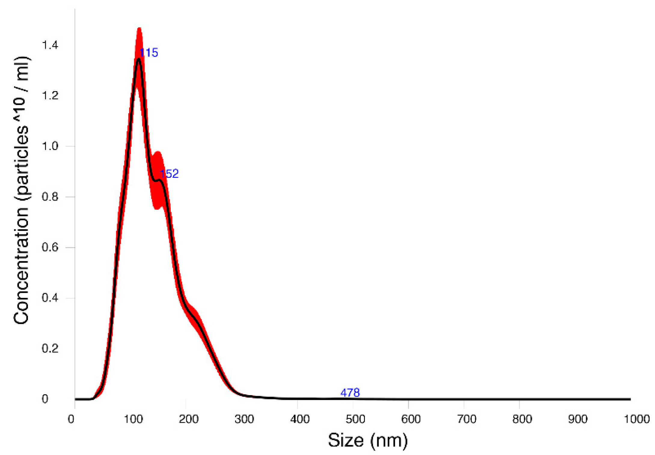
2.2. EVs-Enriched Fraction Characterization

Nanoparticle tracking analysis (NTA), using Nanosight technology, estimated the size distribution and concentration of the particles in the exosomal-enriched fraction (Video S1). The mean size of the particles in the Anisakis EV samples was 141.7 nm (mode, 107.4). Three main numbers of peaks were obtained (main peak, 115 nm), and a concentration of 1.32 × 10¹⁰ particles/mL was reported (Figure 1).
A Western blot with an anti-Alix antibody, performed on the Anisakis EV-enriched fraction, the Anisakis CE, and on the Caco-2 cells as the controls, showed a band at 110 kDa in the positive controls (Caco-2), while in the CE and EVs, the bands were specific but at a higher molecular weight of around 130 kDa (Figure S1), probably due to post-transcriptional modifications of the protein.

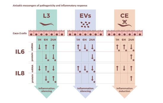
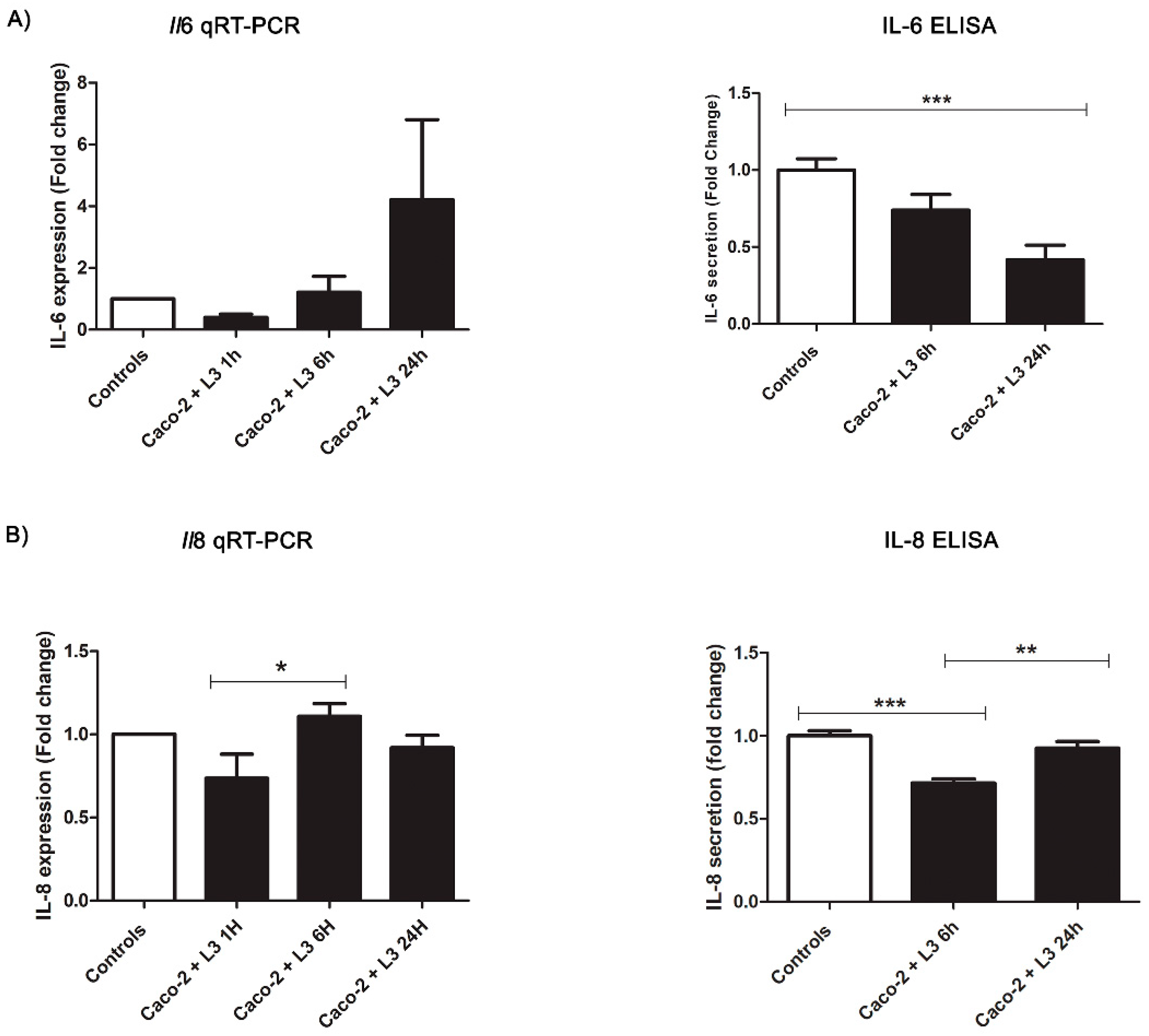
2.3. Intestinal Epithelial Cells’ Response to Live L3 Actions

An additional time point at 6 h was included in the incubation with the L3, in order to monitor its potential mechanical activity on the cells’ monolayer integrity. This time point has been analyzed by ELISA, too.
No morphological changes, such as cytoplasmic vacuolization, shrinkage, plasma membrane blebbing, and chromatin condensation, were observed in the Caco-2 cells after 1 h, 6 h, and 24 h post-contact with the Anisakis products or in the unexposed Caco-2 cells. The challenge with the live L3 showed a progressive decrease of IL-6 in the Caco-2 cells incubated at 6 h (not significant) and 24 h (F(2,37) = 8.143, p = 0.0001) if compared with the non-treated cells. In order to evaluate the gene expression and cellular response over time, qRT-PCR analyses were performed at three different time points (GAPDH Eff: 99.97%, Il-8 Eff: 98.71%, and Il-6 Eff: 90.59%). However, the results obtained for the Il6 gene expression showed no significant changes. On the other hand, levels of the neutrophil chemotactic factor IL-8 (Figure 2) were consistently reduced in the Caco-2 cells incubated with the live L3 for 6 h (F(2,37) = 17.85, p < 0.0001), if compared to the controls, followed by a significant increase at 24 h (6 h vs. 24 h: p < 0.01). However, the IL-8 levels at 24 h did not significantly differ from the controls. Similarly, the gene expression of Il8 showed a slight decrease at 1 h compared to the non-treated cells (not significant), followed by an increase at 6 h (F(2,33) = 3.060, p = 0.0422, 1 h vs. 6 h: p < 0.05) that tended to reach the level of expression as in the controls (controls vs. 6 h = ns, and controls vs. 24 h = ns) (Figure 2).

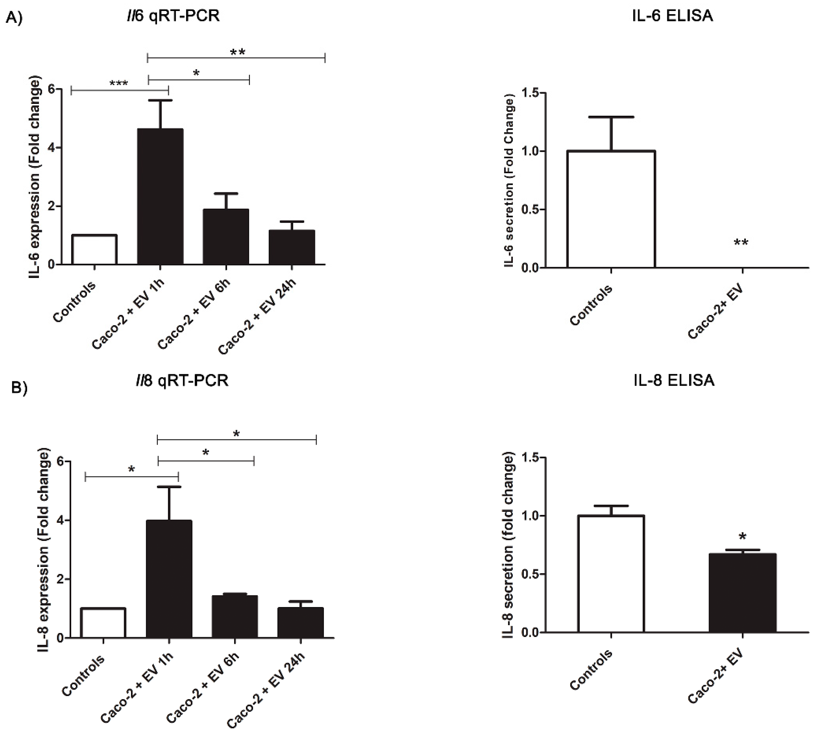
2.4. Intestinal Epithelial Response to Anisakis EVs

The challenges of the Caco-2 cells with the Anisakis EVs revealed a decreasing trend for the two cytokines of interest. In particular, IL-6 was not detected in the ELISA assay compared to the controls (t(29) = 3.42, p = 0.0065). The relative quantification of the Il6 gene expression showed a statistically relevant (F(3,18) = 9.205, p = 0.0007) upregulation at 1 h (p < 0.001), followed by a significant decrease in gene expression at 6 h and 24 h (1 h vs. 6 h: p < 0.05; 1 h vs. 24 h: p < 0.01, respectively), reaching the expression level reported in the controls (Figure 3). A similar pattern was also observed for the IL-8 assays. IL-8 in the Caco-2 cells treated with the EVs after 24 h appeared decreased (t(31) = 2.92, p = 0.0127) compared to the controls, and the Il8 gene expression analysis showed a statistically relevant (F(3,20) = 5.762, p = 0.0052) upregulation at 1 h (p < 0.05) that decreased at the other checkpoints (1 h vs. 6 h: p < 0.05; 1 h vs. 24 h: p < 0.05), reaching the control levels of the gene expression (Figure 3).

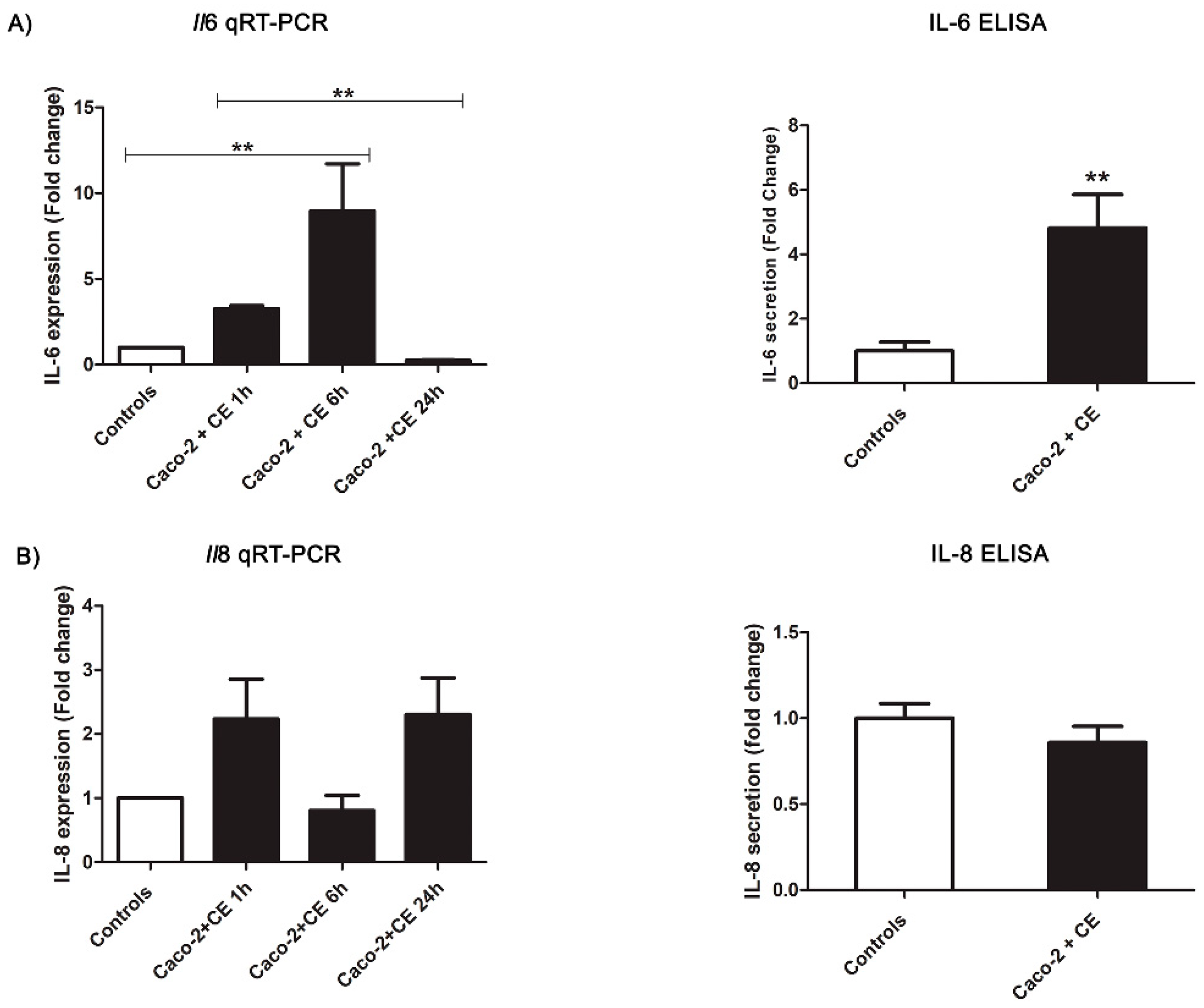
2.5. Intestinal Epithelial Cells’ Response to Anisakis Crude Extract

The number of proteins observed in the CE samples used for the Caco-2 cells challenge was about 900 ng/µL. The Caco-2 cells exposed to the Anisakis CE for 24 h showed a strong, relevant increase in IL-6 secretion (t(22) = 3.64, p = 0.0054) compared to the control group (Figure 4). This evidence was also supported by the qRT-PCR analyses showing an increasing trend in the Il6 gene expression at 1 h (not significant) and 6 h (F(3,20) = 8.143, p = 0.0010). Then, the Il6 expression decreased significantly (6 h vs. 24 h: p < 0.05), reaching the expression level reported in the controls (Figure 4). Interestingly, the CE slightly affected the IL-8 secretion, showing a mild but not significant decrease in the ELISA test compared to the controls (t(24) = 1.49, p = 0.1612) (Figure 4). According to that, a slight decrease in the Il8 gene expression was observed at 6 h, with respect to the other checkpoints analyzed (1 h and 24 h), however, without statistical support (F(3,20) = 2.805, p = 0.0661) (Figure 4).

3. Discussion

Helminths, multicellular worm parasites including nematodes, cestodes, and trematodes, are able to establish long-lasting chronic infections, indicating the successful manipulation of the host immune system, as they need to survive in inhospitable environments during infections. A co-evolutive adaptation is not likely to be applicable to anisakiasis, as humans are accidental hosts; however, despite studies aimed to explore Anisakis ability in modulating humans’ inflammatory and immune response increasing [35,45,46], the fine molecular mechanisms related to its pathogenesis and clinical outcomes are still mostly obscure.
Many parasitic nematodes cannot be easily cultured, and existing in vitro and in vivo models do not sufficiently recount the human background and disease. To date, only two studies used Caco-2 cells to investigate human anisakiasis, without a focus on the cytokines’ release [36,37,38,39,40,41,42]. Hence, the present investigation was performed using Caco-2 cells as a suitable in vitro model able to recreate a microenvironment for Anisakis infection. Accordingly, we tested three challenges mimicking two pivotal phases of anisakiasis: (i). active penetration of the tissue (L3) and communication with the intestinal epitheliums (EVs) and (ii). the larval dying (CE). The present study is the first attempt to explore the cellular response after exposure to the EVs derived from the infective third-stage larvae of the zoonotic nematode Anisakis spp. (A. pegreffii, A. simplex s.s., and their hybrid form).
The evidence, here collected about inflammatory pathways after challenges with Anisakis-derived products, revealed interesting and variable outcomes of the cytokines’ production and gene expression. A few hours after ingestion, the L3 release of proteolytic enzymes and chemotactic factors to adhere to and penetrate the mucosa and submucosa [47,48] induced hemorrhagic and erosive lesions [7] associated with a production of excretory/secretory factors able to interact with the human first line of defense [49]. The first cells to encounter the invading Anisakis L3 were the epithelial cells, a physical barrier also equipped with microbial-detection mechanisms, signaling the circuits and inflammatory mediators [50]. Surprisingly, the present data showed that the Caco-2 cells were not significantly triggered by the presence of the live L3. Hence, the decrease in the IL-6 and IL-8 cytokines’ quantity that was observed could, therefore, reflect a first strategy by which the parasite modulates the epithelial barrier response triggered by the initial contact with the host, quickly returning the system to homeostasis and keeping the host healthy for successful long-lived infections. Such a concept was also described in the phylogenetic-related ascaridoid Ascaris suum [51], in which the transcriptional analysis of L3 incubated with porcine intestinal epithelial cells demonstrated a low magnitude of inflammatory response characterized mainly by IL-8 and NF-kB suppression. Our results, based on ELISA and qRT-PCR analyses, showed live L3 inducing an early and transient downregulation of the host cytokines’ gene expression and protein release. Moreover, the effect of the IL-6 amount was longer than that of IL-8. Despite the significant reduction at 6 h, IL-8 turned to the basal level at 24 h. This is in agreement with a previous study in which IL-8 mRNA alterations were not detected in patient sera exposed to Anisakis simplex [52]. To our knowledge, only Napolitano et al., 2018, have investigated IL-6 secretion in the Anisakis L3-human interaction. Immature and mature dendritic cells (DCs) incubated with seven live specimens of L3 for six days showed an increase in IL-6 secretion and a less reactive phenotype of DC not sufficient to drive a Th2/Th17 immune response [35]. In the present study, consistent outcomes about a suppressive action of live L3 on the first line of human defense were obtained, in this case, based on the decrease of the IL-6 amount.
Besides the live L3, their derived EVs are an additional important factor to consider in the early pathogenicity exerted by Anisakis. EVs are a newly discovered vehicle of early communication between the host and pathogens, since they determine strong immunomodulatory effects [53], as demonstrated for other parasitic nematodes [29,30,32,54,55]. Anisakis live L3 are able to release EVs, as recently demonstrated [56], also, in the same experimental condition here performed [57]. The EVs-enriched fraction used in the present study is the result of the incubation of numerous larval specimens (a pool of 16 to 20 larvae), while usually a single L3 infects a human host. Consequently, a stronger activity of such EVs-enriched fraction in comparison with a single larva is expected. According to this hypothesis and to published data [32,54], we did not detect IL-6 and we observed a decrease in IL-8 in the ELISA at 24 h. However, the expression of the Il6 and Il8 genes showed a different trend. The exposure to EVs induced an early upregulation of both genes, followed by a progressive downregulation over time, even if not significant, compared to the basal level of gene expression in the controls. Based on these results, the attenuation of IL-6 and IL-8 may have occurred via a post-transcriptional mechanism, potentially due to proteases, as observed in other intestinal parasites [58] and usually released also by Anisakis [47], or to other potential regulatory factors such as miRNAs transported in EVs. Alternative explanations may be related to the time points selected that may not be representative of the precise time of the cytokines’ mRNA downregulation. A crucial factor to consider is the timing of the EVs’ uptake by the cells. Heligmosomoides polygyrus EVs are better internalized by epithelial cells (MODE-K) at 24 h (55%) compared to 1 h (10%) [54]; Trichuris muris EVs are internalized by murine colonic organoids within 3 h at 37 °C [30], and Anisakis-derived EVs are internalized by human macrophage-Like THP-1 cells, but a precise indication of the uptake timing is lacking [56]. In this scenario, the early upregulation of the Il6 and Il8 gene expressions, here observed, could be mainly due to the interaction between the host cellular receptors and the Anisakis extracellular EVs’ surface proteins, rather than a function associated with the EVs’ content. Consequently, the observed immunomodulatory effect exerted by live L3-releasing EVs on intestinal cells may be characterized by several not-exclusive mechanisms of action.
A different scenario is detectable in the CE challenge regarding IL-6 production. CE represents a mixture of L3 components (endotoxins, proteases, metalloproteases, and somatic proteins) that may be clinically compared to the moment of L3 death (around 14 days after ingestion) associated with the parasite’s expulsion or granuloma formation and chronic inflammation [7]. Available in vitro studies described a pro-inflammatory activity exerted by Anisakis parasitic products, including the upregulation of oxidative stress, the barrier’s integrity alteration, inhibition of apoptosis-related biomarkers, and inflammatory induction [34,35,36,42]. According to that, our data about Caco-2 cells exposed to CE showed a strong upregulation of IL-6 release and in the Il6 gene expression. This pro-inflammatory enhancement could represent a trigger for the host immune response, able to activate and recruit immune cells (macrophages, DCs, eosinophils, and neutrophils) to the site of damage, determining the arrest or the disease progression (chronic inflammation and granulomas). Additionally, such data are in agreement with those obtained by previous in vivo and in vitro studies on Anisakis CE [34,35,59]. These studies highlighted a pathological condition related to chronic inflammation and, in turn, to a potential progressive increased risk of the host’s DNA damage and cancer, as demonstrated for other helminths [60]. To date, only two studies have investigated these neglected aspects in the framework of anisakiasis. The tumorigenic potential of Anisakis was explored using hamster ovary cells and Sprague–Dawley rats, revealing an increase in cell proliferation, a reduction of apoptosis, and changes in the expression of serum cancer-related miRNAs in rats [59]. Moreover, an increased level of P53 and ROS in the fibroblast cell line, HS-68, treated with Anisakis excretory/secretory products and CE was observed [34]. Interestingly, our results showed that CE did not particularly affect the Il8 gene expression and secretion by Caco-2 cells, suggesting IL-8 as a non-crucial factor involved in anisakiasis. Numerous parasite-derived factors have been reported to recruit, often selectively, neutrophils or eosinophils, and the somatic extracts of A. simplex did not elicit a measurable chemotactic response in the neutrophils, as previously suggested [61].

4. Materials and Methods

4.1. Parasites Sampling

Anisakis live third-stage infective larvae (L3) were collected from the visceral cavity of 21 European hakes Merluccius merluccius purchased at markets between 2018 and 2021 and captured from area FAO 37. L3 were washed three times in a 0.22 µm filtered phosphate buffer saline (PBS). After the experiments, all the L3 used for the study were stored at −20 °C for subsequent identification of species through a molecular diagnostic key based on the PCR-RFLP of the internal transcribed spacers of the nuclear ribosomal DNA, according to D’Amelio et al., 2000 [62].

4.2. Crude Extract

A pool of L3 (n = three) was used for obtaining crude extract samples. CE was prepared as follows: PBS 10X was added to each sample and then homogenated with pestles. The mixture (0.13 mg/mL) was centrifuged at 13,500 rpm for 30 min. at 4 °C, according to Mattiucci et al., 2017 [63]. Lastly, the supernatant was used for the challenge with Caco-2 cells, whereas the pellets were stored in 70% ethanol for the subsequent identification of species. The protein concentration of samples was evaluated using a Qubit4 (Thermo Fisher Scientific, Waltham, MA, USA).

4.3. Isolation and EVs-Enriched Fraction Characterization

Pools of L3 (n = 20) were incubated in RPMI with 1X Pen/Strep for 24 h at 37 °C with 5% CO2. After incubation, the culture media were collected to isolate the exosomal-enriched fraction of the EVs using an ExoQuick kit (System Biosciences, Palo Alto, CA, USA) in accordance with the manufacturer’s protocol. The samples obtained were diluted in a 0.22 µm filtered PBS and immediately used for incubation with Caco-2 cells, while the larvae were stored for the subsequent identification of species.
The size distribution and concentration of particles in the recovered fractions after the exosome enrichment were measured using nanoparticle tracking analysis (NTA), using a Nanosight NS300 (Malvern Panalytical, Malvern, UK). Five measurements were performed with a 60 s duration for each measurement and the data were analyzed using NTA software version 3.4.
To further characterize the presence of extracellular vesicles in our samples, Western blotting analysis of the isolated EVs against the cytosolic EVs’ marker ALG-2-interacting protein X (Alix) antibodies were undertaken. The protein concentration of the EV samples was evaluated using a Qubit4 (Thermo Fisher Scientific, Waltham, MA, USA). The CE sample (used as the larval control), Caco-2 cells (used as a positive control for the antibody), and the EV samples were lysed in a 5X (CE and EVs) and 1X (Caco-2 cells) sample buffer (50 mMTris–HCl pH 6.8, 2% SDS, 10% glycerol, 5% β-mercaptoethanol). About 8 µg of proteins were resolved on a 12% Tris–glycine SDS-PAGE and transferred onto nitrocellulose membranes (Hybond-P GE-Healthcare, Sigma-Aldrich St. Louis, MO, USA). Western blot analyses were performed using polyclonal, rabbit anti-Alix (1:500, TBS-T, 5% NFDM) from Thermo Fisher Scientific, USA (Catalog # PA5-52873). Anti-rabbit antibody conjugated to horseradish peroxidase (Bio-Rad) was used as a secondary antibody (1:2000). Blots were visualized by an enhanced chemiluminescence system (GE-Healthcare Bio-Sciences, Milan, Italy).

4.4. Cell Culture and Challenging Experiments

Caco-2 cells (ATCC HTB-37) were grown in a Dulbecco’s modified Eagle’s medium (DMEM) supplemented with 10% fetal bovine serum and grown at 37 °C in the presence of 5% CO2. Cells were seeded at a density of 1 × 10⁵ cell/mL in 35 mm cell culture dishes and grown for 48 h prior to being challenged with CE and EVs. Otherwise, cells were grown for 14 days (full-confluent and polarized monolayers) before being incubated with live L3 larvae.
To ensure that the Caco-2 monolayer was not affected and disrupted by the larval mechanical action, we performed two-time points for ELISA incubation (6 h and 24 h) while for the CE and EVs challenge, a single time point at 24 h was assessed. To evaluate a potential temporal dynamic early host–pathogen interaction on the cytokines’ gene expression (Ebner et al., 2018) [51], three-time points at 1 h, 6 h, and 24 h for the qRT-PCR were assessed. After the indicated time points, cell supernatants were collected and stored at −80 °C for ELISA analyses, while cells were lysed and whole cell extracts were used for qRT-PCR analyses.

4.5. Cytokines’ Measurements

Supernatants from cells challenged with live Anisakis L3, EVs, and CE were analyzed with ELISA assays (Thermo Fisher Scientific, Milan, Italy) to determine IL-6 and IL-8 amounts, according to the manufacturer’s protocol.

4.6. Relative Quantification of Gene Expression by Real-Time PCR Analyses

Total RNA was obtained from Caco-2 cells using a TRIsure™ reagent (Bioline, London, UK), and contaminating genomic DNA was removed using a Turbo DNA-free kit (Life Technologies, Carlsbad, CA, USA). The amount of RNA was evaluated by spectrophotometric measurements using the Take3 module of the plate reader, BioTek SynergyHT and GEN5™, and the RNA integrity was evaluated through a run in an agarose gel 1.5%, stained with Syber safe (Invitrogen Waltham, Waltham, MA, USA). A total amount of 1µg RNA was reverse-transcribed for each sample, using SuperScript II RT (Invitrogen Waltham, Waltham, MA, USA) and OligodT (Invitrogen Waltham, Waltham, MA, USA) according to the manufacturer’s protocol. Target genes for the relative quantification by real-time PCR were IL-6 and IL-8, using GAPDH as the endogenous control. cDNA templates were mixed with a 2× PowerUp™ SYBR™ Green Master Mix (Applied Biosystem, Foster City, CA, USA) and specific primers, according to Borkowski et al. (Borkowski et al., 2014). Reactions included an initial holding stage of 2 min at 50 °C and of 2 min at 95 °C, followed by 40 cycles of PCR (95 °C, 15 s; 60 °C, 1 min); a final stage for melting curves was included to verify the specificity of the amplifications. A fold change in the expression level was calculated using the ∆∆Ct (Delta–Delta Ct) method.

4.7. Statistical Analysis

ELISA protocols for IL-6 and IL-8 accounted for the calibrated reference, according to the manufacturer. The relative quantifications of Il6 and Il8 gene expression in RT-PCR were obtained using GADPH as a reference gene. The statistical significance of data was determined using a Student’s paired t-test to analyze statistical differences between two groups and a one-way analysis of variance (ANOVA) followed by a Bonferroni’s multiple comparison test to compare three or more groups. Statistical significance was considered from p ≤ 0.05.

5. Conclusions

In conclusion, the results here obtained showed an intricate interplay between the parasite and host; the live, active larva and its released EVs tried to silence the host’s immune response at the intestinal epithelium level to find a long-lasting niche to remain alive. Later on, a dying larva could induce the activation of the immune strategy of the host, leading to parasite expulsion, eosinophilia, and/or granuloma formation. Furthermore, the three challenges, representing different pathology phases, showed how the effects produced by Anisakis on the human host can be manifold, as also suggested in previous studies (Messina et al., 2016) [34]. In this scenario, it would be interesting to deeply investigate the molecular mechanisms at the base of this fine play of roles, expanding the research to other factors that could add important information about anisakiasis, as well as about the tumorigenic potential of these parasites.

Supplementary Materials

The following supporting information can be downloaded at: https://www.mdpi.com/article/10.3390/pathogens11101214/s1, Video S1: EVs’ NTA; Figure S1: Western Blot ALIX (CE: crude extract; EVs: extracellular vesicles; Caco-2 cells; L: ladder).

Author Contributions

Conceptualization: S.C., S.D. and I.B.; methodology: I.B., D.S., C.A., A.P. and M.S.; software: S.C., I.B. and D.S.; validation: S.C., I.B. and D.S.; formal analysis: S.C. and I.B.; resources: S.C., S.D. and A.T.P.; writing—original draft preparation: I.B.; writing—review and editing: all authors. All authors have read and agreed to the published version of the manuscript.

Funding

This research was funded by the Sapienza University of Rome, grant number RP11916B69CBB411 to S.C. and grant number AR12117A7B3BE784 to I.B.

Institutional Review Board Statement

Not applicable.

Informed Consent Statement

Not applicable.

Data Availability Statement

Not applicable.

Acknowledgments

We thank Carlo Taccari for his graphical support.

Conflicts of Interest

The authors declare no conflict of interest.

References

  1. Klimpel, S.; Palm, H.W. Anisakid Nematode (Ascaridoidea) Life Cycles and Distribution: Increasing Zoonotic Potential in the Time of Climate Change? In Progress in Parasitology; Parasitology Research Monographs, 2; Mehlhorn, H., Ed.; Springer: Berlin/Heidelberg, Germany, 2011. [Google Scholar]
  2. Panel EFSA. On biological hazards (BIOHAZ) scientific opinion on risk assessment of parasites in fishery products. EFSA J. 2010, 8, 1543. [Google Scholar]
  3. Cong, W.; Elsheikha, H.M. Biology, Epidemiology, Clinical Features, Diagnosis, and Treatment of Selected Fish-borne Parasitic Zoonoses. Yale J. Biol. Med. 2021, 30, 297–309. [Google Scholar]
  4. Audicana, M.T.; Ansotegui, I.J.; de Corres, L.F.; Kennedy, M.W. Anisakis simplex: Dangerous-dead and alive? Trends Parasitol. 2002, 18, 20–25. [Google Scholar] [CrossRef]
  5. Arizono, N.; Yamada, M.; Tegoshi, T.; Yoshikawa, M. Anisakis simplex sensu stricto and Anisakis pegreffii: Biological characteristics and pathogenetic potential in human anisakiasis. Foodborne Pathog. Dis. 2012, 9, 517–521. [Google Scholar] [CrossRef] [PubMed]
  6. Hrabar, J.; Trumbić, Z.; Bočina, I.; Buselić, I.; Vrbatović, A.; Mladineo, I. Interplay between proinflammatory cytokines, miRNA, and tissue lesions in Anisakis-infected Sprague-Dawley rats. PLoS Negl. Trop Dis. 2019, 13, e0007397. [Google Scholar] [CrossRef]
  7. Audicana, M.T.; Kennedy, M.W. Anisakis simplex: From obscure infectious worm to inducer of immune hypersensitivity. Clin. Microb. Rev. 2008, 21, 360–379. [Google Scholar] [CrossRef]
  8. Nieuwenhuizen, N.E.; Lopata, A.L. Anisakis—A food-borne parasite that triggers allergic host defences. Int. J. Parasitol. 2013, 43, 1047–1057. [Google Scholar] [CrossRef] [PubMed]
  9. D’Amelio, S.; Lombardo, F.; Pizzarelli, A.; Bellini, I.; Cavallero, S. Advances in Omic Studies Drive Discoveries in the Biology of Anisakid Nematodes. Genes 2020, 11, 801. [Google Scholar] [CrossRef] [PubMed]
  10. Suzuki, J.; Murata, R.; Kodo, Y. Current Status of Anisakiasis and Anisakis Larvae in Tokyo, Japan. Food Saf. 2021, 9, 89–100. [Google Scholar] [CrossRef]
  11. Mattiucci, S.; Palomba, M.; Cavallero, S.; D’Amelio, S. Anisakiasis. In Helminth Infections and Their Impact on Global Public Health, 2nd ed; Bruschi, F., Ed.; Springer: Berlin/Heidelberg, Germany, 2022; pp. 451–495. [Google Scholar]
  12. Jerončić, A.; Nonković, D.; Vrbatović, A.; Hrabar, J.; Bušelić, I.; Martínez-Sernández, V.; Rocamonde, S.A.L.; Ubeira, F.M.; Jaman, S.; Jeličić, E.Č.; et al. Anisakis Sensitization in the Croatian fish processing workers: Behavioral instead of occupational risk factors? PLoS Negl. Trop Dis. 2020, 14, e0008038. [Google Scholar] [CrossRef]
  13. Yera, H.; Fréalle, É.; Dutoit, E.; Dupouy-Camet, J. A national retrospective survey of anisakidosis in France (2010–2014): Decreasing incidence, female predominance, and emerging allergic potential. Parasite 2018, 25, 23. [Google Scholar] [CrossRef]
  14. Herrador, Z.; Daschner, Á.; Perteguer, M.J.; Benito, A. Epidemiological Scenario of Anisakidosis in Spain Based on Associated Hospitalizations: The Tip of the Iceberg. Clin. Infect. Dis. 2019, 69, 69–76. [Google Scholar] [CrossRef]
  15. Cavallero, S.; Martini, A.; Migliara, G.; De Vito, C.; Iavicoli, S.; D’Amelio, S. Anisakiasis in Italy: Analysis of hospital discharge records in the years 2005–2015. PLoS ONE 2018, 13, e0208772. [Google Scholar] [CrossRef] [PubMed]
  16. Bao, M.; Pierce, G.J.; Pascual, S.; González-Muñoz, M.; Mattiucci, S.; Mladineo, I.; Cipriani, P.; Bušelić, I.; Strachan, J.C. Assessing the risk of an emerging zoonosis of worldwide concern: Anisakiasis. Sci. Rep. 2017, 7, 43699. [Google Scholar] [CrossRef] [PubMed]
  17. Surapaitoon, A.; Suttiprapa, S.; Mairiang, E.; Khuntikeo, N.; Pairojkul, C.; Bethony, J.; Brindley, P.J.; Sripa, B. Subsets of Inflammatory Cytokine Gene Polymorphisms are Associated with Risk of Carcinogenic Liver Fluke Opisthorchis viverrini-Associated Advanced Periductal Fibrosis and Cholangiocarcinoma. Korean J. Parasitol. 2017, 55, 295–304. [Google Scholar] [CrossRef] [PubMed]
  18. Dematei, A.; Fernandes, R.; Soares, R.; Alves, H.; Richter, J.; Botelho, M.C. Angiogenesis in Schistosoma haematobium-associated urinary bladder cancer. APMIS 2017, 125, 1056–1062. [Google Scholar] [CrossRef]
  19. Kim, T.S.; Pak, J.H.; Kim, J.B.; Bahk, Y.Y. Clonorchis sinensis, an oriental liver fluke, as a human biological agent of cholangiocarcinoma: A brief review. BMB Rep. 2016, 49, 590–597. [Google Scholar] [CrossRef]
  20. Sonoda, H.; Yamamoto, K.; Ozeki, K.; Inoye, H.; Toda, S.; Maehara, Y. An Anisakis larva attached to early gastric cancer: Report of a case. Surg. Today 2015, 45, 1321–1325. [Google Scholar] [CrossRef]
  21. Murata, Y.; Ando, K.; Usui, M.; Sugiyama, H.; Hayashi, A.; Tanemura, A.; Kuriyama, N.; Kishiwada, M.; Mizuno, S.; Sakurai, H.; et al. A case of hepatic anisakiasis caused by Pseudoterranova decipiens mimicking metastatic liver cancer. BMC Infect. Dis. 2018, 18, 619. [Google Scholar] [CrossRef]
  22. Kita, R.; Hashida, H.; Uryuhara, K.; Kaihara, S. Hepatic anisakiasis mimicking metastatic liver tumour. Int. J. Surg. 2019, 60, 209–212. [Google Scholar] [CrossRef]
  23. Rawla, P.; Sunkara, T.; Barsouk, A. Epidemiology of colorectal cancer: Incidence, mortality, survival, and risk factors. Prz. Gastroenterol. 2019, 14, 89–103. [Google Scholar] [CrossRef]
  24. Aibinu, I.E.; Smooker, P.M.; Lopata, A.L. Anisakis Nematodes in Fish and Shellfish- from infection to allergies. Int. J. Parasitol. Parasites Wildl. 2019, 6, 384–393. [Google Scholar] [CrossRef]
  25. Petithory, J.C.; Paugam, B.; Buyet-Rousset, P.; Paugam, A. Anisakis simplex, a cofactor of gastric cancer? Lancet 1990, 336, 1002. [Google Scholar] [CrossRef]
  26. Mineta, S.; Shimanuki, K.; Sugiura, A.; Tsuchiya, Y.; Kaneko, M.; Sugiyama, Y.; Akimaru, K.; Tajiri, T. Chronic anisakiasis of the ascending colon associated with carcinoma. J. Nippon. Med. Sch. 2006, 73, 169–174. [Google Scholar] [CrossRef] [PubMed]
  27. Garcia-Perez, J.C.; Rodríguez-Perez, R.; Ballestero, A.; Zuloaga, J.; Fernandez-Puntero, B.; Arias-Díaz, J.; Caballero, M.L. Previous Exposure to the Fish Parasite Anisakis as a Potential Risk Factor for Gastric or Colon Adenocarcinoma. Medicine 2015, 94, e1699. [Google Scholar] [CrossRef] [PubMed]
  28. Coakley, G.; Maizels, R.M.; Buck, A.H. Exosomes and Other Extracellular Vesicles: The New Communicators in Parasite Infections. Trends Parasitol. 2015, 31, 477–489. [Google Scholar] [CrossRef]
  29. Hansen, E.P.; Fromm, B.; Andersen, S.D.; Marcilla, A.; Andersen, K.L.; Borup, A.; Williams, A.R.; Jex, A.R.; Gasser, R.B.; Young, N.D.; et al. Exploration of extracellular vesicles from Ascaris suum provides evidence of parasite–host cross talk. J. Extracell. Vesicles 2019, 8, 1578116. [Google Scholar] [CrossRef]
  30. Eichenberger, R.M.; Hasanuzzaman Talukder, M.D.; Field, M.A.; Wangchuk, P.; Giacomin, P.; Loukas, A.; Sotillo, J. Characterization of Trichuris muris secreted proteins and extracellular vesicles provides new insights into host–parasite communication. J. Extracell. Vesicles 2018, 7, 1428004. [Google Scholar] [CrossRef]
  31. Saliminejad, K.; Khorram Khorshid, H.R.; Soleymani Fard, S.; Ghaffari, S.H. An overview of microRNAs: Biology, functions, therapeutics, and analysis methods. J. Cell Physiol. 2019, 234, 5451–5465. [Google Scholar] [CrossRef]
  32. Buck, A.; Coakley, G.; Simbari, F.; McSorley, H.J.; Quintana, J.F.; Le Bihan, T.; Kumar, S.; Abreu-Goodger, C.; Lear, M.; Harcus, Y.; et al. Exosomes secreted by nematode parasites transfer small RNAs to mammalian cells and modulate innate immunity. Nat. Commun. 2014, 5, 5488. [Google Scholar] [CrossRef]
  33. Zakeri, A.; Hansen, E.P.; Andersen, S.D.; Williams, A.R.; Nejsum, P. Immunomodulation by Helminths: Intracellular Pathways and Extracellular Vesicles. Front. Immunol. 2018, 9, 2349. [Google Scholar] [CrossRef]
  34. Messina, C.M.; Pizzo, F.; Santulli, A.; Bušelić, I.; Boban, M.; Orhanović, S.; Mladineo, I. Anisakis pegreffii (Nematoda: Anisakidae) products modulate oxidative stress and apoptosis-related biomarkers in human cell lines. Parasit Vectors 2016, 9, 607. [Google Scholar] [CrossRef]
  35. Napoletano, C.; Mattiucci, S.; Colantoni, A.; Battisti, F.; Zizzari, I.G.; Rahimi, H.; Nuti, M.; Rughetti, A. Anisakis pegreffii impacts differentiation and function of human dendritic cells. Parasite Immunol. 2018, 40, e12527. [Google Scholar] [CrossRef]
  36. Speciale, A.; Trombetta, D.; Saija, A.; Panebianco, A.; Giarratana, F.; Ziino, G.; Minciullo, P.L.; Cimino, F.; Gangemi, S. Exposure to Anisakis extracts can induce inflammation on in vitro cultured human colonic cells. Parasitol. Res. 2017, 116, 2471–2477. [Google Scholar] [CrossRef]
  37. Lea, T. Caco-2 Cell Line. In The Impact of Food Bioactives on Health: In Vitro and Ex Vivo Models; Verhoeckx, K., Cotter, P., López-Expósito, I., Kleiveland, C., Lea, T., Mackie, A., Requena, T., Swiatecka, D., Wichers, H., Eds.; Springer: Cham, Switzerland, 2015. [Google Scholar]
  38. Bautista, D.; Rodríguez, L.S.; Franco, M.A.; Angel, J.; Barreto, A. Caco-2 cells infected with rotavirus release extracellular vesicles that express markers of apoptotic bodies and exosomes. Cell Stress Chaperones 2015, 20, 697–708. [Google Scholar] [CrossRef]
  39. Ambrosi, C.; Pompili, M.; Scribano, D.; Limongi, D.; Petrucca, A.; Cannavacciuolo, S. The Shigella flexneri OspB effector: An early immunomodulator. Int. J. Med. Microbiol. 2015, 305, 75–84. [Google Scholar] [CrossRef]
  40. Chaiyadet, S.; Smout, M.; Johnson, M.; Whitchurch, C.; Turnbull, L.; Kaewkes, S.; Sotillo, J.; Loukas, A.; Sripa, B. Excretory/secretory products of the carcinogenic liver fluke are endocytosed by human cholangiocytes and drive cell proliferation and IL6 production. Int. J. Parasitol. 2015, 45, 773–781. [Google Scholar] [CrossRef]
  41. Ma’ayeh, S.Y.; Knörr, L.; Sköld, K.; Garnham, A.; Ansell, B.R.E.; Jex, A.R.; Svard, S.G. Responses of the Differentiated Intestinal Epithelial Cell Line Caco-2 to Infection with the Giardia intestinalis GS Isolate. Front. Cell Infect. Microbiol. 2018, 8, 244. [Google Scholar] [CrossRef] [PubMed]
  42. Carballeda-Sangiao, N.; Sánchez-Alonso, I.; Navas, A.; Arcos, S.C.; de Palencia, P.F.; Careche, M.; González-Muñoz, M. Anisakis simplex products impair intestinal epithelial barrier function and occludin and zonula occludens-1 localisation in differentiated Caco-2 cells. PLoS Negl. Trop Dis. 2020, 14, e0008462. [Google Scholar] [CrossRef] [PubMed]
  43. Ha, H.; Debnath, B.; Neamati, N. Role of the CXCL8-CXCR1/2 Axis in Cancer and Inflammatory Diseases. Theranostics 2017, 7, 1543. [Google Scholar] [CrossRef] [PubMed]
  44. Jones, S.A. Directing transition from innate to acquired immunity: Defining a role for IL-6. J. Immunol. 2005, 175, 3463–3468. [Google Scholar] [CrossRef]
  45. Rodero, M.; Cuéllar, C. Modulation by Anisakis simplex antigen of inflammatory response generated in experimental autoimmune encephalomyelitis. Int. Immunopharmacol. 2021, 90, 107241. [Google Scholar] [CrossRef] [PubMed]
  46. Zamora, V.; Andreu-Ballester, J.C.; Rodero, M.; Cuéllar, C. Anisakis simplex: Immunomodulatory effects of larval antigens on the activation of Toll like Receptors. Int. Immunopharmacol. 2021, 100, 1567–5769. [Google Scholar] [CrossRef] [PubMed]
  47. Kim, J.H.; Kim, J.O.; Jeon, C.H.; Nam, U.H.; Subramaniyam, S.; Yoo, S.I.; Park, J.H. Comparative transcriptome analyses of the third and fourth stage larvae of Anisakis simplex (Nematoda: Anisakidae). Mol. Biochem. Parasitol. 2018, 226, 24–33. [Google Scholar] [CrossRef] [PubMed]
  48. Kochanowski, M.; Dąbrowska, J.; Różycki, M.; Sroka, J.; Karamon, J.; Bełcik, A.; Korpysa-Dzirba, W.; Cencek, T. Proteomic Profiling and In Silico Characterization of the Secretome of Anisakis simplex Sensu Stricto L3 Larvae. Pathogens 2022, 11, 246. [Google Scholar] [CrossRef] [PubMed]
  49. Mehrdana, F.; Buchmann, K. Excretory/secretory products of anisakid nematodes: Biological and pathological roles. Acta Vet. Scand. 2017, 59, 42. [Google Scholar] [CrossRef]
  50. Hammad, H.; Lambrecht, B.N. Barrier Epithelial Cells and the Control of Type 2 Immunity. Immunity 2015, 43, 29–40. [Google Scholar] [CrossRef]
  51. Ebner, F.; Kuhring, M.; Radonić, A.; Midha, A.; Renard, B.Y.; Hartmann, S. Silent Witness: Dual-Species Transcriptomics Reveals Epithelial Immunological Quiescence to Helminth Larval Encounter and Fostered Larval Development. Front. Immunol. 2018, 9, 1868. [Google Scholar] [CrossRef]
  52. Del Pozo, V.; Arrieta, I.; Tuñon, T.; Cortegano, I.; Gomez, B.; Cárdaba, B. Immunopathogenesis of human gastrointestinal infection by Anisakis simplex. J. Allergy Clin. Immunol. 1999, 104, 637–643. [Google Scholar] [CrossRef]
  53. Drurey, C.; Lindholm, H.T.; Coakley, G.; Poveda, M.C.; Löser, S.; Doolan, R.; Gerbe, F.; Jay, P.; Harris, N.; Oudhoff, M.J.; et al. Intestinal epithelial tuft cell induction is negated by a murine helminth and its secreted products. J. Exp. Med. 2022, 219, e20211140. [Google Scholar] [CrossRef]
  54. Coakley, G.; McCaskill, J.L.; Borger, J.G.; McSorley, H.J.; Maizels, R.M.; Buck, A.H. Extracellular Vesicles from a Helminth Parasite Suppress Macrophage Activation and Constitute an Effective Vaccine for Protective Immunity. Cell Rep. 2017, 19, 1545–1557. [Google Scholar] [CrossRef]
  55. Ricciardi, A.; Bennuru, S.; Tariq, S.; Kaur, S.; Wu, W.; Elkahloun, A.G.; Arakelyan, A.; Shaik, J.; Dorward, D.W.; Nutman, T.B.; et al. Extracellular vesicles released from the filarial parasite Brugia malayi downregulate the host mTOR pathway. PLoS Negl. Trop Dis. 2021, 15, e0008884. [Google Scholar] [CrossRef]
  56. Boysen, A.T.; Whitehead, B.; Stensballe, A.; Carnerup, A.; Nylander, T.; Nejsum, P. Fluorescent Labeling of Helminth Extracellular Vesicles Using an In Vivo Whole Organism. Approach Biomed. 2020, 8, 213. [Google Scholar] [CrossRef]
  57. Cavallero, S.; Bellini, I.; Pizzarelli, A.; Arcà, B.; D’Amelio, S. A miRNAs catalogue from third-stage larvae and extracellular vesicles of Anisakis pegreffii provides new clues for host-parasite interplay. Sci. Rep. 2022, 12, 9667. [Google Scholar] [CrossRef]
  58. Cotton, J.A.; Bhargava, A.; Ferraz, J.G.; Yates, R.M.; Beck, P.L.; Buret, A.G. Giardia duodenalis cathepsin B proteases degrade intestinal epithelial interleukin-8 and attenuate interleukin-8-induced neutrophil chemotaxis. Infect Immun. 2014, 82, 2772–2787. [Google Scholar] [CrossRef]
  59. Corcuera, M.T.; Rodríguez-Bobada, C.; Zuloaga, J.; Gómez-Aguado, F.; Rodríguez-Perez, R.; Mendizabal, Á.; González, P.; Arias-Díaz, J.; Caballero, M.L. Exploring tumourigenic potential of the parasite Anisakis: A pilot study. Parasitol. Res. 2018, 117, 3127–3136. [Google Scholar] [CrossRef] [PubMed]
  60. Brindley, P.J.; Loukas, A. Helminth infection–induced malignancy. PLoS Pathog. 2017, 13, e1006393. [Google Scholar] [CrossRef]
  61. Falcone, F.H.; Rossi, A.G.; Sharkey, R.; Brown, A.P.; Pritchard, D.I.; Maizels, R.M. Ascaris suum-Derived Products Induce Human Neutrophil Activation via a G Protein-Coupled Receptor That Interacts with the Interleukin-8 Receptor Pathway. Infect. Immun. 2001, 69, 4007–4018. [Google Scholar] [CrossRef] [PubMed]
  62. D’Amelio, S.; Mathiopoulos, K.D.; Santos, C.P.; Pugachev, O.N.; Webb, S.C.; Picanço, M.; Paggi, L. Genetic markers in ribosomal DNA for the identification of members of the genus Anisakis (Nematoda: Ascaridoidea) defined by polymerase-chain-reaction-based restriction fragment length polymorphism. Int. J. Parasitol. 2000, 30, 223–226. [Google Scholar] [CrossRef]
  63. Mattiucci, S.; Colantoni, A.; Crisafi, B.; Mori-Ubaldini, F.; Caponi, L.; Fazii, P.; Nascetti, G.; Bruschi, F. IgE sensitization to Anisakis pegreffii in Italy: Comparison of two methods for the diagnosis of allergic anisakiasis. Parasite Immunol. 2017, 39, e12440. [Google Scholar] [CrossRef] [PubMed]
Figure 1. Finite track length adjustment (FTLA) concentration/size image for nanoparticle tracking analysis (NTA) of extracellular vesicles secreted by third-stage larvae of Anisakis spp. (Anisakis simplex sensu stricto, Anisakis pegreffii, and the hybrid form). The number of particles is intended as ^10.
Pathogens 11 01214 g001
Figure 2. Anisakis live larvae (L3) modulation of cytokines’ secretion in Caco-2 cell monolayers after different times of incubation (1 h, 6 h, and 24 h). (A) Il6 gene expression and IL-6 levels in Caco-2 cell monolayers. (B) Il8 gene expression and IL-8 levels in Caco-2 cell monolayers. Data are expressed as a fold change compared to the control samples and as means ± SEM (standard error mean). Significance was evaluated using an ANOVA test and Bonferroni multiple comparison test. * p ≤ 0.05, ** p ≤ 0.01, and *** p ≤ 0.001.
Pathogens 11 01214 g002
Figure 3. Anisakis extracellular vesicles (EVs) modulation of cytokines’ secretion in Caco-2 cell monolayers after different times of incubation (1 h, 6 h, and 24 h). (A) Il6 gene expression and IL-6 levels in Caco-2 cell monolayers. (B) Il8 gene expression and IL-8 levels in the Caco-2 cell monolayer. Data are expressed as a fold change compared to the control samples and as means ± SEM (standard error mean). Significance was evaluated using an ANOVA test and Bonferroni multiple comparison test and a Student’s t-test pairing for the Caco-2 cells’ controls vs. the Caco-2 cells exposed. * p ≤ 0.05, ** p ≤ 0.01, and *** p ≤ 0.001.
Pathogens 11 01214 g003
Figure 4. Anisakis crude extract (CE) modulation of cytokines’ secretion in Caco-2 cell monolayers after different times of incubation (1 h, 6 h, and 24 h). (A) Il6 gene expression and IL-6 levels in Caco-2 cell monolayers. (B) Il8 gene expression and IL-8 levels in Caco-2 cell monolayers. Data are expressed as a fold change compared to the control samples and as means ± SEM (standard error mean). Significance was evaluated using an ANOVA test and Bonferroni multiple comparison test and a Student’s t-test pairing for the Caco-2 cells’ controls vs. the Caco-2 cells exposed. ** p ≤ 0.01.
Pathogens 11 01214 g004
Publisher’s Note: MDPI stays neutral with regard to jurisdictional claims in published maps and institutional affiliations.

