Next Issue
Volume 12, February
Previous Issue
Volume 11, December


Viruses, Volume 12, Issue 1 (January 2020) – 125 articles

Cover Story (view full-size image): 
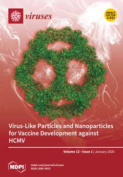
Presenting antigen as a repetitive array in a multivalent fashion on virus-like particles (VLP) or nanoparticles (NP) drives a stronger immune response than free antigen. This effect mainly derives from stronger B cell activation through antigen-driven cross-linking of B cell receptors (BCRs) and potentially modified antigen trafficking. In this issue of Viruses, Perotti and Perez review several VLP and NP technologies that could enhance efficacy against the human cytomegalovirus (HCMV).View this paper.
  • Issues are regarded as officially published after their release is announced to the table of contents alert mailing list.
  • You may sign up for e-mail alerts to receive table of contents of newly released issues.
  • PDF is the official format for papers published in both, html and pdf forms. To view the papers in pdf format, click on the "PDF Full-text" link, and use the free Adobe Reader to open them.
Order results
Result details
Section
Select all
Export citation of selected articles as:
12 pages, 1879 KiB  
Article
Characterization of the Immune Response of MERS-CoV Vaccine Candidates Derived from Two Different Vectors in Mice
by Entao Li, Feihu Yan, Pei Huang, Hang Chi, Shengnan Xu, Guohua Li, Chuanyu Liu, Na Feng, Hualei Wang, Yongkun Zhao, Songtao Yang and Xianzhu Xia
Viruses 2020, 12(1), 125; https://doi.org/10.3390/v12010125 - 20 Jan 2020
Cited by 29 | Viewed by 7042
Abstract
Middle East respiratory syndrome (MERS) is an acute, high-mortality-rate, severe infectious disease caused by an emerging MERS coronavirus (MERS-CoV) that causes severe respiratory diseases. The continuous spread and great pandemic potential of MERS-CoV make it necessarily important to develop effective vaccines. We previously [...] Read more.
Middle East respiratory syndrome (MERS) is an acute, high-mortality-rate, severe infectious disease caused by an emerging MERS coronavirus (MERS-CoV) that causes severe respiratory diseases. The continuous spread and great pandemic potential of MERS-CoV make it necessarily important to develop effective vaccines. We previously demonstrated that the application of Gram-positive enhancer matrix (GEM) particles as a bacterial vector displaying the MERS-CoV receptor-binding domain (RBD) is a very promising MERS vaccine candidate that is capable of producing potential neutralization antibodies. We have also used the rabies virus (RV) as a viral vector to design a recombinant vaccine by expressing the MERS-CoV S1 (spike) protein on the surface of the RV. In this study, we compared the immunological efficacy of the vaccine candidates in BALB/c mice in terms of the levels of humoral and cellular immune responses. The results show that the rabies virus vector-based vaccine can induce remarkably earlier antibody response and higher levels of cellular immunity than the GEM particles vector. However, the GEM particles vector-based vaccine candidate can induce remarkably higher antibody response, even at a very low dose of 1 µg. These results indicate that vaccines constructed using different vaccine vector platforms for the same pathogen have different rates and trends in humoral and cellular immune responses in the same animal model. This discovery not only provides more alternative vaccine development platforms for MERS-CoV vaccine development, but also provides a theoretical basis for our future selection of vaccine vector platforms for other specific pathogens. Full article
(This article belongs to the Section SARS-CoV-2 and COVID-19)
Show Figures

Graphical abstract

9 pages, 2939 KiB  
Article
Identification of a Novel Papillomavirus Associated with Squamous Cell Carcinoma in a Domestic Cat
by Maura Carrai, Kate Van Brussel, Mang Shi, Ci-Xiu Li, Wei-Shan Chang, John S. Munday, Katja Voss, Alicia McLuckie, David Taylor, Andrew Laws, Edward C. Holmes, Vanessa R. Barrs and Julia A. Beatty
Viruses 2020, 12(1), 124; https://doi.org/10.3390/v12010124 - 20 Jan 2020
Cited by 21 | Viewed by 9968
Abstract
Papillomaviruses infect the skin and mucosal surfaces of diverse animal hosts with consequences ranging from asymptomatic colonization to highly malignant epithelial cancers. Increasing evidence suggests a role for papillomaviruses in the most common cutaneous malignancy of domestic cats, squamous cell carcinoma (SCC). Using [...] Read more.
Papillomaviruses infect the skin and mucosal surfaces of diverse animal hosts with consequences ranging from asymptomatic colonization to highly malignant epithelial cancers. Increasing evidence suggests a role for papillomaviruses in the most common cutaneous malignancy of domestic cats, squamous cell carcinoma (SCC). Using total DNA sequencing we identified a novel feline papillomavirus in a nasal biopsy taken from a cat presenting with both nasal cavity lymphoma and recurrent squamous cell carcinoma affecting the nasal planum. We designate this novel virus as Felis catus papillomavirus 6 (FcaPV6). The complete FcaPV6 7453 bp genome was similar to those of other feline papillomaviruses and phylogenetic analysis revealed that it was most closely related to FcaPV3, although was distinct enough to represent a new viral type. Classification of FcaPV6 in a new genus alongside FcaPVs 3, 4 and 5 is supported. Archived excisional biopsy of the SCC, taken 20 months prior to presentation, was intensely positive on p16 immunostaining. FcaPV6, amplified using virus-specific, but not consensus, PCR, was the only papillomavirus detected in DNA extracted from the SCC. Conversely, renal lymphoma, sampled at necropsy two months after presentation, tested negative on FcaPV6-specific PCR. In sum, using metagenomics we demonstrate the presence of a novel feline papillomavirus in association with cutaneous squamous cell carcinoma. Full article
(This article belongs to the Special Issue Feline Viruses and Viral Diseases)
Show Figures

Figure 1

12 pages, 3441 KiB  
Brief Report
Multi-Approach Investigation Regarding the West Nile Virus Situation in Hungary, 2018
by Brigitta Zana, Károly Erdélyi, Anna Nagy, Eszter Mezei, Orsolya Nagy, Mária Takács, Tamás Bakonyi, Petra Forgách, Orsolya Korbacska-Kutasi, Orsolya Fehér, Péter Malik, Krisztina Ursu, Péter Kertész, Anett Kepner, Máté Martina, Tamás Süli, Zsófia Lanszki, Gábor Endre Tóth, Anett Kuczmog, Balázs Somogyi, Ferenc Jakab and Gábor Kemenesiadd Show full author list remove Hide full author list
Viruses 2020, 12(1), 123; https://doi.org/10.3390/v12010123 - 20 Jan 2020
Cited by 11 | Viewed by 6008
Abstract
The West Nile virus is endemic in multiple European countries and responsible for several epidemics throughout the European region. Its evolution into local or even widespread epidemics is driven by multiple factors from genetic diversification of the virus to environmental conditions. The year [...] Read more.
The West Nile virus is endemic in multiple European countries and responsible for several epidemics throughout the European region. Its evolution into local or even widespread epidemics is driven by multiple factors from genetic diversification of the virus to environmental conditions. The year of 2018 was characterized by an extraordinary increase in human and animal cases in the Central-Eastern European region, including Hungary. In a collaborative effort, we summarized and analyzed the genetic and serologic data of WNV infections from multiple Hungarian public health institutions, universities, and private organizations. We compared human and veterinary serologic data, along with NS5 and NS3 gene sequence data through 2018. Wild birds were excellent indicator species for WNV circulation in each year. Our efforts resulted in documenting the presence of multiple phylogenetic subclades with Balkans and Western-European progenitor sequences of WNV circulating among human and animal populations in Hungary prior to and during the 2018 epidemic. Supported by our sequence and phylogenetic data, the epidemic of 2018 was not caused by recently introduced WNV strains. Unfortunately, Hungary has no country-wide integrated surveillance system which would enable the analysis of related conditions and provide a comprehensive epidemiological picture. The One Health approach, involving multiple institutions and experts, should be implemented in order to fully understand ecological background factors driving the evolution of future epidemics. Full article
(This article belongs to the Special Issue West Nile Virus 2019)
Show Figures

Figure 1

9 pages, 2286 KiB  
Communication
5,6-Dichloro-2-Phenyl-Benzotriazoles: New Potent Inhibitors of Orthohantavirus
by Giuseppina Sanna, Sandra Piras, Silvia Madeddu, Bernardetta Busonera, Boris Klempa, Paola Corona, Roberta Ibba, Gabriele Murineddu, Antonio Carta and Roberta Loddo
Viruses 2020, 12(1), 122; https://doi.org/10.3390/v12010122 - 20 Jan 2020
Cited by 13 | Viewed by 4413
Abstract
Orthohantaviruses, previously known as hantaviruses (family Hantaviridae, order Bunyavirales), are emerging zoonoses hosted by different rodent and insectivore species. Orthohantaviruses are transmitted by aerosolized excreta (urine, saliva and feces) of their reservoir hosts. When transmitted to humans, they cause hemorrhagic fever with renal [...] Read more.
Orthohantaviruses, previously known as hantaviruses (family Hantaviridae, order Bunyavirales), are emerging zoonoses hosted by different rodent and insectivore species. Orthohantaviruses are transmitted by aerosolized excreta (urine, saliva and feces) of their reservoir hosts. When transmitted to humans, they cause hemorrhagic fever with renal syndrome (HFRS) in Asia and Europe and hantavirus (cardio) pulmonary syndrome (HPS) in the Americas. Clinical studies have shown that early treatments of HFRS patients with ribavirin (RBV) improve prognosis. Nevertheless, there is the need for urgent development of specific antiviral drugs. In the search for new RNA virus inhibitors, we recently identified a series of variously substituted 5,6-dichloro-1(2)-phenyl-1(2)H-benzo[d][1,2,3]triazole derivatives active against the human respiratory syncytial virus (HRSV). Interestingly, several 2-phenyl-benzotriazoles resulted in fairly potent inhibitors of the Hantaan virus in a chemiluminescence focus reduction assay (C-FRA) showing an EC50 = 4–5 µM, ten-fold more active than ribavirin. Currently, there are no FDA approved drugs for the treatment of orthohantavirus infections. Antiviral activities and cytotoxicity profiles suggest that 5,6-dichloro-1(2)-phenyl-1(2)H-benzo[d][1,2,3]triazoles could be promising candidates for further investigation as a potential treatment of hantaviral diseases. Full article
(This article belongs to the Special Issue Antiviral Agents)
Show Figures

Graphical abstract

19 pages, 1687 KiB  
Review
Microtubules in Polyomavirus Infection
by Lenka Horníková, Kateřina Bruštíková and Jitka Forstová
Viruses 2020, 12(1), 121; https://doi.org/10.3390/v12010121 - 18 Jan 2020
Cited by 11 | Viewed by 3846
Abstract
Microtubules, part of the cytoskeleton, are indispensable for intracellular movement, cell division, and maintaining cell shape and polarity. In addition, microtubules play an important role in viral infection. In this review, we summarize the role of the microtubules’ network during polyomavirus infection. Polyomaviruses [...] Read more.
Microtubules, part of the cytoskeleton, are indispensable for intracellular movement, cell division, and maintaining cell shape and polarity. In addition, microtubules play an important role in viral infection. In this review, we summarize the role of the microtubules’ network during polyomavirus infection. Polyomaviruses usurp microtubules and their motors to travel via early and late acidic endosomes to the endoplasmic reticulum. As shown for SV40, kinesin-1 and microtubules are engaged in the release of partially disassembled virus from the endoplasmic reticulum to the cytosol, and dynein apparently assists in the further disassembly of virions prior to their translocation to the cell nucleus—the place of their replication. Polyomavirus gene products affect the regulation of microtubule dynamics. Early T antigens destabilize microtubules and cause aberrant mitosis. The role of these activities in tumorigenesis has been documented. However, its importance for productive infection remains elusive. On the other hand, in the late phase of infection, the major capsid protein, VP1, of the mouse polyomavirus, counteracts T-antigen-induced destabilization. It physically binds microtubules and stabilizes them. The interaction results in the G2/M block of the cell cycle and prolonged S phase, which is apparently required for successful completion of the viral replication cycle. Full article
(This article belongs to the Special Issue Regulation and Exploitation of Microtubules by Viruses)
Show Figures

Figure 1

11 pages, 2057 KiB  
Case Report
Using the LN34 Pan-Lyssavirus Real-Time RT-PCR Assay for Rabies Diagnosis and Rapid Genetic Typing from Formalin-Fixed Human Brain Tissue
by Rene Edgar Condori, Michael Niezgoda, Griselda Lopez, Carmen Acosta Matos, Elinna Diaz Mateo, Crystal Gigante, Claire Hartloge, Altagracia Pereira Filpo, Joseph Haim, Panayampalli Subbian Satheshkumar, Brett Petersen, Ryan Wallace, Victoria Olson and Yu Li
Viruses 2020, 12(1), 120; https://doi.org/10.3390/v12010120 - 18 Jan 2020
Cited by 9 | Viewed by 3470
Abstract
Human rabies post mortem diagnostic samples are often preserved in formalin. While immunohistochemistry (IHC) has been routinely used for rabies antigen detection in formalin-fixed tissue, the formalin fixation process causes nucleic acid fragmentation that may affect PCR amplification. This study reports the diagnosis [...] Read more.
Human rabies post mortem diagnostic samples are often preserved in formalin. While immunohistochemistry (IHC) has been routinely used for rabies antigen detection in formalin-fixed tissue, the formalin fixation process causes nucleic acid fragmentation that may affect PCR amplification. This study reports the diagnosis of rabies in an individual from the Dominican Republic using both IHC and the LN34 pan-lyssavirus real-time RT-PCR assay on formalin-fixed brain tissue. The LN34 assay generates a 165 bp amplicon and demonstrated higher sensitivity than traditional PCR. Multiple efforts to amplify nucleic acid fragments larger than 300 bp using conventional PCR were unsuccessful, probably due to RNA fragmentation. Sequences generated from the LN34 amplicon linked the case to the rabies virus (RABV) strain circulating in the Ouest Department of Haiti to the border region between Haiti and the Dominican Republic. Direct sequencing of the LN34 amplicon allowed rapid and low-cost rabies genetic typing. Full article
(This article belongs to the Special Issue Rabies Virus: Knowledge Gaps and Challenges to Elimination)
Show Figures

Figure 1

21 pages, 454 KiB  
Editorial
Acknowledgement to Reviewers of Viruses in 2019
by Viruses Editorial Office
Viruses 2020, 12(1), 119; https://doi.org/10.3390/v12010119 - 17 Jan 2020
Cited by 1 | Viewed by 3585
Abstract
The editorial team greatly appreciates the reviewers who have dedicated their considerable time and expertise to the journal’s rigorous editorial process over the past 12 months, regardless of whether the papers are finally published or not [...] Full article
18 pages, 4440 KiB  
Article
Integrated Analysis of Differentially Expressed miRNAs and mRNAs in Goat Skin Fibroblast Cells in Response to Orf Virus Infection Reveals That cfa-let-7a Regulates Thrombospondin 1 Expression
by Feng Pang, Xinying Wang, Zhen Chen, Zhenxing Zhang, Mengmeng Zhang, Chengqiang Wang, Xiaohong Yang, Qi An, Li Du and Fengyang Wang
Viruses 2020, 12(1), 118; https://doi.org/10.3390/v12010118 - 17 Jan 2020
Cited by 5 | Viewed by 3128
Abstract
Orf is a zoonotic disease that has caused huge economic losses globally. Systematical analysis of dysregulated cellular micro RNAs (miRNAs) in response to Orf virus (ORFV) infection has not been reported. In the current study, miRNA sequencing and RNA sequencing (RNA-seq) were performed [...] Read more.
Orf is a zoonotic disease that has caused huge economic losses globally. Systematical analysis of dysregulated cellular micro RNAs (miRNAs) in response to Orf virus (ORFV) infection has not been reported. In the current study, miRNA sequencing and RNA sequencing (RNA-seq) were performed in goat skin fibroblast (GSF) cells at 0, 18, and 30 h post infection (h.p.i). We identified 140 and 221 differentially expressed (DE) miRNAs at 18 and 30 h.p.i, respectively. We also identified 729 and 3961 DE genes (DEGs) at 18 and 30 h.p.i, respectively. GO enrichment analysis indicates enrichment of apoptotic regulation, defense response to virus, immune response, and inflammatory response at both time points. DE miRNAs and DEGs with reverse expression were used to construct miRNA-gene networks. Seven DE miRNAs and seven DEGs related to “negative regulation of viral genome replication” were identified. These were validated by RT-qPCR. Cfa-let-7a, a significantly upregulated miRNA, was found to repress Thrombospondin 1 (THBS1) mRNA and protein expression by directly targeting the THBS1 3′ untranslated region. THBS1 has been reported to induce apoptosis; therefore, the cfa-let-7a-THBS1 axis may play an important role in cellular apoptosis during ORFV infection. This study provides new insights into ORFV and host cell interaction mechanisms. Full article
(This article belongs to the Section Animal Viruses)
Show Figures

Figure 1

20 pages, 3332 KiB  
Review
Microtubules in Influenza Virus Entry and Egress
by Caitlin Simpson and Yohei Yamauchi
Viruses 2020, 12(1), 117; https://doi.org/10.3390/v12010117 - 17 Jan 2020
Cited by 28 | Viewed by 13402
Abstract
Influenza viruses are respiratory pathogens that represent a significant threat to public health, despite the large-scale implementation of vaccination programs. It is necessary to understand the detailed and complex interactions between influenza virus and its host cells in order to identify successful strategies [...] Read more.
Influenza viruses are respiratory pathogens that represent a significant threat to public health, despite the large-scale implementation of vaccination programs. It is necessary to understand the detailed and complex interactions between influenza virus and its host cells in order to identify successful strategies for therapeutic intervention. During viral entry, the cellular microenvironment presents invading pathogens with a series of obstacles that must be overcome to infect permissive cells. Influenza hijacks numerous host cell proteins and associated biological pathways during its journey into the cell, responding to environmental cues in order to successfully replicate. The cellular cytoskeleton and its constituent microtubules represent a heavily exploited network during viral infection. Cytoskeletal filaments provide a dynamic scaffold for subcellular viral trafficking, as well as virus-host interactions with cellular machineries that are essential for efficient uncoating, replication, and egress. In addition, influenza virus infection results in structural changes in the microtubule network, which itself has consequences for viral replication. Microtubules, their functional roles in normal cell biology, and their exploitation by influenza viruses will be the focus of this review. Full article
(This article belongs to the Special Issue Regulation and Exploitation of Microtubules by Viruses)
Show Figures

Figure 1

13 pages, 2324 KiB  
Article
Real Time Analysis of Bovine Viral Diarrhea Virus (BVDV) Infection and Its Dependence on Bovine CD46
by Christiane Riedel, Hann-Wei Chen, Ursula Reichart, Benjamin Lamp, Vibor Laketa and Till Rümenapf
Viruses 2020, 12(1), 116; https://doi.org/10.3390/v12010116 - 17 Jan 2020
Cited by 10 | Viewed by 4173
Abstract
Virus attachment and entry is a complex interplay of viral and cellular interaction partners. Employing bovine viral diarrhea virus (BVDV) encoding an mCherry-E2 fusion protein (BVDVE2-mCherry), being the first genetically labelled member of the family Flaviviridae applicable for the analysis of [...] Read more.
Virus attachment and entry is a complex interplay of viral and cellular interaction partners. Employing bovine viral diarrhea virus (BVDV) encoding an mCherry-E2 fusion protein (BVDVE2-mCherry), being the first genetically labelled member of the family Flaviviridae applicable for the analysis of virus particles, the early events of infection—attachment, particle surface transport, and endocytosis—were monitored to better understand the mechanisms underlying virus entry and their dependence on the virus receptor, bovine CD46. The analysis of 801 tracks on the surface of SK6 cells inducibly expressing fluorophore labelled bovine CD46 (CD46fluo) demonstrated the presence of directed, diffusive, and confined motion. 26 entry events could be identified, with the majority being associated with a CD46fluo positive structure during endocytosis and occurring more than 20 min after virus addition. Deletion of the CD46fluo E2 binding domain (CD46fluo∆E2bind) did not affect the types of motions observed on the cell surface but resulted in a decreased number of observable entry events (2 out of 1081 tracks). Mean squared displacement analysis revealed a significantly increased velocity of particle transport for directed motions on CD46fluo∆E2bind expressing cells in comparison to CD46fluo. These results indicate that the presence of bovine CD46 is only affecting the speed of directed transport, but otherwise not influencing BVDV cell surface motility. Instead, bovine CD46 seems to be an important factor during uptake, suggesting the presence of additional cellular proteins interacting with the virus which are able to support its transport on the virus surface. Full article
(This article belongs to the Special Issue Bovine Viral Diarrhea Virus and Related Pestiviruses)
Show Figures

Figure 1

12 pages, 1413 KiB  
Article
Population- and Variant-Based Genome Analyses of Viruses from Vaccine-Derived Rabies Cases Demonstrate Product Specific Clusters and Unique Patterns
by Sten Calvelage, Marcin Smreczak, Anna Orłowska, Conrad Martin Freuling, Thomas Müller, Christine Fehlner-Gardiner, Susan Nadin-Davis, Dirk Höper and Paweł Trębas
Viruses 2020, 12(1), 115; https://doi.org/10.3390/v12010115 - 17 Jan 2020
Cited by 6 | Viewed by 2843
Abstract
Rabies in wildlife has been successfully controlled in parts of Europe and North America using oral rabies vaccination, i.e., the distribution of baits containing live-attenuated virus strains. Occasionally, these vaccines caused vaccine virus-induced rabies cases. To elucidate the mechanisms of genetic selection and [...] Read more.
Rabies in wildlife has been successfully controlled in parts of Europe and North America using oral rabies vaccination, i.e., the distribution of baits containing live-attenuated virus strains. Occasionally, these vaccines caused vaccine virus-induced rabies cases. To elucidate the mechanisms of genetic selection and the effect of viral populations on these rabies cases, a next generation sequencing approach as well as comprehensive data analyses of the genetic diversity of Street Alabama Dufferin (SAD) and ERA vaccine virus strains and vaccine-induced rabies cases from Canada and several European countries were conducted. As a result, twelve newly generated sets of sequencing data from Canada and Poland were added to a pool of previously investigated samples. While the population-based analysis showed a segregation of viruses of ERA vaccine-induced rabies cases from those of SAD Bern original (SAD Bernorig)-derived rabies cases, the in-depth variant analysis revealed three distinct combinations of selected variants for the ERA vaccine-induced cases, suggesting the presence of multiple replication-competent haplotypes in the investigated ERA-BHK21 vaccine. Our findings demonstrate the potential of a deep sequencing approach in combination with comprehensive analyses on the consensus, population, and variant level. Full article
(This article belongs to the Special Issue Rabies Virus: Knowledge Gaps and Challenges to Elimination)
Show Figures

Figure 1

15 pages, 7297 KiB  
Article
Systematic Identification of Host Immune Key Factors Influencing Viral Infection in PBL of ALV-J Infected SPF Chicken
by Manman Dai, Shibing Li, Keyi Shi, Jiayu Liao, Hui Sun and Ming Liao
Viruses 2020, 12(1), 114; https://doi.org/10.3390/v12010114 - 16 Jan 2020
Cited by 20 | Viewed by 3369
Abstract
Although research related to avian leukosis virus subgroup J (ALV-J) has lasted for more than a century, the systematic identification of host immune key factors against ALV-J infection has not been reported. In this study, we establish an infection model in which four-week-old [...] Read more.
Although research related to avian leukosis virus subgroup J (ALV-J) has lasted for more than a century, the systematic identification of host immune key factors against ALV-J infection has not been reported. In this study, we establish an infection model in which four-week-old SPF chickens are infected with ALV-J strain CHN06, after which the host immune response is detected. We found that the expression of two antiviral interferon-stimulated genes (ISGs) (Mx1 and IFIT5) were increased in ALV-J infected peripheral blood lymphocytes (PBL). A significant CD8+ T cell response induced by ALV-J appeared as early as seven days post-infection (DPI), and humoral immunity starting from 21 DPI differed greatly in the time scale of induction level. Meanwhile, the ALV-J viremia was significantly decreased before antibody production at 14 DPI, and eliminated at 21 DPI under a very low antibody level. The up-regulated CD8+ T cell in the thymus (14DPI) and PBL (7 DPI and 21 DPI) was detected, indicating that the thymus may provide the output of CD8+ T cell to PBL, which was related to virus clearance. Besides, up-regulated chemokine CXCLi1 at 7 DPI in PBL was observed, which may be related to the migration of the CD8+ T cell from the thymus to PBL. More importantly, the CD8 high+ T cell response of the CD8αβ phenotype may produce granzyme K, NK lysin, or IFN-γ for clearing viruses. These findings provide novel insights and direction for developing effective ALV-J vaccines. Full article
(This article belongs to the Special Issue T Cell-Mediated Antiviral Immunity)
Show Figures

Figure 1

25 pages, 3900 KiB  
Article
Interferon-Gamma Modulation of the Local T Cell Response to Alphavirus Encephalomyelitis
by Victoria K. Baxter and Diane E. Griffin
Viruses 2020, 12(1), 113; https://doi.org/10.3390/v12010113 - 16 Jan 2020
Cited by 18 | Viewed by 5082
Abstract
Infection of mice with Sindbis virus (SINV) provides a model for examining the role of the immune response to alphavirus infection of the central nervous system (CNS). Interferon-gamma (IFN-γ) is an important component of this response, and we show that SINV-infected differentiated neurons [...] Read more.
Infection of mice with Sindbis virus (SINV) provides a model for examining the role of the immune response to alphavirus infection of the central nervous system (CNS). Interferon-gamma (IFN-γ) is an important component of this response, and we show that SINV-infected differentiated neurons respond to IFN-γ in vitro by induction of antiviral genes and suppression of virus replication. To determine the in vivo effects of IFN-γ on SINV clearance and T cell responses, C57BL/6 mice lacking IFN-γ or IFN-γ receptor-1 were compared to wild-type (WT) mice after intracranial SINV infection. In WT mice, IFN-γ was first produced in the CNS by natural killer cells and then by CD4+ and CD8+ T cells. Mice with impaired IFN-γ signaling initiated clearance of viral RNA earlier than WT mice associated with CNS entry of more granzyme B-producing CD8+ T cells. However, these mice established fewer CD8+ tissue-resident memory T (TRM) cells and were more likely to experience reactivation of viral RNA synthesis late after infection. Therefore, IFN-γ suppresses the local development of granzyme B-expressing CD8+ T cells and slows viral RNA clearance but promotes CD8+ TRM cell establishment. Full article
(This article belongs to the Special Issue T Cell-Mediated Antiviral Immunity)
Show Figures

Graphical abstract

28 pages, 8155 KiB  
Article
Herpes Simplex Virus Type-2 Paralyzes the Function of Monocyte-Derived Dendritic Cells
by Linda Grosche, Petra Mühl-Zürbes, Barbara Ciblis, Adalbert Krawczyk, Christine Kuhnt, Lisa Kamm, Alexander Steinkasserer and Christiane Silke Heilingloh
Viruses 2020, 12(1), 112; https://doi.org/10.3390/v12010112 - 16 Jan 2020
Cited by 10 | Viewed by 4638
Abstract
Herpes simplex viruses not only infect a variety of different cell types, including dendritic cells (DCs), but also modulate important cellular functions in benefit of the virus. Given the relevance of directed immune cell migration during the initiation of potent antiviral immune responses, [...] Read more.
Herpes simplex viruses not only infect a variety of different cell types, including dendritic cells (DCs), but also modulate important cellular functions in benefit of the virus. Given the relevance of directed immune cell migration during the initiation of potent antiviral immune responses, interference with DC migration constitutes a sophisticated strategy to hamper antiviral immunity. Notably, recent reports revealed that HSV-1 significantly inhibits DC migration in vitro. Thus, we aimed to investigate whether HSV-2 also modulates distinct hallmarks of DC biology. Here, we demonstrate that HSV-2 negatively interferes with chemokine-dependent in vitro migration capacity of mature DCs (mDCs). Interestingly, rather than mediating the reduction of the cognate chemokine receptor expression early during infection, HSV-2 rapidly induces β2 integrin (LFA-1)-mediated mDC adhesion and thereby blocks mDC migration. Mechanistically, HSV-2 triggers the proteasomal degradation of the negative regulator of β2 integrin activity, CYTIP, which causes the constitutive activation of LFA-1 and thus mDC adhesion. In conclusion, our data extend and strengthen recent findings reporting the reduction of mDC migration in the context of a herpesviral infection. We thus hypothesize that hampering antigen delivery to secondary lymphoid organs by inhibition of mDC migration is an evolutionary conserved strategy among distinct members of Herpesviridae. Full article
(This article belongs to the Special Issue Dendritic Cells and Antiviral Defense)
Show Figures

Figure 1

14 pages, 636 KiB  
Article
Host Range Evolution of Potyviruses: A Global Phylogenetic Analysis
by Benoît Moury and Cécile Desbiez
Viruses 2020, 12(1), 111; https://doi.org/10.3390/v12010111 - 16 Jan 2020
Cited by 32 | Viewed by 5014
Abstract
Virus host range, i.e., the number and diversity of host species of viruses, is an important determinant of disease emergence and of the efficiency of disease control strategies. However, for plant viruses, little is known about the genetic or ecological factors involved in [...] Read more.
Virus host range, i.e., the number and diversity of host species of viruses, is an important determinant of disease emergence and of the efficiency of disease control strategies. However, for plant viruses, little is known about the genetic or ecological factors involved in the evolution of host range. Using available genome sequences and host range data, we performed a phylogenetic analysis of host range evolution in the genus Potyvirus, a large group of plant RNA viruses that has undergone a radiative evolution circa 7000 years ago, contemporaneously with agriculture intensification in mid Holocene. Maximum likelihood inference based on a set of 59 potyviruses and 38 plant species showed frequent host range changes during potyvirus evolution, with 4.6 changes per plant species on average, including 3.1 host gains and 1.5 host loss. These changes were quite recent, 74% of them being inferred on the terminal branches of the potyvirus tree. The most striking result was the high frequency of correlated host gains occurring repeatedly in different branches of the potyvirus tree, which raises the question of the dependence of the molecular and/or ecological mechanisms involved in adaptation to different plant species. Full article
(This article belongs to the Special Issue The Complexity of the Potyviral Interaction Network)
Show Figures

Figure 1

41 pages, 1120 KiB  
Review
Bright and Early: Inhibiting Human Cytomegalovirus by Targeting Major Immediate-Early Gene Expression or Protein Function
by Catherine S. Adamson and Michael M. Nevels
Viruses 2020, 12(1), 110; https://doi.org/10.3390/v12010110 - 16 Jan 2020
Cited by 36 | Viewed by 7807
Abstract
The human cytomegalovirus (HCMV), one of eight human herpesviruses, establishes lifelong latent infections in most people worldwide. Primary or reactivated HCMV infections cause severe disease in immunosuppressed patients and congenital defects in children. There is no vaccine for HCMV, and the currently approved [...] Read more.
The human cytomegalovirus (HCMV), one of eight human herpesviruses, establishes lifelong latent infections in most people worldwide. Primary or reactivated HCMV infections cause severe disease in immunosuppressed patients and congenital defects in children. There is no vaccine for HCMV, and the currently approved antivirals come with major limitations. Most approved HCMV antivirals target late molecular processes in the viral replication cycle including DNA replication and packaging. “Bright and early” events in HCMV infection have not been exploited for systemic prevention or treatment of disease. Initiation of HCMV replication depends on transcription from the viral major immediate-early (IE) gene. Alternative transcripts produced from this gene give rise to the IE1 and IE2 families of viral proteins, which localize to the host cell nucleus. The IE1 and IE2 proteins are believed to control all subsequent early and late events in HCMV replication, including reactivation from latency, in part by antagonizing intrinsic and innate immune responses. Here we provide an update on the regulation of major IE gene expression and the functions of IE1 and IE2 proteins. We will relate this insight to experimental approaches that target IE gene expression or protein function via molecular gene silencing and editing or small chemical inhibitors. Full article
(This article belongs to the Special Issue Antiviral Agents)
Show Figures

Figure 1

15 pages, 796 KiB  
Review
Prophylactic Hepatitis E Vaccines: Antigenic Analysis and Serological Evaluation
by Yike Li, Xiaofen Huang, Zhigang Zhang, Shaowei Li, Jun Zhang, Ningshao Xia and Qinjian Zhao
Viruses 2020, 12(1), 109; https://doi.org/10.3390/v12010109 - 16 Jan 2020
Cited by 15 | Viewed by 4740
Abstract
Hepatitis E virus (HEV) infection causes sporadic outbreaks of acute hepatitis worldwide. HEV was previously considered to be restricted to resource-limited countries with poor sanitary conditions, but increasing evidence implies that HEV is also a public health problem in developed countries and regions. [...] Read more.
Hepatitis E virus (HEV) infection causes sporadic outbreaks of acute hepatitis worldwide. HEV was previously considered to be restricted to resource-limited countries with poor sanitary conditions, but increasing evidence implies that HEV is also a public health problem in developed countries and regions. Fortunately, several vaccine candidates based on virus-like particles (VLPs) have progressed into the clinical development stage, and one of them has been approved in China. This review provides an overview of the current HEV vaccine pipeline and future development with the emphasis on defining the critical quality attributes for the well-characterized vaccines. The presence of clinically relevant epitopes on the VLP surface is critical for eliciting functional antibodies against HEV infection, which is the key to the mechanism of action of the prophylactic vaccines against viral infections. Therefore, the epitope-specific immunochemical assays based on monoclonal antibodies (mAbs) for HEV vaccine antigen are critical methods in the toolbox for epitope characterization and for in vitro potency assessment. Moreover, serological evaluation methods after immunization are also discussed as biomarkers for clinical performance. The vaccine efficacy surrogate assays are critical in the preclinical and clinical stages of VLP-based vaccine development. Full article
(This article belongs to the Special Issue Virus-Like Particle Vaccines)
Show Figures

Figure 1

14 pages, 1869 KiB  
Article
In Vitro Hepatitis C Virus Infection and Hepatic Choline Metabolism
by Kaelan Gobeil Odai, Conor O’Dwyer, Rineke Steenbergen, Tyler A. Shaw, Tyler M. Renner, Peyman Ghorbani, Mojgan Rezaaifar, Shauna Han, Marc-André Langlois, Angela M. Crawley, Rodney S. Russell, John P. Pezacki, D. Lorne Tyrrell and Morgan D. Fullerton
Viruses 2020, 12(1), 108; https://doi.org/10.3390/v12010108 - 16 Jan 2020
Cited by 7 | Viewed by 3807
Abstract
Choline is an essential nutrient required for normal neuronal and muscular development, as well as homeostatic regulation of hepatic metabolism. In the liver, choline is incorporated into the main eukaryotic phospholipid, phosphatidylcholine (PC), and can enter one-carbon metabolism via mitochondrial oxidation. Hepatitis C [...] Read more.
Choline is an essential nutrient required for normal neuronal and muscular development, as well as homeostatic regulation of hepatic metabolism. In the liver, choline is incorporated into the main eukaryotic phospholipid, phosphatidylcholine (PC), and can enter one-carbon metabolism via mitochondrial oxidation. Hepatitis C virus (HCV) is a hepatotropic positive-strand RNA virus that similar to other positive-strand RNA viruses and can impact phospholipid metabolism. In the current study we sought to interrogate if HCV modulates markers of choline metabolism following in vitro infection, while subsequently assessing if the inhibition of choline uptake and metabolism upon concurrent HCV infection alters viral replication and infectivity. Additionally, we assessed whether these parameters were consistent between cells cultured in fetal bovine serum (FBS) or human serum (HS), conditions known to differentially affect in vitro HCV infection. We observed that choline transport in FBS- and HS-cultured Huh7.5 cells is facilitated by the intermediate affinity transporter, choline transporter-like family (CTL). HCV infection in FBS, but not HS-cultured cells diminished CTL1 transcript and protein expression at 24 h post-infection, which was associated with lower choline uptake and lower incorporation of choline into PC. No changes in other transporters were observed and at 96 h post-infection, all differences were normalized. Reciprocally, limiting the availability of choline for PC synthesis by use of a choline uptake inhibitor resulted in increased HCV replication at this early stage (24 h post-infection) in both FBS- and HS-cultured cells. Finally, in chronic infection (96 h post-infection), inhibiting choline uptake and metabolism significantly impaired the production of infectious virions. These results suggest that in addition to a known role of choline kinase, the transport of choline, potentially via CTL1, might also represent an important and regulated process during HCV infection. Full article
(This article belongs to the Section Viral Immunology, Vaccines, and Antivirals)
Show Figures

Graphical abstract

22 pages, 1707 KiB  
Review
Virus Metagenomics in Farm Animals: A Systematic Review
by Kirsty T. T. Kwok, David F. Nieuwenhuijse, My V. T. Phan and Marion P. G. Koopmans
Viruses 2020, 12(1), 107; https://doi.org/10.3390/v12010107 - 16 Jan 2020
Cited by 44 | Viewed by 8765
Abstract
A majority of emerging infectious diseases are of zoonotic origin. Metagenomic Next-Generation Sequencing (mNGS) has been employed to identify uncommon and novel infectious etiologies and characterize virus diversity in human, animal, and environmental samples. Here, we systematically reviewed studies that performed viral mNGS [...] Read more.
A majority of emerging infectious diseases are of zoonotic origin. Metagenomic Next-Generation Sequencing (mNGS) has been employed to identify uncommon and novel infectious etiologies and characterize virus diversity in human, animal, and environmental samples. Here, we systematically reviewed studies that performed viral mNGS in common livestock (cattle, small ruminants, poultry, and pigs). We identified 2481 records and 120 records were ultimately included after a first and second screening. Pigs were the most frequently studied livestock and the virus diversity found in samples from poultry was the highest. Known animal viruses, zoonotic viruses, and novel viruses were reported in available literature, demonstrating the capacity of mNGS to identify both known and novel viruses. However, the coverage of metagenomic studies was patchy, with few data on the virome of small ruminants and respiratory virome of studied livestock. Essential metadata such as age of livestock and farm types were rarely mentioned in available literature, and only 10.8% of the datasets were publicly available. Developing a deeper understanding of livestock virome is crucial for detection of potential zoonotic and animal pathogens and One Health preparedness. Metagenomic studies can provide this background but only when combined with essential metadata and following the “FAIR” (Findable, Accessible, Interoperable, and Reusable) data principles. Full article
(This article belongs to the Special Issue Viromics: Approaches, Advances, and Applications)
Show Figures

Figure 1

8 pages, 1548 KiB  
Article
Modeling Challenges of Ebola Virus–Host Dynamics during Infection and Treatment
by Daniel S. Chertow, Louis Shekhtman, Yoav Lurie, Richard T. Davey, Theo Heller and Harel Dahari
Viruses 2020, 12(1), 106; https://doi.org/10.3390/v12010106 - 16 Jan 2020
Cited by 6 | Viewed by 3386
Abstract
Mathematical modeling of Ebola virus (EBOV)–host dynamics during infection and treatment in vivo is in its infancy due to few studies with frequent viral kinetic data, lack of approved antiviral therapies, and limited insight into the timing of EBOV infection of cells and [...] Read more.
Mathematical modeling of Ebola virus (EBOV)–host dynamics during infection and treatment in vivo is in its infancy due to few studies with frequent viral kinetic data, lack of approved antiviral therapies, and limited insight into the timing of EBOV infection of cells and tissues throughout the body. Current in-host mathematical models simplify EBOV infection by assuming a single homogeneous compartment of infection. In particular, a recent modeling study assumed the liver as the largest solid organ targeted by EBOV infection and predicted that nearly all cells become refractory to infection within seven days of initial infection without antiviral treatment. We compared our observations of EBOV kinetics in multiple anatomic compartments and hepatocellular injury in a critically ill patient with Ebola virus disease (EVD) with this model’s predictions. We also explored the model’s predictions, with and without antiviral therapy, by recapitulating the model using published inputs and assumptions. Our findings highlight the challenges of modeling EBOV–host dynamics and therapeutic efficacy and emphasize the need for iterative interdisciplinary efforts to refine mathematical models that might advance understanding of EVD pathogenesis and treatment. Full article
(This article belongs to the Special Issue Medical Advances in Viral Hemorrhagic Fever Research)
Show Figures

Figure 1

13 pages, 2216 KiB  
Article
Detection and Molecular Characterization of Picobirnaviruses (PBVs) in the Mongoose: Identification of a Novel PBV Using an Alternative Genetic Code
by Alyssa Kleymann, Anne A. M. J. Becker, Yashpal S. Malik, Nobumichi Kobayashi and Souvik Ghosh
Viruses 2020, 12(1), 99; https://doi.org/10.3390/v12010099 - 15 Jan 2020
Cited by 25 | Viewed by 3408
Abstract
We report high rates of detection (35.36%, 29/82) of genogroup-I (GI) picobirnaviruses (PBVs) in non-diarrheic fecal samples from the small Indian mongoose (Urva auropunctata). In addition, we identified a novel PBV-like RNA-dependent RNA polymerase (RdRp) gene sequence that uses an alternative [...] Read more.
We report high rates of detection (35.36%, 29/82) of genogroup-I (GI) picobirnaviruses (PBVs) in non-diarrheic fecal samples from the small Indian mongoose (Urva auropunctata). In addition, we identified a novel PBV-like RNA-dependent RNA polymerase (RdRp) gene sequence that uses an alternative mitochondrial genetic code (that of mold or invertebrate) for translation. The complete/nearly complete gene segment-2/RdRp gene sequences of seven mongoose PBV GI strains and the novel PBV-like strain were obtained by combining a modified non-specific primer-based amplification method with conventional RT-PCRs, facilitated by the inclusion of a new primer targeting the 3′-untranslated region (UTR) of PBV gene segment-2. The mongoose PBV and PBV-like strains retained the various features that are conserved in gene segment-2/RdRps of other PBVs. However, high genetic diversity was observed among the mongoose PBVs within and between host species. This is the first report on detection of PBVs in the mongoose. Molecular characterization of the PBV and PBV-like strains from a new animal species provided important insights into the various features and complex diversity of PBV gene segment-2/putative RdRps. The presence of the prokaryotic ribosomal binding site in the mongoose PBV genomes, and analysis of the novel PBV-like RdRp gene sequence that uses an alternative mitochondrial genetic code (especially that of mold) for translation corroborated recent speculations that PBVs may actually infect prokaryotic or fungal host cells. Full article
(This article belongs to the Section Animal Viruses)
Show Figures

Figure 1

20 pages, 4256 KiB  
Article
Distinct MCM10 Proteasomal Degradation Profiles by Primate Lentiviruses Vpr Proteins
by Hao Chang, Lowela Siarot, Ryosuke Matsuura, Chieh-Wen Lo, Hirotaka Sato, Hiroyuki Otsuki and Yoko Aida
Viruses 2020, 12(1), 98; https://doi.org/10.3390/v12010098 - 15 Jan 2020
Cited by 5 | Viewed by 3081
Abstract
Viral protein R (Vpr) is an accessory protein found in various primate lentiviruses, including human immunodeficiency viruses type 1 and 2 (HIV-1 and HIV-2) as well as simian immunodeficiency viruses (SIVs). Vpr modulates many processes during viral lifecycle via interaction with several of [...] Read more.
Viral protein R (Vpr) is an accessory protein found in various primate lentiviruses, including human immunodeficiency viruses type 1 and 2 (HIV-1 and HIV-2) as well as simian immunodeficiency viruses (SIVs). Vpr modulates many processes during viral lifecycle via interaction with several of cellular targets. Previous studies showed that HIV-1 Vpr strengthened degradation of Mini-chromosome Maintenance Protein10 (MCM10) by manipulating DCAF1-Cul4-E3 ligase in proteasome-dependent pathway. However, whether Vpr from other primate lentiviruses are also associated with MCM10 degradation and the ensuing impact remain unknown. Based on phylogenetic analyses, a panel of primate lentiviruses Vpr/x covering main virus lineages was prepared. Distinct MCM10 degradation profiles were mapped and HIV-1, SIVmus and SIVrcm Vprs induced MCM10 degradation in proteasome-dependent pathway. Colocalization and interaction between MCM10 with these Vprs were also observed. Moreover, MCM10 2-7 interaction region was identified as a determinant region susceptible to degradation. However, MCM10 degradation did not alleviate DNA damage response induced by these Vpr proteins. MCM10 degradation by HIV-1 Vpr proteins was correlated with G2/M arrest, while induction of apoptosis and oligomerization formation of Vpr failed to alter MCM10 proteolysis. The current study demonstrated a distinct interplay pattern between primate lentiviruses Vpr proteins and MCM10. Full article
(This article belongs to the Section Animal Viruses)
Show Figures

Figure 1

27 pages, 6039 KiB  
Article
Reporter Assays for Ebola Virus Nucleoprotein Oligomerization, Virion-Like Particle Budding, and Minigenome Activity Reveal the Importance of Nucleoprotein Amino Acid Position 111
by Aaron E. Lin, William E. Diehl, Yingyun Cai, Courtney L. Finch, Chidiebere Akusobi, Robert N. Kirchdoerfer, Laura Bollinger, Stephen F. Schaffner, Elizabeth A. Brown, Erica Ollmann Saphire, Kristian G. Andersen, Jens H. Kuhn, Jeremy Luban and Pardis C. Sabeti
Viruses 2020, 12(1), 105; https://doi.org/10.3390/v12010105 - 15 Jan 2020
Cited by 7 | Viewed by 4817
Abstract
For highly pathogenic viruses, reporter assays that can be rapidly performed are critically needed to identify potentially functional mutations for further study under maximal containment (e.g., biosafety level 4 [BSL-4]). The Ebola virus nucleoprotein (NP) plays multiple essential roles during the viral life [...] Read more.
For highly pathogenic viruses, reporter assays that can be rapidly performed are critically needed to identify potentially functional mutations for further study under maximal containment (e.g., biosafety level 4 [BSL-4]). The Ebola virus nucleoprotein (NP) plays multiple essential roles during the viral life cycle, yet few tools exist to study the protein under BSL-2 or equivalent containment. Therefore, we adapted reporter assays to measure NP oligomerization and virion-like particle (VLP) production in live cells and further measured transcription and replication using established minigenome assays. As a proof-of-concept, we examined the NP-R111C substitution, which emerged during the 2013–2016 Western African Ebola virus disease epidemic and rose to high frequency. NP-R111C slightly increased NP oligomerization and VLP budding but slightly decreased transcription and replication. By contrast, a synthetic charge-reversal mutant, NP-R111E, greatly increased oligomerization but abrogated transcription and replication. These results are intriguing in light of recent structures of NP oligomers, which reveal that the neighboring residue, K110, forms a salt bridge with E349 on adjacent NP molecules. By developing and utilizing multiple reporter assays, we find that the NP-111 position mediates a complex interplay between NP’s roles in protein structure, virion budding, and transcription and replication. Full article
(This article belongs to the Collection Advances in Ebolavirus, Marburgvirus, and Cuevavirus Research)
Show Figures

Figure 1

22 pages, 13651 KiB  
Article
An RNA Thermometer Activity of the West Nile Virus Genomic 3′-Terminal Stem-Loop Element Modulates Viral Replication Efficiency during Host Switching
by Alexandra Meyer, Marie Freier, Tobias Schmidt, Katja Rostowski, Juliane Zwoch, Hauke Lilie, Sven-Erik Behrens and Susann Friedrich
Viruses 2020, 12(1), 104; https://doi.org/10.3390/v12010104 - 15 Jan 2020
Cited by 15 | Viewed by 3765
Abstract
The 3′-terminal stem-loop (3′SL) of the RNA genome of the flavivirus West Nile (WNV) harbors, in its stem, one of the sequence elements that are required for genome cyclization. As cyclization is a prerequisite for the initiation of viral replication, the 3′SL was [...] Read more.
The 3′-terminal stem-loop (3′SL) of the RNA genome of the flavivirus West Nile (WNV) harbors, in its stem, one of the sequence elements that are required for genome cyclization. As cyclization is a prerequisite for the initiation of viral replication, the 3′SL was proposed to act as a replication silencer. The lower part of the 3′SL is metastable and confers a structural flexibility that may regulate the switch from the linear to the circular conformation of the viral RNA. In the human system, we previously demonstrated that a cellular RNA-binding protein, AUF1 p45, destabilizes the 3′SL, exposes the cyclization sequence, and thus promotes flaviviral genome cyclization and RNA replication. By investigating mutant RNAs with increased 3′SL stabilities, we showed the specific conformation of the metastable element to be a critical determinant of the helix-destabilizing RNA chaperone activity of AUF1 p45 and of the precision and efficiency of the AUF1 p45-supported initiation of RNA replication. Studies of stability-increasing mutant WNV replicons in human and mosquito cells revealed that the cultivation temperature considerably affected the replication efficiencies of the viral RNA variants and demonstrated the silencing effect of the 3′SL to be temperature dependent. Furthermore, we identified and characterized mosquito proteins displaying similar activities as AUF1 p45. However, as the RNA remodeling activities of the mosquito proteins were found to be considerably lower than those of the human protein, a potential cell protein-mediated destabilization of the 3′SL was suggested to be less efficient in mosquito cells. In summary, our data support a model in which the 3′SL acts as an RNA thermometer that modulates flavivirus replication during host switching. Full article
(This article belongs to the Special Issue Flavivirus Replication and Pathogenesis)
Show Figures

Figure 1

15 pages, 3737 KiB  
Article
Real-Time Analysis of Individual Ebola Virus Glycoproteins Reveals Pre-Fusion, Entry-Relevant Conformational Dynamics
by Natasha D. Durham, Angela R. Howard, Ramesh Govindan, Fernando Senjobe, J. Maximilian Fels, William E. Diehl, Jeremy Luban, Kartik Chandran and James B. Munro
Viruses 2020, 12(1), 103; https://doi.org/10.3390/v12010103 - 15 Jan 2020
Cited by 14 | Viewed by 5062
Abstract
The Ebola virus (EBOV) envelope glycoprotein (GP) mediates the fusion of the virion membrane with the membrane of susceptible target cells during infection. While proteolytic cleavage of GP by endosomal cathepsins and binding of the cellular receptor Niemann-Pick C1 protein (NPC1) are essential [...] Read more.
The Ebola virus (EBOV) envelope glycoprotein (GP) mediates the fusion of the virion membrane with the membrane of susceptible target cells during infection. While proteolytic cleavage of GP by endosomal cathepsins and binding of the cellular receptor Niemann-Pick C1 protein (NPC1) are essential steps for virus entry, the detailed mechanisms by which these events promote membrane fusion remain unknown. Here, we applied single-molecule Förster resonance energy transfer (smFRET) imaging to investigate the structural dynamics of the EBOV GP trimeric ectodomain, and the functional transmembrane protein on the surface of pseudovirions. We show that in both contexts, pre-fusion GP is dynamic and samples multiple conformations. Removal of the glycan cap and NPC1 binding shift the conformational equilibrium, suggesting stabilization of conformations relevant to viral fusion. Furthermore, several neutralizing antibodies enrich alternative conformational states. This suggests that these antibodies neutralize EBOV by restricting access to GP conformations relevant to fusion. This work demonstrates previously unobserved dynamics of pre-fusion EBOV GP and presents a platform with heightened sensitivity to conformational changes for the study of GP function and antibody-mediated neutralization. Full article
(This article belongs to the Special Issue Mechanisms of Viral Fusion and Applications in Antivirals)
Show Figures

Figure 1

19 pages, 2418 KiB  
Review
Contribution of Dendritic Cells in Protective Immunity against Respiratory Syncytial Virus Infection
by Hi Eun Jung, Tae Hoon Kim and Heung Kyu Lee
Viruses 2020, 12(1), 102; https://doi.org/10.3390/v12010102 - 15 Jan 2020
Cited by 18 | Viewed by 12563
Abstract
Respiratory syncytial virus (RSV) is a major cause of severe respiratory disease in infants and the elderly. The socioeconomic burden of RSV infection is substantial because it leads to serious respiratory problems, subsequent hospitalization, and mortality. Despite its clinical significance, a safe and [...] Read more.
Respiratory syncytial virus (RSV) is a major cause of severe respiratory disease in infants and the elderly. The socioeconomic burden of RSV infection is substantial because it leads to serious respiratory problems, subsequent hospitalization, and mortality. Despite its clinical significance, a safe and effective vaccine is not yet available to prevent RSV infection. Upon RSV infection, lung dendritic cells (DCs) detecting pathogens migrate to the lymph nodes and activate the adaptive immune response. Therefore, RSV has evolved various immunomodulatory strategies to inhibit DC function. Due to the capacity of RSV to modulate defense mechanisms in hosts, RSV infection results in inappropriate activation of immune responses resulting in immunopathology and frequent reinfection throughout life. This review discusses how DCs recognize invading RSV and induce adaptive immune responses, as well as the regulatory mechanisms mediated by RSV to disrupt DC functions and ultimately avoid host defenses. Full article
(This article belongs to the Special Issue Dendritic Cells and Antiviral Defense)
Show Figures

Figure 1

11 pages, 1261 KiB  
Article
Hepatitis C Virus Affects Tuberculosis-Specific T Cells in HIV-Negative Patients
by Mohamed Ahmed El-Mokhtar, Sherein G. Elgendy, Abeer Sharaf Eldin, Elham Ahmed Hassan, Ali Abdel Azeem Hasan, Muhamad R. Abdel Hameed, Douaa Sayed and Eman H. Salama
Viruses 2020, 12(1), 101; https://doi.org/10.3390/v12010101 - 15 Jan 2020
Cited by 8 | Viewed by 2777
Abstract
The occurrence of tuberculosis (TB) and hepatitis C virus (HCV) infections in the same patient presents a unique clinical challenge. The impact of HCV infection on the immune response to TB remains poorly investigated in TB+/HCV+ patients. This study was [...] Read more.
The occurrence of tuberculosis (TB) and hepatitis C virus (HCV) infections in the same patient presents a unique clinical challenge. The impact of HCV infection on the immune response to TB remains poorly investigated in TB+/HCV+ patients. This study was conducted to evaluate the impact of HCV on the T-cell-mediated immune response to TB in coinfected patients. Sixty-four patients with active TB infections were screened for coinfection with HCV. The expression of immune activation markers IFN-γ, CD38, and HLA-DR on TB-specific CD4+ T cells was evaluated by flow cytometry in TB-monoinfected patients, TB/HCV-coinfected patients, and healthy controls. IL-2, IL-4, IFN-γ, TNF-α, and IL-10 levels were measured using ELISA. The end-of-treatment response to anti-TB therapy was recorded for both patient groups. Significantly lower levels of CD4+IFN-γ+CD38+ and CD4+IFN-γ+HLA-DR+ T cells were detected in TB/HCV-coinfected patients compared to TB monoinfected patients and controls. TB+/HCV+-coinfected patients showed higher serum levels of IL-10. The baseline frequencies of TB-specific activated T-cell subsets did not predict the response to antituberculous therapy in TB+/HCV+ patients. We concluded that different subsets of TB-specific CD4+ T cells in TB/HCV-infected individuals are partially impaired in early-stage HCV infection. This was combined with increased serum IL-10 level. Such immune modulations may represent a powerful risk factor for disease progression in patients with HCV/TB coinfection. Full article
(This article belongs to the Section Animal Viruses)
Show Figures

Figure 1

14 pages, 3251 KiB  
Article
Development of a Multiplex RT-qPCR for the Detection of Different Clades of Avian Influenza in Poultry
by Tran Bac Le, Hye Kwon Kim, Woonsung Na, Van Phan Le, Min-Suk Song, Daesub Song, Dae Gwin Jeong and Sun-Woo Yoon
Viruses 2020, 12(1), 100; https://doi.org/10.3390/v12010100 - 15 Jan 2020
Cited by 9 | Viewed by 4755
Abstract
Since the initial detection of H5N1, a highly pathogenic avian influenza (HPAI) virus, in 1996 in China, numerous HPAI H5 lineages have been classified, and they continue to pose a threat to animal and human health. In this study, we developed a novel [...] Read more.
Since the initial detection of H5N1, a highly pathogenic avian influenza (HPAI) virus, in 1996 in China, numerous HPAI H5 lineages have been classified, and they continue to pose a threat to animal and human health. In this study, we developed a novel primer/probe set that can be employed to simultaneously detect pan-H5 HPAI and two clades, 2.3.2.1 and 2.3.4.4, of H5Nx viruses using reverse transcription quantitative polymerase chain reaction (RT-qPCR). The sensitivity and specificity of these primer sets and probes were confirmed with a number of different subtypes of influenza virus and the H5-HA gene plasmid DNA. In particular, the multiplex RT-qPCR assay was successfully applied to the simultaneous detection of H5 HPAI and different virus clades in clinical field samples from a poultry farm. Therefore, this multiplex assay and a novel detection primer set and probes will be useful for the laboratory diagnosis and epidemiological field studies of different circulating H5 HPAI virus clades in poultry and migratory wild birds. Full article
(This article belongs to the Section Animal Viruses)
Show Figures

Figure 1

11 pages, 546 KiB  
Article
Chronic Hepatitis B Virus Infection Associated with Increased Colorectal Cancer Risk in Taiwanese Population
by Fu-Hsiung Su, Thi Nga Le, Chih-Hsin Muo, Sister Arlene Te, Fung-Chang Sung and Chih-Ching Yeh
Viruses 2020, 12(1), 97; https://doi.org/10.3390/v12010097 - 14 Jan 2020
Cited by 21 | Viewed by 3448
Abstract
Chronic hepatitis B virus (HBV) infections and colorectal cancer (CRC) are prevalent in Taiwan. We carried out a population-based case-control study to assess the association between HBV infection and CRC risk. Using the National Health Insurance Research Database of Taiwan, we identified 69,478 [...] Read more.
Chronic hepatitis B virus (HBV) infections and colorectal cancer (CRC) are prevalent in Taiwan. We carried out a population-based case-control study to assess the association between HBV infection and CRC risk. Using the National Health Insurance Research Database of Taiwan, we identified 69,478 newly diagnosed patients with CRC from 2005 to 2011. We further randomly selected 69,478 age- and gender-matched controls without CRC from the same database. Odds ratios (ORs) were calculated to evaluate the association between chronic HBV infection and CRC using a logistic regression analysis. HBV infection was found to be associated with the risk of CRC (OR = 1.27, 95% confidence interval (CI) = 1.20–1.33). This relationship was similar in men and women. Age-specific analysis revealed that the CRC risk associated with HBV decreased with age. The adjusted ORs for patients aged <55, 55–64, and 65–74 years were 1.63 (95% CI = 1.48–1.79), 1.24 (95% CI = 1.13–1.37), and 1.02 (95% = 0.92–1.13), respectively. In conclusion, this study suggests that chronic HBV infection is significantly associated with an increased risk of CRC. Monitoring the risk of CRC development in young patients with HBV infection is crucial. Full article
(This article belongs to the Section Animal Viruses)
Show Figures

Figure 1

23 pages, 3398 KiB  
Article
Origin of Bluetongue Virus Serotype 8 Outbreak in Cyprus, September 2016
by Paulina Rajko-Nenow, Vasiliki Christodoulou, William Thurston, Honorata M. Ropiak, Savvas Savva, Hannah Brown, Mehnaz Qureshi, Konstantinos Alvanitopoulos, Simon Gubbins, John Flannery and Carrie Batten
Viruses 2020, 12(1), 96; https://doi.org/10.3390/v12010096 - 14 Jan 2020
Cited by 8 | Viewed by 4171
Abstract
In September 2016, clinical signs, indicative of bluetongue, were observed in sheep in Cyprus. Bluetongue virus serotype 8 (BTV-8) was detected in sheep, indicating the first incursion of this serotype into Cyprus. Following virus propagation, Nextera XT DNA libraries were sequenced on the [...] Read more.
In September 2016, clinical signs, indicative of bluetongue, were observed in sheep in Cyprus. Bluetongue virus serotype 8 (BTV-8) was detected in sheep, indicating the first incursion of this serotype into Cyprus. Following virus propagation, Nextera XT DNA libraries were sequenced on the MiSeq instrument. Full-genome sequences were obtained for five isolates CYP2016/01-05 and the percent of nucleotide sequence (% nt) identity between them ranged from 99.92% to 99.95%, which corresponded to a few (2–5) amino acid changes. Based on the complete coding sequence, the Israeli ISR2008/13 (98.42–98.45%) was recognised as the closest relative to CYP2016/01-05. However, the phylogenetic reconstruction of CYP2016/01-05 revealed that the possibility of reassortment in several segments: 4, 7, 9 and 10. Based on the available sequencing data, the incursion BTV-8 into Cyprus most likely occurred from the neighbouring countries (e.g., Israel, Lebanon, Syria, or Jordan), where multiple BTV serotypes were co-circulating rather than from Europe (e.g., France) where a single BTV-8 serotype was dominant. Supporting this hypothesis, atmospheric dispersion modelling identified wind-transport events during July–September that could have allowed the introduction of BTV-8 infected midges from Lebanon, Syria or Israel coastlines into the Larnaca region of Cyprus. Full article
(This article belongs to the Section Animal Viruses)
Show Figures

Figure 1

Previous Issue
Next Issue
Back to TopTop