Next Issue
Volume 12, January
Previous Issue
Volume 11, November


Viruses, Volume 11, Issue 12 (December 2019) – 85 articles

Cover Story (view full-size image): 
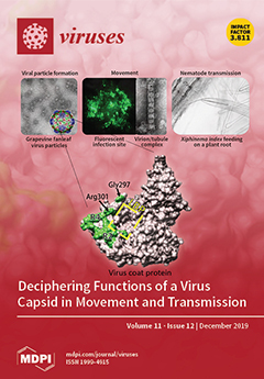
To investigate the role of viral capsid residues in transmission by a plant-feeding nematode, we need mutants able to infect plants systemically. We report on our progression from a grapevine fanleaf virus mutant impaired in encapsidation and movement to the identification of a new transmission determinant composed of region R4 and Arg301 of the coat protein. It lies in close proximity to a charged cavity, likely constituting the binding pocket (LBP) of the virus in the nematode’s mouthpart.View this paper.
  • Issues are regarded as officially published after their release is announced to the table of contents alert mailing list.
  • You may sign up for e-mail alerts to receive table of contents of newly released issues.
  • PDF is the official format for papers published in both, html and pdf forms. To view the papers in pdf format, click on the "PDF Full-text" link, and use the free Adobe Reader to open them.
Order results
Result details
Section
Select all
Export citation of selected articles as:
17 pages, 3537 KiB  
Article
Homburgvirus LP-018 Has a Unique Ability to Infect Phage-Resistant Listeria monocytogenes
by Yaxiong Song, Tracey L. Peters, Daniel W. Bryan, Lauren K. Hudson and Thomas G. Denes
Viruses 2019, 11(12), 1166; https://doi.org/10.3390/v11121166 - 17 Dec 2019
Cited by 14 | Viewed by 2893
Abstract
Listeria phage LP-018 is the only phage from a diverse collection of 120 phages able to form plaques on a phage-resistant Listeria monocytogenes strain lacking rhamnose in its cell wall teichoic acids. The aim of this study was to characterize phage LP-018 and [...] Read more.
Listeria phage LP-018 is the only phage from a diverse collection of 120 phages able to form plaques on a phage-resistant Listeria monocytogenes strain lacking rhamnose in its cell wall teichoic acids. The aim of this study was to characterize phage LP-018 and to identify what types of mutations can confer resistance to LP-018. Whole genome sequencing and transmission electron microscopy revealed LP-018 to be a member of the Homburgvirus genus. One-step-growth curve analysis of LP-018 revealed an eclipse period of ~60–90 min and a burst size of ~2 PFU per infected cell. Despite slow growth and small burst size, LP-018 can inhibit the growth of Listeria monocytogenes at a high multiplicity of infection. Ten distinct LP-018-resistant mutants were isolated from infected Listeria monocytogenes 10403S and characterized by whole genome sequencing. In each mutant, a single mutation was identified in either the LMRG_00278 or LMRG_01613 encoding genes. Interesting, LP-018 was able to bind to a representative phage-resistant mutant with a mutation in each gene, suggesting these mutations confer resistance through a mechanism independent of adsorption inhibition. Despite forming plaques on the rhamnose deficient 10403S mutant, LP-018 showed reduced binding efficiency, and we did not observe inhibition of the strain under the conditions tested. Two mutants of LP-018 were also isolated and characterized, one with a single SNP in a gene encoding a BppU domain protein that likely alters its host range. LP-018 is shown to be a unique Listeria phage that, with additional evaluation, may be useful in biocontrol applications that aim to reduce the emergence of phage resistance. Full article
(This article belongs to the Section Bacterial Viruses)
Show Figures

Figure 1

27 pages, 1622 KiB  
Review
Microtubule-Dependent Trafficking of Alphaherpesviruses in the Nervous System: The Ins and Outs
by Drishya Diwaker and Duncan W. Wilson
Viruses 2019, 11(12), 1165; https://doi.org/10.3390/v11121165 - 17 Dec 2019
Cited by 16 | Viewed by 4547
Abstract
The Alphaherpesvirinae include the neurotropic pathogens herpes simplex virus and varicella zoster virus of humans and pseudorabies virus of swine. These viruses establish lifelong latency in the nuclei of peripheral ganglia, but utilize the peripheral tissues those neurons innervate for productive replication, spread, [...] Read more.
The Alphaherpesvirinae include the neurotropic pathogens herpes simplex virus and varicella zoster virus of humans and pseudorabies virus of swine. These viruses establish lifelong latency in the nuclei of peripheral ganglia, but utilize the peripheral tissues those neurons innervate for productive replication, spread, and transmission. Delivery of virions from replicative pools to the sites of latency requires microtubule-directed retrograde axonal transport from the nerve terminus to the cell body of the sensory neuron. As a corollary, during reactivation newly assembled virions must travel along axonal microtubules in the anterograde direction to return to the nerve terminus and infect peripheral tissues, completing the cycle. Neurotropic alphaherpesviruses can therefore exploit neuronal microtubules and motors for long distance axonal transport, and alternate between periods of sustained plus end- and minus end-directed motion at different stages of their infectious cycle. This review summarizes our current understanding of the molecular details by which this is achieved. Full article
(This article belongs to the Special Issue Regulation and Exploitation of Microtubules by Viruses)
Show Figures

Figure 1

18 pages, 2477 KiB  
Article
Zika Virus Surveillance at the Human–Animal Interface in West-Central Brazil, 2017–2018
by Alex Pauvolid-Corrêa, Helver Gonçalves Dias, Laura Marina Siqueira Maia, Grasiela Porfírio, Thais Oliveira Morgado, Gilberto Sabino-Santos, Paula Helena Santa Rita, Wanessa Teixeira Gomes Barreto, Gabriel Carvalho de Macedo, Jaire Marinho Torres, Wesley Arruda Gimenes Nantes, Filipe Martins Santos, William Oliveira de Assis, Andreza Castro Rucco, Rafael Mamoru dos Santos Yui, João Bosco Vilela Campos, Renato Rodrigues Leandro e Silva, Raquel da Silva Ferreira, Nilvanei Aparecido da Silva Neves, Michell Charlles de Souza Costa, Leticia Ramos Martins, Emerson Marques de Souza, Michellen dos Santos Carvalho, Marina Gonçalves Lima, Fernanda de Cássia Gonçalves Alves, Luiz Humberto Guimarães Riquelme-Junior, Luan Luiz Batista Figueiró, Matheus Fernandes Gomes de Santana, Luiz Gustavo Rodrigues Oliveira Santos, Samara Serra Medeiros, Larissa Lopes Seino, Emily Hime Miranda, José Henrique Rezende Linhares, Vanessa de Oliveira Santos, Stephanie Almeida da Silva, Kelly Araújo Lúcio, Viviane Silva Gomes, Alexandre de Araújo Oliveira, Julia dos Santos Silva, William de Almeida Marques, Marcio Schafer Marques, José Junior França de Barros, Letícia Campos, Dinair Couto-Lima, Claudia Coutinho Netto, Christine Strüssmann, Nicholas Panella, Emily Hannon, Barbara Cristina de Macedo, Júlia Ramos de Almeida, Karen Ramos Ribeiro, Maria Carolina Barros de Castro, Larissa Pratta Campos, Ana Paula Rosa dos Santos, Isabelle Marino de Souza, Mateus de Assis Bianchini, Sandra Helena Ramiro Correa, Renato Ordones Baptista Luz, Ananda dos Santos Vieira, Luzia Maria de Oliveira Pinto, Elzinandes Azeredo, Luiz Tadeu Moraes Figueiredo, Jeronimo Augusto Fonseca Alencar, Sheila Maria Barbosa de Lima, Heitor Miraglia Herrera, Renata Dezengrini Shlessarenko, Flavia Barreto dos Santos, Ana Maria Bispo de Filippis, Stephanie Salyer, Joel Montgomery and Nicholas Komaradd Show full author list remove Hide full author list
Viruses 2019, 11(12), 1164; https://doi.org/10.3390/v11121164 - 16 Dec 2019
Cited by 15 | Viewed by 6800
Abstract
Zika virus (ZIKV) was first discovered in 1947 in Uganda but was not considered a public health threat until 2007 when it found to be the source of epidemic activity in Asia. Epidemic activity spread to Brazil in 2014 and continued to spread [...] Read more.
Zika virus (ZIKV) was first discovered in 1947 in Uganda but was not considered a public health threat until 2007 when it found to be the source of epidemic activity in Asia. Epidemic activity spread to Brazil in 2014 and continued to spread throughout the tropical and subtropical regions of the Americas. Despite ZIKV being zoonotic in origin, information about transmission, or even exposure of non-human vertebrates and mosquitoes to ZIKV in the Americas, is lacking. Accordingly, from February 2017 to March 2018, we sought evidence of sylvatic ZIKV transmission by sampling whole blood from approximately 2000 domestic and wild vertebrates of over 100 species in West-Central Brazil within the active human ZIKV transmission area. In addition, we collected over 24,300 mosquitoes of at least 17 genera and 62 species. We screened whole blood samples and mosquito pools for ZIKV RNA using pan-flavivirus primers in a real-time reverse-transcription polymerase chain reaction (RT-PCR) in a SYBR Green platform. Positives were confirmed using ZIKV-specific envelope gene real-time RT-PCR and nucleotide sequencing. Of the 2068 vertebrates tested, none were ZIKV positive. Of the 23,315 non-engorged mosquitoes consolidated into 1503 pools tested, 22 (1.5%) with full data available showed some degree of homology to insect-specific flaviviruses. To identify previous exposure to ZIKV, 1498 plasma samples representing 62 species of domestic and sylvatic vertebrates were tested for ZIKV-neutralizing antibodies by plaque reduction neutralization test (PRNT90). From these, 23 (1.5%) of seven species were seropositive for ZIKV and negative for dengue virus serotype 2, yellow fever virus, and West Nile virus, suggesting potential monotypic reaction for ZIKV. Results presented here suggest no active transmission of ZIKV in non-human vertebrate populations or in alternative vector candidates, but suggest that vertebrates around human populations have indeed been exposed to ZIKV in West-Central Brazil. Full article
(This article belongs to the Special Issue Emerging Arboviruses)
Show Figures

Figure 1

20 pages, 2667 KiB  
Article
Genomic and Proteomic Characterization of Bacteriophage BH1 Spontaneously Released from Probiotic Lactobacillus rhamnosus Pen
by Piotr Jarocki, Elwira Komoń-Janczara, Marcin Podleśny, Oleksandr Kholiavskyi, Monika Pytka and Monika Kordowska-Wiater
Viruses 2019, 11(12), 1163; https://doi.org/10.3390/v11121163 - 16 Dec 2019
Cited by 9 | Viewed by 3298
Abstract
Lactobacillus rhamnosus Pen is a human endogenous strain used for the production of probiotic formula, which is effective in the prevention of antibiotic-associated diarrhoea. Our study showed that this probiotic strain releases bacteriophage BH1 without the addition of any inducing agent. Our research [...] Read more.
Lactobacillus rhamnosus Pen is a human endogenous strain used for the production of probiotic formula, which is effective in the prevention of antibiotic-associated diarrhoea. Our study showed that this probiotic strain releases bacteriophage BH1 without the addition of any inducing agent. Our research revealed that phage BH1 has a circular genome with a length of 40721 nt and a GC content of 44.8%. The genome of phage BH1 possesses 57 open reading frames which could be divided into functional modules associated with DNA packaging, morphogenesis, lysis, integration, genetic switch, and replication. In spite of similarity in morphology and genomic organization, comparative analysis revealed substantial genetic diversity and mosaic genomic architecture among phages described for the Lactobacillus casei group. Additionally, qPCR and ddPCR analysis confirmed earlier microscopic observations indicating that L. rhamnosus Pen liberates bacteriophage particles during growth. This occurs spontaneously, and is not a result of external inducing factors. For samples collected after 4 and 24 h of L. rhamnosus Pen culture, the number of attB and attP copies increased 2.5 and 12 times, respectively. This phenomenon, by introducing resistance to other phages or enhancing the biofilm-forming capabilities, may increase the survivability of microorganisms in their natural ecological niche. Conversely, spontaneous phage induction may be an important virulence factor for bacteria, posing a potential threat for the human host. Full article
(This article belongs to the Section Bacterial Viruses)
Show Figures

Figure 1

14 pages, 1375 KiB  
Brief Report
Nuclear Transit and HIV LTR Binding of NF-κB Subunits Held by IκB Proteins: Implications for HIV-1 Activation
by Sohrab Z. Khan, Sofia Gasperino and Steven L. Zeichner
Viruses 2019, 11(12), 1162; https://doi.org/10.3390/v11121162 - 16 Dec 2019
Cited by 6 | Viewed by 2516
Abstract
No effective therapy to eliminate the HIV latently infected cell reservoir has been developed. One approach, “shock and kill”, employs agents that activate HIV, subsequently killing the activated infected cells and/or virus. Shock and kill requires agents that safely and effectively activate HIV. [...] Read more.
No effective therapy to eliminate the HIV latently infected cell reservoir has been developed. One approach, “shock and kill”, employs agents that activate HIV, subsequently killing the activated infected cells and/or virus. Shock and kill requires agents that safely and effectively activate HIV. One class of activation agents works through classical NF-κB pathways, but global NF-κB activators are non-specific and toxic. There exist two major IκBs: IκBα, and IκBε, which hold activating NF-κB subunits in the cytoplasm, releasing them for nuclear transit upon cell stimulation. IκBα was considered the main IκB responsible for gene expression regulation, including HIV activation. IκBε is expressed in cells constituting much of the latent HIV reservoir, and IκBε knockout mice have a minimal phenotype, suggesting that IκBε could be a valuable target for HIV activation and reservoir depletion. We previously showed that targeting IκBε yields substantial increases in HIV expression. Here, we show that IκBε holds c-Rel and p65 activating NF-κB subunits in the cytoplasm, and that targeting IκBε with siRNA produces a strong increase in HIV expression associated with enhanced c-Rel and p65 transit to the nucleus and binding to the HIV LTR of the activating NF-κBs, demonstrating a mechanism through which targeting IκBε increases HIV expression. The findings suggest that it may be helpful to develop HIV activation approaches, acting specifically to target IκBε and its interactions with the NF-κBs. Full article
(This article belongs to the Section Animal Viruses)
Show Figures

Figure 1

16 pages, 1015 KiB  
Review
Mechanistic Insights into Chemoresistance Mediated by Oncogenic Viruses in Lymphomas
by Jungang Chen, Samantha Kendrick and Zhiqiang Qin
Viruses 2019, 11(12), 1161; https://doi.org/10.3390/v11121161 - 16 Dec 2019
Cited by 7 | Viewed by 3671
Abstract
Viral lymphomagenesis induced by infection with oncogenic viruses, such as Kaposi’s sarcoma associated herpesvirus (KSHV), Epstein–Barr virus (EBV) and human T-cell leukemia virus (HTLV-1), represents a group of aggressive malignancies with a diverse range of pathological features. Combined chemotherapy remains the standard of [...] Read more.
Viral lymphomagenesis induced by infection with oncogenic viruses, such as Kaposi’s sarcoma associated herpesvirus (KSHV), Epstein–Barr virus (EBV) and human T-cell leukemia virus (HTLV-1), represents a group of aggressive malignancies with a diverse range of pathological features. Combined chemotherapy remains the standard of care for these virus-associated lymphomas; however, frequent chemoresistance is a barrier to achieving successful long-term disease-free survival. There is increasing evidence that indicates virus-associated lymphomas display more resistance to cytotoxic chemotherapeutic agents than that observed in solid tumors. Although the tumor microenvironment and genetic changes, such as key oncogene mutations, are closely related to chemoresistance, some studies demonstrate that the components of oncogenic viruses themselves play pivotal roles in the multidrug chemoresistance of lymphoma cells. In this review, we summarize recent advances in the understanding of the mechanisms through which oncogenic viruses mediate lymphoma cell chemoresistance, with a particular focus on KSHV and EBV, two major oncogenic viruses. We also discuss the current challenges to overcome these obstacles in the treatment of virus-associated lymphomas. Full article
(This article belongs to the Section Animal Viruses)
Show Figures

Figure 1

14 pages, 2773 KiB  
Article
IFI16 Inhibits Porcine Reproductive and Respiratory Syndrome Virus 2 Replication in a MAVS-Dependent Manner in MARC-145 Cells
by Xiaobo Chang, Xibao Shi, Xiaozhuan Zhang, Li Wang, Xuewu Li, Aiping Wang, Ruiguang Deng, Enmin Zhou and Gaiping Zhang
Viruses 2019, 11(12), 1160; https://doi.org/10.3390/v11121160 - 16 Dec 2019
Cited by 20 | Viewed by 3006
Abstract
Porcine reproductive and respiratory syndrome virus (PRRSV) is a single-stranded positive-sense RNA virus, and the current strategies for controlling PRRSV are limited. Interferon gamma-inducible protein 16 (IFI16) has been reported to have a broader role in the regulation of the type I interferons [...] Read more.
Porcine reproductive and respiratory syndrome virus (PRRSV) is a single-stranded positive-sense RNA virus, and the current strategies for controlling PRRSV are limited. Interferon gamma-inducible protein 16 (IFI16) has been reported to have a broader role in the regulation of the type I interferons (IFNs) response to RNA and DNA viruses. However, the function of IFI16 in PRRSV infection is unclear. Here, we revealed that IFI16 acts as a novel antiviral protein against PRRSV-2. IFI16 could be induced by interferon-beta (IFN-β). Overexpression of IFI16 could significantly suppress PRRSV-2 replication, and silencing the expression of endogenous IFI16 by small interfering RNAs led to the promotion of PRRSV-2 replication in MARC-145 cells. Additionally, IFI16 could promote mitochondrial antiviral signaling protein (MAVS)-mediated production of type I interferon and interact with MAVS. More importantly, IFI16 exerted anti-PRRSV effects in a MAVS-dependent manner. In conclusion, our data demonstrated that IFI16 has an inhibitory effect on PRRSV-2, and these findings contribute to understanding the role of cellular proteins in regulating PRRSV replication and may have implications for the future antiviral strategies. Full article
(This article belongs to the Section Animal Viruses)
Show Figures

Figure 1

18 pages, 4456 KiB  
Article
Ecology of West Nile Virus in the Danube Delta, Romania: Phylogeography, Xenosurveillance and Mosquito Host-Feeding Patterns
by Alexandru Tomazatos, Stephanie Jansen, Stefan Pfister, Edina Török, Iulia Maranda, Cintia Horváth, Lujza Keresztes, Marina Spînu, Egbert Tannich, Hanna Jöst, Jonas Schmidt-Chanasit, Daniel Cadar and Renke Lühken
Viruses 2019, 11(12), 1159; https://doi.org/10.3390/v11121159 - 14 Dec 2019
Cited by 17 | Viewed by 5795
Abstract
The ecology of West Nile virus (WNV) in the Danube Delta Biosphere Reserve (Romania) was investigated by combining studies on the virus genetics, phylogeography, xenosurveillance and host-feeding patterns of mosquitoes. Between 2014 and 2016, 655,667 unfed and 3842 engorged mosquito females were collected [...] Read more.
The ecology of West Nile virus (WNV) in the Danube Delta Biosphere Reserve (Romania) was investigated by combining studies on the virus genetics, phylogeography, xenosurveillance and host-feeding patterns of mosquitoes. Between 2014 and 2016, 655,667 unfed and 3842 engorged mosquito females were collected from four sampling sites. Blood-fed mosquitoes were negative for WNV-RNA, but two pools of unfed Culex pipiens s.l./torrentium collected in 2014 were tested positive. Our results suggest that Romania experienced at least two separate WNV lineage 2 introductions: from Africa into Danube Delta and from Greece into south-eastern Romania in the 1990s and early 2000s, respectively. The genetic diversity of WNV in Romania is primarily shaped by in situ evolution. WNV-specific antibodies were detected for 19 blood-meals from dogs and horses, but not from birds or humans. The hosts of mosquitoes were dominated by non-human mammals (19 species), followed by human and birds (23 species). Thereby, the catholic host-feeding pattern of Culex pipiens s.l./torrentium with a relatively high proportion of birds indicates the species’ importance as a potential bridge vector. The low virus prevalence in combination with WNV-specific antibodies indicate continuous, but low activity of WNV in the Danube Delta during the study period. Full article
(This article belongs to the Section Animal Viruses)
Show Figures

Figure 1

24 pages, 20656 KiB  
Article
A Novel Interaction Network Used by Potyviruses in Virus–Host Interactions at the Protein Level
by Marjo Ala-Poikela, Minna-Liisa Rajamäki and Jari P.T. Valkonen
Viruses 2019, 11(12), 1158; https://doi.org/10.3390/v11121158 - 14 Dec 2019
Cited by 18 | Viewed by 5082
Abstract
Host proteins that are central to infection of potyviruses (genus Potyvirus; family Potyviridae) include the eukaryotic translation initiation factors eIF4E and eIF(iso)4E. The potyviral genome-linked protein (VPg) and the helper component proteinase (HCpro) interact with each other and with eIF4E and eIF(iso)4E [...] Read more.
Host proteins that are central to infection of potyviruses (genus Potyvirus; family Potyviridae) include the eukaryotic translation initiation factors eIF4E and eIF(iso)4E. The potyviral genome-linked protein (VPg) and the helper component proteinase (HCpro) interact with each other and with eIF4E and eIF(iso)4E and proteins are involved in the same functions during viral infection. VPg interacts with eIF4E/eIF(iso)4E via the 7-methylguanosine cap-binding region, whereas HCpro interacts with eIF4E/eIF(iso)4E via the 4E-binding motif YXXXXLΦ, similar to the motif in eIF4G. In this study, HCpro and VPg were found to interact in the nucleus, nucleolus, and cytoplasm in cells infected with the potyvirus potato virus A (PVA). In the cytoplasm, interactions between HCpro and VPg occurred in punctate bodies not associated with viral replication vesicles. In addition to HCpro, the 4E-binding motif was recognized in VPg of PVA. Mutations in the 4E-binding motif of VPg from PVA weakened interactions with eIF4E and heavily reduced PVA virulence. Furthermore, mutations in the 4G-binding domain of eIF4E reduced interactions with VPg and abolished interactions with HCpro. Thus, HCpro and VPg can both interact with eIF4E using the 4E-binding motif. Our results suggest a novel interaction network used by potyviruses to interact with host plants via translation initiation factors. Full article
(This article belongs to the Special Issue The Complexity of the Potyviral Interaction Network)
Show Figures

Figure 1

21 pages, 3536 KiB  
Article
Viral Diversity of Microbats within the South West Botanical Province of Western Australia
by Diana Prada, Victoria Boyd, Michelle L. Baker, Mark O’Dea and Bethany Jackson
Viruses 2019, 11(12), 1157; https://doi.org/10.3390/v11121157 - 13 Dec 2019
Cited by 18 | Viewed by 5396
Abstract
Bats are known reservoirs of a wide variety of viruses that rarely result in overt clinical disease in the bat host. However, anthropogenic influences on the landscape and climate can change species assemblages and interactions, as well as undermine host-resilience. The cumulative result [...] Read more.
Bats are known reservoirs of a wide variety of viruses that rarely result in overt clinical disease in the bat host. However, anthropogenic influences on the landscape and climate can change species assemblages and interactions, as well as undermine host-resilience. The cumulative result is a disturbance of bat–pathogen dynamics, which facilitate spillover events to sympatric species, and may threaten bat communities already facing synergistic stressors through ecological change. Therefore, characterisation of viral pathogens in bat communities provides important basal information to monitor and predict the emergence of diseases relevant to conservation and public health. This study used targeted molecular techniques, serological assays and next generation sequencing to characterise adenoviruses, coronaviruses and paramyxoviruses from 11 species of insectivorous bats within the South West Botanical Province of Western Australia. Phylogenetic analysis indicated complex ecological interactions including virus–host associations, cross-species infections, and multiple viral strains circulating concurrently within selected bat populations. Additionally, we describe the entire coding sequences for five alphacoronaviruses (representing four putative new species), and one novel adenovirus. Results indicate that viral burden (both prevalence and richness) is not homogeneous among species, with Chalinolobus gouldii identified as a key epidemiological element within the studied communities. Full article
(This article belongs to the Section Animal Viruses)
Show Figures

Figure 1

17 pages, 3747 KiB  
Article
Pathogenic Characteristics of a Porcine Astrovirus Strain Isolated in China
by Qingli Fang, Cui Wang, Huan Liu, Qingping Wu, Siying Liang, Minli Cen, Qinting Dong, Yingyi Wei, Ying Chen, Kang Ouyang, Zuzhang Wei and Weijian Huang
Viruses 2019, 11(12), 1156; https://doi.org/10.3390/v11121156 - 13 Dec 2019
Cited by 25 | Viewed by 3496
Abstract
Astroviral infection is considered to be one of the causes of mammalian diarrheal diseases. It has been shown that astrovirus infections cause varying degrees of diarrhea in turkeys and mice. However, the pathogenesis of porcine astrovirus is unknown. In this study, the virulence [...] Read more.
Astroviral infection is considered to be one of the causes of mammalian diarrheal diseases. It has been shown that astrovirus infections cause varying degrees of diarrhea in turkeys and mice. However, the pathogenesis of porcine astrovirus is unknown. In this study, the virulence of a cytopathic porcine astrovirus (PAstV) strain (PAstV1-GX1) isolated from the PK-15 cell line was tested using seven-day-old nursing piglets. The results showed that PAstV1-GX1 infection could cause mild diarrhea, growth retardation, and damage of the villi of the small intestinal mucosa. However, all the above symptoms could be restored within 7 to 10days post inoculation (dpi). To evaluate the innate immunity response of PAstV in vivo, the alteration of inflammatory cytokine expression in piglets infected with PAstV1-GX1 was determined using quantitative real-time reverse transcription polymerase chain reaction (qRT-PCR). The mRNA expression levels of the IFNβ and ISG54 were found to be significantly elevated in virus-infected piglets. In contrast, expression of IFNλ was downregulated in piglets infected with PAstV1-GX1. In addition, the mRNA expression of the tight junction protein 1 and 2 and zonula occludin 1, which are associated with the intestinal barrier permeability, were affected after PAstV1 infection. The present study adds to our understanding of the pathogenic mechanism of PAstV and has established an animal model for further study of pig astrovirus infection. Full article
(This article belongs to the Special Issue Endemic and Emerging Swine Viruses)
Show Figures

Figure 1

20 pages, 5187 KiB  
Article
Distinct Lineages of Feline Parvovirus Associated with Epizootic Outbreaks in Australia, New Zealand and the United Arab Emirates
by Kate Van Brussel, Maura Carrai, Carrie Lin, Mark Kelman, Laura Setyo, Danielle Aberdein, Juliana Brailey, Michelle Lawler, Simone Maher, Ildiko Plaganyi, Emily Lewis, Adele Hawkswell, Andrew B. Allison, Joanne Meers, Vito Martella, Julia A. Beatty, Edward C. Holmes, Nicola Decaro and Vanessa R. Barrs
Viruses 2019, 11(12), 1155; https://doi.org/10.3390/v11121155 - 13 Dec 2019
Cited by 26 | Viewed by 5864
Abstract
Feline panleukopenia (FPL), a frequently fatal disease of cats, is caused by feline parvovirus (FPV) or canine parvovirus (CPV). We investigated simultaneous outbreaks of FPL between 2014 and 2018 in Australia, New Zealand and the United Arab Emirates (UAE) where FPL outbreaks had [...] Read more.
Feline panleukopenia (FPL), a frequently fatal disease of cats, is caused by feline parvovirus (FPV) or canine parvovirus (CPV). We investigated simultaneous outbreaks of FPL between 2014 and 2018 in Australia, New Zealand and the United Arab Emirates (UAE) where FPL outbreaks had not been reported for several decades. Case data from 989 cats and clinical samples from additional 113 cats were obtained to determine the cause of the outbreaks and epidemiological factors involved. Most cats with FPL were shelter-housed, 9 to 10 weeks old at diagnosis, unvaccinated, had not completed a primary vaccination series or had received vaccinations noncompliant with current guidelines. Analysis of parvoviral VP2 sequence data confirmed that all FPL cases were caused by FPV and not CPV. Phylogenetic analysis revealed that each of these outbreaks was caused by a distinct FPV, with two virus lineages present in eastern Australia and virus movement between different geographical locations. Viruses from the UAE outbreak formed a lineage of unknown origin. FPV vaccine virus was detected in the New Zealand cases, highlighting the difficulty of distinguishing the co-incidental shedding of vaccine virus in vaccinated cats. Inadequate vaccination coverage in shelter-housed cats was a common factor in all outbreaks, likely precipitating the multiple re-emergence of infection events. Full article
(This article belongs to the Special Issue Feline Viruses and Viral Diseases)
Show Figures

Figure 1

14 pages, 1822 KiB  
Article
Antigenic and Pathogenic Characteristics of QX-Type Avian Infectious Bronchitis Virus Strains Isolated in Southwestern China
by Shuyun Li, Lijing Du, Jing Xia, Jiteng Du, Guojin You, Yiping Wen, Xiaobo Huang, Qing Zhao, Xinfeng Han, Qigui Yan, Rui Wu, Min Cui, Sanjie Cao and Yong Huang
Viruses 2019, 11(12), 1154; https://doi.org/10.3390/v11121154 - 13 Dec 2019
Cited by 14 | Viewed by 3597
Abstract
The QX-type avian infectious bronchitis virus (IBV) is still a prevalent genotype in Southwestern China. To analyze the antigenicity and pathogenicity characteristics of the dominant genotype strains (QX-type), S1 gene sequence analysis, virus cross-neutralization tests, and pathogenicity test of eight QX-type IBV isolates [...] Read more.
The QX-type avian infectious bronchitis virus (IBV) is still a prevalent genotype in Southwestern China. To analyze the antigenicity and pathogenicity characteristics of the dominant genotype strains (QX-type), S1 gene sequence analysis, virus cross-neutralization tests, and pathogenicity test of eight QX-type IBV isolates were conducted. Sequence analysis showed that the nucleotide homology between the eight strains was high, but distantly related to H120 and 4/91 vaccine strains. Cross-neutralization tests showed that all eight strains isolated from 2015 and 2017 belonged to the same serotype, but exhibited antigenic variations over time. The pathogenicity test of the five QX-type IBV isolates showed that only three strains, CK/CH/SC/DYW/16, CK/CH/SC/MS/17, and CK/CH/SC/GH/15, had a high mortality rate with strong respiratory and renal pathogenicity, whereas CK/CH/SC/PZ/17 and CK/CH/SC/DYYJ/17 caused only mild clinical symptoms and tissue lesions. Our results indicate that the prevalent QX-type IBVs displayed antigenic variations and pathogenicity difference. These findings may provide reference for research on the evolution of IBV and vaccine preparation of infectious bronchitis (IB). Full article
(This article belongs to the Section Animal Viruses)
Show Figures

Figure 1

25 pages, 2716 KiB  
Article
Modelling Vector Transmission and Epidemiology of Co-Infecting Plant Viruses
by Linda J. S. Allen, Vrushali A. Bokil, Nik J. Cunniffe, Frédéric M. Hamelin, Frank M. Hilker and Michael J. Jeger
Viruses 2019, 11(12), 1153; https://doi.org/10.3390/v11121153 - 13 Dec 2019
Cited by 18 | Viewed by 4566
Abstract
Co-infection of plant hosts by two or more viruses is common in agricultural crops and natural plant communities. A variety of models have been used to investigate the dynamics of co-infection which track only the disease status of infected and co-infected plants, and [...] Read more.
Co-infection of plant hosts by two or more viruses is common in agricultural crops and natural plant communities. A variety of models have been used to investigate the dynamics of co-infection which track only the disease status of infected and co-infected plants, and which do not explicitly track the density of inoculative vectors. Much less attention has been paid to the role of vector transmission in co-infection, that is, acquisition and inoculation and their synergistic and antagonistic interactions. In this investigation, a general epidemiological model is formulated for one vector species and one plant species with potential co-infection in the host plant by two viruses. The basic reproduction number provides conditions for successful invasion of a single virus. We derive a new invasion threshold which provides conditions for successful invasion of a second virus. These two thresholds highlight some key epidemiological parameters important in vector transmission. To illustrate the flexibility of our model, we examine numerically two special cases of viral invasion. In the first case, one virus species depends on an autonomous virus for its successful transmission and in the second case, both viruses are unable to invade alone but can co-infect the host plant when prevalence is high. Full article
(This article belongs to the Special Issue Plant Virus Transmission by Vectors)
Show Figures

Figure 1

12 pages, 11982 KiB  
Article
Tilapia Lake Virus Does Not Hemagglutinate Avian and Piscine Erythrocytes and NH4Cl Does Not Inhibit Viral Replication In Vitro
by Augustino Alfred Chengula, Stephen Mutoloki, Øystein Evensen and Hetron Mweemba Munang’andu
Viruses 2019, 11(12), 1152; https://doi.org/10.3390/v11121152 - 12 Dec 2019
Cited by 9 | Viewed by 4813
Abstract
Tilapia lake virus (TiLV) is a negative-sense single-stranded RNA (-ssRNA) icosahedral virus classified to be the only member in the family Amnoonviridae. Although TiLV segment-1 shares homology with the influenza C virus PB1 and has four conserved motifs similar to influenza A, B, [...] Read more.
Tilapia lake virus (TiLV) is a negative-sense single-stranded RNA (-ssRNA) icosahedral virus classified to be the only member in the family Amnoonviridae. Although TiLV segment-1 shares homology with the influenza C virus PB1 and has four conserved motifs similar to influenza A, B, and C polymerases, it is unknown whether there are other properties shared between TiLV and orthomyxovirus. In the present study, we wanted to determine whether TiLV agglutinated avian and piscine erythrocytes, and whether its replication was inhibited by lysosomotropic agents, such as ammonium chloride (NH4Cl), as seen for orthomyxoviruses. Our findings showed that influenza virus strain A/Puerto Rico/8 (PR8) was able to hemagglutinate turkey (Meleagris gallopavo), Atlantic salmon (Salmo salar L), and Nile tilapia (Oreochromis niloticus) red blood cells (RBCs), while infectious salmon anemia virus (ISAV) only agglutinated Atlantic salmon, but not turkey or tilapia, RBCs. In contrast to PR8 and ISAV, TiLV did not agglutinate turkey, Atlantic salmon, or tilapia RBCs. qRT-PCR analysis showed that 30 mM NH4Cl, a basic lysosomotropic agent, neither inhibited nor enhanced TiLV replication in E-11 cells. There was no difference in viral quantities in the infected cells with or without NH4Cl treatment during virus adsorption or at 1, 2, and 3 h post-infection. Given that hemagglutinin proteins that bind RBCs also serve as ligands that bind host cells during virus entry leading to endocytosis in orthomyxoviruses, the data presented here suggest that TiLV may use mechanisms that are different from orthomyxoviruses for entry and replication in host cells. Therefore, future studies should seek to elucidate the mechanisms used by TiLV for entry into host cells and to determine its mode of replication in infected cells. Full article
(This article belongs to the Section Animal Viruses)
Show Figures

Figure 1

13 pages, 2506 KiB  
Article
Efficacy of FDA-Approved Anti-Inflammatory Drugs Against Venezuelan Equine Encephalitis Virus Infection
by Kenneth Risner, Aslaa Ahmed, Allison Bakovic, Stephanie Kortchak, Nishank Bhalla and Aarthi Narayanan
Viruses 2019, 11(12), 1151; https://doi.org/10.3390/v11121151 - 12 Dec 2019
Cited by 14 | Viewed by 3349
Abstract
Venezuelan equine encephalitis virus (VEEV) is a category B select agent pathogen that can be aerosolized. Infections in murine models and humans can advance to an encephalitic phenotype which may result in long-term neurological complications or death. No specific FDA-approved treatments or vaccines [...] Read more.
Venezuelan equine encephalitis virus (VEEV) is a category B select agent pathogen that can be aerosolized. Infections in murine models and humans can advance to an encephalitic phenotype which may result in long-term neurological complications or death. No specific FDA-approved treatments or vaccines are available for the treatment or prevention of VEEV infection. Neurotropic viral infections have two damaging components: neuronal death caused by viral replication, and damage from the subsequent inflammatory response. Reducing the level of inflammation may lessen neurological tissue damage that often arises following VEEV infection. In this study, three commercially available anti-inflammatory drugs, Celecoxib, Rolipram, and Tofacitinib, were evaluated for antiviral activity in an astrocyte and a microglial model of VEEV infection. The inhibitors were tested against the vaccine strain VEEV TC-83, as well as the wild-type VEEV Trinidad donkey strain. Celecoxib, Tofacitinib, and Rolipram significantly decreased viral titers both after pre-treatment and post-treatment of infected cells. VEEV Trinidad Donkey (TrD) titers were reduced 6.45-fold in cells treated with 50 µM of Celecoxib, 2.45-fold when treated with 50 µM of Tofacitinib, and 1.81-fold when treated with 50 µM of Rolipram. Celecoxib was also shown to decrease inflammatory gene expression in the context of TC-83 infection. Overall, Celecoxib demonstrated potency as a countermeasure strategy that slowed VEEV infection and infection-induced inflammation in an in vitro model. Full article
(This article belongs to the Section Viral Immunology, Vaccines, and Antivirals)
Show Figures

Graphical abstract

2 pages, 168 KiB  
Reply
Reply to Comments by Yih et al. (Exposure to Hantavirus is a Risk Factor Associated with Kidney Diseases in Sri Lanka: A Cross-Sectional Study)
by Yomani D. Sarathkumara, Chandika D. Gamage, Sithumini Lokupathirage, Devinda S. Muthusinghe, Nishantha Nanayakkara, Lishanthe Gunarathne, Kenta Shimizu, Yoshimi Tsuda, Jiro Arikawa and Kumiko Yoshimatsu
Viruses 2019, 11(12), 1150; https://doi.org/10.3390/v11121150 - 11 Dec 2019
Cited by 1 | Viewed by 1999
Abstract
Dear Drs. W. Katherine Yih, Martin Kulldorff, Jessica H. Leibler, David J. Friedman, and Daniel R. Brooks [...] Full article
(This article belongs to the Special Issue Hantaviruses)
15 pages, 2245 KiB  
Article
A Novel Bacterium-Like Particle-Based Vaccine Displaying the SUDV Glycoprotein Induces Potent Humoral and Cellular Immune Responses in Mice
by Shengnan Xu, Cuicui Jiao, Hongli Jin, Wujian Li, Entao Li, Zengguo Cao, Zhikang Shi, Feihu Yan, Shengnan Zhang, Hongbin He, Hang Chi, Na Feng, Yongkun Zhao, Yuwei Gao, Songtao Yang, Jianzhong Wang, Hualei Wang and Xianzhu Xia
Viruses 2019, 11(12), 1149; https://doi.org/10.3390/v11121149 - 11 Dec 2019
Cited by 11 | Viewed by 3029
Abstract
Sudan virus (SUDV) causes severe lethal hemorrhagic fever in humans and nonhuman primates. The most effective and economical way to protect against Sudan ebolavirus disease is prophylactic vaccination. However, there are no licensed vaccines to prevent SUDV infections. In this study, a bacterium-like [...] Read more.
Sudan virus (SUDV) causes severe lethal hemorrhagic fever in humans and nonhuman primates. The most effective and economical way to protect against Sudan ebolavirus disease is prophylactic vaccination. However, there are no licensed vaccines to prevent SUDV infections. In this study, a bacterium-like particle (BLP)-based vaccine displaying the extracellular domain of the SUDV glycoprotein (eGP) was developed based on a gram-positive enhancer matrix-protein anchor (GEM-PA) surface display system. Expression of the recombinant GEM-displayed eGP (eGP-PA-GEM) was verified by Western blotting and immunofluorescence assays. The SUDV BLPs (SBLPs), which were mixed with Montanide ISA 201VG plus Poly (I:C) combined adjuvant, could induce high SUDV GP-specific IgG titers of up to 1:40,960 and robust virus-neutralizing antibody titers reached 1:460. The SBLP also elicited T-helper 1 (Th1) and T-helper 2 (Th2) cell-mediated immunity. These data indicate that the SBLP subunit vaccine has the potential to be developed into a promising candidate vaccine against SUDV infections. Full article
(This article belongs to the Section Viral Immunology, Vaccines, and Antivirals)
Show Figures

Graphical abstract

12 pages, 2069 KiB  
Article
A Novel and Divergent Gyrovirus with Unusual Genomic Features Detected in Wild Passerine Birds from a Remote Rainforest in French Guiana
by Daniel A. Truchado, José Manuel Diaz-Piqueras, Esperanza Gomez-Lucia, Ana Doménech, Borja Milá, Javier Pérez-Tris, Jonas Schmidt-Chanasit, Daniel Cadar and Laura Benítez
Viruses 2019, 11(12), 1148; https://doi.org/10.3390/v11121148 - 11 Dec 2019
Cited by 19 | Viewed by 3520
Abstract
Sequence-independent amplification techniques have become important tools for virus discovery, metagenomics, and exploration of viral diversity at the global scale, especially in remote areas. Here, we describe the detection and genetic characterization of a novel gyrovirus, named GyV11, present in cloacal, oral, and [...] Read more.
Sequence-independent amplification techniques have become important tools for virus discovery, metagenomics, and exploration of viral diversity at the global scale, especially in remote areas. Here, we describe the detection and genetic characterization of a novel gyrovirus, named GyV11, present in cloacal, oral, and blood samples from neotropical wild birds in French Guiana. The molecular epidemiology revealed the presence of GyV11 only in passerine birds from three different species at a low prevalence (0.73%). This is the first characterization and prevalence study of a gyrovirus carried out in resident wild bird populations in a remote region, and provides evidence of the fecal–oral route transmission and local circulation of the virus. The molecular phylogeny of gyroviruses reveals the existence of two distinct gyrovirus lineages in which GyV11 is phylogenetically distinct from previously reported gyroviruses. Furthermore, GyV11 is placed basal in the gyrovirus phylogeny, likely owing to its ancestral origin and marked divergence. This study also provides important insights into the ecology, epidemiology, and genomic features of gyroviruses in a remote neotropical rainforest. The pathogenesis of this virus in avian species or whether GyV11 can infect humans and/or chickens needs to be further investigated. Full article
(This article belongs to the Section Animal Viruses)
Show Figures

Figure 1

1 pages, 161 KiB  
Comment
Comment on Sarathkumara et al.: Exposure to Hantavirus is a Risk Factor Associated with Kidney Diseases in Sri Lanka: A Cross Sectional Study
by W. Katherine Yih, Martin Kulldorff, Jessica H. Leibler, David J. Friedman and Daniel R. Brooks
Viruses 2019, 11(12), 1147; https://doi.org/10.3390/v11121147 - 11 Dec 2019
Viewed by 2165
Abstract
In a recent paper, Sarathkumara et al [...] Full article
(This article belongs to the Special Issue Hantaviruses)
21 pages, 7486 KiB  
Article
From a Movement-Deficient Grapevine Fanleaf Virus to the Identification of a New Viral Determinant of Nematode Transmission
by Lorène Belval, Aurélie Marmonier, Corinne Schmitt-Keichinger, Sophie Gersch, Peggy Andret-Link, Véronique Komar, Emmanuelle Vigne, Olivier Lemaire, Christophe Ritzenthaler and Gérard Demangeat
Viruses 2019, 11(12), 1146; https://doi.org/10.3390/v11121146 - 11 Dec 2019
Cited by 5 | Viewed by 4152
Abstract
Grapevine fanleaf virus (GFLV) and arabis mosaic virus (ArMV) are nepoviruses responsible for grapevine degeneration. They are specifically transmitted from grapevine to grapevine by two distinct ectoparasitic dagger nematodes of the genus Xiphinema. GFLV and ArMV move from cell to cell as [...] Read more.
Grapevine fanleaf virus (GFLV) and arabis mosaic virus (ArMV) are nepoviruses responsible for grapevine degeneration. They are specifically transmitted from grapevine to grapevine by two distinct ectoparasitic dagger nematodes of the genus Xiphinema. GFLV and ArMV move from cell to cell as virions through tubules formed into plasmodesmata by the self-assembly of the viral movement protein. Five surface-exposed regions in the coat protein called R1 to R5, which differ between the two viruses, were previously defined and exchanged to test their involvement in virus transmission, leading to the identification of region R2 as a transmission determinant. Region R4 (amino acids 258 to 264) could not be tested in transmission due to its requirement for plant systemic infection. Here, we present a fine-tuning mutagenesis of the GFLV coat protein in and around region R4 that restored the virus movement and allowed its evaluation in transmission. We show that residues T258, M260, D261, and R301 play a crucial role in virus transmission, thus representing a new viral determinant of nematode transmission. Full article
(This article belongs to the Special Issue Plant Virus Transmission by Vectors)
Show Figures

Figure 1

14 pages, 2221 KiB  
Article
Fatty Acids Regulate Porcine Reproductive and Respiratory Syndrome Virus Infection via the AMPK-ACC1 Signaling Pathway
by Siwen Long, Yanrong Zhou, Dongcheng Bai, Wanjun Hao, Bohan Zheng, Shaobo Xiao and Liurong Fang
Viruses 2019, 11(12), 1145; https://doi.org/10.3390/v11121145 - 10 Dec 2019
Cited by 15 | Viewed by 4134
Abstract
Lipids play a crucial role in the replication of porcine reproductive and respiratory syndrome virus (PRRSV), a porcine virus that is endemic throughout the world. However, little is known about the effect of fatty acids (FAs), a type of vital lipid, on PRRSV [...] Read more.
Lipids play a crucial role in the replication of porcine reproductive and respiratory syndrome virus (PRRSV), a porcine virus that is endemic throughout the world. However, little is known about the effect of fatty acids (FAs), a type of vital lipid, on PRRSV infection. In this study, we found that treatment with a FA biosynthetic inhibitor significantly inhibited PRRSV propagation, indicating the necessity of FAs for optimal replication of PRRSV. Further study revealed that 5′-adenosine monophosphate (AMP)-activated protein kinase (AMPK), a key kinase antagonizing FA biosynthesis, was strongly activated by PRRSV and the pharmacological activator of AMPK exhibited anti-PRRSV activity. Additionally, we found that acetyl-CoA carboxylase 1 (ACC1), the first rate-limiting enzyme in the FA biosynthesis pathway, was phosphorylated (inactive form) by PRRSV-activated AMPK, and active ACC1 was required for PRRSV proliferation, suggesting that the PRRSV infection induced the activation of the AMPK–ACC1 pathway, which was not conducive to PRRSV replication. This work provides new evidence about the mechanisms involved in host lipid metabolism during PRRSV infection and identifies novel potential antiviral targets for PRRSV. Full article
(This article belongs to the Special Issue Endemic and Emerging Swine Viruses)
Show Figures

Figure 1

17 pages, 1891 KiB  
Article
Feline Infectious Peritonitis as a Systemic Inflammatory Disease: Contribution of Liver and Heart to the Pathogenesis
by Alexandra J Malbon, Sonja Fonfara, Marina L Meli, Shelley Hahn, Herman Egberink and Anja Kipar
Viruses 2019, 11(12), 1144; https://doi.org/10.3390/v11121144 - 10 Dec 2019
Cited by 15 | Viewed by 6486
Abstract
Feline infectious peritonitis (FIP) is a fatal immune-mediated disease of cats, induced by feline coronavirus (FCoV). A combination of as yet poorly understood host and viral factors combine to cause a minority of FCoV-infected cats to develop FIP. Clinicopathological features include fever, vasculitis, [...] Read more.
Feline infectious peritonitis (FIP) is a fatal immune-mediated disease of cats, induced by feline coronavirus (FCoV). A combination of as yet poorly understood host and viral factors combine to cause a minority of FCoV-infected cats to develop FIP. Clinicopathological features include fever, vasculitis, and serositis, with or without effusions; all of which indicate a pro-inflammatory state with cytokine release. As a result, primary immune organs, as well as circulating leukocytes, have thus far been of most interest in previous studies to determine the likely sources of these cytokines. Results have suggested that these tissues alone may not be sufficient to induce the observed inflammation. The current study therefore focussed on the liver and heart, organs with a demonstrated ability to produce cytokines and therefore with huge potential to exacerbate inflammatory processes. The IL-12:IL-10 ratio, a marker of the immune system’s inflammatory balance, was skewed towards the pro-inflammatory IL-12 in the liver of cats with FIP. Both organs were found to upregulate mRNA expression of the inflammatory triad of cytokines IL-1β, IL-6, and TNF-α in FIP. This amplifying step may be one of the missing links in the pathogenesis of this enigmatic disease. Full article
(This article belongs to the Special Issue Feline Viruses and Viral Diseases)
Show Figures

Figure 1

17 pages, 3450 KiB  
Article
New Genus Fibralongavirus in Siphoviridae Phages of Staphylococcus pseudintermedius
by Michal Zeman, Pavol Bárdy, Veronika Vrbovská, Pavel Roudnický, Zbyněk Zdráhal, Vladislava Růžičková, Jiří Doškař and Roman Pantůček
Viruses 2019, 11(12), 1143; https://doi.org/10.3390/v11121143 - 10 Dec 2019
Cited by 6 | Viewed by 3793
Abstract
Bacteriophages of the significant veterinary pathogen Staphylococcus pseudintermedius are rarely described morphologically and genomically in detail, and mostly include phages of the Siphoviridae family. There is currently no taxonomical classification for phages of this bacterial species. Here we describe a new phage designated [...] Read more.
Bacteriophages of the significant veterinary pathogen Staphylococcus pseudintermedius are rarely described morphologically and genomically in detail, and mostly include phages of the Siphoviridae family. There is currently no taxonomical classification for phages of this bacterial species. Here we describe a new phage designated vB_SpsS_QT1, which is related to phage 2638A originally described as a Staphylococcus aureus phage. Propagating strain S. aureus 2854 of the latter was reclassified by rpoB gene sequencing as S. pseudintermedius 2854 in this work. Both phages have a narrow but different host range determined on 54 strains. Morphologically, both of them belong to the family Siphoviridae, share the B1 morphotype, and differ from other staphylococcal phage genera by a single long fibre at the terminus of the tail. The complete genome of phage vB_SpsS_QT1 was sequenced with the IonTorrent platform and expertly annotated. Its linear genome with cohesive ends is 43,029 bp long and encodes 60 predicted genes with the typical modular structure of staphylococcal siphophages. A global alignment found the genomes of vB_SpsS_QT1 and 2638A to share 84% nucleotide identity, but they have no significant similarity of nucleotide sequences with other phage genomes available in public databases. Based on the morphological, phylogenetic, and genomic analyses, a novel genus Fibralongavirus in the family Siphoviridae is described with phage species vB_SpsS_QT1 and 2638A. Full article
(This article belongs to the Section Bacterial Viruses)
Show Figures

Figure 1

8 pages, 779 KiB  
Article
Molecular Survey and Phylogenetic Analysis of Atypical Porcine Pestivirus (APPV) Identified in Swine and Wild Boar from Northern Italy
by Enrica Sozzi, Cristian Salogni, Davide Lelli, Ilaria Barbieri, Ana Moreno, Giovanni Loris Alborali and Antonio Lavazza
Viruses 2019, 11(12), 1142; https://doi.org/10.3390/v11121142 - 10 Dec 2019
Cited by 18 | Viewed by 2669
Abstract
Atypical porcine pestivirus (APPV) is a newly recognized member of the Flaviviridae family. This novel porcine pestivirus was first described in 2015 in the USA, where it has been associated with congenital tremor type A-II in new-born piglets. APPV is widely distributed in [...] Read more.
Atypical porcine pestivirus (APPV) is a newly recognized member of the Flaviviridae family. This novel porcine pestivirus was first described in 2015 in the USA, where it has been associated with congenital tremor type A-II in new-born piglets. APPV is widely distributed in domestic pigs in Europe and Asia. In this study, a virological survey was performed in Northern Italy to investigate the presence of APPV using molecular methods. Testing of 360 abortion samples from pig herds revealed two APPV strains from distinct provinces in the Lombardy region and testing of 430 wild boar blood samples revealed three strains, one from Lombardy and two from Emilia Romagna. The nucleotide sequencing of a fragment of the nonstructural protein 3-coding region revealed a high similarity to the previously detected European strains (Spanish, German, and Italian) of APPV. Full article
(This article belongs to the Special Issue Bovine Viral Diarrhea Virus and Related Pestiviruses)
Show Figures

Figure 1

16 pages, 1423 KiB  
Article
Porcine Circovirus Type 2 Rep Enhances IL-10 Production in Macrophages via Activation of p38-MAPK Pathway
by Xingchen Wu, Xiaoya Wang, Tengfei Shi, Le Luo, Dan Qiao, Zhenyu Wang, Cong Han, Qian Du, Dewen Tong and Yong Huang
Viruses 2019, 11(12), 1141; https://doi.org/10.3390/v11121141 - 10 Dec 2019
Cited by 14 | Viewed by 3228
Abstract
Porcine circovirus type 2 (PCV2) is one of the major threats to pig farms worldwide. Although PCV2 has been identified to promote IL-10 production, the detailed regulatory roles of PCV2 Rep for IL-10 production remain unclear. Herein, we first found that PCV2 Rep, [...] Read more.
Porcine circovirus type 2 (PCV2) is one of the major threats to pig farms worldwide. Although PCV2 has been identified to promote IL-10 production, the detailed regulatory roles of PCV2 Rep for IL-10 production remain unclear. Herein, we first found that PCV2 Rep, rather than PCV1 Rep, enhanced IL-10 expression at the later phase of PCV2 infection in porcine alveolar macrophages (PAMs). Furthermore, we found that PCV2 Rep directly activated the p38-MAPK pathway to promote transcription factors NF-κB p50 and Sp1 binding to the il10 promoter, but PCV1 Rep did not. During PCV2 infection, however, PCV2 Rep promoted the binding activities of NF-κB p50 and Sp1 with the il10 promoter only at the later phase of PCV2 infection, since Rep proteins only expressed at the later phase of the infection. Moreover, silence of the thymine DNA glycosylase (TDG), a Rep-binding protein, significantly reduced the binding activities of NF-κB p50 and Sp1 with il10 promoter, resulting in the reduction of IL-10 production in PCV2-inoculated PAMs at the later phase of infection. Taken together, our results demonstrate that Rep proteins enhance IL-10 production during PCV2 infection of PAMs via activation of p38-MAPK pathways, in which host TDG is a critical mediator. Full article
(This article belongs to the Section Animal Viruses)
Show Figures

Figure 1

22 pages, 4555 KiB  
Article
Three YXXL Sequences of a Bovine Leukemia Virus Transmembrane Protein are Independently Required for Fusion Activity by Controlling Expression on the Cell Membrane
by Ryosuke Matsuura, Kazunori Inabe, Hiroyuki Otsuki, Kazuo Kurokawa, Naoshi Dohmae and Yoko Aida
Viruses 2019, 11(12), 1140; https://doi.org/10.3390/v11121140 - 10 Dec 2019
Cited by 5 | Viewed by 3268
Abstract
Bovine leukemia virus (BLV), which is closely related to human T-cell leukemia viruses, is the causative agent of enzootic bovine leukosis, the most common neoplastic disease of cattle. The transmembrane subunit of the BLV envelope glycoprotein, gp30, contains three completely conserved YXXL sequences [...] Read more.
Bovine leukemia virus (BLV), which is closely related to human T-cell leukemia viruses, is the causative agent of enzootic bovine leukosis, the most common neoplastic disease of cattle. The transmembrane subunit of the BLV envelope glycoprotein, gp30, contains three completely conserved YXXL sequences that fit an endocytic sorting motif. The two N-terminal YXXL sequences are reportedly critical for viral infection. However, their actual function in the viral life cycle remains undetermined. Here, we identified the novel roles of each YXXL sequence. Syncytia formation ability was upregulated by a single mutation of the tyrosine (Tyr) residue in any of the three YXXL sequences, indicating that each YXXL sequence is independently able to regulate the fusion event. The alteration resulted from significantly high expression of gp51 on the cell surface, thereby decreasing the amount of gp51 in early endosomes and further revealing that the three YXXL sequences are independently required for internalization of the envelope (Env) protein, following transport to the cell surface. Moreover, the 2nd and 3rd YXXL sequences contributed to Env protein incorporation into the virion by functionally distinct mechanisms. Our findings provide new insights regarding the three YXXL sequences toward the BLV viral life cycle and for developing new anti-BLV drugs. Full article
(This article belongs to the Section Animal Viruses)
Show Figures

Figure 1

16 pages, 2023 KiB  
Article
Detection of Multiple Variants of Grapevine Fanleaf Virus in Single Xiphinema index Nematodes
by Shahinez Garcia, Jean-Michel Hily, Véronique Komar, Claude Gertz, Gérard Demangeat, Olivier Lemaire and Emmanuelle Vigne
Viruses 2019, 11(12), 1139; https://doi.org/10.3390/v11121139 - 10 Dec 2019
Cited by 11 | Viewed by 56336
Abstract
Grapevine fanleaf virus (GFLV) is responsible for a widespread disease in vineyards worldwide. Its genome is composed of two single-stranded positive-sense RNAs, which both show a high genetic diversity. The virus is transmitted from grapevine to grapevine by the ectoparasitic nematode Xiphinema index [...] Read more.
Grapevine fanleaf virus (GFLV) is responsible for a widespread disease in vineyards worldwide. Its genome is composed of two single-stranded positive-sense RNAs, which both show a high genetic diversity. The virus is transmitted from grapevine to grapevine by the ectoparasitic nematode Xiphinema index. Grapevines in diseased vineyards are often infected by multiple genetic variants of GFLV but no information is available on the molecular composition of virus variants retained in X. index following nematodes feeding on roots. In this work, aviruliferous X. index were fed on three naturally GFLV-infected grapevines for which the virome was characterized by RNAseq. Six RNA-1 and four RNA-2 molecules were assembled segregating into four and three distinct phylogenetic clades of RNA-1 and RNA-2, respectively. After 19 months of rearing, single and pools of 30 X. index tested positive for GFLV. Additionally, either pooled or single X. index carried multiple variants of the two GFLV genomic RNAs. However, the full viral genetic diversity found in the leaves of infected grapevines was not detected in viruliferous nematodes, indicating a genetic bottleneck. Our results provide new insights into the complexity of GFLV populations and the putative role of X. index as reservoirs of virus diversity. Full article
(This article belongs to the Special Issue Plant Virus Transmission by Vectors)
Show Figures

Figure 1

13 pages, 1995 KiB  
Article
Chimeric Capsid Proteins Impact Transduction Efficiency of Haploid Adeno-Associated Virus Vectors
by Zheng Chai, Xintao Zhang, Amanda Lee Dobbins, Ellie Azure Frost, R. Jude Samulski and Chengwen Li
Viruses 2019, 11(12), 1138; https://doi.org/10.3390/v11121138 - 09 Dec 2019
Cited by 6 | Viewed by 4422
Abstract
Our previous studies have demonstrated that haploid AAV vectors made from capsids of two different serotypes induced high transduction and prevented serotype-specific antibody binding. In this study, we explored the transduction efficiency of several haploid viruses, which were made from the VP1/VP2 of [...] Read more.
Our previous studies have demonstrated that haploid AAV vectors made from capsids of two different serotypes induced high transduction and prevented serotype-specific antibody binding. In this study, we explored the transduction efficiency of several haploid viruses, which were made from the VP1/VP2 of one serotype and VP3 of another compatible serotype. After systemic injection of 2 × 10¹⁰ vg of AAV vectors into mice, the haploid AAV vectors, composed of VP1/VP2 from serotypes 8 or 9, and VP3 from AAV2, displayed a two to seven-fold increase in liver transduction compared with those of parental AAV2 vectors. Furthermore, a chimeric AAV2/8 VP1/VP2 with N-terminus of VP1/VP2 from AAV2 and C-terminus (VP3 domain) from AAV8 was constructed, and produced the haploid vector 28m-2VP3 with AAV2 VP3. The haploid 28m-2VP3 vector showed a five-fold higher transduction than that of the vectors composed solely of AAV2 VPs. Remarkably, the 28m-2VP3 vectors also induced a significant increase in transgene expression compared to the vectors composed of AAV8 VP1/VP2 with AAV2 VP3. The results suggest that the difference in the VP1/VP2 N-terminal region between AAV2 and AAV8 may allow better “communication” between the VP1/VP2 N-terminus of AAV2 with its cognate VP3. Similarly, the haploid vectors, VP1/VP2 from serotypes 8 or 9 and VP3 from AAV3, achieved higher transductions in multiple tissue types beyond typical tropism compared with those of AAV3 vectors. Consistently, higher vector genome copy numbers were detected in these tissues, indicating that an incorporation of non-cognate VP1/VP2 might influence the cellular tropism of the haploid vectors. However, there was no significant difference or even decreased transductions when compared with those of parental AAV8 or AAV9 vectors. In summary, these studies provide insight into current development strategies of AAV vectors that can increase AAV transduction across multiple tissues. Full article
(This article belongs to the Section Animal Viruses)
Show Figures

Figure 1

15 pages, 6301 KiB  
Article
Genetic Characterization and Molecular Evolution of Urban Seoul Virus in Southern China
by Qianqian Su, Yi Chen, Meng Li, Jiajun Ma, Bo Wang, Jing Luo and Hongxuan He
Viruses 2019, 11(12), 1137; https://doi.org/10.3390/v11121137 - 09 Dec 2019
Cited by 14 | Viewed by 2924
Abstract
Seoul virus (SEOV), which causes hemorrhagic fever with renal syndrome (HFRS) in humans, has spread all over the world, especially in mainland China. Understanding basic mechanisms of SEOV evolution is essential to better combat and prevent viral diseases. Here, we examined SEOV prevalence [...] Read more.
Seoul virus (SEOV), which causes hemorrhagic fever with renal syndrome (HFRS) in humans, has spread all over the world, especially in mainland China. Understanding basic mechanisms of SEOV evolution is essential to better combat and prevent viral diseases. Here, we examined SEOV prevalence and evolution in the residential area of four districts in Guangzhou city, China. The carriage of SEOV was observed in 33.33% of the sampled rodents, with 35.96% of the sampled Rattus norvegicus and 13.33% of R. tanezumi. Based on the comprehensive analyses of large (L), medium (M), and small (S) segments, our study first demonstrated that the genetic characterization of urban SEOV was shaped by high nucleotide substitution rates, purifying selection, and recombination. Additionally, we detected mutational saturation in the S segment of SEOV, which may lead to the biases of genetic divergence and substitution rates in our study. Importantly, we have filled the gap of SEOV evolution in the urban area. The genetic variation of SEOV may highlight the risk of HFRS, which merits further investigation. Full article
(This article belongs to the Special Issue Hantaviruses)
Show Figures

Figure 1

Previous Issue
Next Issue
Back to TopTop