Next Issue
Volume 11, December
Previous Issue
Volume 11, October


Viruses, Volume 11, Issue 11 (November 2019) – 109 articles

Cover Story (view full-size image): 
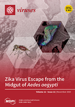
The Zika virus circulates between vertebrate hosts and mosquito vectors, both of which need to be productively infected. During mosquito infection, the virus encounters several tissue barriers, one of them between the midgut and other tissues. We investigated how Zika virus is overcoming this midgut escape barrier. We revealed the mesh width of the midgut basal lamina as a critical factor, which remains enlarged after bloodmeal digestion, enabling the virus to escape from the organ.View this paper.
 
  • Issues are regarded as officially published after their release is announced to the table of contents alert mailing list.
  • You may sign up for e-mail alerts to receive table of contents of newly released issues.
  • PDF is the official format for papers published in both, html and pdf forms. To view the papers in pdf format, click on the "PDF Full-text" link, and use the free Adobe Reader to open them.
Order results
Result details
Section
Select all
Export citation of selected articles as:
20 pages, 6525 KiB  
Article
A Novel Benthic Phage Infecting Shewanella with Strong Replication Ability
by Zengmeng Wang, Jiulong Zhao, Long Wang, Chengcheng Li, Jianhui Liu, Lihua Zhang and Yongyu Zhang
Viruses 2019, 11(11), 1081; https://doi.org/10.3390/v11111081 - 19 Nov 2019
Cited by 12 | Viewed by 3660
Abstract
The coastal sediments were considered to contain diverse phages playing important roles in driving biogeochemical cycles based on genetic analysis. However, till now, benthic phages in coastal sediments were very rarely isolated, which largely limits our understanding of their biological characteristics. Here, we [...] Read more.
The coastal sediments were considered to contain diverse phages playing important roles in driving biogeochemical cycles based on genetic analysis. However, till now, benthic phages in coastal sediments were very rarely isolated, which largely limits our understanding of their biological characteristics. Here, we describe a novel lytic phage (named Shewanella phage S0112) isolated from the coastal sediments of the Yellow Sea infecting a sediment bacterium of the genus Shewanella. The phage has a very high replication capability, with the burst size of ca. 1170 phage particles per infected cell, which is 5–10 times higher than that of most phages isolated before. Meanwhile, the latent period of this phage is relatively longer, which might ensure adequate time for phage replication. The phage has a double-stranded DNA genome comprising 62,286 bp with 102 ORFs, ca. 60% of which are functionally unknown. The expression products of 16 ORF genes, mainly structural proteins, were identified by LC-MS/MS analysis. Besides the general DNA metabolism and structure assembly genes in the phage genome, there is a cluster of auxiliary metabolic genes that may be involved in 7-cyano-7-deazaguanine (preQ0) biosynthesis. Meanwhile, a pyrophosphohydrolase (MazG) gene being considered as a regulator of programmed cell death or involving in host stringer responses is inserted in this gene cluster. Comparative genomic and phylogenetic analysis both revealed a great novelty of phage S0112. This study represents the first report of a benthic phage infecting Shewanella, which also sheds light on the phage–host interactions in coastal sediments. Full article
(This article belongs to the Special Issue Phage Ecology)
Show Figures

Figure 1

18 pages, 2221 KiB  
Article
Characterization of Klebsiella pneumoniae ST11 Isolates and Their Interactions with Lytic Phages
by Demeng Tan, Yiyuan Zhang, Mengjun Cheng, Shuai Le, Jingmin Gu, Juan Bao, Jinhong Qin, Xiaokui Guo and Tongyu Zhu
Viruses 2019, 11(11), 1080; https://doi.org/10.3390/v11111080 - 19 Nov 2019
Cited by 42 | Viewed by 7153
Abstract
The bacterial pathogen Klebsiella pneumoniae causes urinary tract infections in immunocompromised patients. Generally, the overuse of antibiotics contributes to the potential development and the spread of antibiotic resistance. In fact, certain strains of K. pneumoniae are becoming increasingly resistant to antibiotics, making infection [...] Read more.
The bacterial pathogen Klebsiella pneumoniae causes urinary tract infections in immunocompromised patients. Generally, the overuse of antibiotics contributes to the potential development and the spread of antibiotic resistance. In fact, certain strains of K. pneumoniae are becoming increasingly resistant to antibiotics, making infection by these strains more difficult to treat. The use of bacteriophages to control pathogens may offer a non-antibiotic-based approach to treat multidrug-resistant (MDR) infections. However, a detailed understanding of phage–host interactions is crucial in order to explore the potential success of phage-therapy for treatment. In this study, we investigated the molecular epidemiology of nine carbapenemase-producing K. pneumoniae isolates from a local hospital in Shanghai, China. All strain isolates belong to sequence type 11 (ST11) and harbor the blaKPC-2 gene. The S1-PFGE (S1 nuclease pulsed field gel electrophoresis) pattern of the isolates did not show any relationship to the multilocus sequence typing (MLST) profiles. In addition, we characterized phage 117 and phage 31 and assessed the potential application of phage therapy in treating K. pneumoniae infections in vitro. The results of morphological and genomic analyses suggested that both phages are affiliated to the T7 virus genus of the Podoviridae family. We also explored phage–host interactions during growth in both planktonic cells and biofilms. The phages’ heterogeneous lytic capacities against K. pneumoniae strains were demonstrated experimentally. Subsequent culture and urine experiments with phage 117 and host Kp36 initially demonstrated a strong lytic activity of the phages. However, rapid regrowth was observed following the initial lysis which suggests that phage resistant mutants were selected in the host populations. Additionally, a phage cocktail (117 + 31) was prepared and investigated for antimicrobial activity. In Luria Broth (LB) cultures, we observed that the cocktail showed significantly higher antimicrobial activity than phage 117 alone, but this was not observed in urine samples. Together, the results demonstrate the potential therapeutic value of phages in treating K. pneumoniae urinary tract infections. Full article
(This article belongs to the Special Issue Bacteriophages and Biofilms)
Show Figures

Figure 1

16 pages, 4103 KiB  
Article
Identification of B-Cell Epitopes with Potential to Serologicaly Discrimnate Dengue from Zika Infections
by Alice F. Versiani, Raissa Prado Rocha, Tiago A. O. Mendes, Glauco C. Pereira, Jordana Graziella A. Coelho dos Reis, Daniella C. Bartholomeu and Flávio G. da Fonseca
Viruses 2019, 11(11), 1079; https://doi.org/10.3390/v11111079 - 19 Nov 2019
Cited by 7 | Viewed by 3515
Abstract
Dengue is currently one of the most important arbovirus infections worldwide. Early diagnosis is important for disease outcome, particularly for those afflicted with the severe forms of infection. The goal of this work was to identify conserved and polymorphic linear B-cell Dengue virus [...] Read more.
Dengue is currently one of the most important arbovirus infections worldwide. Early diagnosis is important for disease outcome, particularly for those afflicted with the severe forms of infection. The goal of this work was to identify conserved and polymorphic linear B-cell Dengue virus (DENV) epitopes that could be used for diagnostic purposes. To this end, we aligned the predicted viral proteome of the four DENV serotype and performed in silico B-cell epitope mapping. We developed a script in Perl integrating alignment and prediction information to identify potential serotype-specific epitopes. We excluded epitopes that were similarly present in the yellow fever and zika viruses’ proteomes. A total of 15 polymorphic and nine conserved peptides among DENV serotypes were selected. Peptides were spotted on cellulose membranes and tested against sera from rabbits that were monoinfected with each DENV serotype. Although serotype-specific peptides failed to recognize any sera, three conserved peptides were recognized by all anti-dengue sera and were included on an ELISA test employing a well-characterized human sera bank. Of the three peptides, one was able to efficiently identify sera from all four DENV serotypes and to discriminate them from Zika virus positive sera. Full article
(This article belongs to the Special Issue Emerging Viruses: Surveillance, Prevention, Evolution and Control)
Show Figures

Figure 1

9 pages, 1113 KiB  
Review
Cellular Attachment and Entry Factors for Chikungunya Virus
by Barbara S. Schnierle
Viruses 2019, 11(11), 1078; https://doi.org/10.3390/v11111078 - 19 Nov 2019
Cited by 40 | Viewed by 6169
Abstract
Chikungunya virus (CHIKV) is clinically the most relevant member of the Alphavirus genus. Like alphaviruses in general, CHIKV has the capacity to infect a large variety of cells, tissues, and species. This broad host tropism of CHIKV indicates that the virus uses a [...] Read more.
Chikungunya virus (CHIKV) is clinically the most relevant member of the Alphavirus genus. Like alphaviruses in general, CHIKV has the capacity to infect a large variety of cells, tissues, and species. This broad host tropism of CHIKV indicates that the virus uses a ubiquitously expressed receptor to infect cells. This review summarizes the current knowledge available on cellular CHIKV receptors and the attachment factors used by CHIKV. Full article
(This article belongs to the Section Viral Immunology, Vaccines, and Antivirals)
Show Figures

Figure 1

10 pages, 231 KiB  
Review
HBV Infection in HIV-Driven Immune Suppression
by Loredana Sarmati and Vincenzo Malagnino
Viruses 2019, 11(11), 1077; https://doi.org/10.3390/v11111077 - 19 Nov 2019
Cited by 28 | Viewed by 5174
Abstract
Worldwide, approximately 10% of all human immunodeficiency virus (HIV)-infected people are also chronically coinfected with hepatitis B virus (HBV). HBV infection has a poor prognosis in HIV-positive people and has been documented by an increased risk of developing chronic HBV infection (CHB), progression [...] Read more.
Worldwide, approximately 10% of all human immunodeficiency virus (HIV)-infected people are also chronically coinfected with hepatitis B virus (HBV). HBV infection has a poor prognosis in HIV-positive people and has been documented by an increased risk of developing chronic HBV infection (CHB), progression to liver fibrosis and end-stage liver disease (ESLD) and evolution of hepatocellular carcinoma (HCC). Furthermore, in HIV patients, HBV-resolved infection is often associated with the appearance of HBV-DNA, which configures occult HBV infection (OBI) as a condition to be explored in coinfected patients. In this narrative review we summarize the main aspects of HBV infection in HIV-positive patients, emphasizing the importance of carefully considering the coinfected patient in the context of therapeutic strategies of antiretroviral therapy. Full article
(This article belongs to the Special Issue Hepatitis B Virus Reactivation)
17 pages, 2348 KiB  
Review
Review on Outbreak Dynamics, the Endemic Serotypes, and Diversified Topotypic Profiles of Foot and Mouth Disease Virus Isolates in Ethiopia from 2008 to 2018
by Ashenafi Kiros Wubshet, Junfei Dai, Qian Li and Jie Zhang
Viruses 2019, 11(11), 1076; https://doi.org/10.3390/v11111076 - 18 Nov 2019
Cited by 17 | Viewed by 5725
Abstract
Foot and mouth disease (FMD) endemicity in Ethiopia’s livestock remains an ongoing cause for economic concern, with new topotypes still arising even in previously unaffected areas. FMD outbreaks occur every year almost throughout the country. Understanding the outbreak dynamics, endemic serotypes, and lineage [...] Read more.
Foot and mouth disease (FMD) endemicity in Ethiopia’s livestock remains an ongoing cause for economic concern, with new topotypes still arising even in previously unaffected areas. FMD outbreaks occur every year almost throughout the country. Understanding the outbreak dynamics, endemic serotypes, and lineage profiles of FMD in this country is very critical in designing control and prevention programs. For this, detailed information on outbreak dynamics in Ethiopia needs to be understood clearly. In this article, therefore, we review the spatial and temporal patterns and dynamics of FMD outbreaks from 2008 to 2018. The circulating serotypes and the topotypic profiles of the virus are also discussed. FMD outbreak data were obtained from; reports of MoARD (Ministry of Agriculture and Rural Development)/MoLF (Ministry of livestock and Fishery, NVI (National Veterinary Institute), and NAHDIC (National Animal Health Diagnostic and Investigation Center); published articles; MSc works; PhD theses; and documents from international organizations. To effectively control and prevent FMD outbreaks, animal health agencies should focus on building surveillance systems that can quickly identify and control ongoing outbreaks and implement efficient preventive measures. Full article
(This article belongs to the Special Issue Emerging Viruses: Surveillance, Prevention, Evolution and Control)
Show Figures

Figure 1

12 pages, 1826 KiB  
Article
Molecular Detection and Characterization of the First Cowpox Virus Isolate Derived from a Bank Vole
by Kathrin Jeske, Saskia Weber, Florian Pfaff, Christian Imholt, Jens Jacob, Martin Beer, Rainer G. Ulrich and Donata Hoffmann
Viruses 2019, 11(11), 1075; https://doi.org/10.3390/v11111075 - 18 Nov 2019
Cited by 12 | Viewed by 3592
Abstract
Cowpox virus (CPXV) is a zoonotic orthopoxvirus (OPV) that infects a wide range of mammals. CPXV-specific DNA and antibodies were detected in different vole species, such as common voles (Microtus arvalis) and bank voles (Myodes glareolus). Therefore, voles are [...] Read more.
Cowpox virus (CPXV) is a zoonotic orthopoxvirus (OPV) that infects a wide range of mammals. CPXV-specific DNA and antibodies were detected in different vole species, such as common voles (Microtus arvalis) and bank voles (Myodes glareolus). Therefore, voles are the putative main reservoir host of CPXV. However, CPXV was up to now only isolated from common voles. Here we report the detection and isolation of a bank vole-derived CPXV strain (GerMygEK 938/17) resulting from a large-scale screening of bank voles collected in Thuringia, Germany, during 2017 and 2018. Phylogenetic analysis using the complete viral genome sequence indicated a high similarity of the novel strain to CPXV clade 3 and to OPV “Abatino” but also to Ectromelia virus (ECTV) strains. Phenotypic characterization of CPXV GerMygEK 938/17 using inoculation of embryonated chicken eggs displayed hemorrhagic pock lesions on the chorioallantoic membrane that are typical for CPXV but not for ECTV. CPXV GerMygEK 938/17 replicated in vole-derived kidney cell lines but at lower level than on Vero76 cell line. In conclusion, the first bank vole-derived CPXV isolate provides new insights into the genetic variability of CPXV in the putative reservoir host and is a valuable tool for further studies about CPXV-host interaction and molecular evolution of OPV. Full article
(This article belongs to the Section Animal Viruses)
Show Figures

Figure 1

22 pages, 5552 KiB  
Article
Characterization and Pathogenicity of the Porcine Deltacoronavirus Isolated in Southwest China
by Yujia Zhao, Huan Qu, Jingfei Hu, Jiayu Fu, Rui Chen, Cheng Li, Sanjie Cao, Yiping Wen, Rui Wu, Qin Zhao, Qigui Yan, Xintian Wen and Xiaobo Huang
Viruses 2019, 11(11), 1074; https://doi.org/10.3390/v11111074 - 18 Nov 2019
Cited by 32 | Viewed by 4249
Abstract
Porcine deltacoronavirus (PDCoV) is a newly emerging enteric pathogen in swine that causes diarrhea in neonatal piglets and creates an additional economic burden on porcine industries in Asia and North America. In this study, a PDCoV isolate, CHN-SC2015, was isolated from Sichuan Province [...] Read more.
Porcine deltacoronavirus (PDCoV) is a newly emerging enteric pathogen in swine that causes diarrhea in neonatal piglets and creates an additional economic burden on porcine industries in Asia and North America. In this study, a PDCoV isolate, CHN-SC2015, was isolated from Sichuan Province in southwest China. The isolate was characterized by a cytopathic effect, immunofluorescence, and electron microscopy. CHN-SC2015 titers in LLC-PK cells ranged from 104.31 to 108.22 TCID50/mL during the first 30 passages. During serial passage, 11 nucleotide mutations occurred in the S gene, resulting in nine amino acid changes. A whole genome sequencing analysis demonstrated that CHN-SC2015 shares 97.5%–99.1% identity with 59 reference strains in GenBank. Furthermore, CHN-SC2015 contained 6-nt deletion and 9-nt insertion in the ORF1ab gene, 3-nt deletion in the S gene and 11-nt deletion in its 3′UTR compared with other reference strains available in GenBank. A phylogenetic analysis showed that CHN-SC2015 is more closely related to other PDCoV strains in China than to the strains from Southeast Asia, USA, Japan, and South Korea, indicating the diversity of genetic relationships and regional and epidemic characteristics among these strains. A recombination analysis indicated that CHN-SC2015 experienced recombination events between SHJS/SL/2016 and TT-1115. In vivo infection demonstrated that CHN-SC2015 is highly pathogenic to sucking piglets, causing diarrhea, vomiting, dehydration, and death. Virus was shed daily in the feces of infected piglets and upon necropsy, was found distributed in the gastrointestinal tract and in multiple organs. CHN-SC2015 is the first systematically characterized strain from southwest China hitherto reported. Our results enrich the body of information on the epidemiology, pathogenicity and molecular evolution associated with PDCoV. Full article
Show Figures

Figure 1

17 pages, 1527 KiB  
Review
Towards Understanding KSHV Fusion and Entry
by Stephen J. Dollery
Viruses 2019, 11(11), 1073; https://doi.org/10.3390/v11111073 - 18 Nov 2019
Cited by 20 | Viewed by 5032
Abstract
How viruses enter cells is of critical importance to pathogenesis in the host and for treatment strategies. Over the last several years, the herpesvirus field has made numerous and thoroughly fascinating discoveries about the entry of alpha-, beta-, and gamma-herpesviruses, giving rise to [...] Read more.
How viruses enter cells is of critical importance to pathogenesis in the host and for treatment strategies. Over the last several years, the herpesvirus field has made numerous and thoroughly fascinating discoveries about the entry of alpha-, beta-, and gamma-herpesviruses, giving rise to knowledge of entry at the amino acid level and the realization that, in some cases, researchers had overlooked whole sets of molecules essential for entry into critical cell types. Herpesviruses come equipped with multiple envelope glycoproteins which have several roles in many aspects of infection. For herpesvirus entry, it is usual that a collective of glycoproteins is involved in attachment to the cell surface, specific interactions then take place between viral glycoproteins and host cell receptors, and then molecular interactions and triggers occur, ultimately leading to viral envelope fusion with the host cell membrane. The fact that there are multiple cell and virus molecules involved with the build-up to fusion enhances the diversity and specificity of target cell types, the cellular entry pathways the virus commandeers, and the final triggers of fusion. This review will examine discoveries relating to how Kaposi’s sarcoma-associated herpesvirus (KSHV) encounters and binds to critical cell types, how cells internalize the virus, and how the fusion may occur between the viral membrane and the host cell membrane. Particular focus is given to viral glycoproteins and what is known about their mechanisms of action. Full article
(This article belongs to the Special Issue Viral Entry Pathways)
Show Figures

Figure 1

14 pages, 1097 KiB  
Article
Support for the Transmission-Clearance Trade-Off Hypothesis from a Study of Zika Virus Delivered by Mosquito Bite to Mice
by Kathryn A. Hanley, Sasha R. Azar, Rafael K. Campos, Nikos Vasilakis and Shannan L. Rossi
Viruses 2019, 11(11), 1072; https://doi.org/10.3390/v11111072 - 18 Nov 2019
Cited by 11 | Viewed by 3458
Abstract
Evolutionary theory indicates that virus virulence is shaped by a trade-off between instantaneous rate of transmission and duration of infection. For most viruses, infection is curtailed by immune clearance, but there are few empirical tests of the transmission–clearance trade-off hypothesis. We exposed A129 [...] Read more.
Evolutionary theory indicates that virus virulence is shaped by a trade-off between instantaneous rate of transmission and duration of infection. For most viruses, infection is curtailed by immune clearance, but there are few empirical tests of the transmission–clearance trade-off hypothesis. We exposed A129 mice to bites from groups of 1, 2–4, or 6–9 Aedes albopictus mosquitoes infected with Zika virus (ZIKV). We predicted that a higher number of infectious mosquito bites would deliver a higher total dose of the virus, and that increasing dose would result in earlier onset, higher magnitude, and shorter duration of viremia, as well as a more robust neutralizing antibody response. We found that increases in the number of mosquito bites delivered resulted in significantly different virus replication dynamics with higher, earlier peak titers. All mice experienced a transient weight loss following infection, but the nadir in weight loss was delayed in the mice that received the highest number of bites. Viremia persisted past the period of measurement in this study, so we did not capture its duration. However, the association at the level of the individual mouse between the estimated virus dose delivered and neutralizing antibody titer was remarkably strong, supporting the transmission–clearance trade-off hypothesis. Full article
(This article belongs to the Section Animal Viruses)
Show Figures

Figure 1

19 pages, 14637 KiB  
Article
Survey and Characterization of Jingmen Tick Virus Variants
by Ender Dinçer, Sabri Hacıoğlu, Sırrı Kar, Nergis Emanet, Annika Brinkmann, Andreas Nitsche, Aykut Özkul, Yvonne-Marie Linton and Koray Ergünay
Viruses 2019, 11(11), 1071; https://doi.org/10.3390/v11111071 - 17 Nov 2019
Cited by 38 | Viewed by 5474
Abstract
We obtained a Jingmen tick virus (JMTV) isolate, following inoculation of a tick pool with detectable Crimean-Congo hemorrhagic fever virus (CCHFV) RNA. We subsequently screened 7223 ticks, representing 15 species in five genera, collected from various regions in Anatolia and eastern Thrace, Turkey. [...] Read more.
We obtained a Jingmen tick virus (JMTV) isolate, following inoculation of a tick pool with detectable Crimean-Congo hemorrhagic fever virus (CCHFV) RNA. We subsequently screened 7223 ticks, representing 15 species in five genera, collected from various regions in Anatolia and eastern Thrace, Turkey. Moreover, we tested specimens from various patient cohorts (n = 103), and canine (n = 60), bovine (n = 20) and avian specimens (n = 65). JMTV nucleic acids were detected in 3.9% of the tick pools, including those from several tick species from the genera Rhipicephalus and Haemaphysalis, and Hyalomma marginatum, the main vector of CCHFV in Turkey. Phylogenetic analysis supported two separate clades, independent of host or location, suggesting ubiquitous distribution in ticks. JMTV was not recovered from any human, animal or bird specimens tested. Near-complete viral genomes were sequenced from the prototype isolate and from three infected tick pools. Genome topology and functional organization were identical to the members of Jingmen group viruses. Phylogenetic reconstruction of individual viral genome segments and functional elements further supported the close relationship of the strains from Kosovo. We further identified probable recombination events in the JMTV genome, involving closely-related strains from Anatolia or China. Full article
(This article belongs to the Special Issue Emerging Arboviruses)
Show Figures

Figure 1

19 pages, 3632 KiB  
Article
A Multi-Omics Study of Chicken Infected by Nephropathogenic Infectious Bronchitis Virus
by Puzhi Xu, Ping Liu, Changming Zhou, Yan Shi, Qingpeng Wu, Yitian Yang, Guyue Li, Guoliang Hu and Xiaoquan Guo
Viruses 2019, 11(11), 1070; https://doi.org/10.3390/v11111070 - 16 Nov 2019
Cited by 21 | Viewed by 7332
Abstract
Chicken gout resulting from nephropathogenic infectious bronchitis virus (NIBV) has become a serious kidney disease problem in chicken worldwide with alterations of the metabolic phenotypes in multiple metabolic pathways. To investigate the mechanisms in chicken responding to NIBV infection, we examined the global [...] Read more.
Chicken gout resulting from nephropathogenic infectious bronchitis virus (NIBV) has become a serious kidney disease problem in chicken worldwide with alterations of the metabolic phenotypes in multiple metabolic pathways. To investigate the mechanisms in chicken responding to NIBV infection, we examined the global transcriptomic and metabolomic profiles of the chicken’s kidney using RNA-seq and GC–TOF/MS, respectively. Furthermore, we analyzed the alterations in cecal microorganism composition in chickens using 16S rRNA-seq. Integrated analysis of these three phenotypic datasets further managed to create correlations between the altered kidney transcriptomes and metabolome, and between kidney metabolome and gut microbiome. We found that 2868 genes and 160 metabolites were deferentially expressed or accumulated in the kidney during NIBV infection processes. These genes and metabolites were linked to NIBV-infection related processes, including immune response, signal transduction, peroxisome, purine, and amino acid metabolism. In addition, the comprehensive correlations between the kidney metabolome and cecal microbial community showed contributions of gut microbiota in the progression of NIBV-infection. Taken together, our research comprehensively describes the host responses during NIBV infection and provides new clues for further dissection of specific gene functions, metabolite affections, and the role of gut microbiota during chicken gout. Full article
(This article belongs to the Section Animal Viruses)
Show Figures

Figure 1

17 pages, 3185 KiB  
Article
In Vivo Characterization of Tick-Borne Encephalitis Virus in Bank Voles (Myodes glareolus)
by Anna Michelitsch, Birke Andrea Tews, Christine Klaus, Malena Bestehorn-Willmann, Gerhard Dobler, Martin Beer and Kerstin Wernike
Viruses 2019, 11(11), 1069; https://doi.org/10.3390/v11111069 - 15 Nov 2019
Cited by 13 | Viewed by 3242
Abstract
Tick-borne encephalitis is the most important tick-transmitted zoonotic virus infection in Eurasia, causing severe neurological symptoms in humans. The causative agent, the tick-borne encephalitis virus (TBEV), circulates between ticks and a variety of mammalian hosts. To study the interaction between TBEV and one [...] Read more.
Tick-borne encephalitis is the most important tick-transmitted zoonotic virus infection in Eurasia, causing severe neurological symptoms in humans. The causative agent, the tick-borne encephalitis virus (TBEV), circulates between ticks and a variety of mammalian hosts. To study the interaction between TBEV and one of its suspected reservoir hosts, bank voles of the Western evolutionary lineage were inoculated subcutaneously with either one of eight TBEV strains or the related attenuated Langat virus, and were euthanized after 28 days. In addition, a subset of four strains was characterized in bank voles of the Carpathian linage. Six bank voles were inoculated per strain, and were housed together in groups of three with one uninfected in-contact animal each. Generally, most bank voles did not show any clinical signs over the course of infection. However, one infected bank vole died and three had to be euthanized prematurely, all of which had been inoculated with the identical TBEV strain (Battaune 17-H9, isolated in 2017 in Germany from a bank vole). All inoculated animals seroconverted, while none of the in-contact animals did. Viral RNA was detected via real-time RT-PCR in the whole blood samples of 31 out of 74 inoculated and surviving bank voles. The corresponding serum sample remained PCR-negative in nearly all cases (29/31). In addition, brain and/or spine samples tested positive in 11 cases, mostly correlating with a positive whole blood sample. Our findings suggest a good adaption of TBEV to bank voles, combining in most cases a low virulence phenotype with detectable virus replication and hinting at a reservoir host function of bank voles for TBEV. Full article
(This article belongs to the Special Issue Flavivirus Replication and Pathogenesis)
Show Figures

Figure 1

35 pages, 878 KiB  
Review
Diagnosis of Feline Infectious Peritonitis: A Review of the Current Literature
by Sandra Felten and Katrin Hartmann
Viruses 2019, 11(11), 1068; https://doi.org/10.3390/v11111068 - 15 Nov 2019
Cited by 82 | Viewed by 24746
Abstract
Feline infectious peritonitis (FIP) is a fatal disease that poses several challenges for veterinarians: clinical signs and laboratory changes are non-specific, and there are two pathotypes of the etiologic agent feline coronavirus (FCoV), sometimes referred to as feline enteric coronavirus (FECV) and feline [...] Read more.
Feline infectious peritonitis (FIP) is a fatal disease that poses several challenges for veterinarians: clinical signs and laboratory changes are non-specific, and there are two pathotypes of the etiologic agent feline coronavirus (FCoV), sometimes referred to as feline enteric coronavirus (FECV) and feline infectious peritonitis virus (FIPV) that vary fundamentally in their virulence, but are indistinguishable by a number of diagnostic methods. This review focuses on all important steps every veterinary practitioner has to deal with and new diagnostic tests that can be considered when encountering a cat with suspected FIP with the aim to establish a definitive diagnosis. It gives an overview on all available direct and indirect diagnostic tests and their sensitivity and specificity reported in the literature in different sample material. By providing summarized data for sensitivity and specificity of each diagnostic test and each sample material, which can easily be accessed in tables, this review can help to facilitate the interpretation of different diagnostic tests and raise awareness of their advantages and limitations. Additionally, diagnostic trees depict recommended diagnostic steps that should be performed in cats suspected of having FIP based on their clinical signs or clinicopathologic abnormalities. These steps can easily be followed in clinical practice. Full article
(This article belongs to the Special Issue Feline Viruses and Viral Diseases)
Show Figures

Figure 1

12 pages, 5050 KiB  
Article
Efficient Expression and Processing of Ebola Virus Glycoprotein Induces Morphological Changes in BmN Cells but Cannot Rescue Deficiency of Bombyx Mori Nucleopolyhedrovirus GP64
by Jinshan Huang, Na Liu, Fanbo Xu, Ellen Ayepa, Charles Amanze, Luping Sun, Yaqin Shen, Miao Yang, Shuwen Yang, Xingjia Shen and Bifang Hao
Viruses 2019, 11(11), 1067; https://doi.org/10.3390/v11111067 - 15 Nov 2019
Cited by 6 | Viewed by 4062
Abstract
Ebola virus (EBOV) disease outbreaks have resulted in many fatalities, yet no licensed vaccines are available to prevent infection. Recombinant glycoprotein (GP) production may contribute to finding a cure for Ebola virus disease, which is the key candidate protein for vaccine preparation. To [...] Read more.
Ebola virus (EBOV) disease outbreaks have resulted in many fatalities, yet no licensed vaccines are available to prevent infection. Recombinant glycoprotein (GP) production may contribute to finding a cure for Ebola virus disease, which is the key candidate protein for vaccine preparation. To explore GP1,2 expression in BmN cells, EBOV-GP1,2 with its native signal peptide or the GP64 signal peptide was cloned and transferred into a normal or gp64 null Bombyx mori nucleopolyhedrovirus (BmNPV) bacmid via transposition. The infectivity of the recombinant bacmids was investigated after transfection, expression and localization of EBOV-GP were investigated, and cell morphological changes were analyzed by TEM. The GP64 signal peptide, but not the GP1,2 native signal peptide, caused GP1,2 localization to the cell membrane, and the differentially localized GP1,2 proteins were cleaved into GP1 and GP2 fragments in BmN cells. GP1,2 expression resulted in dramatic morphological changes in BmN cells in the early stage of infection. However, GP1,2 expression did not rescue GP64 deficiency in BmNPV infection. This study provides a better understanding of GP expression and processing in BmN cells, which may lay a foundation for EBOV-GP expression using the BmNPV baculovirus expression system. Full article
(This article belongs to the Collection Advances in Ebolavirus, Marburgvirus, and Cuevavirus Research)
Show Figures

Figure 1

6 pages, 497 KiB  
Communication
Treatment of Highly Pathogenic H7N9 Virus-Infected Mice with Baloxavir Marboxil
by Maki Kiso, Seiya Yamayoshi, Yuri Furusawa, Masaki Imai and Yoshihiro Kawaoka
Viruses 2019, 11(11), 1066; https://doi.org/10.3390/v11111066 - 15 Nov 2019
Cited by 7 | Viewed by 3986
Abstract
Viral neuraminidase inhibitors show limited efficacy in mice infected with H7N9 influenza A viruses isolated from humans. Although baloxavir marboxil protected mice from lethal challenge infection with a low pathogenic avian influenza H7N9 virus isolated from a human, its efficacy in mice infected [...] Read more.
Viral neuraminidase inhibitors show limited efficacy in mice infected with H7N9 influenza A viruses isolated from humans. Although baloxavir marboxil protected mice from lethal challenge infection with a low pathogenic avian influenza H7N9 virus isolated from a human, its efficacy in mice infected with a recent highly pathogenic version of H7N9 human isolates is unknown. Here, we examined the efficacy of baloxavir marboxil in mice infected with a highly pathogenic human H7N9 virus, A/Guangdong/17SF003/2016. Treatment of infected mice with a single 1.5 mg/kg dose of baloxavir marboxil protected mice from the highly pathogenic human H7N9 virus infection as effectively as oseltamivir treatment at 50 mg/kg twice a day for five days. Daily treatment for five days at 15 or 50 mg/kg of baloxavir marboxil showed superior therapeutic efficacy, largely preventing virus replication in respiratory organs. These results indicate that baloxavir marboxil is a valuable candidate treatment for human patients suffering from highly pathogenic H7N9 virus infection. Full article
(This article belongs to the Section Viral Immunology, Vaccines, and Antivirals)
Show Figures

Figure 1

11 pages, 482 KiB  
Review
Schmallenberg Disease—A Newly Emerged Culicoides-Borne Viral Disease of Ruminants
by Abaineh D. Endalew, Bonto Faburay, William C. Wilson and Juergen A. Richt
Viruses 2019, 11(11), 1065; https://doi.org/10.3390/v11111065 - 15 Nov 2019
Cited by 21 | Viewed by 4690
Abstract
First appearing in 2011 in Northern Europe, Schmallenberg virus (SBV), an Orthobunyavirus of the Simbu serogroup, is associated with clinical disease mainly in ruminants such as cattle, sheep and goats. The clinical signs are characterized by abortion and congenital deformities in newborns. The [...] Read more.
First appearing in 2011 in Northern Europe, Schmallenberg virus (SBV), an Orthobunyavirus of the Simbu serogroup, is associated with clinical disease mainly in ruminants such as cattle, sheep and goats. The clinical signs are characterized by abortion and congenital deformities in newborns. The virus is transmitted by Culicoides midges of the Obsoletus complex. SBV infection induces a solid protective immunity that persists for at least 4 or 6 years in sheep and cattle, respectively. SBV infection can be diagnosed directly by real-time RT-qPCR and virus isolation or indirectly by serological assays. Three vaccines are commercially available in Europe. This article provides a comprehensive literature review on this emerging disease regarding pathogenesis, transmission, diagnosis, control and prevention. This review also highlights that although much has been learned since SBV’s first emergence, there are still areas that require further study to devise better mitigation strategies. Full article
(This article belongs to the Special Issue Virus-Vector-Host Interactions of Culicoides-Borne Diseases)
Show Figures

Figure 1

17 pages, 2601 KiB  
Article
Erythromycin Estolate Inhibits Zika Virus Infection by Blocking Viral Entry as a Viral Inactivator
by Xiaohuan Wang, Shuai Xia, Peng Zou and Lu Lu
Viruses 2019, 11(11), 1064; https://doi.org/10.3390/v11111064 - 15 Nov 2019
Cited by 12 | Viewed by 4330
Abstract
Recently, Zika virus (ZIKV) has attracted much attention in consideration of its association with severe neurological complications including fetal microcephaly. However, there are currently no prophylactic vaccines or therapeutic drugs approved for clinical treatments of ZIKV infection. To determine the potential anti-ZIKV inhibitors, [...] Read more.
Recently, Zika virus (ZIKV) has attracted much attention in consideration of its association with severe neurological complications including fetal microcephaly. However, there are currently no prophylactic vaccines or therapeutic drugs approved for clinical treatments of ZIKV infection. To determine the potential anti-ZIKV inhibitors, we screened a library of clinical drugs with good safety profiles. Erythromycin estolate (Ery-Est), one of the macrolide antibiotics, was found to effectively inhibit ZIKV infection in different cell types and significantly protect A129 mice from ZIKV-associated neurological signs and mortality. Through further investigation, Ery-Est was verified to inhibit ZIKV entry by disrupting the integrity of the viral membrane which resulted in the loss of ZIKV infectivity. Furthermore, Ery-Est also showed inhibitory activity against dengue virus (DENV) and yellow fever virus (YFV). Thus, Ery-Est may be a promising drug for patients with ZIKV infection, particularly pregnant women. Full article
(This article belongs to the Special Issue Viral Entry Pathways)
Show Figures

Figure 1

16 pages, 1940 KiB  
Article
Molecular Dynamics Simulation Reveals Exposed Residues in the Ligand-Binding Domain of the Low-Density Lipoprotein Receptor that Interacts with Vesicular Stomatitis Virus-G Envelope
by Faisal A. Al-Allaf, Zainularifeen Abduljaleel, Mohiuddin M. Taher, Ahmed A. H. Abdellatif, Mohammad Athar, Neda M. Bogari, Mohammed N. Al-Ahdal, Futwan Al-Mohanna, Zuhair N. Al-Hassnan, Kamal H. Y. Alzabeedi, Talib M. Banssir and Abdellatif Bouazzaoui
Viruses 2019, 11(11), 1063; https://doi.org/10.3390/v11111063 - 15 Nov 2019
Cited by 4 | Viewed by 3366
Abstract
Familial hypercholesterolemia (FH) is an autosomal dominant disease most often caused by mutations in the low-density lipoprotein receptor (LDLR) gene, which consists of 18 exons spanning 45 kb and codes for a precursor protein of 860 amino acids. Mutations in the LDLR gene [...] Read more.
Familial hypercholesterolemia (FH) is an autosomal dominant disease most often caused by mutations in the low-density lipoprotein receptor (LDLR) gene, which consists of 18 exons spanning 45 kb and codes for a precursor protein of 860 amino acids. Mutations in the LDLR gene lead to a reduced hepatic clearance of LDL as well as a high risk of coronary artery disease (CAD) and sudden cardiac death (SCD). Recently, LDLR transgenes have generated interest as potential therapeutic agents. However, LDLR packaging using a lentiviral vector (LVV) system pseudotyped with a vesicular stomatitis virus (VSV)-G envelope is not efficient. In this study, we modified the LVV system to improve transduction efficiency and investigated the LDLR regions responsible for transduction inhibition. Transduction efficiency of 293T cells with a 5′-LDLReGFP-3′ fusion construct was only 1.55% compared to 42.32% for the eGFP construct. Moreover, co-expression of LDLR affected eGFP packaging. To determine the specific region of the LDLR protein responsible for packaging inhibition, we designed constructs with mutations or sequential deletions at the 3′ and 5′ ends of LDLR cDNA. All constructs except one without the ligand-binding domain (LBD) (pWoLBD–eGFP) resulted in low transduction efficiency, despite successful packaging of viral RNA in the VSV envelope, as confirmed through RT-PCR. When we evaluated a direct interaction between LDLR and the VSV envelope glycoprotein using MD simulation and protein–protein interactions, we uncovered Val119, Thr120, Thr67, and Thr118 as exposed residues in the LDLR receptor that interact with the VSV protein. Together, our results suggest that the LBD of LDLR interacts with the VSV-G protein during viral packaging, which significantly reduces transduction efficiency. Full article
(This article belongs to the Section Animal Viruses)
Show Figures

Figure 1

18 pages, 1731 KiB  
Review
Parechovirus A Pathogenesis and the Enigma of Genotype A-3
by Adithya Sridhar, Eveliina Karelehto, Lieke Brouwer, Dasja Pajkrt and Katja C. Wolthers
Viruses 2019, 11(11), 1062; https://doi.org/10.3390/v11111062 - 14 Nov 2019
Cited by 26 | Viewed by 4336
Abstract
Parechovirus A is a species in the Parechovirus genus within the Picornaviridae family that can cause severe disease in children. Relatively little is known on Parechovirus A epidemiology and pathogenesis. This review aims to explore the Parechovirus A literature and highlight the differences [...] Read more.
Parechovirus A is a species in the Parechovirus genus within the Picornaviridae family that can cause severe disease in children. Relatively little is known on Parechovirus A epidemiology and pathogenesis. This review aims to explore the Parechovirus A literature and highlight the differences between Parechovirus A genotypes from a pathogenesis standpoint. In particular, the curious case of Parechovirus-A3 and the genotype-specific disease association will be discussed. Finally, a brief outlook on Parechovirus A research is provided. Full article
(This article belongs to the Special Issue Human Picornaviruses)
Show Figures

Figure 1

20 pages, 5689 KiB  
Article
The U3 and Env Proteins of Jaagsiekte Sheep Retrovirus and Enzootic Nasal Tumor Virus Both Contribute to Tissue Tropism
by María C. Rosales Gerpe, Laura P. van Lieshout, Jakob M. Domm, Joelle C. Ingrao, Jodre Datu, Scott R. Walsh, Darrick L. Yu, Jondavid de Jong, Peter J. Krell and Sarah K. Wootton
Viruses 2019, 11(11), 1061; https://doi.org/10.3390/v11111061 - 14 Nov 2019
Cited by 4 | Viewed by 2919
Abstract
Jaagsiekte sheep retrovirus (JSRV) and enzootic nasal tumor virus (ENTV) are small-ruminant betaretroviruses that share high nucleotide and amino acid identity, utilize the same cellular receptor, hyaluronoglucosaminidase 2 (Hyal2) for entry, and transform tissues with their envelope (Env) glycoprotein; yet, they target discrete [...] Read more.
Jaagsiekte sheep retrovirus (JSRV) and enzootic nasal tumor virus (ENTV) are small-ruminant betaretroviruses that share high nucleotide and amino acid identity, utilize the same cellular receptor, hyaluronoglucosaminidase 2 (Hyal2) for entry, and transform tissues with their envelope (Env) glycoprotein; yet, they target discrete regions of the respiratory tract—the lung and nose, respectively. This distinct tissue selectivity makes them ideal tools with which to study the pathogenesis of betaretroviruses. To uncover the genetic determinants of tropism, we constructed JSRV–ENTV chimeric viruses and produced lentivectors pseudotyped with the Env proteins from JSRV (Jenv) and ENTV (Eenv). Through the transduction and infection of lung and nasal turbinate tissue slices, we observed that Hyal2 expression levels strongly influence ENTV entry, but that the long terminal repeat (LTR) promoters of these viruses are likely responsible for tissue-specificity. Furthermore, we show evidence of ENTV Env expression in chondrocytes within ENTV-infected nasal turbinate tissue, where Hyal2 is highly expressed. Our work suggests that the unique tissue tropism of JSRV and ENTV stems from the combined effort of the envelope glycoprotein-receptor interactions and the LTR and provides new insight into the pathogenesis of ENTV. Full article
(This article belongs to the Section Animal Viruses)
Show Figures

Figure 1

21 pages, 917 KiB  
Review
Relating GPI-Anchored Ly6 Proteins uPAR and CD59 to Viral Infection
by Jingyou Yu, Vaibhav Murthy and Shan-Lu Liu
Viruses 2019, 11(11), 1060; https://doi.org/10.3390/v11111060 - 14 Nov 2019
Cited by 12 | Viewed by 4458
Abstract
The Ly6 (lymphocyte antigen-6)/uPAR (urokinase-type plasminogen activator receptor) superfamily protein is a group of molecules that share limited sequence homology but conserved three-fingered structures. Despite diverse cellular functions, such as in regulating host immunity, cell adhesion, and migration, the physiological roles of these [...] Read more.
The Ly6 (lymphocyte antigen-6)/uPAR (urokinase-type plasminogen activator receptor) superfamily protein is a group of molecules that share limited sequence homology but conserved three-fingered structures. Despite diverse cellular functions, such as in regulating host immunity, cell adhesion, and migration, the physiological roles of these factors in vivo remain poorly characterized. Notably, increasing research has focused on the interplays between Ly6/uPAR proteins and viral pathogens, the results of which have provided new insight into viral entry and virus–host interactions. While LY6E (lymphocyte antigen 6 family member E), one key member of the Ly6E/uPAR-family proteins, has been extensively studied, other members have not been well characterized. Here, we summarize current knowledge of Ly6/uPAR proteins related to viral infection, with a focus on uPAR and CD59. Our goal is to provide an up-to-date view of the Ly6/uPAR-family proteins and associated virus–host interaction and viral pathogenesis. Full article
(This article belongs to the Special Issue Mechanisms of Viral Fusion and Applications in Antivirals)
Show Figures

Figure 1

18 pages, 868 KiB  
Review
Mosquitoes of North-Western Europe as Potential Vectors of Arboviruses: A Review
by Jean-Philippe Martinet, Hubert Ferté, Anna-Bella Failloux, Francis Schaffner and Jérôme Depaquit
Viruses 2019, 11(11), 1059; https://doi.org/10.3390/v11111059 - 14 Nov 2019
Cited by 70 | Viewed by 5671
Abstract
Background: The intensification of trade and travel is linked to the growing number of imported cases of dengue, chikungunya or Zika viruses into continental Europe and to the expansion of invasive mosquito species such as Aedes albopictus and Aedes japonicus. Local outbreaks [...] Read more.
Background: The intensification of trade and travel is linked to the growing number of imported cases of dengue, chikungunya or Zika viruses into continental Europe and to the expansion of invasive mosquito species such as Aedes albopictus and Aedes japonicus. Local outbreaks have already occurred in several European countries. Very little information exists on the vector competence of native mosquitoes for arboviruses. As such, the vectorial status of the nine mosquito species largely established in North-Western Europe (Aedes cinereus and Aedes geminus, Aedes cantans, Aedes punctor, Aedes rusticus, Anopheles claviger s.s., Anopheles plumbeus, Coquillettidia richiardii, Culex pipiens s.l., and Culiseta annulata) remains mostly unknown. Objectives: To review the vector competence of both invasive and native mosquito populations found in North-Western Europe (i.e., France, Belgium, Germany, United Kingdom, Ireland, The Netherlands, Luxembourg and Switzerland) for dengue, chikungunya, Zika, West Nile and Usutu viruses. Methods: A bibliographical search with research strings addressing mosquito vector competence for considered countries was performed. Results: Out of 6357 results, 119 references were related to the vector competence of mosquitoes in Western Europe. Eight species appear to be competent for at least one virus. Conclusions: Aedes albopictus is responsible for the current outbreaks. The spread of Aedes albopictus and Aedes japonicus increases the risk of the autochthonous transmission of these viruses. Although native species could contribute to their transmission, more studies are still needed to assess that risk. Full article
(This article belongs to the Special Issue Emerging Arboviruses)
Show Figures

Figure 1

11 pages, 518 KiB  
Article
Use of Monocyte-Derived Macrophage Culture Increases Zika Virus Isolation Rate from Human Plasma
by Emilia Sippert, Bruno C. Rocha, Felipe L. Assis, Suzan Ok and Maria Rios
Viruses 2019, 11(11), 1058; https://doi.org/10.3390/v11111058 - 14 Nov 2019
Cited by 3 | Viewed by 2655
Abstract
Viral isolation is desirable for many reasons, including development of diagnostic assays and reference materials, and for virology basic research. Zika virus (ZIKV) isolation from clinical samples is challenging, but isolates are known to infect various cell lines. Here, we evaluated suitability of [...] Read more.
Viral isolation is desirable for many reasons, including development of diagnostic assays and reference materials, and for virology basic research. Zika virus (ZIKV) isolation from clinical samples is challenging, but isolates are known to infect various cell lines. Here, we evaluated suitability of Vero, C6/36 and JEG-3 as host cells, for direct isolation of ZIKV from human plasma. We also assessed the use of primary monocyte-derived macrophages (MDMs) culture to enhance ZIKV isolation from human plasma samples followed by virus expansion in Vero, C6/36 and JEG-3 cultures. Direct inoculation of cell lines with 42 ZIKV-RNA positive samples resulted in isolation rates of 9.52% (4/42) in Vero and C6/36, and of 7.14% (3/42) in JEG-3 cells. Inoculation of plasma in MDMs followed by supernatant testing by TaqMan RT-PCR, resulted in 33/42 (78.57%) ZIKV-RNA-positive supernatants, which expansion in cell lines increased isolation rates to 24.24% (8/33) in Vero and to 27.27% (9/33) in C6/36 and JEG-3 regardless of the presence of ZIKV-antibody. Isolates generated in JEG-3 cells were also produced in Vero and C6/36 with similar viral titers. These results suggest that efficiency of ZIKV isolation from human plasma can be enhanced when MDM culture is used before viral expansion in cell lines. Full article
(This article belongs to the Special Issue Emerging Arboviruses)
Show Figures

Figure 1

14 pages, 1597 KiB  
Article
A Toxicity Screening Approach to Identify Bacteriophage-Encoded Anti-Microbial Proteins
by Ushanandini Mohanraj, Xing Wan, Cindy M. Spruit, Mikael Skurnik and Maria I. Pajunen
Viruses 2019, 11(11), 1057; https://doi.org/10.3390/v11111057 - 14 Nov 2019
Cited by 11 | Viewed by 5432
Abstract
The rapid emergence of antibiotic resistance among many pathogenic bacteria has created a profound need to discover new alternatives to antibiotics. Bacteriophages, the viruses of microbes, express special proteins to overtake the metabolism of the bacterial host they infect, the best known of [...] Read more.
The rapid emergence of antibiotic resistance among many pathogenic bacteria has created a profound need to discover new alternatives to antibiotics. Bacteriophages, the viruses of microbes, express special proteins to overtake the metabolism of the bacterial host they infect, the best known of which are involved in bacterial lysis. However, the functions of majority of bacteriophage encoded gene products are not known, i.e., they represent the hypothetical proteins of unknown function (HPUFs). In the current study we present a phage genomics-based screening approach to identify phage HPUFs with antibacterial activity with a long-term goal to use them as leads to find unknown targets to develop novel antibacterial compounds. The screening assay is based on the inhibition of bacterial growth when a toxic gene is expression-cloned into a plasmid vector. It utilizes an optimized plating assay producing a significant difference in the number of transformants after ligation of the toxic and non-toxic genes into a cloning vector. The screening assay was first tested and optimized using several known toxic and non-toxic genes. Then, it was applied to screen 94 HPUFs of bacteriophage φR1-RT, and identified four HPUFs that were toxic to Escherichia coli. This optimized assay is in principle useful in the search for bactericidal proteins of any phage, and also opens new possibilities to understanding the strategies bacteriophages use to overtake bacterial hosts. Full article
(This article belongs to the Section Bacterial Viruses)
Show Figures

Figure 1

18 pages, 4416 KiB  
Article
Zika Virus Dissemination from the Midgut of Aedes aegypti is Facilitated by Bloodmeal-Mediated Structural Modification of the Midgut Basal Lamina
by Yingjun Cui, DeAna G. Grant, Jingyi Lin, Xiudao Yu and Alexander W. E. Franz
Viruses 2019, 11(11), 1056; https://doi.org/10.3390/v11111056 - 14 Nov 2019
Cited by 23 | Viewed by 5668
Abstract
The arboviral disease cycle requires that key tissues in the arthropod vector become persistently infected with the virus. The midgut is the first organ in the mosquito that needs to be productively infected with an orally acquired virus. Following midgut infection, the virus [...] Read more.
The arboviral disease cycle requires that key tissues in the arthropod vector become persistently infected with the virus. The midgut is the first organ in the mosquito that needs to be productively infected with an orally acquired virus. Following midgut infection, the virus then disseminates to secondary tissues including the salivary glands. Once these are productively infected, the mosquito is able to transmit the virus to a vertebrate host. Recently, we described the midgut dissemination pattern for chikungunya virus in Aedes aegypti. Here we assess the dissemination pattern in the same mosquito species for Zika virus (ZIKV), a human pathogenic virus belonging to the Flaviviridae. ZIKV infection of secondary tissues, indicative of dissemination from the midgut, was not observed before 72 h post infectious bloodmeal (pibm). Virion accumulation at the midgut basal lamina (BL) was only sporadic, although at 96–120 h pibm, virions were frequently observed between strands of the BL indicative of their dissemination. Our data suggest that ZIKV dissemination from the mosquito midgut occurs after digestion of the bloodmeal. Using gold-nanoparticles of 5 nm and 50 nm size, we show that meal ingestion leads to severe midgut tissue distention, causing the mesh width of the BL to remain enlarged after complete digestion of the meal. This could explain how ZIKV can exit the midgut via the BL after bloodmeal digestion. Ingestion of a subsequent, non-infectious bloodmeal five days after acquisition of an initial, dengue 4 virus containing bloodmeal resulted in an increased number of virions present in the midgut epithelium adjacent to the BL. Thus, subsequent bloodmeal ingestion by an infected mosquito may primarily stimulate de novo synthesis of virions leading to increased viral titers in the vector. Full article
(This article belongs to the Special Issue Emerging Arboviruses)
Show Figures

Figure 1

16 pages, 8383 KiB  
Article
PCV2 Regulates Cellular Inflammatory Responses through Dysregulating Cellular miRNA-mRNA Networks
by Chang Li, Yumei Sun, Jing Li, Changsheng Jiang, Wei Zeng, Hao Zhang, Shengxian Fan and Qigai He
Viruses 2019, 11(11), 1055; https://doi.org/10.3390/v11111055 - 13 Nov 2019
Cited by 10 | Viewed by 2958
Abstract
Porcine circovirus type 2 (PCV2) is closely linked to postweaning multisystemic wasting syndrome (PMWS) and other PCV-associated diseases (PCVADs), which influence the global pig industry. MicroRNAs (miRNAs) are evolutionarily conserved classes of endogenous small non-coding RNA that regulate almost every cellular process. According [...] Read more.
Porcine circovirus type 2 (PCV2) is closely linked to postweaning multisystemic wasting syndrome (PMWS) and other PCV-associated diseases (PCVADs), which influence the global pig industry. MicroRNAs (miRNAs) are evolutionarily conserved classes of endogenous small non-coding RNA that regulate almost every cellular process. According to our previous transcription study, PCV2 infection causes up-regulation of genes related to inflammation. To reveal the function of miRNAs in PCV2 infection and PCV2-encoded miRNAs, next generation sequencing and data analysis was performed to explore miRNA expression in PCV2-infected cells and non-infected cells. Data analysis found some small RNAs matched the PCV2 genome but PCV2 does not express miRNAs in an in vitro infection (PK-15 cells). More than 297 known and 427 novel miRNAs were identified, of which 44 miRNAs were differently expressed (DE). The pathways of inflammation mediated by chemokine and cytokine signaling pathway (P00031), were more perturbed in PCV2-infected cells than in mock controls. DE miRNAs and DE mRNA interaction network clearly revealed that PCV2 regulates the cellular inflammatory response through dysregulating the cellular miRNA-mRNA network. MiRNA overexpression and down-expression results demonstrated that miRNA dysregulation could affect PCV2 infection-induced cellular inflammatory responses. Our study revealed that host miRNA can be dysregulated by PCV2 infection and play an important role in PCV2-modulated inflammation. Full article
(This article belongs to the Section Animal Viruses)
Show Figures

Figure 1

16 pages, 3995 KiB  
Article
Delmarva (DMV/1639) Infectious Bronchitis Virus (IBV) Variants Isolated in Eastern Canada Show Evidence of Recombination
by Mohamed S. H. Hassan, Davor Ojkic, Carla S. Coffin, Susan C. Cork, Frank van der Meer and Mohamed Faizal Abdul-Careem
Viruses 2019, 11(11), 1054; https://doi.org/10.3390/v11111054 - 13 Nov 2019
Cited by 38 | Viewed by 4626
Abstract
Infectious bronchitis virus (IBV) infection in chickens can lead to an economically important disease, namely, infectious bronchitis (IB). New IBV variants are continuously emerging, which complicates vaccination-based IB control. In this study, five IBVs were isolated from clinical samples submitted to a diagnostic [...] Read more.
Infectious bronchitis virus (IBV) infection in chickens can lead to an economically important disease, namely, infectious bronchitis (IB). New IBV variants are continuously emerging, which complicates vaccination-based IB control. In this study, five IBVs were isolated from clinical samples submitted to a diagnostic laboratory in Ontario, Canada, and subjected to detailed molecular characterization. Analysis of the spike (S)1 gene showed that these five IBVs were highly related to the Delmarva (DMV/1639) strain (~97.0% nucleotide sequence similarity) that was firstly isolated from an IB outbreak in the Delmarva peninsula, United States of America (USA), in 2011. However, the complete genomic sequence analysis showed a 93.5–93.7% similarity with the Connecticut (Conn) vaccine strain, suggesting that Conn-like viruses contributed to the evolution of the five Canadian IBV/DMV isolates. A SimPlot analysis of the complete genomic sequence showed evidence of recombination for at least three different IBV strains, including a Conn vaccine-like strain, a 4/91 vaccine-like strain, and one strain that is yet-unidentified. The unidentified strain may have contributed the genomic regions of the S, 3, and membrane (M) genes of the five Canadian IBV/DMV isolates. The study outcomes add to the existing knowledge about involvement of recombination in IBV evolution. Full article
(This article belongs to the Special Issue Avian Respiratory Viruses)
Show Figures

Figure 1

5 pages, 168 KiB  
Editorial
New Insights into Parvovirus Research
by Giorgio Gallinella
Viruses 2019, 11(11), 1053; https://doi.org/10.3390/v11111053 - 13 Nov 2019
Cited by 3 | Viewed by 3109
Abstract
The family Parvoviridae includes an ample and most diverse collection of viruses. Exploring the biological diversity and the inherent complexity in these apparently simple viruses has been a continuous commitment for the scientific community since their first discovery more than fifty years ago. [...] Read more.
The family Parvoviridae includes an ample and most diverse collection of viruses. Exploring the biological diversity and the inherent complexity in these apparently simple viruses has been a continuous commitment for the scientific community since their first discovery more than fifty years ago. The Special Issue of ‘Viruses’ dedicated to the ‘New Insights into Parvovirus Research’ aimed at presenting a ‘state of the art’ in many aspects of research in the field, at collecting the newest contributions on unresolved issues, and at presenting new approaches exploiting systemic (-omic) methodologies. Full article
(This article belongs to the Special Issue New Insights into Parvovirus Research)
18 pages, 2683 KiB  
Article
Characterization of Brain Inflammation, Apoptosis, Hypoxia, Blood-Brain Barrier Integrity and Metabolism in Venezuelan Equine Encephalitis Virus (VEEV TC-83) Exposed Mice by In Vivo Positron Emission Tomography Imaging
by Thomas M. Bocan, Robert G. Stafford, Jennifer L. Brown, Justice Akuoku Frimpong, Falguni Basuli, Bradley S. Hollidge, Xiang Zhang, Natarajan Raju, Rolf E. Swenson and Darci R. Smith
Viruses 2019, 11(11), 1052; https://doi.org/10.3390/v11111052 - 13 Nov 2019
Cited by 14 | Viewed by 3481
Abstract
Traditional pathogenesis studies of alphaviruses involves monitoring survival, viremia, and pathogen dissemination via serial necropsies; however, molecular imaging shifts this paradigm and provides a dynamic assessment of pathogen infection. Positron emission tomography (PET) with PET tracers targeted to study neuroinflammation (N, [...] Read more.
Traditional pathogenesis studies of alphaviruses involves monitoring survival, viremia, and pathogen dissemination via serial necropsies; however, molecular imaging shifts this paradigm and provides a dynamic assessment of pathogen infection. Positron emission tomography (PET) with PET tracers targeted to study neuroinflammation (N,N-diethyl-2-[4-phenyl]-5,7-dimethylpyrazolo[1,5-a]pyrimidine-3-acetamide, [18F]DPA-714), apoptosis (caspase-3 substrate, [18F]CP-18), hypoxia (fluormisonidazole, [18F]FMISO), blood–brain barrier (BBB) integrity ([18F]albumin), and metabolism (fluorodeoxyglucose, [18F]FDG) was performed on C3H/HeN mice infected intranasally with 7000 plaque-forming units (PFU) of Venezuelan equine encephalitis virus (VEEV) TC-83. The main findings are as follows: (1) whole-brain [18F]DPA-714 and [18F]CP-18 uptake increased three-fold demonstrating, neuroinflammation and apoptosis, respectively; (2) [18F]albumin uptake increased by 25% across the brain demonstrating an altered BBB; (3) [18F]FMISO uptake increased by 50% across the whole brain indicating hypoxic regions; (4) whole-brain [18F]FDG uptake was unaffected; (5) [18F]DPA-714 uptake in (a) cortex, thalamus, striatum, hypothalamus, and hippocampus increased through day seven and decreased by day 10 post exposure, (b) olfactory bulb increased at day three, peaked day seven, and decreased day 10, and (c) brain stem and cerebellum increased through day 10. In conclusion, intranasal exposure of C3H/HeN mice to VEEV TC-83 results in both time-dependent and regional increases in brain inflammation, apoptosis, and hypoxia, as well as modest decreases in BBB integrity; however, it has no effect on brain glucose metabolism. Full article
(This article belongs to the Section Animal Viruses)
Show Figures

Figure 1

Previous Issue
Next Issue
Back to TopTop