


Pathogens 2023, 12(11), 1349; https://doi.org/10.3390/pathogens12111349 - 14 Nov 2023
Viewed by 918
Abstract
►
Show Figures
Mastitis is one of the most important diseases in dairy cows, leading to substantial economic losses associated with decreased milk production and quality. Early detection of changes in metabolic and milk parameters is crucial for maintaining animal welfare and milk quality. This study
[...] Read more.
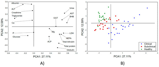
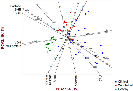
Mastitis is one of the most important diseases in dairy cows, leading to substantial economic losses associated with decreased milk production and quality. Early detection of changes in metabolic and milk parameters is crucial for maintaining animal welfare and milk quality. This study aimed to detect patterns in metabolic and milk composition parameters in Serbian dairy cows affected by mastitis. It also examined the relationship between these factors in cows with clinical and subclinical mastitis, as well as in healthy cows. This study included 60 Holstein-Friesian cows with the same body score condition that were in the same lactation phase. They were divided into three groups of 20: clinical and subclinical mastitis and a control group of healthy cows. The categorization was based on clinical udder health and the California mastitis test. Blood serum metabolic profiles were measured using a Rayto spectrophotometer (Shenzhen, China), and milk composition was determined using MilcoScanTM (Foss, Hilleroed, Denmark) and FossomaticTM (Foss, Hilleroed, Denmark) instruments. Significant increases in non-esterified fatty acids (NEFAs), beta-hydroxybutyrate (BHB), total protein, globulin, urea, total bilirubin, magnesium, and enzyme activity were noted in mastitis-affected cows compared to healthy ones. Additionally, mastitis-affected cows had higher total protein and globulin levels and increased somatic cell counts (SCCs), while albumin concentrations were decreased. Furthermore, a negative correlation between total protein and lactose suggested inflammation leading to reduced lactose levels due to cell damage, infection, and lactose use by mastitis pathogens. Hence, indicators of the energy and protein status of the metabolic profile, together with the chemical composition of milk, may be significant diagnostic tools for detecting, monitoring, and predicting the outcome of mastitis in cows.
Full article
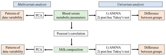
Figure 1

