materials-logo

Journal Browser

Journal Browser

Laser Processing for Bioengineering

A topical collection in Materials (ISSN 1996-1944). This collection belongs to the section "Manufacturing Processes and Systems".

Viewed by 48877

Editors


E-Mail Website
Guest Editor
Dental Surgery Department, Wroclaw Medical University, 50-367 Wrocław, Poland
Interests: oral sugery; periosurgery; laser treatment; dental lasers; lasers in oral and perio surgery; soft tissue; bone regeneration; periimplantitis; recession; 3D customized bone blocks; orthosurgery

Topical Collection Information

Dear Colleagues,

We are delighted to be able to invite you to submit a manuscript to the forthcoming Special Issue, “Laser Processing for Bioengineering”, for the journal Materials (IF: 2.972). This Special Issue is covering a wide variety of topics, from basic and applied research to laboratory and pre-clinical applications related to laser processing for applications in bioengineering and medicine.

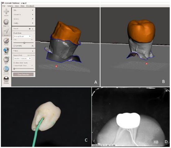
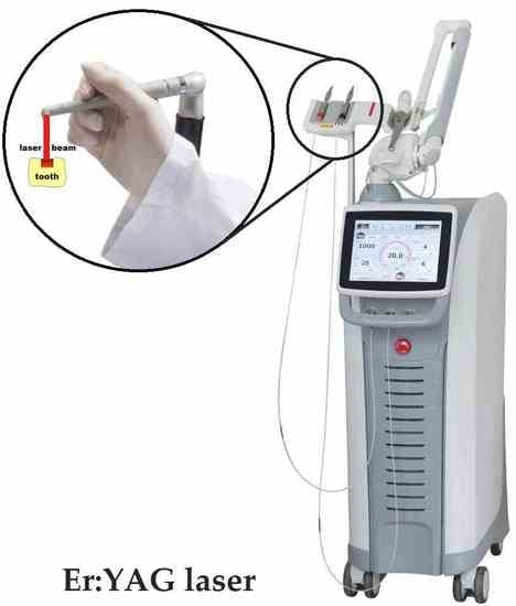
The high-level laser beam interaction with different materials contributes to advanced material processing procedures, among others shaping of surfaces, surface patterning, surface laser micromachining, surface cleaning, drilling and cutting, surface treatment, and laser sintering. The application of laser processing methods allows unique applications in bioengineering for clinical use in medicine, veterinary, and dentistry. Nowadays, a wide brand of different laser wavelengths is used for various therapeutical reasons in surgery, ophthalmology, and dentistry. Laser surface shaping is used for the treatment of patients needing orthopedic and dental implants.

In this Special Issue, the most advanced research and improvements in laser processing for bioengineering applications will be accepted.

Special attention will be paid to new procedures and approaches of laser processing for bioengineering, pre-clinical and clinical application of lasers on adhesion effect and materials properties in conservative, endodontic, prosthetic, orthodontic and periodontal treatment.

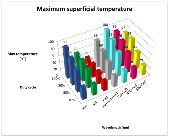
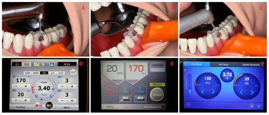
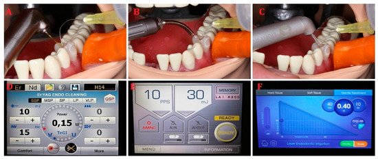
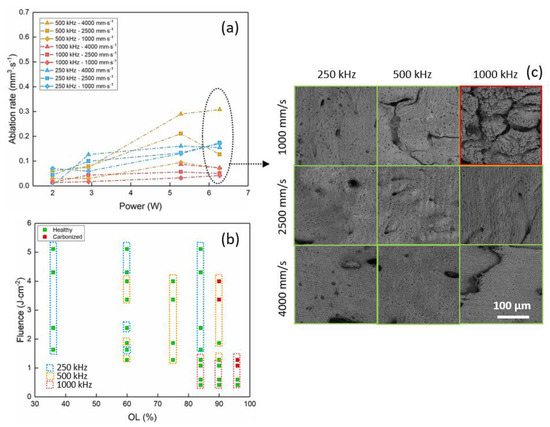
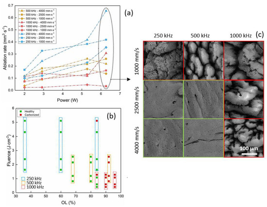
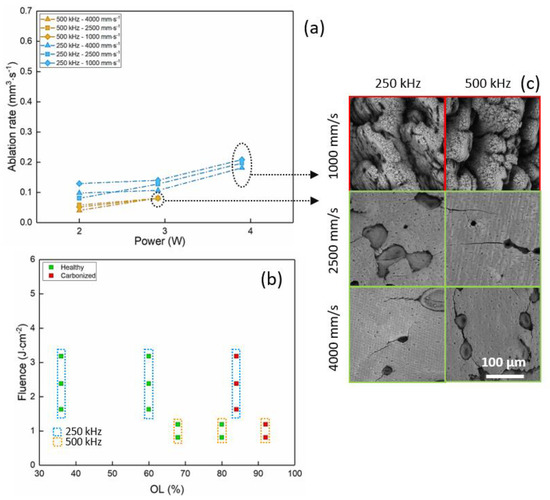
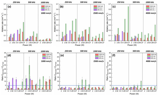
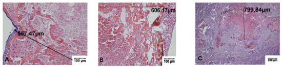
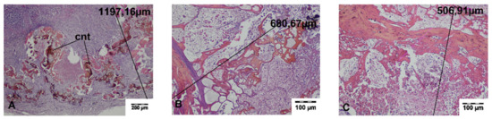
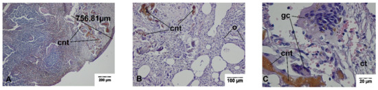
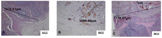
Study displaying the effects of laser light and describing precisely the laser parameters, e.g., power, energy, fluence, power density, wavelength, pulse duration, repetition rate, and exposure time will also be considered. (Please read the reference: DOI: 10.7860/JCDR/2015/15561.6955).

Prof. Marzena Dominiak
Dr. Jacek Matys
Guest Editors

Manuscript Submission Information

Manuscripts should be submitted online at www.mdpi.com by registering and logging in to this website. Once you are registered, click here to go to the submission form. Manuscripts can be submitted until the deadline. All submissions that pass pre-check are peer-reviewed. Accepted papers will be published continuously in the journal (as soon as accepted) and will be listed together on the collection website. Research articles, review articles as well as short communications are invited. For planned papers, a title and short abstract (about 100 words) can be sent to the Editorial Office for announcement on this website.

Submitted manuscripts should not have been published previously, nor be under consideration for publication elsewhere (except conference proceedings papers). All manuscripts are thoroughly refereed through a single-blind peer-review process. A guide for authors and other relevant information for submission of manuscripts is available on the Instructions for Authors page. Materials is an international peer-reviewed open access semimonthly journal published by MDPI.

Please visit the Instructions for Authors page before submitting a manuscript. The Article Processing Charge (APC) for publication in this open access journal is 2600 CHF (Swiss Francs). Submitted papers should be well formatted and use good English. Authors may use MDPI's English editing service prior to publication or during author revisions.

Keywords

  • laser processing
  • bioengineering
  • laser surface cleaning
  • laser surface pattering
  • laser surface shaping
  • surgery
  • titanium
  • zirconia
  • implants
  • orthopedic
  • dentistry
  • surface adhesion
  • materials adhesion
  • materials properties
  • conservative dentistry
  • prosthetic dentistry
  • endodontics
  • orthodontics
  • periodontal treatment

Published Papers (18 papers)

2022

Jump to: 2021, 2020, 2019

12 pages, 477 KiB  
Review
Orthodontic Bracket Removal Using LASER-Technology—A Short Systematic Literature Review of the Past 30 Years
by Anca Mesaroș, Michaela Mesaroș and Smaranda Buduru
Materials 2022, 15(2), 548; https://doi.org/10.3390/ma15020548 - 12 Jan 2022
Cited by 8 | Viewed by 1865
Abstract
Background: Since fixed orthodontic treatment is widely spread and one of its inconveniences is bracket removal, as this affects enamel integrity as well as being a cause of discomfort to the patient, studies have searched for the most adequate bracket removal technique, many [...] Read more.
Background: Since fixed orthodontic treatment is widely spread and one of its inconveniences is bracket removal, as this affects enamel integrity as well as being a cause of discomfort to the patient, studies have searched for the most adequate bracket removal technique, many of them focusing on using laser-technology. Methods: Our review focused on articles published investigating methods of orthodontic bracket removal using laser technology in the last 30 years. Results: 19 relevant studies were taken into consideration after a thorough selection. Different types of laser devices, with specific settings and various testing conditions were tested and the investigators presented their pertinent conclusions. Conclusions: Most studies were performed using ceramic brackets and the best results in terms of prevention of enamel loss, temperature stability for the tooth as well as reduced chair time were obtained with Er:YAG lasers. Full article
Show Figures

Figure 1

2021

Jump to: 2022, 2020, 2019

12 pages, 607 KiB  
Review
Fibroblasts Adhesion to Laser-Modified Titanium Surfaces—A Systematic Review
by Julia Kensy, Maciej Dobrzyński, Rafał Wiench, Kinga Grzech-Leśniak and Jacek Matys
Materials 2021, 14(23), 7305; https://doi.org/10.3390/ma14237305 - 29 Nov 2021
Cited by 7 | Viewed by 1983
Abstract
Objective: Laser treatment has been recently introduced in many fields of implant dentistry. The systematic review tried to address the question: “How does laser modification of titanium surface influence fibroblast adhesion?”. Methods: An electronic search of the PubMed and Scopus databases was performed. [...] Read more.
Objective: Laser treatment has been recently introduced in many fields of implant dentistry. The systematic review tried to address the question: “How does laser modification of titanium surface influence fibroblast adhesion?”. Methods: An electronic search of the PubMed and Scopus databases was performed. The following keywords were used: (laser) AND (fibroblast) AND (titanium) AND (implant OR disc) AND (proliferation OR adhesion). Initially, 136 studies were found. Ten studies met the inclusion criteria and were included in the review. All studies chosen to be included in the review were considered to have a low risk of bias. Results: Studies included in the review varied with laser parameters or ways of observing fibroblast behavior. Studies showed that fibroblasts tend to take different shapes and create extensions on modified surfaces and that their metabolic activity is more intense. One study concentrated on laser application and showed that three-directional laser application is the most successful in terms of fibroblast adhesion. Studies which concentrated more on laser parameters showed that too low energy density (lower or equal to 0.75 J/cm2) does not influence fibroblast adhesion. Increasing the energy density over 0.75 J/cm2 causes better cell adhesion of fibroblasts to the laser-modified sample. One included study focused on increasing titanium surface wettability, which also positively influenced cell adhesion. Conclusion: The studies included in the review proved a positive effect of laser-modified titanium surfaces on fibroblast adhesion. However, the application of an appropriate laser energy dose is crucial. Full article
Show Figures

Figure 1

13 pages, 71424 KiB  
Article
Local Effects of a 1940 nm Thulium-Doped Fiber Laser and a 1470 nm Diode Laser on the Pulmonary Parenchyma: An Experimental Study in a Pig Model
by Maciej Janeczek, Zbigniew Rybak, Anna Lipińska, Jolanta Bujok, Albert Czerski, Maria Szymonowicz, Maciej Dobrzyński, Jacek Świderski and Bogusława Żywicka
Materials 2021, 14(18), 5457; https://doi.org/10.3390/ma14185457 - 21 Sep 2021
Cited by 4 | Viewed by 1661
Abstract
The lungs are a common site of metastases from malignant tumors. Their removal with a minimal but safe tissue margin is essential for the long-term survival of patients. The aim of this study was to evaluate the usefulness of a 1940 nm thulium-doped [...] Read more.
The lungs are a common site of metastases from malignant tumors. Their removal with a minimal but safe tissue margin is essential for the long-term survival of patients. The aim of this study was to evaluate the usefulness of a 1940 nm thulium-doped fiber laser (TDFL) and a 1470 nm diode laser (DL) in a pig model of lung surgery that involved the incision and excision of lung tissue. Histopathological analysis was performed on days 0 and 7 after surgery. Neither TDFL nor DL caused significant perioperative or postoperative bleeding. Histological analysis revealed the presence of carbonized necrotic tissue, mixed fibrin–cellular exudate in the superficial zone of thermal damage and bands of deeper thermal changes. The mean total width of thermal damage on day 0 was 499.46 ± 61.44 and 937.39 ± 109.65 µm for TDFL and DL, respectively. On day 7, cell activation and repair processes were visible. The total width of thermal damage was 2615.74 ± 487.17 µm for TDFL vs. 6500.34 ±1118.02 µm for DL. The superficial zone of thermal damage was narrower for TDFL on both days 0 and 7. The results confirm the effectiveness of both types of laser in cutting and providing hemostasis in the lungs. TDFL caused less thermal damage to the lung parenchyma than DL. Full article
Show Figures

Figure 1

10 pages, 1241 KiB  
Article
SEM Evaluation of Tooth Surface after a Composite Filling Removal Using Er:YAG Laser, Drills with and without Curettes, and Optional EDTA or NaOCl Conditioning
by Jan Kiryk, Jacek Matys, Kinga Grzech-Leśniak, Marzena Dominiak, Małgorzata Małecka, Piotr Kuropka, Rafał J. Wiglusz and Maciej Dobrzyński
Materials 2021, 14(16), 4469; https://doi.org/10.3390/ma14164469 - 10 Aug 2021
Cited by 6 | Viewed by 2135
Abstract
(1) Background: This study aimed to evaluate the microporosity of the tooth surface structure adjacent to the cemento-enamel junction (CEJ) after the removal of composite fillings with a drill in comparison with removal by an Er:YAG laser and after cleaning with a periodontal [...] Read more.
(1) Background: This study aimed to evaluate the microporosity of the tooth surface structure adjacent to the cemento-enamel junction (CEJ) after the removal of composite fillings with a drill in comparison with removal by an Er:YAG laser and after cleaning with a periodontal curette, chemical EDTA and NaOCl (sodium hypochlorite) conditioning. (2) Methods: The research material consisted of 30 extracted premolars with cervical composite fillings. The teeth were divided into six groups according to the method of tooth preparation: group G1 (n = 5)—a diamond drill; group G2 (n = 5)—a diamond drill + curette; group G3 (n = 5)—a diamond drill + 24% EDTA (PrefGel, Straumann, Switzerland); group G4 (n = 5)—an Er:YAG laser (LightWalker, Fotona, Ljubljana, Slovenia) set with the following parameters: power: 1.65 W (composite removal, CR), 1.2 (tooth conditioning, TC), energy: 110 mJ (CR), 80 mJ (TC), frequency: 15 Hz, pulse duration: 50 μs, tip diameter: 1 mm, air/fluid cooling: 4, distance 1.5 mm, energy density: 14.01 J/cm2 (CR), 10.19 J/cm2 (TC); group G5 (n = 5)—an Er:YAG laser + 2% sodium hypochlorite (NaOCl); group G6 (n = 5)—an Er:YAG laser + 5.25% NaOCl. In each tooth, three cavities were made and subjected to analysis. The dentin surface was evaluated using a scanning electron microscope (SEM). (3) Results: Groups G1 and G2 exhibited mechanical damage to the tooth surface structure caused by the rotary motion of a diamond drill. The SEM image showed a smear layer that could only be removed chemically using 24% EDTA gel (group G3). The tooth surfaces prepared with the Er:YAG laser (groups G4–G6) revealed a homogeneous structure without damage along with open dentinal tubules (without smear layer) and visible denaturation of collagen fibers. The sodium hypochlorite (NaOCl) conditioning did not increase the visibility of dentinal tubules. (4) Conclusions: Dentin surfaces have open dentinal tubules after removal of the composite filling using the Er:YAG laser and therefore do not require additional NaOCl conditioning. Full article
Show Figures

Figure 1

13 pages, 2567 KiB  
Article
The Effect of Er:YAG Lasers on the Reduction of Aerosol Formation for Dental Workers
by Kinga Grzech-Leśniak and Jacek Matys
Materials 2021, 14(11), 2857; https://doi.org/10.3390/ma14112857 - 26 May 2021
Cited by 14 | Viewed by 2910
Abstract
Infection prevention in dental practice plays a major role, especially during the COVID-19 pandemic. This study aimed to measure the quantity of aerosol released during various dental procedures (caries and prosthetic treatment, debonding of orthodontic brackets, root canal irrigation) while employing the Er:YAG [...] Read more.
Infection prevention in dental practice plays a major role, especially during the COVID-19 pandemic. This study aimed to measure the quantity of aerosol released during various dental procedures (caries and prosthetic treatment, debonding of orthodontic brackets, root canal irrigation) while employing the Er:YAG lasers combined with a high-volume evacuator, HVE or salivary ejector, SE. The mandibular second premolar was extracted due to standard orthodontic therapy and placed in a dental manikin, to simulate typical treatment conditions. The particle counter was used to measure the aerosol particles (0.3–10.0 μm) at three different sites: dental manikin and operator’s and assistant’s mouth area. The study results showed that caries’ treatment and dental crown removal with a high-speed handpiece and the use of the SE generated the highest aerosol quantity at each measured site. All three tested Er:YAG lasers significantly reduced the number of aerosol particles during caries’ treatment and ceramic crown debonding compared the conventional handpieces, p < 0.05. Furthermore, the Er:YAG lasers generated less aerosol during orthodontic bracket debonding and root canal irrigation in contrast to the initial aerosol quantity measured in the dental office. The use of the Er:YAG lasers during dental treatments significantly generates less aerosol in the dental office setting, which reduces the risk of transmission of viruses or bacteria. Full article
Show Figures

Graphical abstract

13 pages, 2644 KiB  
Article
Ablation of Bone Tissue by Femtosecond Laser: A Path to High-Resolution Bone Surgery
by Laura Gemini, Samy Al-Bourgol, Guillaume Machinet, Aboubakr Bakkali, Marc Faucon and Rainer Kling
Materials 2021, 14(9), 2429; https://doi.org/10.3390/ma14092429 - 07 May 2021
Cited by 9 | Viewed by 3111
Abstract
Femtosecond lasers allow for high-precision, high-quality ablation of biological tissues thanks to their capability of minimizing the thermal loads into the irradiated material. Nevertheless, reported ablation rates remain still too limited to enable their exploitation on a clinical level. This study demonstrates the [...] Read more.
Femtosecond lasers allow for high-precision, high-quality ablation of biological tissues thanks to their capability of minimizing the thermal loads into the irradiated material. Nevertheless, reported ablation rates remain still too limited to enable their exploitation on a clinical level. This study demonstrates the possibility to upscale the process of fs laser ablation of bone tissue by employing industrially available fs laser sources. A comprehensive parametric study is presented in order to optimize the bone tissue ablation rate while maintaining the tissue health by avoiding excessive thermal loads. Three different absorption regimes are investigated by employing fs laser sources at 1030 nm, 515 nm and 343 nm. The main differences in the three different wavelength regimes are discussed by comparing the evolution of the ablation rate and the calcination degree of the laser ablated tissue. The maximum of the ablation rate is obtained in the visible regime of absorption where a maximum value of 0.66 mm3/s is obtained on a non-calcined tissue for the lowest laser repetition rate and the lowest spatial overlap between successive laser pulses. In this regime, the hemoglobin present in the fresh bone tissue is the main chromophore involved in the absorption process. To the best of our knowledge, this is the highest ablation rate obtained on porcine femur upon fs laser ablation. Full article
Show Figures

Graphical abstract

11 pages, 2677 KiB  
Article
The Effect of Er:YAG Laser on a Shear Bond Strength Value of Orthodontic Brackets to Enamel—A Preliminary Study
by Jan Kiryk, Jacek Matys, Anna Nikodem, Karolina Burzyńska, Kinga Grzech-Leśniak, Marzena Dominiak and Maciej Dobrzyński
Materials 2021, 14(9), 2093; https://doi.org/10.3390/ma14092093 - 21 Apr 2021
Cited by 7 | Viewed by 1768
Abstract
We sought to evaluate the effects of Er:YAG laser (LightTouch, LightInstruments, Israel) conditioning on enamel roughness and shear bond strength of orthodontic brackets on enamel. Eighteen human molars (n = 9) and premolars (n = 9), were divided into 3 groups depending on [...] Read more.
We sought to evaluate the effects of Er:YAG laser (LightTouch, LightInstruments, Israel) conditioning on enamel roughness and shear bond strength of orthodontic brackets on enamel. Eighteen human molars (n = 9) and premolars (n = 9), were divided into 3 groups depending on the enamel conditioning method; Er:YAG laser (G1, n = 6), conventional etching with 37% orthophosphoric acid (G2, n = 6), Er:YAG laser combined with conventional etching (G3, n = 6). Er:YAG laser parameters were as follows: energy: 100 mJ, frequency: 10 Hz, exposure time: 10 s, applicator diameter: 600 μm, fluence: 35.37 J/cm2, distance: 1 mm away from a tooth, cooling: 80%. An MTS 858 MiniBionix® machine was used to determine the shear bond strength (MTS System, Eden Prairie, MN, USA). The enamel structure was assessed using X-ray microtomography (SkyScan 1172, Bruker, Kontich, Belgium). The highest values of shear bond strength were obtained in the G3 group (9.23 ± 2.38 MPa) and the lowest values in the G2 group (6.44 ± 2.11 MPa) (p < 0.05). A significant change in the enamel surface was noted after applying laser, reaching up to 9% of enamel thickness, which was not observed in the etched samples. Moreover, the Er:YAG laser-irradiated enamel surface was characterized by the greatest roughness. The combined use of an Er:YAG laser with a conventional etching improves the adhesion of composite materials to the tooth. Full article
Show Figures

Figure 1

11 pages, 2486 KiB  
Article
Usefulness of Thulium-Doped Fiber Laser and Diode Laser in Zero Ischemia Kidney Surgery—Comparative Study in Pig Model
by Bogusława Żywicka, Jolanta Bujok, Maciej Janeczek, Albert Czerski, Maria Szymonowicz, Maciej Dobrzyński, Jacek Świderski and Zbigniew Rybak
Materials 2021, 14(8), 2000; https://doi.org/10.3390/ma14082000 - 16 Apr 2021
Cited by 4 | Viewed by 2069
Abstract
Background: The aim of this study was to evaluate the usefulness of a thulium-doped fiber laser and a diode laser in zero ischemia kidney surgery, by carrying out a comparative study in a pig model. Material and methods: Research was carried out on [...] Read more.
Background: The aim of this study was to evaluate the usefulness of a thulium-doped fiber laser and a diode laser in zero ischemia kidney surgery, by carrying out a comparative study in a pig model. Material and methods: Research was carried out on 12 pigs weighing 30 kg each. A thulium-doped fiber laser (TDFL) and a diode laser (DL) operating at wavelengths of 1940 and 1470 nm, respectively, were used. The cut sites were assessed both macroscopically and microscopically. The zone of thermal damage visible in the histopathological preparations was divided into superficial and total areas. Results: During partial nephrectomy, moderate to minimal bleeding was observed, which did not require additional hemostatic measures. All animals survived the procedure. On day 0, the total thermal damage depth was 837.8 µm for the TDFL and 1175.0 µm for the DL. On day 7, the depths were 1556.2 and 2301.7 µm, respectively. On day 14, the overall thermal damage depth for the DL was the greatest (6800 µm). The width of the superficial zone was significantly reduced on days 7 and 14 after TDFL application. Conclusion: Both lasers are suitable for partial wedge nephrectomy without ischemia in pigs. The TDFL produced similar or better hemostasis than the DL, with a smaller zone of thermal damage and, therefore, seems more suitable for application in human medicine. Full article
Show Figures

Figure 1

1 pages, 159 KiB  
Addendum
Addendum: Żywicka, B., et al. Comparison of a 1940 nm Thulium-Doped Fiber Laser and a 1470 nm Diode Laser for Cutting Efficacy and Hemostasis in a Pig Model of Spleen Surgery. Materials 2020, 13, 1167
by Bogusława Żywicka, Zbigniew Rybak, Maciej Janeczek, Albert Czerski, Jolanta Bujok, Maria Szymonowicz, Maciej Dobrzyński, Mariusz Korczyński and Jacek Świderski
Materials 2021, 14(4), 966; https://doi.org/10.3390/ma14040966 - 18 Feb 2021
Viewed by 1440
Abstract
The authors would like to add the following sentence to the “Funding” section of their article [...] Full article

2020

Jump to: 2022, 2021, 2019

12 pages, 1271 KiB  
Article
Retrieval of Prefabricated Zirconia Crowns with Er,Cr:YSGG Laser from Primary and Permanent Molars
by Janina Golob Deeb, Connor McCall, Caroline K. Carrico, William O. Dahlke and Kinga Grzech-Leśniak
Materials 2020, 13(23), 5569; https://doi.org/10.3390/ma13235569 - 07 Dec 2020
Cited by 12 | Viewed by 2815
Abstract
(1) Background: Prefabricated zirconia crowns are used to restore teeth in children. The purpose of this study was to evaluate the removal of these crowns with the erbium, chromium: yttrium-scandium-gallium-garnet (Er,Cr:YSGG) laser; (2) Methods: A total of 12 primary and 12 permanent teeth [...] Read more.
(1) Background: Prefabricated zirconia crowns are used to restore teeth in children. The purpose of this study was to evaluate the removal of these crowns with the erbium, chromium: yttrium-scandium-gallium-garnet (Er,Cr:YSGG) laser; (2) Methods: A total of 12 primary and 12 permanent teeth were prepared and prefabricated zirconia crowns were passively fitted and cemented with two resin modified glass-ionomer (RMGI) cements. Surface areas of prepared teeth and crowns were calculated. Crowns were removed using two laser settings: 4.5 Watts, 15 Hertz, 20 water/20 air, and 5 Watts, 15 Hertz, 50 water/50 air. The retrieval time and temperature changes were tested recorded. Data were analyzed using ANOVA with Tukey’s adjusted post hoc pairwise comparison t-test; (3) Results: The average time for crown removal was: 3 min, 47.7 s for permanent; and 2 min 5 s for primary teeth. The mean temperature changes were 2.48 °C (SD = 1.43) for permanent; and 3.14 °C (SD = 1.88) for primary teeth. The time to debond was significantly positively correlated with tooth inner surface area and volume, outer crown volume, and the cement volume; (4) Conclusions: Use of the Er,Cr:YSGG laser is an effective, safe and non-invasive method to remove prefabricated zirconia crowns cemented with RMGI cements from permanent and primary teeth. Full article
Show Figures

Figure 1

16 pages, 1752 KiB  
Article
Assessment of Effects of Laser Light Combining Three Wavelengths (450, 520 and 640 nm) on Temperature Increase and Depth of Tissue Lesions in an Ex Vivo Study
by Kamil Jurczyszyn, Witold Trzeciakowski, Zdzisław Woźniak, Piotr Ziółkowski and Mateusz Trafalski
Materials 2020, 13(23), 5340; https://doi.org/10.3390/ma13235340 - 25 Nov 2020
Cited by 3 | Viewed by 1911
Abstract
Background: Lasers are widely used in medicine in soft and hard tissue surgeries and biostimulation. Studies found in literature typically compare the effects of single-wavelength lasers on tissues or cell cultures. In our study, we used a diode laser capable of emitting three [...] Read more.
Background: Lasers are widely used in medicine in soft and hard tissue surgeries and biostimulation. Studies found in literature typically compare the effects of single-wavelength lasers on tissues or cell cultures. In our study, we used a diode laser capable of emitting three components of visible light (640 nm, red; 520 nm, green; 450 nm, blue) and combining them in a single beam. The aim of the study was to assess the effects of laser radiation in the visible spectrum on tissue in vitro, depending on the wavelength and pulse width. Methods: All irradiations were performed using the same output power (1.5 W). We used various duty cycles: 10, 50, 80 and 100% with 100 Hz frequency. Maximum superficial temperature, rate of temperature increase and lesion depth were investigated. Results: Maximum superficial temperature was observed for 450 + 520 nm irradiation (100% duty cycle). The highest rate of increase of temperature was noted for 450 + 520 nm (100% duty cycle). Maximum lesion depth was observed in case of three-wavelength irradiation (450 + 520 + 640 nm) for 100, 80 and 50% duty cycles. Conclusions: The synergistic effect of two-wavelength (450 + 520 nm) irradiation was observed in case of maximum temperature measurement. The deepest depth of lesion was noted after three-wavelength irradiation (450 + 520 + 640 nm). Full article
Show Figures

Figure 1

13 pages, 2614 KiB  
Article
Dental Aerosol as a Hazard Risk for Dental Workers
by Jacek Matys and Kinga Grzech-Leśniak
Materials 2020, 13(22), 5109; https://doi.org/10.3390/ma13225109 - 12 Nov 2020
Cited by 47 | Viewed by 4877
Abstract
Standard dental procedures, when using a water coolant and rotary instruments, generate aerosols with a significantly higher number of various dangerous pathogens (viruses, bacteria, and fungi). Reducing the amount of aerosols to a minimum is mandatory, especially during the new coronavirus disease, COVID-19. [...] Read more.
Standard dental procedures, when using a water coolant and rotary instruments, generate aerosols with a significantly higher number of various dangerous pathogens (viruses, bacteria, and fungi). Reducing the amount of aerosols to a minimum is mandatory, especially during the new coronavirus disease, COVID-19. The study aimed to evaluate the amount of aerosol generated during standard dental procedures such as caries removal (using dental bur on a high and low-speed handpiece and Er:YAG laser), ultrasonic scaling, and tooth polishing (using silicon rubber on low-speed handpiece) combined with various suction systems. The airborne aerosols containing particles in a range of 0.3–10.0 μm were measured using the PC200 laser particle counter (Trotec GmbH, Schwerin, Germany) at three following sites, manikin, operator, and assistant mouth, respectively. The following suction systems were used to remove aerosols: saliva ejector, high volume evacuator, saliva ejector with extraoral vacuum, high volume evacuator with extraoral vacuum, Zirc® evacuator (Mr.Thirsty One-Step®), and two customized high volume evacuators (white and black). The study results showed that caries removal with a high-speed handpiece and saliva ejector generates the highest amount of spray particles at each measured site. The aerosol measurement at the manikin mouth showed the highest particle amount during caries removal with the low and high-speed handpiece. The results for the new high volume evacuator (black) and the Zirc® evacuator showed the lowest increase in aerosol level during caries removal with a high-speed handpiece. The Er:YAG laser used for caries removal produced the lowest aerosol amount at the manikin mouth level compared to conventional dental handpieces. Furthermore, ultrasonic scaling caused a minimal aerosol rise in terms of the caries removal with bur. The Er:YAG laser and the new wider high volume evacuators improved significantly suction efficiency during dental treatment. The use of new suction systems and the Er:YAG laser allows for the improvement of biological safety in the dental office, which is especially crucial during the COVID-19 pandemic. Full article
Show Figures

Figure 1

10 pages, 1421 KiB  
Article
Removal of Composite Restoration from the Root Surface in the Cervical Region Using Er: YAG Laser and Drill—In Vitro Study
by Wojciech Zakrzewski, Maciej Dobrzynski, Piotr Kuropka, Jacek Matys, Malgorzata Malecka, Jan Kiryk, Zbigniew Rybak, Marzena Dominiak, Kinga Grzech-Lesniak, Katarzyna Wiglusz and Rafal J. Wiglusz
Materials 2020, 13(13), 3027; https://doi.org/10.3390/ma13133027 - 07 Jul 2020
Cited by 12 | Viewed by 2799
Abstract
Background: Recently, the defects of the tooth surface in the cervical region are often restored using composite filling materials. It should meet the needs of the patients regarding esthetics and material stability. The aim of the study was to analyze the tooth root [...] Read more.
Background: Recently, the defects of the tooth surface in the cervical region are often restored using composite filling materials. It should meet the needs of the patients regarding esthetics and material stability. The aim of the study was to analyze the tooth root surface at the cervical region after the removal of the composite filling material by means of the Erbium-doped Yttrium Aluminium Garnet (Er: YAG) laser or drill using the scanning electron microscopy (SEM) and fluorescence microscopy. Materials and Methods: For the purposes of this study, 14 premolar teeth (n = 14) were removed due to orthodontic reasons. The rectangular shape cavities with 3 mm in width and 1.5 mm in height were prepared with a 0.8 mm bur on high-speed contra-angle in the tooth surface just below cemento-enamel junction (CEJ) and filled with the composite material. The composite material was removed with the Er: YAG laser at a power of 3.4 W, energy 170 mJ, frequency 20 Hz, pulse duration 300 μs, tip diameter 0.8 mm, air/fluid cooling 3 mL/s, and time of irradiation: 6 sec, at a distance from teeth of 2 mm (G1 group, n = 7) or a high-speed contra-angle bur (G2 group, n = 7). After the removal of composite material, the surfaces of teeth were examined using the scanning electron microscopy (SEM) and fluorescence microscopy. Results: The Er: YAG irradiation allowed to remove completely the composite material from the tooth cavity. The study confirmed, that the ends of collagen fibers were only partially denatured after the Er: YAG laser application. Conclusion: It has been proved that using the Er: YAG laser is an effective and safe method of composite removal for the dentin surface. Full article
Show Figures

Graphical abstract

12 pages, 1982 KiB  
Article
Effect of 808 nm Semiconductor Laser on the Stability of Orthodontic Micro-Implants: A Split-Mouth Study
by Jacek Matys, Rafał Flieger, Tomasz Gedrange, Krzysztof Janowicz, Bartosz Kempisty, Kinga Grzech-Leśniak and Marzena Dominiak
Materials 2020, 13(10), 2265; https://doi.org/10.3390/ma13102265 - 14 May 2020
Cited by 15 | Viewed by 2777
Abstract
Background: To evaluate the effect of photobiomodulation (PBM) on orthodontic micro-implants (n = 44; 14 women, 8 men). Methods: PBM with 808 nm diode laser was applied immediately, 3, 6, 9, 12, 15, and 30 days post the implantation. Results were assessed within [...] Read more.
Background: To evaluate the effect of photobiomodulation (PBM) on orthodontic micro-implants (n = 44; 14 women, 8 men). Methods: PBM with 808 nm diode laser was applied immediately, 3, 6, 9, 12, 15, and 30 days post the implantation. Results were assessed within same time frames and additionally after 60 days to check for implants stability using the Periotest device. Patients pain experiences following the first day post-treatment and potential loss of micro-implants after 60 days were recorded. The procedure involved insertion of mini-implants in the maxilla for the laser group (L, n = 22) and negative control group (C, n = 22). Irradiation was carried buccally and palatally with respect to the maxillary ridge (2 points). The energy per point was 4 J (8 J/cm2), total dose was 56 J. Results: Patients did not report significant differences in terms of pain experiences comparing the L and C groups (p = 0.499). At 30 days post-treatment, higher secondary stability of implants was observed in the laser group (Periotest Test Value, PTV 6.32 ± 3.62), in contrast to the controls (PTV 11.34 ± 5.76) (p = 0.004). At 60 days post-treatment, significantly higher stability was recorded in the laser group (PTV 6.55 ± 4.66) compared with the controls, PTV (10.95 ± 4.77) (p = 0.009). Conclusions: Application of the 808 nm diode laser increased secondary micro-implant stability. Full article
Show Figures

Figure 1

10 pages, 552 KiB  
Article
Q-Switch Nd:YAG Laser-Assisted Elimination of Multi-Species Biofilm on Titanium Surfaces
by Melanie Namour, Tim Verspecht, Marwan El Mobadder, Wim Teughels, Andre Peremans, Samir Nammour and Eric Rompen
Materials 2020, 13(7), 1573; https://doi.org/10.3390/ma13071573 - 29 Mar 2020
Cited by 11 | Viewed by 2091
Abstract
(1) Background: The relatively high prevalence of peri-implantitis (PI) and the lack of a standard method for decontamination of the dental implant surface have pushed us to conduct further research in the field. Bacterial biofilms were found to play a primordial role in [...] Read more.
(1) Background: The relatively high prevalence of peri-implantitis (PI) and the lack of a standard method for decontamination of the dental implant surface have pushed us to conduct further research in the field. Bacterial biofilms were found to play a primordial role in the etiology of PI. Therefore, the aim is to evaluate the efficacy of a laser-assisted elimination of biofilm protocol in the removal of a multi-species biofilm on titanium surfaces. (2) Methods: In total, 52 titanium discs (grade 4) were used. The study group consisted of 13 titanium disks contaminated with multi-species biofilms and subsequently irradiated with the laser (T + BF + L). The control groups consisted of the following types of titanium disks: 13 contaminated with multi-species biofilms (T + BF), 13 sterile and irradiated (T + L), 13 sterile and untreated (T). Q-Switch Nd:YAG laser Irradiation parameters were the following: energy density equal to 0.597 J/cm2 per pulse, power equal to 270 milliwatt per pulse, 2.4 mm of spot diameter, and 10 Hz repetition rate for pulse duration of six nanoseconds (ns). The laser irradiation was made during 2 s of total time in non-contact and at 0.5 mm away from the titanium disc surface. After treatment, presence of biofilms on the disks was evaluated by staining with crystal violet (CV), which was measured as optical density at six hundred thirty nm, and statistical analyses were done. (3) Results: the optical density values were 0.004 ± 0.004 for the study group T + BF + L, 0.120 ± 0.039 for group T + BF, 0.006 ± 0.003 for group T + L, and 0.007 ± 0.007 for group T. For the study group, laser treatment resulted in a total elimination of the biofilm, with mean values statistically significantly lower than those of contaminated titanium surfaces and similar to those of sterile titanium surfaces. (4) Conclusions: Our irradiation protocol provided a significant elimination of the multi-species biofilm on titanium surfaces. Laser treated titanium surfaces were biofilm-free, similar to the sterile ones. Full article
Show Figures

Figure 1

13 pages, 4713 KiB  
Article
Comparison of A 1940 nm Thulium-Doped Fiber Laser and A 1470 nm Diode Laser for Cutting Efficacy and Hemostasis in A Pig Model of Spleen Surgery
by Bogusława Żywicka, Zbigniew Rybak, Maciej Janeczek, Albert Czerski, Jolanta Bujok, Maria Szymonowicz, Maciej Dobrzyński, Mariusz Korczyński and Jacek Świderski
Materials 2020, 13(5), 1167; https://doi.org/10.3390/ma13051167 - 05 Mar 2020
Cited by 14 | Viewed by 3157
Abstract
Partial and total splenectomies are associated with a high risk of substantial blood loss. Lasers operating at wavelengths strongly absorbed by water have the potential to improve hemostasis and cut while providing a narrow zone of thermal damage. The aim of this study [...] Read more.
Partial and total splenectomies are associated with a high risk of substantial blood loss. Lasers operating at wavelengths strongly absorbed by water have the potential to improve hemostasis and cut while providing a narrow zone of thermal damage. The aim of this study is to compare a thulium-doped fiber laser (TDFL) emitting a wavelength of 1940 nm and a diode laser (DL) operating at 1470 nm for spleen surgery in a pig model. A partial splenectomy and spleen incisions were made in 12 animals using the two laser devices. The hemostasis was evaluated visually during surgeries. Post-mortem and histopathological evaluations were done on days 0, 7, and 14 following surgery. Neither TDFL nor DL caused bleeding on day 0 or delayed bleeding. On day 14, pale streaks at the site of incision were slightly wider after cutting with DL than with TDFL. Histological analysis revealed a carbonized zone with exudation and a deeper zone of thermal tissue damage on day 0. The width of the thermal changes was 655.26 ± 107.70 μm for TDFL and 1413.37 ± 111.85 μm for DL. On day 7, a proliferation of fibroblasts and splenocytes was visible, as well as a formation of multinucleated giant cells adjacent to the residues of carbonization. The zone of thermal damage was broader for DL (1157.5 ± 262.77 μm) than for TDFL (682.22 ± 116.58 μm). On day 14, cutting sites were filled with connective and granulation tissues with the residues of carbonization. The zone of thermal damage was narrower for TDFL (761.65 ± 34.3 μm) than for DL (1609.82 ± 202.22 μm). Thus, both lasers are efficient in spleen surgery, providing good hemostasis. However, TDFL produces a narrower zone of thermal damage, which suggests its better efficiency for spleen surgery, especially when performing more precise procedures. Full article
Show Figures

Figure 1

15 pages, 5869 KiB  
Article
Effect of Laser Pulse Overlap and Scanning Line Overlap on Femtosecond Laser-Structured Ti6Al4V Surfaces
by Georg Schnell, Ulrike Duenow and Hermann Seitz
Materials 2020, 13(4), 969; https://doi.org/10.3390/ma13040969 - 21 Feb 2020
Cited by 50 | Viewed by 4796
Abstract
Surface structuring is a key factor for the tailoring of proper cell attachment and the improvement of the bone-implant interface anchorage. Femtosecond laser machining is especially suited to the structuring of implants due to the possibility of creating surfaces with a wide variety [...] Read more.
Surface structuring is a key factor for the tailoring of proper cell attachment and the improvement of the bone-implant interface anchorage. Femtosecond laser machining is especially suited to the structuring of implants due to the possibility of creating surfaces with a wide variety of nano- and microstructures. To achieve a desired surface topography, different laser structuring parameters can be adjusted. The scanning strategy, or rather the laser pulse overlap and scanning line overlap, affect the surface topography in an essential way, which is demonstrated in this study. Ti6Al4V samples were structured using a 300 fs laser source with a wavelength of 1030 nm. Laser pulse overlap and scanning line overlap were varied between 40% and 90% over a wide range of fluences (F from 0.49 to 12.28 J/cm²), respectively. Four different main types of surface structures were obtained depending on the applied laser parameters: femtosecond laser-induced periodic surface structures (FLIPSS), micrometric ripples (MR), micro-craters, and pillared microstructures. It could also be demonstrated that the exceedance of the strong ablation threshold of Ti6Al4V strongly depends on the scanning strategy. The formation of microstructures can be achieved at lower levels of laser pulse overlap compared to the corresponding value of scanning line overlap due to higher heat accumulation in the irradiated area during laser machining. Full article
Show Figures

Figure 1

2019

Jump to: 2022, 2021, 2020

13 pages, 2852 KiB  
Article
Temperature Changes and SEM Effects of Three Different Implants-Abutment Connection during Debridement with Er:YAG Laser: An Ex Vivo Study
by Jacek Matys, Umberto Romeo, Krzysztof Mroczka, Kinga Grzech-Leśniak and Marzena Dominiak
Materials 2019, 12(22), 3748; https://doi.org/10.3390/ma12223748 - 14 Nov 2019
Cited by 11 | Viewed by 2742
Abstract
The study aimed to evaluate a temperature increase in, and damage to, titanium implants during flapless laser debridement. The study analyzed 15 implants with various implant–abutment connections: a two-piece implant (n = 4) with a screw abutment (IA—Implant–Abutment) and a one-piece implant with [...] Read more.
The study aimed to evaluate a temperature increase in, and damage to, titanium implants during flapless laser debridement. The study analyzed 15 implants with various implant–abutment connections: a two-piece implant (n = 4) with a screw abutment (IA—Implant–Abutment) and a one-piece implant with a ball type fixture (BTF, n = 4) or fix type fixture (FTF, n = 4). The implants were placed in porcine mandibles 2 mm over a bone crest to imitate a peri-implantitis. The implants were debrided in contact mode for 60 s with a Er:YAG laser at fluence of 9.95 J/cm2 (G1 group: 50 mJ/30 Hz); 19.89 J/cm2 (G2 group: 100 mJ/30 Hz); 39.79 J/cm2 (G3 group: 200 mJ/30 Hz), or a scaler with a ceramic tip (G4 control group: 4 W/20 Hz). The temperature was measured with thermocouples at implant and abutment levels. The damage in the titanium surface (n = 3, non-irradiated implants from each type) was assessed using SEM (Scanning Electron Microscopy). The temperature increase at the implant level for the laser was higher at IA in contrast with FTF and BTF. (p < 0.05) The temperature change at the abutment level was lower for the scaler in contrast to Er:YAG laser at FTF. (p < 0.0002) Er:YAG laser didn’t increase the temperature by 10 °C at 100 mJ/30 Hz and 50 mJ/30 Hz. Based on SEM analysis, cracks occurred on the surface of two-piece implants and were more pronounced. Cracks and the melting of the titanium surface of two-piece implants cleaned with Er:YAG laser at 100 or 200 mJ were observed. The specimens treated with the ultrasonic scaler with a plastic curette showed the remaining dark debris on the titanium surface. We recommend using Er:YAG laser at 50 mJ/30 Hz during flapless implants debridement. Full article
Show Figures

Figure 1

Back to TopTop